Note: Descriptions are shown in the official language in which they were submitted.
CA 02912150 2015-11-10
P1135PCAUV4
1
A CATHETER SYSTEM
FIELD
Various aspects of the invention relate to catheter systems and related
methods and
components therefor.
BACKGROUND
A catheter is a tube insertable into a patient, so that part of the tube is in
the patient and
another part is external the patient, to establish fluid communication with
one or more
sites within the patient. By way of example, nerve block catheters are used to
deliver
anaesthetic to nerves whereas other catheters are used for withdrawing blood
samples,
administering medication into the blood stream or measuring pressure.
To so measure blood pressure, the catheter is inserted into a blood vessel and
connected to a blood pressure transducer (or blood pressure "sensor" ¨ the
terms are
used interchangeably herein) to establish fluid communication between the
vessel and
the transducer. A typical blood pressure transducer contains a membrane which
moves
with pressure changes. This movement then generates an electrical signal which
is
translated into the measured pressure displayed on the monitor on the
transducer.
During arterial pressure measurement, the compliance of commercial transducers
is
such that blood moves in and out of the end of the catheter during the beat-to-
beat
pressure change of the pulse.
Some existing catheters take the form of a simple tube being open at both of
its ends,
whereas others have a closed distal end and a set of side openings.
Nerve block catheters are conventionally inserted by:
1. inserting into the patient a tubular needle;
=
CA 02912150 2015-11-10
P1135PCAUV4
2
2. threading the catheter through the tubular needle;
3. withdrawing the tubular needle leaving a free open end of the catheter
projecting from the patient; and
4. fitting a connector to the projecting free end of the catheter to
connect the
catheter to a fluid source e.g. a syringe loaded with anaesthetic (whereas
other catheters might be connected to a fluid destination e.g. a vacuum
source for drawing a blood sample).
The present inventor has recognised that the proper functioning of catheters
is
sometimes adversely impacted by the catheter kinking, the buildup of material
(e.g.
blood clotting), leakage of injected fluid back along the exterior of the
catheter and/or by
the catheter inadvertently (partly or wholly) being withdrawn from the
patient.
Accordingly the various aspects of the present invention aim to at least
partly address
one or more of these problems, or at least to provide alternatives for those
concerned
with catheter systems and their use.
It is not admitted that any of the information in this patent specification is
common
general knowledge, or that the person skilled in the art could be reasonably
expected to
ascertain or understand it, regard it as relevant or combine it in any way at
the priority
date.
SUMMARY
One aspect of the invention provides a method, of fluidly communicating with a
blood
vessel within a patient, including
inserting into the blood vessel a catheter; and
inserting into the catheter a stylet configured such that, when so inserted,
at least a
portion of the stylet is spaced from the catheter such that the at least
portion and the
CA 02912150 2015-11-10
P1135PCAUV4
3
catheter together define at least one elongate void for conveying fluid along
the catheter
at least one of to or from the blood vessel.
The blood vessel may be an artery.
Another aspect of the invention provides a method of monitoring blood pressure
including so fluidly communicating with the blood vessel. This method
preferably
includes delivering fluid, e.g. saline, via the catheter to the blood vessel
to resist clotting.
The stylet may include a shaped portion positioned to be within the patient
and shaped
to act upon to remove build-up from the catheter. Preferably the shaped
portion has a
shape complementary to a cylindrical interior of the catheter, and most
preferably is
shaped to obstruct flow through the catheter.
The method may further include manipulating the stylet to remove build-up,
from the
catheter, within the patient, e.g. to remove build-up, from an open end of the
catheter.
Preferably the method includes, after inserting the stylet, leaving for a
period of time the
stylet in place. Preferably the period is at least one hour. Optionally the
inserting into the
patient a catheter and the inserting into the catheter a stylet are during an
attendance to
the patient; and the method further includes leaving the stylet and catheter
in place until
a subsequent attendance to the patient.
The stylet may be replaced with another stylet.
The inserting into the patient a catheter preferably includes inserting into
the patient a
needle externally carrying the catheter; and withdrawing the needle.
Another aspect of the invention provides a catheter system including
a catheter; and
a stylet;
= =
CA 02912150 2015-11-10
P1135PCAUV4
4
the stylet including an elongate portion and a shaped portion;
the elongate portion being receivable within the catheter;
the elongate portion and the catheter being co-operably configured such that,
when the
elongate portion is so received, at least a portion of the elongate portion is
spaced from
the catheter such that the at least portion and the catheter together define
at least one
elongate void for conveying fluid along the catheter at least one of to or
from one or
more sites within a patient;
the shaped portion being positioned to be within the patient.
Preferably the shaped portion is shaped to obstruct the catheter. The shaped
portion
may be a bulbous portion and is preferably in substance at an end of the
stylet.
The catheter may have an open end positionable within the patient in which
case the
stylet is preferably advanceable within the catheter to move the shaped
portion at least
to the open end, when the open end is within the patient, to remove build-up
at the open
end.
It is preferred that at least a rearward portion of an exterior of the shaped
portion
rearwardly converges.
Another aspect of the invention provides a catheter system including
a catheter; and
a stylet;
the stylet including an elongate portion;
the elongate portion being receivable within the catheter;
CA 02912150 2015-11-10
P11 3 SPCA UV4
the catheter having an open end positionable within the patient; and
the stylet being long enough to be manipulated external the patient to remove
build-up
from the open end within the patient.
The system may further include a needle, e.g. a short beveled nerve block
needle, for
5 inserting the catheter into the patient, in which case the needle is
preferably receivable
within the catheter.
Another aspect of the invention provides a catheter system including
a catheter;
a needle; and
a stylet;
the stylet including an elongate portion;
the elongate portion being receivable within the catheter;
the elongate portion and the catheter being co-operably configured such that,
when the
elongate portion is so received, at least a portion of the elongate portion is
spaced from
the catheter such that the at least portion and the catheter together define
at least one
elongate void for conveying fluid along the catheter at least one of to or
from one or
more sites within a patient;
the needle being receivable within the catheter to insert the catheter into
the patient.
The stylet preferably includes a connector for sealingly engaging an end of
the catheter
external the patient to fluidly connect the void(s) with at least one of a
fluid source or a
fluid destination.
CA 02912150 2015-11-10
P1135PCAUV4
6
Optionally the catheter has one or more side openings positionable within the
patient to
fluidly connect the void(s) to one or more of the sites within the patient.
The system may be a nerve block catheter system or a blood vessel catheter
system.
Preferably the system, of any of the foregoing aspects of the invention, is
individually
packaged in a package such that the system is sterile upon removal of the
system from
the package.
Another aspect of the invention provides a stylet for a catheter system;
the stylet including an elongate portion and a shaped portion;
the elongate portion being receivable within a catheter and configured such
that, when
the elongate portion is so received, at least a portion of the elongate
portion is spaced
from the catheter such that the at least portion and the catheter together
define at least
one elongate void for conveying fluid along the catheter at least one of to or
from one or
more sites within a patient;
the shaped portion being positioned to be within the patient.
Preferably the shaped portion has a shape complementary to a cylindrical
interior of the
catheter to obstruct the catheter.
Another aspect of the invention provides a method, of fluidly communicating
with one or
more sites within a patient, including
inserting into the patient a catheter;
inserting into the catheter a stylet.
The method preferably includes manipulating the stylet to position the shaped
portion
between openings, of the catheter, within the patient. The stylet may be
manipulated to
CA 02912150 2015-11-10
P1 1 3 SPCA UV4
7
move the shaped portion so as to remove build-up, from the catheter, within
the patient,
e.g. to remove build-up, from an open end of the catheter, within the patient.
Preferably
the stylet is manipulated to move the shaped portion beyond an or the open
end, of the
catheter, within the patient. The method may include at least one of advancing
and
retracting the stylet to establish fluid communication via selected ones of a
plurality of
side openings along the catheter.
Another aspect of the invention provides a method, of fluidly communicating
with one or
more sites within a patient, including
inserting into the patient a catheter;
inserting into the catheter the stylet;
manipulating the stylet to remove build-up, from the catheter, within the
patient.
The method preferably includes, after inserting the catheter, leaving for a
period of time
the catheter in place.
Another aspect of the invention provides a method, of fluidly communicating
with one or
more sites within a patient, including
inserting into the patient a catheter; and
inserting into the catheter a stylet configured such that, when so inserted,
at least a
portion of the stylet is spaced from the catheter such that the at least
portion and the
catheter together define the at least one elongate void for conveying fluid
along the
catheter at least one of to or from one or more sites within a patient; then
leaving for a period the stylet in place.
Preferably the period is at least one hour.
=
CA 02912150 2015-11-10
P1135PCAUV4
8
Optionally the inserting into the patient a catheter and the inserting into
the catheter a
stylet are during an attendance to the patient; and
the method further includes leaving the stylet and catheter in place until a
subsequent
attendance to the patient.
Another aspect of the invention provides a method, of fluidly communicating
with one or
more sites within a patient, including
inserting into the patient a catheter; then
obstructing flow through the catheter at a location within the patient.
Preferably the obstructing is at a location along the catheter to establish
fluid
communication via selected ones of a plurality of side openings along the
catheter.
Preferably the inserting into the patient a catheter includes
inserting into the patient a needle externally carrying the catheter; and
withdrawing the needle.
Another aspect of the invention provides a method, of fluidly communicating
with one or
more sites within a patient, including
inserting into the patient a catheter;
inserting into the catheter a stylet;
wherein the inserting into the patient a catheter includes
inserting into the patient a needle externally carrying the catheter; and
withdrawing the needle.
CA 02912150 2015-11-10
P1135PCAUV4
9
Another aspect of the invention provides a method of administering a nerve
block
including
fluidly communicating with one or more nerves;
supplying at least one anesthetic to the nerve(s) via a catheter.
Another aspect of the invention provides a method of monitoring arterial blood
pressure
including fluidly communicating with a blood vessel, e.g. an artery.
Another aspect of the invention provides a method of removing build-up, from a
catheter, within a patient,
the method including inserting into the catheter a stylet; and
manipulating the stylet.
Another aspect of the invention provides a stylet, for a catheter system,
individually
packaged in a package;
the stylet including an elongate portion;
the elongate portion being receivable within a catheter and configured such
that, when
the elongate portion is so received, at least a portion of the elongate
portion is spaced
from the catheter such that the at least portion and the catheter together
define at least
one elongate void for conveying fluid along the catheter at least one of to or
from one or
more sites within a patient;
the stylet and the package being sterile such that the stylet is sterile upon
removal of the
stylet from the package.
CA 02912150 2015-11-10
P1135PCAUV4
BRIEF DESCRIPTION OF DRAWINGS
Figures 1 to 6 are schematic side views illustrating a preferred method of
inserting and
operating a preferred catheter system;
Figure 7 schematically illustrates another catheter system in use;
5 Figure 8 is a schematic cross section view of an arterial catheter system
in situ;
Figure 9 is a transverse cross section view of a catheter and a stylet;
Figure 10 is a perspective view of a kinked catheter on test;
Figures 11 and 12 chart blood pressure signal attenuation as a function of
kinking angle
for various stylet sizes; and
10 Figures 13 to 17 are blood pressure wave forms comparing a catheter
system on test to
an unkinked stylet-less catheter.
DESCRIPTION OF EMBODIMENTS
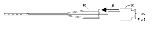
The catheter system of Figures 1 to 6 includes a catheter 10, a needle 20, and
a stylet
30.
The catheter 10 includes an elongate thin walled tubular body 11 formed of
suitably
pliable polymer material. The distal (or forward) end of the body 11
terminates at simple
open end 12. The other (or proximal or rearward) end of the body 11 terminates
in a
rearwardly flared, connector receiving, portion 14. In this example, the
connector
receiving portion is integrally formed with the body 11. Preferably, the
portion 14 is a
female luer connection.
To suit nerve blocking applications the catheter preferably has an outer
diameter of 16g
to 20g. Arterial line catheters are preferably 18g to 22g. In this example,
the catheter
CA 02912150 2015-11-10
P1135PCAUV4
11
has an outer diameter of 20g (0.902mm, 0.0355in) and is dimensioned to
accommodate
a 22g needle.= To aid in insertion, the outer diameter may be conically
tapered at the tip
12 to define a lead-in.
The needle 20 includes a needle body 21. The body 21 is a straight cylindrical
tube of
rigid metallic construction. In this example, the needle 20 is a short beveled
nerve block
needle. The distal end of the body 21 terminates at a penetrating tip 22,
which in this
example includes a single oblique planar face the edges of which are sharp.
The body 21 is receivable within the catheter 10 such that the catheter 10 is
externally
carried by the needle 20. The catheter 10 is fitted to the needle 20 in the
manner of a
sleeve. In particular, the portion 11 forms a close fitting sleeve about the
body 21 and is
supported by the body 21 during insertion into the patient.
The proximal end of the body 21 is rigidly mounted within a connector 23. The
connector
23 includes a forwardly projecting cylindrical boss 24. The boss 24 is
concentric with the
body 21 and dimensioned to be snugly received within, and to sealingly engage,
the
connector receiving portion 14 of the catheter 10 to resist inadvertent
separation of the
needle 20 from the catheter 10.
A rearward end of the needle 20, or more specifically its connector 23,
terminates in a
port 25 co-operable with a tube to fluidly connect to the tube with an
interior of the
needle 21. In an alternate construction, a tube may be directly connected to
the needle
21 during manufacture.
The stylet 30 includes an elongate portion in the form of a long cylindrical
solid rod 31
formed of incompressible, non-porous, semi-rigid plastic or metal. For the
avoidance of
doubt, "rigid" and similar terms as used herein refer to material which does
not deform
appreciably in use, and "semi-rigid" is used in contrast to "freely pliable"
to refer to
materials which deform, but offer appreciable resistance thereto, in use.
CA 02912150 2015-11-10
P1135PCAUV4
12
The distal end of the rod 31 terminates in a shaped formation 32. The shaped
formation
32 is integrally formed with the rod 31 but is distinct therefrom in that it
has an
appreciably different shape. The shaped formation 32 is a bulbous portion
having a
cylindrical exterior shaped to closely fit within the cylindrical interior of
the catheter 10 (or
more specifically its body 11). In this example, the formation 32 has a dome
shaped
rounded leading end.
The proximal end of the rod 31 is rigidly mounted within a connector 33. The
connector
33 is preferably a female to male luer handle and includes a forwardly
projecting
conically tapered cylindrical boss 34 dimensioned to snugly fit within the
connector
receiving portion 14 to sealingly engage the catheter 10 and to resist
inadvertent
separation of the connector 33 and the catheter 10. The catheter 10 has a luer
lock or
similar mechanism to engage the stylet (i.e. the stylet screws into place with
threads).
The boss 34 is a male connector end.
The stylet, or more specifically its connector 33, rearwardly terminates in a
flow port 35
co-operable with a tube to connect the stylet to a fluid source or fluid
destination. In an
alternate construction, a tube may be directly connected to the stylet during
manufacture. The boss 34 is a tubular boss defining an outlet 36 at its
forward end.
Figure 7 schematically illustrates the catheter 10 and stylet 30 inserted into
a patient 40
to perform a fascial plane nerve block called a transversus abdominis plane
block. As
illustrated the patient 40 includes (in order in an inwards direction) skin
layer 41, external
oblique layer 42, internal oblique layer 43, fascia! plane 44 and transversus
abdominis
45. The fascia! plane 44 separates the internal oblique and the transversus
abdominis
and it is within this plane that the nerves lie.
To insert the catheter 10 and stylet 30, first the catheter 10 is inserted.
The catheter 10
is fitted to the needle 20 as in Figure 1 and manipulated to drive the
piercing tip 22 and
to manoeuvre the tip 22 and the distal end portions of the catheter 10 and
needle 20 into
the fascial plane. Positioning of the needle may be guided by electrical
stimulation
CA 02912150 2015-11-10
P1135PCAUV4
13
and/or ultrasound. By way of example, a voltage may be applied to the needle
body 21
so that when its tip 22 (uninsulated by the catheter 10) acts on the nerves
the patient
observably twitches.
The configuration of the piercing tip 22 may be application dependent. Needles
for
arterial puncture are usually sharper (than needles for nerve blocks) having a
Quinke tip
or other tip more suited to vascular access.
Once the subassembly 10, 20 is appropriately positioned, the needle 20 is
withdrawn as
suggested by arrow A in Figure 2. The stylet 30 is then inserted into the
catheter 10. The
rounded leading end of the stylet 30 and the tapered interior of the portion
14 guides the
stylet into the catheter 10, or more specifically its body 11.
In this example, the shaped formation 32 is dimensioned for a snug receipt
within the
body 11 so as to substantially occlude the body 11, although obstruction less
than
substantial occlusion would still be useful.
The catheter 10 and the stylet 30 are co-operably configured whereby when the
stylet
30 is fully advanced the shaped portion 32 is brought into register with the
open end 12
and the boss 34 sealingly engages the connector receiving portion 14. The
cylindrical
exterior of the rod 31 is of lesser diameter than the nominally cylindrical
interior of the
body 11 whereby a nominally annular void is defined between the exterior of
the rod 31
and the interior of the body 11. Of course, given that the body 11 is
flexible, this
nominally annular void would in fact vary in shape along its length, and of
course the
portions of the void defined within the flared connector receiving portion 14
are larger
than the void portions defined within the body 11.
The connector 33 defines one or more flow paths communicating the port 35 with
the
outlet 36 to fluidly communicate the port 35 with the nominally annular void.
Once the subassembly 10, 30 is in situ as in Figure 7 the flexible tube of a
fluid source
comprising the tube and a syringe containing anaesthetic is fitted to the port
35. By
CA 02912150 2015-11-10
P1 1 35PCA UV4
14
advancing the plunger of the syringe fluid is driven through the connector 33
as
suggested by arrow C to emerge from the connector 33 via the outlets 36 into
the
nominally annular void as suggested by arrows D. The fluid is in turn conveyed
along
the voids to the side openings 13 to emerge therefrom at respective sites
within the
fascial plane 44 or next to a nerve as suggested by arrows E.
The shaped formation 32 obstructs the open free end 12 of the catheter 10 and
so
promotes fluid flow through the side openings 13. In this example, the shaped
formation
32 substantially occludes the open free end at 12 and causes outward flow
through the
side openings 13.
The described catheter over needle mode of insertion has been found to lead to
more
secure embedding of the catheter 10 within the patient (i.e. to have higher
resistance to
inadvertent withdrawal of the catheter) than conventional catheter through
needle
techniques.
The use of a stylet as described serves to not only reinforce the catheter 10
against
kinking but also gives the anaesthetist a degree of control over the delivery
pattern of
anaesthetic within the patient. By way of example, the catheter 10 once
inserted may be
used without a stylet, i.e. a fluid source may be directly connected to the
connector
receiving portion 14. By operating the catheter 10 without the stylet 30 a
high proportion
of the supplied anaesthetic would be delivered to the patient via the open
free end 12 of
the catheter thus appreciably varying the delivery pattern. In other variants
of the
disclosed system and method, the delivery pattern may be varied by varying the
location
of the shaped portion 32 relative to the openings 12, 13. For example, in the
illustrated
variant, when the shaped portion 32 is inserted as illustrated, the side
openings 13 are
selected for fluid delivery and the open end 12 is deselected for fluid
delivery. By more
proximally positioning the shaped portion 32 distal ones of the openings 13
can be
deselected. By way of example, if the shaped portion 32 were positioned half
way along
the group of four openings 13 only the proximal two of the openings 13 would
be
selected to convey fluid.
CA 02912150 2015-11-10
P11 35PCA UV4
To so vary the position of the shaped portion 32 a set of stylets of varying
length may be
provided. Preferably each stylet of the set is individually packaged so that a
selected
one of the stylets can be used whilst the others remain sterile.
Alternatively, the rod 31
may be mounted to slide through the connector 33 so that the spacing of the
portion 32
5 from the connector 33 may be varied.
Alternatively the portion 32 may be advanced beyond the open end 12 to open
the end
12 to flow.
Figure 8 schematically illustrates a catheter 110 and stylet 130, both parts
of an arterial
catheter system, in situ in an artery 146. The stylet 130 includes a connector
(not
10 shown) akin to the connector 33 to seal the arterial catheter 110 and
allow fluid to flow
up and down the arterial catheter. By way of example, the catheter may be used
to
deliver medicament, draw a blood sample, or simply for beat-to-beat blood
pressure
measurement.
As the skilled person will understand from the foregoing, to so monitor blood
pressure,
15 the catheter is connected to a suitable pressure sensor external to the
patient.
Early testing suggests that the use of the stylet whilst monitoring blood
pressure is a
radical improvement over existing stylet-less approaches. In particular, this
testing
suggests:
= the problems associated with the catheter kinking are substantially
avoided;
= the fidelity of the system including a 20g catheter can be maintained by
using a
stylet no bigger than about 0.3mm in diameter along its elongate, void-
defining,
portion;
= the system with the stylet in place allows for a flow rate adequate for
blood
sampling; and
CA 02912150 2015-11-10
P1135PCAUV4
16
= that even stylets with occluding ends can be passed into the catheter
even when
there is considerable pressure and flow out the catheter as the stylet is
placed.
When blood pressure is monitored in this way, fluid may be delivered to the
artery 46 via
the catheter 110 to resist clotting within the catheter. Preferably the fluid
is saline and is
most preferably delivered very slowly, e.g. at about 3mL/hour. Optionally the
flushing
fluid may include an anti-coagulant such as Heparin, although generally this
is not the
preferred approach.
Despite such precautions, build-up in the form of clotting can occur about the
catheter
tip 112. To address this build-up, the stylet 130 includes a distinct shaped
portion 132
and is configured for that shaped portion to extend beyond the open end 112.
The stylet
130 is longer than the catheter 110, long enough (relative to the catheter
110) to so
clean the open end of the catheter whilst a portion of the stylet remains
external the
patient to be manipulated by hand. By periodically withdrawing the stylet 130,
the
shaped portion 132 is moved to act upon any such build-up causing it to break
up and
safely move away along the artery before it occludes the catheter 110 or grows
to a
dangerous size. Optionally, the stylet 130 may be fully withdrawn and replaced
by
another stylet. So replacing the stylet refreshes the interior of the catheter
lumen.
The stylet might be so manipulated and replaced periodically, e.g. daily. For
this
purpose, separately packaged stylets and kits of multiple (potentially
identical) stylets
are contemplated. By way of example, a catheter system may include a kit made
up of
three individually packaged stylets to be used over the three days following
the initial
insertion of the catheter.
To emphasise, whilst the exemplary stylet 130 is longer than the catheter 110
and
includes a bulbous end portion 132 to clean the catheter's open end, other
useful
variants are possible. By way of example:
= shorter stylets may be used to clean the interior of the catheter lumen;
CA 02912150 2015-11-10
P1135PCAUV4
17
= a stylet without a bulbous end portion may be manipulated to remove build-
up;
and
= similar modes of cleaning may be applied to catheters having closed ends.
About 0.3mm diameter is thought to be a practical minimum diameter for the
elongate,
void-defining, portion of stylets formed of plastics. Smaller diameters are
thought to be
practical in metallic stylets.
Smaller diameters present less restriction to flow, which for example can lead
to
improved blood pressure measurement. On the other hand, smaller diameters are
more
fragile. Hence the optimum diameter will depend on the application and in
particular on
the strength and flow rate requirements of the application. Generally
speaking:
= longer stylets must be thicker to be sufficiently robust; and
= the fluid path for nerve block applications can be a lot smaller than for
arterial
blood pressure measurement.
For longer nerve block applications (such as the transversus abdominis block),
the stylet
could be up to lmm or so in external diameter to fit down a 16g to 18g
catheter.
The rearward portion 137 of the shaped formation 132 rearwardly converges to
define a
lead-in surface to guide the shaped formation 132 back into the catheter 110
after it has
been extended beyond the open end of the catheter 110. In this example, the
bulbous
end formation 132 has a continuous smoothly curved exterior to minimise build-
up on
the formation 132.
The described catheter systems may be installed and removed from the patient
during a
single attendance to the patient, although it is preferred that the catheter
remain in place
for ongoing use during a lengthy surgery, or even during the patient's entire
hospital
stay. By way of example, the described arterial catheter 110 and its stylet
130 may be
CA 02912150 2015-11-10
P1135PCAUV4
18
left in place to provide continuous monitoring of arterial blood pressure over
a period of
days, or if ongoing monitoring is not required the connection arrangement (not
shown)
external the patient may simply be capped.
The installed components 110, 130 of the disclosed catheter system may
advantageously be left in place well beyond their initial installation. By way
of example,
the catheter may be left in place and then removed during a subsequent
attendance to
the patient prior to leaving hospital, or even removed by the patient after
leaving the
hospital.
The stylet may be produced separately, e.g. to suit existing catheters.
EXPERIMENTAL VALIDATION
The present inventor has tested various catheter systems to address the
following
questions:
1. How may the stylet affect the ease of injection/aspiration through the
combined
stylet/catheter system (for bolus and infusion in nerve block applications and
blood
sampling in arterial line applications)?
2. How does the stylet damp the arterial pressure waveform, and can it reduce
the
damping effect of kinking on the measured waveform? What is the optimal size
of stylet
to minimise damping and maintain kink resistance?
3. Is it feasible to place the arterial stylet down the catheter when arterial
blood
pressure is resisting passage of the stylet?
Method and Apparatus
In view of the movement of blood during blood pressure measurement noted
above, the
present inventor recognised that it was important to test the catheter systems
with a fluid
CA 02912150 2015-11-10
P1135PCAUV4
19
that has a similar viscosity to blood, so that the resistance of the fluid
moving in and out
the end of the system is similar to how it would behave in the artery and
bloodstream of
a patient.
The viscosity of blood is typically between three to four times that of water,
depending
on the concentration of blood factors, particularly the concentration of red
cells. To
simulate blood, a solution of 30% sucrose, dyed with methylene blue to make it
easily
identifiable, was prepared. According to standard tables, this solution has a
viscosity of
3.19 times that of water at 20 degrees Celsius.
An apparatus was constructed to produce the required pressure for testing of
flow and to
create a pressure waveform to simulate the arterial pulse. The "arterial"
pulse was then
measured simultaneously through the test and control catheters. The pressure
generating system included a variable speed electric motor connected to a cam.
Rotation of the cam was translated through a flexible lever into pressure on
and
movement of the barrel of a syringe. The syringe was filled with the sucrose
solution
which was transmitted via a tube to the testing chamber in which the ends of
the test
catheters were placed via sealing rubber membranes. The test catheters were
further
stabilised in position with epoxy resin to prevent inadvertent withdrawal. The
catheters
were positioned within the sucrose solution which was flushed between each
experimental run to ensure it was not contaminated by the 0.9% NaCI (normal
saline)
flush solution from the pressure measuring system. Connected to the pressure
generating syringe was a vertically mounted adjustable syringe on a 3-way
connector,
which, by allowing the sucrose solution to enter and compress the air in the
syringe,
acted to increase and decrease the pulse pressure generated by changing the
compliance of the system.
The pressure measuring system consisted of two commercially available pressure
measuring transducers (Edwards Laboratories), the whole system as essentially
used
for clinical measurement of blood pressures including the flush of low volumes
of normal
saline through a slow flush valve. The transducers were mounted on a board
with 3-way
CA 02912150 2015-11-10
P1135PCAUV4
connectors and tubing between the transducers to enable the simple switching
of the
pressure tubing from connection with one transducer to the other without the
physical
disconnection and reconnection of tubing. This crossover allowed the pressure
from
each catheter to be alternately measured by the alternative transducer. The
signal from
5 the transducers was displayed on a standard clinical monitor using
simultaneous
waveforms in different colours. The numerical readout gave the values of peak
(systolic),
trough (diastolic) and mean pressure after a short averaging interval. The
pressure
waveforms and measurements were recorded by digital photography for analysis.
After
flushing the system to remove air, the transducer flush valve was sealed off
by closing a
10 3-way tap to prevent any fluid loss through back pressure on the valve
or flush through
and contamination of the sucrose solution with normal saline.
The ideal pressure transduction system has characteristics of low compliance
to
minimise movement of blood and normal saline back and forth within the system,
as well
as a high natural resonant frequency to enable accurate measurement of rapidly
15 changing pressures. These physical characteristics of the transducer and
catheter
systems mean they are most accurate measuring small pressure changes at lower
frequencies of change.
The major concern of adding the stylet into the system is that it may impair
the
movement of fluid within the measuring system, causing damping of the trace.
It was
20 therefore decided to test the operation of a variety of sizes of stylets
at high frequencies
and with relatively large pressure changes to accentuate the effect of any
reduction in
fidelity. The motor was therefore adjusted to produce a pressure wave (pulse)
frequency
of 120 to 150 (normal 80-100/min) per minute and the compliance adjusted to a
pulse
pressure of 90-120mm Hg (normal 40-80).
Prototype stylets were prepared using commercially available nylon line of
diameter
between 0.17 and 0.6mm (Maxima GmbH, Geretsried Germany) mounted into a luer
lock fitting male/female combi-stopper (Braun) with a drilled fluid bypass
hole of 1.5mm
diameter, that diameter selected to be a non-limiting part of the pressure and
fluid
CA 02912150 2015-11-10
P1135PCAUV4
21
channel compared to the 0.7mm diameter of the 20g catheter. An occluding bulb
was
created when needed using a small amount of resin on the end of the prototype
stylet.
Investigations included
1. Fluid flow test, measurement of passive flow through the catheter stylet
system to
simulate blood sampling via the catheter and nerve block injections. A 10m1
syringe was
placed vertically connected to a 3-way tap at the hub of the test catheter.
The pressure
was adjusted to 80mm Hg to approximate normal mean arterial pressure and the
time to
fill the syringe to the 5m1 mark was recorded.
2. Damping and kink resistance test. Assessment of the optimal size of
stylet was
done by measuring the damping effect of stylets of various sizes on the
measurement of
simulated arterial pressure. 20g Optiva TM and 20g ArrowTM catheters were
tested with a
variety of nylon stylets from 0.17mm to 0.6mm diameter. The test catheter and
stylet
was progressively kinked from straight (0 degrees) through a right angle (90
degrees) to
kinked as much as possible (180 degrees). Figure 10 illustrates an Optiva TM
catheter
with a stylet kinked to 180 degrees (at a point suggested by the black arrow).
Figure 10 also shows the test apparatus including a rudimentary protractor
with a nail
adjacent its point of convergence. The nail serves as a stop to assist in
manipulating the
catheter system.
Simultaneous pressure readings from the test and control catheters (i.e.
catheters
without any stylet in place) were obtained using separate transducers and
displayed on
a monitor screen. The transducers were swapped between the control and the
stylet
containing catheters and then the result averaged. An average percent
reduction in
pulse pressure was calculated (systolic-diastolic pressure of test catheter
divided by
systolic-diastolic pressure of control catheter * 100) and recorded for each
diameter of
stylet.
CA 02912150 2015-11-10
P1135PCAUV4
22
3. Insertion test. For ease of use, the stylet needs to be passed
relatively easily into
the catheter when it is in the artery, against the pressure of blood in the
vessel. This was
assessed using the previously determined optimal sized stylet. A prototype
nylon stylet
was made with a near occluding bulb on the end to just fit through a 20g
catheter. The
pressure in the catheter was adjusted to 200mm Hg by moving the height of the
reservoir of 30% sucrose solution. While the sucrose solution was free flowing
from the
catheter, the nylon stylet with a bulb on the tip was passed into the catheter
and any
difficulty in insertion noted.
Results
1. Fluid flow test
A. 20q OptivaTM catheter
Stylet Time to fill to 5m1
Control no stylet 13.5 sec
0.6mm 68 secs for 1 ml
0.42mm 45 secs
0.37mm 36 secs
0.30mm 24 secs
0.25mm 26 secs
0.17mm 19 secs
CA 02912150 2015-11-10
P1135PCAUV4
23
B. 20g ArrOWTM Catheter
Stylet Time to fill to 5m1
Control no stylet 15 sec
0.6mm 107 secs for 1 ml
0.42mm 50 secs
0.37mm 37 secs
0.30mm 32 secs
0.25mm 28 secs
0.17mm 24 secs
2. Damping and kink resistance test
Figures 11 and 12 chart the reduction in the measured pulse pressure (as a
percentage
of the pulse pressure measured using the control catheter) plotted on the
vertical axis as
a function of the angle to which the catheter is kinked on the horizontal axis
(from 0
degrees to, fully kinked, 180 degrees). The legends indicate stylet diameters
from nil (no
stylet) to 0.6mm
Figures 13 to 17 show measured pressure waves obtained using 20g Optiva TM
1 0 catheters. Each of these Figures compares the pressure wave (shown in
solid line)
obtained from a test catheter to the pressure wave (shown in dotted line)
obtained from
an unkinked stylet-less control catheter.
CA 02912150 2015-11-10
P1 1 3 SPCA UV4
24
In Figure 13 the catheters are straight (i.e. the angle of kinking is 0
degrees). The test.
catheter is not fitted with a stylet and is thus nominally identical to the
control catheter.
Thus, Figure 13 gives an indication of the expected degree of experimental
error. Figure
14 shows the result of the same stylet-less test catheter being kinked to 120
degrees:
the pressure wave is almost entirely attenuated.
In contrast Figures 15, 16 and 17 respectively show the results of kinking to
0 degrees,
120 degrees and 180 degrees when a 00.3mm stylet is in place. Figure 15 shows
that
when the catheter is straight (as ideally it should be in use), the stylet
causes no
discernible deterioration of the signal.
A comparison of the Figures 14 and 16 demonstrates the benefits of the stylet.
With the
stylet in place, a trace of the pressure wave can be obtained that matches the
trace
obtained from a straight catheter despite a kinking condition (i.e. 120
degrees) at which
the signal from a stylet-less catheter would be almost entirely lost. The
obtained trace
matches the "straight catheter trace" within the degree of experimental error
contemplated in Figure 13.
Figure 17 illustrates that even when the catheter is fully reversed on itself
(i.e. kinked to
180 degrees), a meaningful trace of the pressure wave can be obtained.
In the 20g OptivaTM catheter, all the stylets of 0.42mm diameter and less
performed
almost identically. In the 20g AIIOWTM catheter, the 0.42mm stylet was also
damped
relative to the smaller stylets. The free flow test showed that all the
stylets slowed flow;
in both catheters the 0.37mm stylet slowed the flow by approximately a third
and the
0.3mm stylet slowed the flow by approximately half. Both these outcomes would
be
clinically acceptable, although the less reduction in flow the better.
For both the pressure test and the flow test, a smaller stylet is an
advantage, however in
use the kink resistance and placement is likely to be better with the widest
diameter
stylet. The 0.37mm and 0.3mm stylets were the largest stylets that did not
show
significant damping on the blood pressure transduction test and both were
similar in the
=
CA 02912150 2015-11-10
P1135PCAUV4
test, kinking up to 180 degrees. Of these, the 0.3mm stylet is preferred
because it is
smaller and will be less likely to obstruct the catheter in clinical
conditions.
3. Insertion test
The 0.3mm stylet was modified with a small bulb of glue on the end to create a
near
5 occluding end then tested in the 20g OptivaTM catheter. The sucrose
solution was placed
at a height of 220cm which creates a driving pressure of approximately 175mm
Hg. The
sucrose solution was allowed to flow freely from the hub of a 20g OptivaTM
catheter
inserted into the pressurised solution. The 0.3mm stylet with bulb was
inserted on the
first attempt into the 20g catheter. Once the bulb was in the catheter lumen
and largely
10 occluding it, the insertion proceeded smoothly.
Discussion
This testing demonstrates the marked effect the intra-catheter stylet has on
pressure
measurement of a simulated arterial pulse via commonly used arterial
catheters. The
area of lumen required for an accurate measurement of blood pressure is likely
to be
15 influenced by the shape of the lumen available. A stylet of 0.6mm
diameter in a 20g
lumen of 0.7mm occupies 73% of the available area while a stylet of 0.55mm
occupies
62%, 0.42mm occupies 36%, 0.37mm occupies 28%, 0.3mm occupies 18%, 0.25mm
occupies 13% and the 0.17mm stylet occupies only 6% of the available area.
The fluid flow test showed that all the stylets of 0.42mm and below showed an
20 acceptable rate of passive flow in these 20g catheters, with greater
flow for the smaller
stylets. Even the smallest stylet of 0.17mm diameter occupying only 6% of the
lumen
reduced flow by approximately a third with the 0.42mm stylets reducing flow by
70%.
These figures would all be acceptable clinically as blood sampling time
usually only
takes a few minutes, mostly in setting up the sampling, labelling and other
tasks, while
25 withdrawing the blood is a relatively small portion of the task.
CA 02912150 2015-11-10
P1135PCAUV4
26
All stylets reduced the damping effect of kinking the catheter, although the
larger stylets
over 0.42mm diameter also produced significant damping by their presence. All
the
stylets of 0.37mm and under performed very similarly in the damping and
kinking test.
The 0.42mm stylet showed a reduction in performance with kinking in the
ArrowTM
catheter, particularly at over 90 degrees of kinking. This may be because the
ArrOWTM
catheter is softer than the OptivaTm-and kinks differently, the introducer
needle is slightly
smaller than the OptivaTM suggesting the lumen may be smaller, and it is
slightly longer
which may accentuate any damping from the stylet. There was no lower limit
seen to the
effect of the stylet maintaining a lumen for pressure transduction during
kinking over the
stylets measured. The stylet may maintain a lumen by resisting kinking and
therefore
maintaining a lumen around the unkinked stylet. This effect was seen for the
biggest
stylets, however they occluded so much of the lumen that the trace was damped
in all
positions. The smaller stylets allowed the catheter to apparently kink in a
similar fashion
to the stylet-less catheter with the opposite walls coming together to occlude
the fluid
path. The position of the stylet between the walls however is thought to have
made a
smaller lumen each side of the stylet where the walls of the catheter were
unable to
oppose. Using the simulated blood in this testing, this reduced lumen remained
adequate for blood pressure measurement with only a small reduction in
measured
pulse pressure even with extreme kinking which would be very valuable
clinically where
the arterial catheter has been kinked for example by movement of the patient.
The practical lower limit for the size of a stylet would be determined by the
stiffness of
the stylet required to insert it into an arterial catheter placed into an
artery. A plain stylet
would be inserted much more easily than one with a bulb on the end, however
the bulb
confers other benefits, particularly the ability to clear debris and clot out
of the lumen of
the catheter on removal. The 0.17mm stylet was very fine and would be
impractical to
insert against blood coming from and arterial cannula in situ unless it was
very stiff,
probably a steel wire. A plastic stylet would need to be larger than a steel
stylet as it is
less rigid, however clinicians would be more likely to accept a plastic stylet
as it would
probably conform better to the patient's anatomy and be less likely to
perforate and
damage arteries, nerves or other structures.
CA 02912150 2015-11-10
P1135PCAUV4
27
The 0.3mm stylet was chosen for the insertion test as a compromise, as the
area of the
stylet is only half that of the 0.42mm stylet with better performance on the
fluid flow,
damping and kinking test. If the plastic was suitably rigid, the stylet size
could be
reduced further. The insertion test showed that the 0.3mm stylet could be
feasibly
inserted into a catheter with free flowing arterial blood under pressure
coming out. In
practice, most clinicians occlude the artery and catheter tip proximally to
prevent the free
flow of blood from the catheter during connection of the transducer line. This
would aid
insertion of the stylet as the only time the stylet would be subject to being
pushed
= against the full pressure of arterial blood is when the bulb reaches the
very end of the
catheter in which position the stylet is maximally supported by the catheter
walls,
enabling greater force to be transmitted to the bulb on the tip.