Note: Descriptions are shown in the official language in which they were submitted.
1
Heart monitoring system
Field of the invention
The present invention relates to monitoring vital signs of a user and
especially to a
system, method and a computer program product for monitoring cardiac operation
of a
subject.
Background of the invention
A heart is a hollow tissue formed of cells that are capable of producing a
contraction that
changes the length and shape of the cell. Heart pumps blood in cyclic
contractions through
a network of arteries and veins called the cardiovascular system. As shown in
Figure 1, a
human heart includes four chambers, which are divided by a septum into a right
side (right
atrium RA and right ventricle RV) and a left side (left atrium LA and left
ventricle LV). During
a heartbeat cycle, the right atrium RA receives blood from the veins and pumps
it into the
right ventricle and the right ventricle RV pumps the blood into the lungs for
oxygenation.
The left atrium LA receives the oxygenated blood from the lungs and pumps it
to the left
ventricle LV, and the left ventricle LV pumps the blood into the veins. The
apex AP of the
heart is a portion formed by the inferolateral part of the left ventricle LV.
Various techniques have been developed to provide measurable parameters that
are
indicative of cardiac operation of a monitored subject. Many of these
techniques are
invasive and therefore suitable for advanced medical use only.
In the noninvasive side, echocardiography is a technique that applies
ultrasound to provide
an image of the heart. Echocardiography can be comfortably carried out at the
bedside,
and it has therefore become a widely-used tool for noninvasive studies on
cardiac
mechanics of diseased and healthy
Date Recue/Date Received 2020-05-12
CA 02922364 2016-02-10
PCT/16 201'4/064 377 - 09-03-2015
2
hearts. The produced images require, however, complex and basically
immobile computer equipment and the images need to be interpreted by a
highly trained physician. Ambulatory or long-term monitoring of the cardiac
operation outside the clinical environment by echocardiography is practically
impossible.
Electrocardiography is based on measuring electrical activity of the heart
with
electrodes attached to the surface of the skin of the monitored subject. In,
electrocardiography, wave depolarization of the heart is detected as changes
of
voltage between a pair of electrodes placed in specific positions on the skin.
Typically a number of electrodes are used, and they are arranged in
combination into pairs (leads). Electrocardiograms are very accurate and
widely used, and also allow some computerized interpretation. Proper
placement of the electrodes may, however, be challenging for users without
medical training. In addition, the measurement system typically requires a
computerized system connected with cables to a plurality of self-adhesive pads
that couple through conducting gel to the skin of the monitored subject.
Moving with such wiring is very limited.
Patent publication W02010145009 discloses an apparatus for determining
information indicative of physiological condition of a subject. The apparatus
comprises a sensor device that obtains ballistocardiograph data indicative of
heart motion of the subject, measured along a plurality of spatial axes.
Ballistocardiograph data indicates the extent of mechanical movements of a
body that take place in response to the myocardial activity of the heart. This
ballistocardiograph data is then used to process data that is indicative of
heart
motion of the subject. This prior art method overcomes some of the limitations
of the prior art. However, it has been noted that the linear measurement along
spatial axes is strongly affected by the posture of the monitored subject
during
the measurement. In addition, some characteristics of the heartbeat cycle are
not completely reliably measurable with the linear motion data.
AMENDED SHEET
3
Brief description of the invention
The object of the present invention is to provide a non-invasive cardiac
operation
monitoring solution where at least one of disadvantages of the prior art is
eliminated or at
least alleviated. The objects of the present invention are achieved with a
system, method
and computer program product.
Due to a specific orientation of the myocardial fibers, in a heartbeat cycle
the heart makes
rotation along its long-axis and a wringing (twisting) motion. Torsional
squeezing and
opening of the left ventricle LV caused by heart rotation stands for about 60%
of the stroke
volume of the heart. The rest may be considered to result from the deflection
of a wall
between the left ventricle LV and the left atrium LA, and from the linear
squeezing of the left
ventricle LV from the apex AP.
The present invention discloses a device that includes a sensor of angular
motion
configured to obtain an angular ballistograph signal indicative of rotational
movement of a
chest of a subject. Signal processing means are configured to generate from
this angular
ballistocardiograph signal measured values of an output parameter, which is
indicative of
cardiac operation of the subject. The generated values or parameters can be
used in a
stand-alone system or in combination to improve signals and/or analysis made
in a system
that applies one or more of the prior art techniques.
The signal of a sensor of angular motion is not affected by gravity, which
makes the
measurement practically independent of the position or posture of the
monitored subject. It
has been noted that the external angular motion of the chest is orders of
magnitude larger
than what one would anticipate from the mere extent of the heart rotation and
the ratio
between the size of the
Date Recue/Date Received 2020-05-12
CA 02922364 2016-02-10
PCT/I8 2014/064 377 - 09-03-2015
4
heart and the diameter of the human chest. It has also been noted that the
detection of the angular motion is also relatively insensitive to the location
of
the sensor in respect to the heart. Due to these aspects, accurate
measurements can be made with even one gyroscope, for example
microelectromechanical gyroscope, attached to the chest of the monitored
subject. Microelectromechanical gyroscopes are accurate, small in size and
commercially well available.
These and further advantages of the invention are discussed in more detail in
the following with detailed descriptions of some embodiments of the invention.
Brief description of the figures
In the following the invention will be described in greater detail, in
connection
with preferred embodiments, with reference to the attached drawings, in which
Figure 1 illustrates elements of a human heart;
Figure 2 illustrates functional elements of an embodiment of a monitoring
system;
Figure 3 illustrates functional configuration of a cardiac monitoring
system;
Figure 4 illustrates another exemplary configuration of a cardiac
monitoring system;
Figure 5 illustrates measurement results taken with the system of Figure
4;
Figure 6 illustrates a remote monitoring system including the cardiac
monitoring system;
Figure 7 illustrates an exemplary angular ballistocardiograph signal during
heartbeat cycles;
Figure 8 shows a simplified example of an angular ballistocardiograph
signal;
AMENDED SHEET
CA 02922364 2016-02-10
PCT/IB 2014/064 377- 09-03-2015
Figure 9 illustrates an exemplary output signal corresponding to the
angular ballistocardiograph signal of Figure 7 after a specific matched
filtering;
Figure 10 illustrates a potential AO peak from the signal of Figure 7; and
5 Figure 11 illustrates exemplary values of stroke volume and heartbeat
timestamps measured from a test subject;
Figure 12 illustrates measurements taken simultaneously from one test
subject with various measurement technologies;
Figure 13 illustrates generation of a parameter indicative of atrial
extrasystole of the subject;
Figure 14 shows exemplary time differences (TD) in a case of atrial
fibrillation of the subject;
Figure 15 illustrates amplitude variation of an exemplifying signal in a case
of atrial fibrillation when a person under consideration is breathing;
Figure 16 illustrates an example of an ECG waveform and an angular
ballistocardiogram waveform of an exemplifying signal indicative of
cardiovascular rotation.
Detailed description of some embodiments
The following embodiments are exemplary. Although the specification may refer
to "an", "one", or "some" embodiment(s), this does not necessarily mean that
each such reference is to the same embodiment(s), or that the feature only
applies to a single embodiment. Single features of different embodiments may
be combined to provide further embodiments.
In the following, features of the invention will be described with a simple
example of a device architecture in which various embodiments of the invention
may be implemented. Only elements relevant for illustrating the embodiments
are described in detail. Various implementations of heart monitoring systems
AMENDED SHEET
CA 02922364 2016-02-10
PCT/IB 2014/064 377 - 09-03-2015
,6
and methods comprise elements that are generally known to a person skilled in
the art and may not be specifically described herein.
The monitoring system according to the invention generates one or more
output values for one or more parameters that are indicative of operation of
the heart of a subject. These values may be used as such or be further
processed to indicate condition of the heart of the subject. The monitoring
system is herein disclosed as applied to a human subject. The invention is,
however, applicable to animal species or any type of subject that has a heart
and a body that responsively encloses the heart such that the heartbeat
results
in recoil motion of the body.
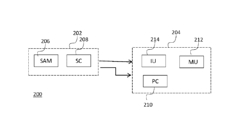
The block chart of Figure 2 illustrates functional elements of an embodiment
of
a monitoring system 200 according to the present Invention. The system
includes a sensor of angular motion configured to obtain an angular
ballistocardiograph signal that is indicative of rotational movement of a
chest of
a subject, and signal processing means configured to generate from the
angular ballistocardiograph signal measured values of an output parameter
that is Indicative of cardiac operation of the subject. These elements may be
implemented as one physical device, for example, a mobile computing device,
like a smartphone, or a tablet. Alternatively, the elements may be included in
two or more electrically or communicatively coupled physical devices of the
system. Figure 2 illustrates an exemplary configuration where the system 200
comprises a sensor unit 202 and a control unit 204. In this example, the
sensor
unit 202 may be considered as an element to be attached to the monitored
subject and the control unit 204 may be considered as an element physically
detached from the monitored subject.
The sensor unit 202 includes a sensor of angular motion 206. The sensor of
angular motion is configured to be attached to the subject to move along
motions of the subject, or part of the subject it is attached to. Rotational
movement or angular motion refers herein to circular movement in which an
AMENDED SHEET
I CA 02922364.2016-02-10
== =
PCT/I8 2014/064 377 - 09-03-2015
7
object progresses in radial orientation to a rotation axis. The sensor of
angular
motion refers here to a functional element that may be exposed to angular
motion of the subject and translate at least one variable of the angular
motion
into an electrical signal. Applicable variables include, for example, position
in
radial orientation, angular velocity and angular acceleration. Rotary motion
of
the heart and the reverse rotary. motion of the surrounding part of the body
of
the subject are oscillatory, so the sensor of angular motion may be configured
to detect both direction and magnitude of an applied variable.
The sensor unit 202 may also include a signal conditioning unit 208 that
manipulates the raw input electrical signal to meet requirements of a next
stage for further processing. Signal conditioning may include, for example,
isolating, filtering, amplifying, and converting a sensor input signal to a
proportional output signal that may be forwarded to another control device or
control system. A signal conditioning unit 208 may also perform some
computation functions such as totalization, integration, pulse-width
modulation,
linearization, and other mathematical operations on a signal. The signal
conditioning unit 208 may alternatively be included in the control unit 204.
The sensor of angular motion is configured to generate a chest motion
signal, an angular ballistocardiograph signal that is indicative of rotational
recoil movement on the chest in response to cardiac operation of the
subject within the chest. Ballistocardiography refers in general to a
technology for measuring movements of a body, which are caused in
response to shifts in the center of the mass of the body during heartbeat
cycles. The chest refers here to a pectoral part of the body in the upper
torso between the neck and the abdomen of the subject. Advantageously,
rotational movement of the chest about an axis parallel to the sagittal
plane of the subject is measured. However, other axes may be applied
within the scope, as well.
The sensor of angular motion 206 may be attached in a desired position
and orientation to the exterior of the chest of the subject with a fastening
AMENDED SHEET
CA 02922364 2016-02-10
PCT/IB 2014/064 377 - 09-03-2015
8
element such that when the underlying part of the chest moves, the
sensor moves accordingly. The fastening element refers here to mechanical
means that may be applied to position the sensor of angular motion 206
into contact with the outer surface of the skin of the user. The fastening
element may be implemented, for example, with an elastic or adjustable
strap. The sensor of angular motion 206 and any electrical wiring required
by its electrical connections may be attached or integrated to the strap.
Other fastening mechanisms may be applied, as well. For example the
fastening element may comprise one or more easily removable adhesive
bands to attach the sensor of angular motion 206 on the skin in the chest
area. Rotational movement of the chest of the subject may alternatively
be detected with a sensor of angular motion coupled to a position in any
other part of the upper torso of the subject. For example, a position in the
backside of the upper torso of the subject may be applied for the purpose.
Such sensor configuration allows measurements without specific fastening
elements. For example, the sensor unit may be incorporated into an
underlay, like a mattress on which the monitored subject may lie without
additional straps and tapes.
A sensor of angular motion typically has a sense direction, which means
that it is configured to sense angular motion about a specific axis of
rotation. This axis of rotation defines the sense direction of the sensor of
angular motion.
It is known that microelectromechanical (MEMS) structures can be applied to
quickly and accurately detect very small changes in physical properties. A
microelectromechanical gyroscope can be applied to quickly and accurately
detect very small angular displacements. Motion has six degrees of freedom:
translations in three orthogonal directions and rotations around three
orthogonal axes. The latter three may be measured by an angular rate sensor,
also known as a gyroscope. MEMS gyroscopes use the Coriolis Effect to
measure the angular rate. When a mass is moving in one direction and
AMENDED SHEET
CA 02922364 2016-02-10
PCT/IB 2014/064 377 - 09-03-2015
9
rotational angular velocity is applied, the mass experiences a force in
orthogonal direction as a result of the Coriolis force. The resulting physical
displacement caused by the Coriolis force may then be read from, for example,
a capacitively, piezoelectrically or piezoresistively sensing structure.
In MEMS gyroscopes the primary motion is typically not continuous rotation as
in conventional ones due to lack of adequate bearings. Instead, mechanical
oscillation may be used as the primary motion. When an oscillating gyroscope
is subjected to an angular motion orthogonal to the direction of the primary
motion, an undulating Coriolis force results. This creates a secondary
oscillation
orthogonal to the primary motion and to the axis of the angular motion, and at
the frequency of the primary oscillation. The amplitude of this coupled
oscillation can be used as the measure of the angular motion.
Being based on Coriolis force, the detected signal of a gyroscope is minimally
affected by gravity. This makes gyrocardiograms far more insensitive to
posture of the monitored subject than, for example, seismocardiograms. The
subject may then freely select a comfortable position for taking a cardiogram
measurement, or to some extent even move during the measurement.
During measurement the position of the sensor should optimally be as
close to the heart as possible and the orientation of the sensor should be
such that the sense direction is aligned as accurately to the axis of
rotation of the body of the subject as possible. In a human subject, axes
parallel to the sagittal plane that passes from ventral to dorsal, and
divides the body into halves may be applied. These requirements for
sensor positioning are easy to understand and implement. The tolerances
in positioning are, in addition, reasonable, which enables fastening of the
sensor unit in, for example, ambulatory environment or by people with
less or no medical training.
Cardiac function typically includes various ventricular directional motions
of narrowing shortening, lengthening, widening and twisting. Despite this
directionality, it has been detected that the recoil effect is relatively
AMENDED SHEET
CA 02922364 2016-02-10
*
PCT/113 2014/064 -
09-03-2015
insensitive to the position and orientation of the sensor unit. One reason
for relative insensitivity to deviations in the orientation is that in theory
the error is proportional to cosine of an angle between the sense direction
of the sensor and a rotation axis of the rotary oscillation of the heart. It
is
5 known that in the neighborhood of zero, cosine is a slowly decreasing
function. One reason for relative insensitivity to position of the sensor is
that different parts of the heart couple differently to the surrounding,
mostly liquid tissue. In addition, a volume of blood flowing into the aorta
contributes to the detected recoil motion of the chest. The inertial
10 volumes beyond the extent of the heart muscle itself balance the recoil
effect such that reasonable deviations in the position and orientation of
the sensor unit can be tolerated. In addition, the detected motion is larger
and thereby provides relatively easily detectable large signals.
The control unit 204 is communicatively coupled to the sensor unit to
input signals generated by the sensor of angular motion for further
processing. Typically the coupling is electrical, allowing both power supply
to the sensor unit, as well as wireline exchange of signals between the
sensor unit and the control unit. The sensor unit may, however, be a
standalone unit with own power supply, and radio interface to the control
unit. On the other hand, the sensor unit and control unit may be
implemented as one integrated physical device.
The control unit 204 is a device that may comprise a processing component
210. The processing component 210 is a combination of one or more
computing devices for performing systematic execution of operations upon
predefined data. The processing component may comprise one or more
arithmetic logic units, a number of special registers and control circuits.
The
processing component may comprise or may be connected to a memory unit
212 that provides a data medium where computer-readable data or
programs, or user data can be stored. The memory unit may comprise one
or more units of volatile or non-volatile memory, for example EEPROM, ROM,
PROM, RAM, DRAM, SRAM, firmware, programmable logic, etc.
AMENDED SHEET
CA 02922364 2016-02-10
PCT/18 2014/064 377 - 09-03-2015
11
The control unit 204 may also comprise, or be connected to an interface
unit 214 that comprises at least one input unit for inputting data to the
internal processes of the control unit, and at least one output unit for
outputting data from the internal processes of the control unit.
If a line interface is applied, the interface unit 214 typically comprises
plug-
in units acting as a gateway for information delivered to its external
connection points and for information fed to the lines connected to its
external connection points. If a radio interface is applied, the interface
unit
214 typically comprises a radio transceiver unit, which includes a transmitter
and a receiver. A transmitter of the radio transceiver unit may receive a
bitstream from the processing component 210, and convert it to a radio
signal for transmission by an antenna. Correspondingly, the radio signals
received by the antenna may be led to a receiver of the radio transceiver
unit, which converts the radio signal into a bitstream that is forwarded for
further processing to the processing component 210. Different line or radio
interfaces may be implemented in one interface unit.
The interface unit 214 may also comprise a user interface with a keypad, a
touch screen, a microphone, or equals for inputting data and a screen, a
touch screen, a loudspeaker, or equals for outputting data to a user of the
device.
The processing component 210 and the interface unit 214 are electrically
interconnected to provide means for performing systematic execution of
operations on the received and/or stored data according to predefined,
essentially programmed processes. These operations comprise the
procedures described herein for the control unit of the monitoring system of
Figure 2.
Figure 3 illustrates functional configuration of a cardiac monitoring system
200 that includes the sensor unit 202 and the control unit 204 of Figure 2.
The sensor unit, attached to the chest of the monitored subject is exposed
to temporary angular motion AMehest of the chest, and undergoes a
AMENDED SHEET
CA 02922364 2016-02-10
. =
PCT/16 2014/064 377 - 09-03-2015
12
corresponding motion am(t). In response to the angular motion am(t), the
sensor unit generates an angular ballistocardiograph signal Sam and
forwards it to the control unit. The control unit includes one or more data
processing functions F1, 12, F3, each of which defines a rule or
correspondence between values of the angular ballistocardiograph signal
Sam and values of output parameters pl, p2, p3 that are indicative of
operational parameters of the heart of the subject. The control unit may
store one or more of these output parameters pl, p2, p3 to a local data
storage for later processing, output one or more of them in one or more
media forms through the user interface of the control unit, or transmit one
or more of them to a remote node for further processing.
Figure 4 illustrates another exemplary configuration where the system
400 is a mobile computing device, a smartphone that incorporates both
the sensor unit and the control unit. Many of the advanced mobile
computing devices today include a gyroscope apparatus, often a multi-
axial gyroscope able to sense angular motion in various directions. The =
signal or signals from the internal gyroscope apparatus may be available,
for example through an application programming interface (API) of the
operating system. An application may be configured to use the gyroscope
signals and the computing means of the mobile computing device, and
thereby form the claimed system. The advantage of using a mobile
computing device system is that the monitoring can be made with a non-
dedicated device, typically available to the user in any case. The user can
easily use, for example, a smartphone to take his/her own
gyrocardiogram to, for example, measure heart rates, detect atrial
fibrillation etc. Furthermore, processing, memory and interface means of
the mobile computing device allow measured data to be stored,
preprocessed or processed locally in the mobile computing device, and/or
to be transmitted to a remote location for further processing, or to be
analyzed, for example by a physician.
AMENDED SHEET
CA 02922364 2016-02-10
PCT/IB 2014/064 377 - 09-03-2015
13
As will be discussed in more detail later on, in monitoring systems the
gyroscope signal may be used in combination with other signal types. The
mobile computing device of Figure 4 may be equipped with, for example,
an ECG monitoring capability by integrating ECG electrodes into a casing
the mobile computing device. Such configuration enables one to combine
ECG and gyroscope signals to determine, for example, cardiac time
intervals.
As illustrated in Figure 4, the mobile computing device 400 may also be
connected with other apparatuses, such as a wrist-type heart rate monitor
402 (smartwatch or similar) or a set of one or two headphones 404
capable of measuring heart rates. The use of signals from two
measurement points makes it possible to determine a pulse (arterial
pressure pulse) transit time from the heart to some specific position, in
these exemplary cases to the wrist or to the ear. When the distance
between these two measurement positions is known, the pulse transit
time can be used to measure various physiological parameters, such as
blood pressure and arterial resistance.
Figure 5 illustrates measurement results taken with the system of Figure
4, i.e. with a smartphone attached to the chest of the user. The
smartphone includes also a multi-axial accelerometer, and curves AccX,
AccY, AccZ represent X- Y- and Z-direction signals from the linear
accelerometer. Curves GyroX, GyroY, GyroZ representangular motion
signals about X-, Y-, and Z-direction aces from a gyroscope apparatus
within the same smartphone. It may be seen that the output signal of the
multi-axial gyroscope is more clear-cut and thus suitable for accurate
analysis than the fuzzy output signal of the multi-axial accelerometer.
Figure 6 illustrates a remote monitoring system including the cardiac
monitoring system of Figure 2. The system may Include a local node 600
that comprises the sensor unit 202 and the control unit 204 of Figure 2. In
AMENDED SHEET
CA 02922364 2016-02-10
PCT/IB 2014/064 377 - 09-03-2015
14
addition, the local node 600 may be communicatively connected to a
remote node 602. The remote node 602 may be, for example, an
application server that provides a monitoring application as a service to
one or more users. One of the aspects monitored with the application may
be the state of the heart of the user. Alternatively, the remote node may
be a personal computing device into which a heart monitoring application
has been installed. The local node may be a dedicated device or
combination of devices including the sensor unit and the control unit
described above. Alternatively, the local node may be implemented as a
sensor unit that interfaces a client application in a multipurpose computer
device (for example a mobile phone, a portable computing device, or
network terminal of a user). A client application in the computer device
may interface the sensor unit and a server application. The server
application may be available in a physical remote node 602, or in a cloud
of remote nodes accessible through a communication network.
While various aspects of the invention may be illustrated and described as
block diagrams, message flow diagrams, flow charts and logic flow diagrams,
or using some other pictorial representation, it is well understood that the
illustrated units, blocks, apparatus, system elements, procedures and
methods may be implemented in, for example, hardware, software,
firmware, special purpose circuits or logic, a computing device or some
combination thereof. Software routines, which may also be called as
program products, are articles of manufacture and can be stored in any
apparatus-readable data storage medium, and they include program
instructions to perform particular predefined tasks. Accordingly,
embodiments of this invention also provide a computer program product,
readable by a computer and encoding instructions for monitoring cardiac
operations of a subject in a device or a system of Figures 2, 3, 4 or 5.
The sensor of angular motion is advantageously a microelectromechanical
device, but other angular motion detection technologies may be applied, as
well. For example, a magnetometer attached to the chest of the subject may
AMENDED SHEET
CA 02922364 2016-02-10
PCT/IB 2014/064 377 - 09-03-2015
be used to determine the change of position of the chest in relation to the
earth's magnetic field.
Noise and other unwanted features may be removed from the raw angular
ballistocardiograph signal Sam with analog or digital filters. A low pass,
5 high pass or band pass filter may be applied. For example, after
converting the analog signal to digital form, a digital low pass filter of the
form
y(t)=(1-k)*y(t-1)+k*x(t) (1)
10 where
y(t) = value of the filtered signal at time step t,
y(t-1) = value of the filtered signal at time step (t-1),
x = value of the unfiltered signal at time step t,
k = filter coefficient,
15 may be applied for the purpose. The filtering may also or alternatively
apply polynomial fitting, for example convolution with a Savitzky-Golay
filter.
The curve of Figure 7 illustrates an exemplary filtered angular
ballistocardiograph signal Sam during heartbeat cycles of a test subject.
The vertical axis represents the magnitude of sensed angular rate in the
specific sense direction, and the horizontal axis represents accumulated
number of time steps or elapsed time. Signal to noise ratio may be
enhanced by means of matched filtering, where the filtered signal is
correlated to a predefined template. The heart motion may be
approximated to constitute a reciprocating motion where the heart twists
in a first direction (here: positive twist), and in an opposite second
direction (here: negative twist). The template may comprise a set of one
or more limits for characteristics of the signal, for example specific
amplitude, time domain feature or frequency domain feature.
AMENDED SHEET
CA 02922364 2016-02-10
PCT/113 2014/064 377 - 09-03-2015
16
As a simple example, matched filtering of the angular ballistocardiograph
signal Sam of Figure 7 may be done by means of signal extreme
(minimum/maximum) values. Figure 8 shows a simplified example of an
angular ballistocardiograph signal Sam. For example, the control unit may
be configured to determine consecutive maximum and minimum values
mxl, mn1, mx2, mn2, mx3, mn3, and determine slopes sl, s2,
between them, as shown in Figure 6.
s1=mx1-mn1
s2= mx2-m n1
s3=mx2-mn2
s4=mx3-mn2
etc.
The matched filtering template may include one or more limits, for
example, to maximum values, minimum values, the values of individual
slopes, or to a combination of slopes. Figure 9 illustrates an exemplary
output signal corresponding to the angular ballistocardiograph signal Sam
of Figure 7 after a specific matched filtering, which will be discussed in
more detail later on.
The control unit may be configured to generate various output parameters.
In the simplest form, a parameter may be indicative of radial orientation of
the heart, angular velocity of the heart, or angular acceleration of the heart
during the twisting motion. This output parameter may correspond to a
measured, conditioned, and filtered angular ballistocardiograph signal Sam
shown in Figure 7 or 9.
Alternatively, or additionally, a parameter may be indicative of the stroke
volume of the heart of the subject. The output parameter may be generated
by determining amplitude of the angular ballistocardiograph signal Sam and
using that as a value to represent the temporal stroke volume. For
example, a peak amplitude, semi-amplitude, or root mean square
amplitude may be used for the purpose. Since the signal is not a pure
AMENDED SHEET
CA 02922364 2016-02-10
PCT/IB 2014/064 377 - 09-03-2015
17 =
symmetric periodic wave, amplitude is advantageously measured in
respect to a defined reference value, for example, from a zero point of the
signal curve. Other reference values may be applied within the scope, as
well.
Alternatively, or additionally, a parameter may be indicative of the
heartbeat of the subject. For example, the output parameter may be
generated by selecting a characteristic point of the angular
ballistocardiograph signal Sam and determining the occurrence of the
characteristic point in consecutive signal sequences. A minimum or =
maximum value of the signal sequence may be applied as the
characteristic point. The occurrence of the characteristic point may be
considered as a time stamp of the heartbeat. A period between two
timestamps may be considered to represent temporary beat-to-beat (B-B)
time of the heart of the subject. The number of timestamps within a
defined period may be applied to indicate heart rate (HR) of the subject.
Alternatively, or additionally, a parameter may be indicative of aortic
opening or closing of the heart of the subject. Aortic opening (AO) and
aortic closing (AC) typically show as peaks in the chest recoil effect. In
measurement systems where the recoil is measured with linear
acceleration means, the AO and AC peaks are quite similar in shape, but
usually the AO peak is higher than the AC peak. For some subjects, the
AO peak and the AC peak may, however, be almost as high, or the AC
peak may even be higher than the AO peak. Also, with linear acceleration
means, the posture of the subject tends to affect the shape of the signal.
Due to this, measurements with linear acceleration means do not
necessarily provide reliable data, especially if the subject may be allowed
to be in various postures. In measurement systems where the recoil is
measured by sensing angular motion with a gyroscope, the AO peak has a
very distinctive shape and is therefore much more reliably distinguishable
from the AC peak in the angular ballistocardiograph signal Sam.
AMENDED SHEET
CA 02922364 2016-02-10
PCT/IB 2014/064 377 - 09-03-2015
18
Referring back to Figures 7 and 9, an emphasized section of the angular
ballistocardiograph signal Sam in Figure 7 includes an AO peak that may be
identified by means of matched filtering mechanism described in general
earlier. Figure 10 illustrates a potential AO peak from the signal of Figure
5. In order to ensure that a valid AO peak is detected, surroundings of the
maximum values of the angular ballistocardiograph signal Sam may be
applied in the matched filtering template. For example, the control unit
may be configured to determine slopes of the signal curve, as described
above, and determine a sum of a defined number of consecutive slopes. If
the defined number is e.g. four, the control unit could compute a sum
Stot=s1+52+53-F54. A valid AO peak may be considered, for example, to
exist in the range that corresponds to a maximum of sums Stot in the
sequence.
Alternatively, or additionally, a parameter may be indicative of another
vital operation that interacts with the cardiac function. Such vital
operation can be, for example, respiration. Figure 11 illustrates exemplary
values of stroke volume and heartbeat timestamps in a signal measured
from a test subject. It may be seen that during respiration, the stroke
volume and beat-to-beat time of the heart typically change. When the
lungs are empty, the stroke volume may reach its maximum values, and
the beat-to-beat time may be lower. When the lungs are full, the stroke
volume values are smaller and the heart beats faster. Accordingly,
breathing of the subject may be seen as periodic modulation of the
angular ballistocardiograph signal Sam. The frequency of the modulation
may be considered to represent the breathing rate of the subject and the
amplitude of the modulation may be considered to represent the depth of
the breathing of the subject.
Other parameters, derivable from the angular ballistocardiograph signal
Sam and applicable for representing state of the cardiac functions of the
subject may be used within the scope, as well.
AMENDED SHEET
CA 02922364 2016-02-10
PCT/IB 2014/064 377 - 09-03-2015
19
Figure 12 illustrates measurements taken simultaneously from one test
subject with the two conventional technologies and with the proposed new
method. The first curve 10 shows an output signal generated with an
electrocardiogram, the second curve 12 shows an output signal generated
with a multi-axial accelerometer (a seismocardiogram, z-axis) and the third
curve 14 shows angular ballistocardiograph signal generated with a multi-
axial gyroscope (y-axis). It may be seen that the occurrences related to
aortic valve opening AO (aortic rotational opening) are more distinguishable
in the proposed angular ballistocardiography signal than in the multi-axial
accelerometer signal.
One or more different types of output parameters may be created in the
system. These parameters may be output from the system or applied in the
system to indicate malfunctions and abnormalities in cardiac operation of the
subject.
In an embodiment, timing of two wave patterns that repeat on the heart-
beat rate of the subject may be applied to indicate abnormal cardiac
operation of the subject. For example, a first signal indicative of
electromagnetic phenomena related to cardiac activity may be extracted
from a first wave pattern that repeats on a heart-beat rate. A second signal
indicative of cardiovascular rotation may be extracted from a second wave
pattern that also repeats on the heart-beat rate. The cardiovascular rotation
may be measured from the rotational movement of the chest of the subject,
as described above. The first signal and the second signal may be used to
form timing data, each timing value of which may be indicative of a time
period from a reference point of the first wave pattern belonging to one
heart-beat period to a reference point of the second wave pattern belonging
to the same heart-beat period. Correlation between the timing data and
pacing data indicative of the heart-beat rate may be used as a parameter
indicative of cardiac (mal)function and (ab)normality.
AMENDED SHEET
CA 02922364 2016-02-10
PCT/IB 2014/064 377 - 09-03-2015
The second wave pattern may be selected such that it represents a response
of the heart to the first wave pattern on the first signal. The first signal
can
represent, for example, an electrocardiograph ECG waveform. The first wave
pattern can be, for example but not necessarily, the R-peak of the ECG
5 waveform shown in Figure 10, and the second wave pattern can be, for
example but not necessarily, the AO peak on the angular
ballistocardiography waveform shown in Figure 12. In this case, the top of
the R-peak can be used as the reference point of the first wave pattern and
the top of the AO-peak can be used as the reference point of the second
10 wave pattern, and values of timing data TD can indicate the time period
from the moment of the top of the R-peak to the moment of the top of the
AO-peak.
The degree of correlation between the timing data and the pacing data can
be expressed, for example but not necessarily, with the aid of a correlation
15 coefficient that can be computed according to the following equation:
C(j) E{(TD PT) x (PD -
where C(j) is the correlation coefficient, E is the expected value operator,,
i.e. E{variable} is the expected value of the variable, TD is the timing data,
pT is the mean of the timing data, PD is the pacing data, pp is the mean of
20 the pacing data, and j is an integer expressing a time-lag of the pacing
data
with respect to the timing data in heart-beat periods. In light of empirical
results, it is advantageous that the pacing data PD has a lag of one heart-
beat period with respect to the timing data TD, i.e. j = 1. In this case, when
the timing data TD relates to a given heart-beat period, the corresponding
pacing data PD relates to the previous heart-beat period. The correlation
coefficient can be expressed in a form ar,p that it is always on the range
from -1 to +1:
ar,p C(j) (aT x
AMENDED SHEET
CA 02922364 2016-02-10
, .
, '
PCT/IB 2014/064 377 - 04-03-2015
21
where GT and O'p are the standard deviations of the timing data and the
pacing data, respectively.
Figure 12 illustrates an exemplifying way to define the timing data TD. In
this exemplifying case, the R-peak appearing on the ECG waveform and
caused by depolarization of the ventricular muscle tissue represents the first
wave pattern 10 repeating on the heart-beat rate, and the AO peak of the
waveform indicative of cardiovascular rotation represents the second wave
pattern 14 repeating on the heart-beat rate. The top of the R-peak may be
applied as the reference point of the first wave pattern and the top of the
.. AO-peak may be applied as the reference point of the second wave pattern.
It is to be noted that the given equation and the method for defining the
timing data are examples only. There are numerous ways for expressing the
possible correlation between the timing data and the pacing data, and the
present invention is not limited to a particular way of expressing the
correlation. Furthermore, it is to be noted that the correlation is - not
necessarily a mathematical quantity but it refers to any of a broad class of
statistical relationships involving dependence, and that the correlation in
its
general sense does not imply or require causation.
As a specific example, Figure 13 illustrates generation of a parameter
indicative of atrial extrasystole of the subject. The two graphs in the left-
hand side of Figure 13 show the first wave pattern 10 and the second wave
pattern 14, as introduced in Figure 10. The graph in the right side shows
empirical values of the timing data TD obtained from these wave patterns.
Each number (1,2,3) in the right-hand graph represents the time difference
between the R-peak of an ECG waveform in the first wave pattern 10 and
the AO-peak of a waveform indicative of cardiovascular rotation in the
second wave pattern 14. As can be seen from the left-hand graphs of Figure
13, the second beat 2 may be considered as atrial extrasystole, and the first
and the third beats may be considered normal. As shown in the right-hand
AMENDED SHEET
CA 02922364 2016-02-10
PCT/IB 2014/064 377 - 09-03-2015
22
graph, the trend of the timing data increases during atrial extrasystole,
whereas in a normal case, the trend is substantially constant or decreasing.
A positive slope of in the right-hand graph in Figure 13 illustrates a
positive
correlation between the timing data and the pacing data. A positive
correlation between the timing data and the pacing data may thus be
applied in or output from the system as a parameter indicative of atrial
extrasystole of the subject.
As another specific example, in light of empirical data, it has been noticed
that, during atrial fibrillation, there is stochastic variation in the time
delay
(TD) between successive heart-beat periods. Figure 14 shows time
differences (TD) between the R-peak of an ECG waveform and the AO -peak
of a waveform indicative of cardiovascular rotation at different heart-beat
rates in an exemplifying case of atrial fibrillation of the subject.
The degree of the above-mentioned variation can be expressed with the aid
of a mathematical variation-quantity that can be computed, for example,
according to the following equation:
v-11 ___ Ega(0-14)2
m M-1 x100%,
PT
where V is the variation quantity, M is the number of timing data values
under consideration at the heart-beat rate under consideration, and
TD(i)
PT __ M =
In light of empirical data, the variation-quantity V can be over 10 % during
atrial fibrillation and about 5 % in a normal case.
The system may thus be configured to produce a signal expressing atrial
fibrillation in response to a situation in which the variation-quantity V is
AMENDED SHEET
CA 02922364 2016-02-10
PCT/IB 2014/064 377 - 09-03-2015
23
greater than a threshold. A suitable value for the threshold can be
determined on the basis of empirical data gathered from a group of patients
and/or other persons. The threshold is not necessary a constant but the
threshold can be changing according to the individual under consideration,
according to time, and/or according to some other factors. It is also possible
to construct a series of thresholds where each threshold represents a specific
probability of atrial fibrillation or some other cardiac malfunction and/or
abnormality.
In another embodiment, amplitude variation, i.e. variation of amplitude of a
wave pattern repeating on the heart-beat rate on the signal may be applied
to indicate abnormal cardiac operation of the subject. Amplitude variation
may be detected from a signal indicative of cardiovascular rotation. The
amplitude variation may be variation of amplitude of a wave pattern
repeating on the heart-beat rate on the signal so that the amplitude
variation includes a plurality of increases of the amplitude and a plurality
of
decreases of the amplitude. An indicator of cardiac malfunction and
abnormality may, at least partly, be determined on the basis of the detected
amplitude variation. The above-mentioned wave pattern can be, for example
but not necessarily, the AO-peak of a waveform indicative of cardiovascular
rotation.
Such cardiac malfunctions and abnormalities, e.g. atrial fibrillation, which
may be sometimes challenging to diagnose, may however cause
irregularities on the waveform of the signal indicative of cardiovascular
rotation. These irregularities may be difficult to detect from waveforms of
one or two heart-beat periods but they may manifest themselves in longer
time periods covering several heart-beat periods so that the amplitude of the
wave pattern repeating on the heart-beat rate varies more strongly than in a
normal case. Therefore, the amplitude variation represents information
indicative of cardiac malfunction and abnormality.
AMENDED SHEET
CA 02922364 2016-02-10
PCT/1B 2014/064 377 - 09-03-2015
24
In another embodiment, time variation may be detected from the signal,
where the time variation is the variation of temporal lengths of heart-beat
periods. The indicator of cardiac malfunction and abnormality can be
determined on the basis of both the amplitude variation and the time
variation in order to improve the reliability of the information indicative of
cardiac malfunctions and abnormalities.
Figure 15 illustrates amplitude variation of an exemplifying signal indicative
of cardiovascular rotation over several successive heartbeats in a case of
atrial fibrillation when a person under consideration is breathing. Figure 16
illustrates an example of an ECG waveform and an angular
ballistocardiogram waveform of an exemplifying signal indicative of
cardiovascular rotation.
The amplitude variation quantity may be applied as a parameter indicative of
cardiac operation and it can be compared to a threshold in order to detect
occurrence of cardiac malfunction and abnormality. The threshold can be
determined on the basis of empirical data gathered from a group of patients
and/or other persons. The threshold is not necessary a constant but the
threshold can be changing according to the individual under consideration,
according to time, and/or according to some other factors. It is also possible
to construct a series of thresholds so that each threshold represents a
specific probability of atrial fibrillation or some other cardiac malfunction
and/or abnormality.
The amplitude variation quantity can be, for example:
RMSp-p - AVEp-p,
where RMSp-p is the root-mean-square "RMS" of the detected peak-to-peak
values and AVER-p is the arithmetic average of the detected peak-to-peak
values of the signal indicative of cardiovascular rotation. For another
example, the strength of the amplitude variation can be expressed with the
aid of the standard deviation of the detected peak-to-peak values, i.e.
AMENDED SHEET
CA 02922364 2016-02-10
PCT/1B 2014/064 377 - 09-03-2015
amplitude variation quantity can be the standard deviation of the detected
peak-to-peak values of the signal indicative of cardiovascular rotation.
It is to be noted that there are numerous ways to express the strength of
the amplitude variation and the present invention is not limited to any
5 particular ways of expressing the strength of the amplitude variation.
For added accuracy reliability and functionality it may, however, be
advantageous to use gyrocardiogram signals in combination with signals
10 generated through other measurement technologies. For example, the
angular ballistocardiograph signal can be used in combination with
conventional linear ballistocardiologic (BCG) measurement data, dynamic
and/or static blood pressure measurement, Photoplethysmography (PPG),
ultrasonic or magnetic measurement equipment or ECG monitors.
15 Combination of the signals may be done in the control unit of the local
node
or in a remote node of Figure 6.
For early and efficient detection of anomalies in the cardiac operation,
angular ballistocardiograph signals of a subject or parameter values generated
from the angular ballistocardiograph signals of the subject may be stored in a
20 local or remote database. The system may then be configured to
automatically
compare fresh data to a selected piece of stored information, and create an
alarm if the deviation of new values from the stored information exceeds a
predefined threshold.
25 It is apparent to a person skilled in the art that as technology advances,
the
basic idea of the invention can be implemented in various ways. The invention
and its embodiments are therefore not restricted to the above examples, but
they may vary within the scope of the claims
AMENDED SHEET