Note: Descriptions are shown in the official language in which they were submitted.
OPHTHALMIC SURGICAL SYSTEMS, METHODS, AND DEVICES
[0001]
BACKGROUND
Field
100021 The disclosure relates generally to the field of ophthalmic
surgery, and more
specifically to ophthalmic surgical systems, methods, and devices.
Description
[0003] The field of ophthalmology has become increasingly important
in today's
society as adults are living longer and older generations comprise a growing
proportion of the
world population. Vision care and the treatment of ocular diseases or
conditions have benefited in
recent years from advancements in both pharmacology and medical device
technologies.
Microsurgical instruments and innovative surgical techniques enable surgeons
to repair or replace
parts of the eye previously considered inaccessible and off-limits. In
particular, console systems
that provide a variety of functions dedicated to a specific set of procedures
(such as vitrectomy or
cataract removal procedures) are now available to surgeons, with improvements
and updates to the
technology occurring on a regular basis. Often times these consoles are very
expensive, requiring
a large capital expenditure by a surgeon, hospital, or ambulatory surgical
center. They also often
have high recurring costs for the single-use disposable elements of the
system, and may have high
maintenance costs as well. The consoles often incorporate a lot of unnecessary
or infrequently
used functionality in order to differentiate from competing products. Hence,
in addition to being
costly, the consoles are often large, heavy, bulky, noisy, power-hungry, and
bloated machines that
contrast sharply with the small, delicate eye they are designed to treat.
Furthermore, the drawbacks
of these systems
- 1 -
Date Recue/Date Received 2021-04-27
CA 02926555 2016-04-05
WO 2015/081262
PCT/US2014/067717
often require them to be located some distance from the surgeon, resulting in
long tubing
sets and/or cables that negatively impact the performance of the system while
increasing
the cost. Hence, there is a need for smaller, more portable, more self-
contained, and more
cost-effective systems that incorporate the major functions required to
perform certain
procedures.
SUMMARY
[0004] The
disclosure herein provides ophthalmic surgical systems, methods,
and devices. In some embodiments, a handheld surgical instrument comprises a
pressure
sensitive button for controlling a surgical function. In some embodiments, the
pressure
sensitive button is positioned circumferentially around a body of the surgical
tool, such
that external pressure applied to the button at any or substantially any
location around the
circumference (or at any location within a predefined range, such as, for
example, about
350, 325, 300, 275, 250, 225, 200, or 180 degrees of the full circumference)
is detectable
by the pressure sensitive button. In some embodiments, a handheld surgical
instrument
comprises a nonelectric button, such as, for example, a pneumatic, hydraulic,
optical,
and/or the like button. In some embodiments, an ophthalmic surgical system is
configured to utilize a reusable base and a disposable sterile surgical tray
coupled thereto.
In some embodiments, some functions are contained within or coupled to the
disposable
tray, such as, for example, fluidics and/or handpieces; and reusable
components, such as,
for example, a power source (for example, electrical, mechanical, hydraulic,
pneumatic,
optical, and/or the like) for the handpicccs located in the reusable base. In
some
embodiments, a custom surgical drape is provided which comprises one or more
functional interfaces enabling a function to pass therethrough. In some
embodiments, the
function configured to pass therethrough may comprise an electrical current,
light,
pneumatic or fluidic coupling, and/or a mechanical coupling or other feature.
In some
embodiments, an ophthalmic surgical system is configured to be automatically
updated or
configured in response to detection of a tag, such as an RFID tag, a near
field
communication device, a memory card/USB, or other storage device, and/or the
like.
[0005] According to
some embodiments, a handheld medical instrument for
surgical procedures comprises: a body having an exterior surface shaped to be
held and
manipulated by a human hand: a surgical tool extending from a distal end of
the body; and
a pressure-sensitive button for controlling operation of the surgical tool,
the pressure-
-2-
CA 02926555 2016-04-05
WO 2015/081262
PCMJS2014/067717
sensitive button comprising an actuation surface positioned adjacent the
exterior surface
of the body, the pressure-sensitive button further comprising a pressure
detection device,
the pressure detection device configured to enable output of a signal for
controlling a
function of the surgical tool, the signal being proportional to a position of
the actuation
surface.
100061 In some
embodiments, the pressure detection device comprises a force
sensitive resistor that changes a resistance based on the position of the
actuation surface.
In some embodiments, the actuation surface extends circumferentially around an
exterior
of the body and is positioned at least partially around a conductive surface
of the force
sensitive resistor. In some embodiments, the pressure detection device
comprises an
optical fiber positioned such that movement of the actuation surface with
respect to the
body causes the optical fiber to deform. In some embodiments, the pressure
detection
device comprises an optical fiber and an optical detection member, wherein
movement of
the actuation surface with respect to the body causes the optical detection
member to
move in a way that affects a light signal of the optical fiber. In some
embodiments, the
pressure detection device comprises a deformable member coupled to the
actuation
surface such that movement of the actuation surface with respect to the body
deforms the
deformable member, causing a change in pressure within the deformable member.
In
some embodiments, the pressure detection device comprises a piezoelectric
material
coupled to the actuation surface such that movement of the actuation surface
with respect
to the body causes deformation of the piezoelectric material. In some
embodiments, the
surgical tool comprises at least one of: an aspiration device, an
endoillumination device, a
laser therapy device, a lens removal device, a trabecular meshwork removal
device, and a
vitreous cutting device. In some embodiments, the controlled function of the
surgical tool
comprises at least one of: a speed and an intensity. For example, the
controlled function
can be configured to be controlling the intensity of infusion pressure or
aspiration
vacuum. In some embodiments, the proportionality of the signal in relation to
the
position of the actuation surface is linear. The term "linear" as used herein
is a broad
term, and unless otherwise indicated, the term can include within its
meanings, without
limitation, a reference to the concept of a variable output that is
proportional to some
input (for example, the applied force or deflection), but in some embodiments,
the term
"linear" can refer to a response that is not necessarily a linearly
proportional response and
can include a non-linear response (for example, logarithmic or exponential
response based
-3-
CA 02926555 2016-04-05
WO 2015/081262
PCMJS2014/067717
on a linear input), and in some embodiments, the term "linear" can refer to a
response that
is a combination of a linear and non-linear response (for example, the initial
range of an
input produces an initial response that is linear and a second range of the
input produces a
response that is non-linear). In some embodiments, the actuation surface is
movable
between a fully outward position and a fully depressed position, wherein the
actuation
surface is biased outward, such that the actuation surface remains in the
fully outward
position until an external force is applied that overcomes a biasing force. In
some
embodiments, the signal is configured to control simultaneously the function
of the
surgical tool and at least one other surgical function. In some embodiments,
the handheld
medical instrument further comprises a second pressure-sensitive button
comprising a
second actuation surface and second pressure detection device configured to
enable
controlling of a second surgical function. In some embodiments, the handheld
medical
instrument further comprises a tether coupled to a surgical tray. In some
embodiments,
the pressure detection device is configured to transmit the signal to a
processor external to
the medical instrument for interpretation of the signal for controlling of the
function of the
surgical tool. In some embodiments, the signal controls the function of the
surgical tool
without the signal being transmitted to a processor external to the medical
instrument for
interpretation. In some embodiments, the body comprises at least one of the
following: an
elongate cylindrical shape and an elongate rounded shape.
[0007] According to
some embodiments, a handheld medical instrument for
surgical procedures comprises: a body having an exterior surface shaped to be
held and
manipulated by a human hand; a surgical tool extending from a distal end of
the body; a
button for controlling operation of the surgical tool, the button positioned
adjacent the
exterior surface of the body, wherein the button comprises a non-electrical
detection
mechanism; and a signal transfer conduit configured to enable output of a
signal from the
non-electrical detection mechanism for controlling a function of the surgical
tool.
[0008] In some
embodiments, the signal transfer conduit comprises an optical
fiber, and the detection mechanism comprises an end surface of the optical
fiber. In some
embodiments, the signal transfer conduit comprises an optical tiber, and the
detection
mechanism comprises an optical detection member, wherein movement of the
optical
detection member with respect to the body affects a light signal of the
optical fiber. In
some embodiments, the signal transfer conduit comprises an optical fiber, and
the
detection mechanism comprises a portion of the optical fiber that is
deformable by
-4-
CA 02926555 2016-04-05
WO 2015/081262
PCMJS2014/067717
movement of an actuation surface with respect to the body. In some
embodiments, the
signal transfer conduit comprises one of a pneumatic and a hydraulic tube, and
the
detection mechanism comprises an opening of the tube or an opening fluidly
coupled to
the tube, the opening positioned adjacent the exterior surface of the body. In
some
embodiments, the signal transfer conduit comprises one of a pneumatic and a
hydraulic
tube, and the detection mechanism comprises an actuation surface movably
coupled to the
body, wherein movement of the actuation surface with respect to the body
causes to
deform one of a portion of the tube and a deformable member fluidly coupled to
the tube.
In some embodiments, the surgical tool comprises at least one of: an
aspiration device, an
endoillumination device, a laser therapy device, a lens removal device, a
trabecular
meshwork removal device, and a vitreous cutting device. In some embodiments,
the
controlled function of the surgical tool comprises at least one of: a speed
and an intensity.
In some embodiments, the handheld medical instrument further comprises a
tether
coupled to a surgical tray. In some embodiments, the pressure detection device
is
configured to transmit the signal to a processor external to the medical
instrument for
interpretation of the signal for controlling of the function of the surgical
tool. In some
embodiments, the body comprises at least one of the following: an elongate
cylindrical
shape and an elongate rounded shape.
100091 According to
some embodiments, a surgical drape for use in a sterile
operating field comprises: a flexible sheet sized to be at least partially
sandwiched
between first and second surgical devices and to at least partially cover the
second
surgical device to maintain a sterile barrier between the first and second
surgical devices;
and at least one access interface integrally formed or coupled to the flexible
sheet,
wherein the access interface is configured to enable at least one of the
following to pass
therethrough while maintaining the sterile barrier: electrical current, light,
a mechanical
coupling, an optical coupling, a fluid coupling, and a pneumatic coupling.
[0010] In some
embodiments, the access interface is positionable at an
electrical interface of the first and second surgical devices, and the access
interface
comprises electrical contacts configured to enable electrical current to pass
therethrough.
In some embodiments, the access interface is positionable at an electrical
interface of the
first and second surgical devices, and the access interface comprises an
anisotropically
conductive material. In some embodiments, the access interface comprises an
optically-
transparent window. In some embodiments, the access interface comprises a
perforated
-5-
CA 02926555 2016-04-05
WO 2015/081262
PCMJS2014/067717
region. In some embodiments, the functional interface comprises a sealing
feature that
forms a seal around the perforated region. In some embodiments, the access
interface
comprises a region to be punctured. In some embodiments, the functional
interface
comprises a sealing feature that forms a seal around the punctured region. In
some
embodiments, the second surgical device comprises a reusable base, and the
first surgical
device comprises a sterile surgical tray configured to be releasably coupled
to the base,
wherein the flexible sheet is form-fitted to the reusable base.
[0011] For purposes of this summary, certain aspects, advantages, and
novel
features of the invention are described herein. It is to be understood that
not necessarily
all such advantages may be achieved in accordance with any particular
embodiment of the
invention. Thus, for example, those skilled in the art will recognize that the
invention
may be embodied or carried out in a manner that achieves one advantage or
group of
advantages as taught herein without necessarily achieving other advantages as
may be
taught or suggested herein.
BRIEF DESCRIPTION OF THE DRAWINGS
[0012] The foregoing and other features, aspects, and advantages of
the
present invention are described in detail below with reference to the drawings
of various
embodiments, which are intended to illustrate and not to limit the invention.
The
drawings comprise the following figures in which:
[0013] FIGS. IA-1F illustrate an embodiment of a surgical tray that
may be
used for an ophthalmic surgical procedure.
[0014] FIGS. 2A-2K illustrate a variety of embodiments of coupling
mechanisms.
[0015] FIGS. 3A-3C illustrates embodiments of functional surgical
drapes.
[0016] FIGS. 4A-4F illustrate embodiments of a modular surgical tray
system.
[0017] FIGS. 5A and 5B illustrate another embodiment of a modular
surgical
tray system.
[0018] FIGS. 6A-6F illustrate another embodiment of a modular surgical
tray
system.
[0019] FIG. 7 illustrates another embodiment of a surgical tray
system.
[0020] FIGS. 8A and 8B illustrate an embodiment of a handpiece having
a
plurality of buttons.
-6-
CA 02926555 2016-04-05
WO 2015/081262
PCMJS2014/067717
[0021] FIGS. 9A-9D
illustrate example embodiments of handpieces
comprising one or more optical buttons.
[0022] FIGS. 10A-
10C illustrate example embodiments of handpieces
comprising pneumatic or hydraulic buttons.
[0023] FIG. 11
illustrates an embodiment of a handpiece comprising a
piezoelectric button.
DETAILED DESCRIPTION OF THE EMBODIMENTS
[0024] Although
several embodiments, examples, and illustrations are
disclosed below, it will be understood by those of ordinary skill in the art
that the
invention described herein extends beyond the specifically disclosed
embodiments,
examples, and illustrations and includes other uses of the invention and
obvious
modifications and equivalents thereof. Embodiments of the invention are
described with
reference to the accompanying figures, wherein like numerals refer to like
elements
throughout. The terminology used in the description presented herein is not
intended to
be interpreted in any limited or restrictive manner simply because it is being
used in
conjunction with a detailed description of certain specific embodiments of the
invention.
In addition, embodiments of the invention can comprise several novel features
and no
single feature is solely responsible for its desirable attributes or is
essential to practicing
the inventions herein described.
Surgical Tray Console
[0025] Some
embodiments comprise a surgical tray or console that is located
adjacent to the surgical site or nearby (e.g. adjacent to or around the
patient's head during
eye surgery). The tray may be U-shaped, L-shaped, or otherwise curved or
angled to
accommodate the anatomy of the surgical site. Some embodiments comprise a
surgical
tray that is mounted, secured, or otherwise attached to the patient gurney,
patient headrest,
surgeon's armrest, or surgical microscope through a temporary, semi-permanent,
or
permanent means. Some embodiments comprise a separate permanent or semi-
permanent
base unit that is securely mounted to the gurney, armrest, or other fixture
such that the
tray can be securely seated or positioned on the base. In some embodiments,
the base
replaces the surgeon's armrest or is mounted to the armrest and is therefore
designed with
the strength to support the surgeon's arms and hands. In other embodiments,
the tray itself
-7-
CA 02926555 2016-04-05
WO 2015/081262
PCMJS2014/067717
mounts directly to the fixture (armrest, gurney, microscope, etc.), for
example using clips,
straps, clamps, or other features to enable a secure mounting.
[0026] FIGS. 1A-1F
illustrate an embodiment of a surgical tray 10 that may be
used for an ophthalmic surgical procedure. FIG. IA illustrates an overhead or
top view of
the surgical tray 10 in use with a patient 2. In this embodiment, the surgical
tray 10
comprises a void or cutout 102 shaped to be positioned around the patient's
head. FIGS.
1B-1F further illustrate the surgical tray 10 and one way the surgical tray 10
may be
mounted to a surgical table, chair, or gurney 13. In some embodiments, a
surgical table
13 as shown in FIG. IC comprises a support 12, such as a wrist support, shown
in more
detail in FIG. 1B. The support 12 comprises a support bar 16 and an end 14
configured to
connect to a head of the surgical table 13. As shown in FIG. 1E, in some
embodiments, a
surgical tray 10 may comprise a top portion 101 configured to mate with a base
portion
104. In the presently illustrated embodiment, the base 104 is desirably
intended as a
mounting structure to enable efficient and configurable mounting of the top
portion 101 to
the table 13. In some embodiments, as further described herein, a base portion
may
comprise more functional features, such as, for example, a motor and/or pump,
electronics, and/or the like. The base 104 may comprise one or more slots 106
or other
features configured to enable the base 104 to attach or couple to the support
112 of the
table 13. In some embodiments, straps are used to hold the base 104 to the
support 112,
with the straps passing through the slots, grooves, or recesses 106. In some
embodiments,
the surgical tray 10 comprises a pad 108 positioned on top of the base 104 to,
among
other things, help remove any slack between the top portion 101 and the base
104 to
maintain a sturdier connection between the top portion 101 and base 104.
[0027] As can be
seen in FIG. IF, in some embodiments, a surgical tray is
configured to slidably engage a base. In this embodiment, the surgical tray
top portion
101 comprises latches 126 which engage the base 104. In some embodiments,
levers or
switches or handles 124 enable a user of the surgical tray 10 to selectively
engage or lock
the top portion 101 in position with the base 104. In some embodiments, the
latches 126
are adjustable to enable the top portion 101 to lock in a plurality of
positions, such as to
accommodate patients of different sizes and/or a preference of the user. For
example, in
the embodiment illustrated in FIG. 1F, which is a bottom view of the surgical
tray 10, the
top portion 101 is illustrated locked in place in a position approximately
halfway to a full
engagement position.
-8-
CA 02926555 2016-04-05
WO 2015/081262
PCMJS2014/067717
[0028] FIG. 1E,
which is an exploded view of the top portion 101, base 104,
and support 112, illustrates several features of the surgical tray 10. For
example, the
surgical tray 10 comprises one or more handpieces or surgical tools 110, in
this
embodiment four handpieces 110. The handpieces 110 may comprise one or more
tools
for performing surgical functions, such as, for example, vitreous cutting,
diathermy or
electrocautery, illumination, and/or the like. In some embodiments, the
handpieces 110
are tethered to the top portion 101 through cables or tethers 112. In some
embodiments, a
cable or tether 112 comprises one or more features, such as, for example,
power
transmission, electronic communication, communication through other methods,
such as
pneumatic or optical, and/or the like.
[0029] The surgical
tray 10 further comprises a plurality of recesses or storage
structures 111 configured to engage the handpieces 110 to hold the handpieces
in place
until they are needed and/or between surgical procedures. The surgical tray 10
further
comprises a plurality of controls 114 to control a plurality of functions of
the surgical tray
10, such as, for example, fluid infusion, oil infusion, air infusion, and/or
the like. The
surgical tray 10 further comprises a power button 116 configured to operate
power to one
or more devices of the surgical tray 10. One or more displays or indicators
and/or light
sources 118 of the surgical tray 10 enable information to be communicated to,
for
example, a user or surgeon during a surgical procedure. In some embodiments,
one or
more displays or indicators 118 may be located separate from the surgical tray
10, for
example on the microscope or on the wall and connected to the tray via a wired
or
wireless connection. The surgical tray 10 further comprises a fluid reservoir
receiver 120
and a balanced salt solution (BSS) bottle, container, sterile enclosure, or
other holder
122. In this embodiment, a motor may be configured to be removable and/or
removably
coupled to a pump head 123, shown in FIG. IF. The pump head further comprises
pump
input and/or output tubes 123. In some embodiments, it may be desirable to
make a
motor removable from the surgical tray 10 and/or pump head 123, so that, for
example, a
relatively expensive and/or higher-quality motor may be utilized, while a rest
of the
surgical tray 10, including the pump head 123, is disposable after a single
procedure or a
predetermined number of procedures. Various embodiments of coupling mechanisms
that
may be used to couple the motor to the pump head 123 are described in further
detail
below with reference to FIGS. 2A-2K.
-9-
100301 In some embodiments, one or more surgical trays disclosed
herein, such as,
for example, the surgical tray 10 illustrated in FIGS. 1A-1F, may comprise one
or more features
similar to and/or one or more features that may operate similarly to those
disclosed in U.S. patent
number 8,568,391, entitled STERILE SURGICAL TRAY.
100311 Different embodiments may comprise removable and/or
nonremovable
electronics that control the functions of the tray. The electronics may
comprise one or more
microcontroller(s) or microprocessor(s); the electronics may include any of a
variety of sensors,
including but not limited to pressure, vacuum, flow, temperature, light
intensity,
voltage/current/power, and inertial measurement. The electronics may also be
designed to be low
cost and therefore disposable after a single use or a limited number of uses.
The electronics may
comprise software or hardware features that prevent the use of the electronics
beyond what was
intended by the manufacturer. For example, the electronics may become
inoperable after a single
use to prevent reuse which can pose a safety risk to the patient (for example,
because the system
is no longer sterile) and protect sales revenue for the manufacturer. The
electronics may also in
some embodiments be designed to work for a limited number of uses, a limited
amount of time, or
until a predefined expiration date. For example, this would be useful to
prevent the use of the
system beyond what is considered reliable (for example, certain components may
have a limited
number of uses before the probability of failure becomes a risk, or the
efficacy or sterility of certain
components of the system may have a limited shelf-life). This could also be
used in a subscription-
style sales model, wherein the surgeon or hospital can purchase additional
credits to use the system
for additional surgical procedures or add/unlock additional functionality of
the system. Some
embodiments may also utilize non-electronic and non-software means of limiting
reuse; for
example the handpieces and/or tray components may be manufactured from
materials that do not
survive autoclave sterilization.
[0032] A tray in some embodiments may incorporate an internal power
supply or
transformer and rectifier that converts AC wall power to lower voltage DC. The
tray may
alternatively utilize a power supply separate from the tray (e.g. a "wall-
wart" transformer or
external brick power supply). The tray may also be powered by one or more
single-use (primary)
batteries (for example, alkaline, lithium manganese, or other chemistry) or
rechargeable
(secondary) batteries (for example, Li-ion, Li-Poly, NiMI-1,
- 10 -
Date Recue/Date Received 2021-04-27
CA 02926555 2016-04-05
WO 2015/081262
PCMJS2014/067717
NiCd, or other chemistry). The batteries may, in some embodiments, be
configured as a
self-contained battery pack that can be removed from the tray itself. The tray
may also
derive power (electrical, pneumatic, and/or otherwise) from a separate console
system to
which the tray is coupled or from the surgical microscope.
[0033] In
embodiments that comprise a reusable permanent or semi-permanent
base that is separate from the tray itself, the base may be designed to
incorporate any one
or several of the following for benefits that include reducing the
manufacturing cost of the
tray, reducing waste, and using higher quality reusable components:
electronics; displays;
sensors (e.g. pressure, flow); power supply; one or more primary or secondary
batteries or
battery packs; pumps or components and sub-assemblies of a pump (e.g. the
motor and
drive circuitry) for example to be used for infusion, aspiration, and/or
driving a pneumatic
or hydraulic instrument; handpiece drive motors (e.g. for moving or rotating a
transmission cable or torque coil connected to a vitreous cutter or other
mechanical
instrument): endoillumination light source: photocoagulation laser. The base
and tray may
implement features that allow the tray to be temporarily but reliably attached
to the base,
as well as to adjust or otherwise translate the position of the tray, for
example to
accommodate different patient geometries. In yet another embodiment, some or
all of
these features may be located in a footpedal (or more than one footpedal) that
is used to
control the functions of the surgical tray and handpieces. The footpedal may
be tethered to
the tray and/or handpieces through electrical connections (e.g. a cable
assembly),
pneumatic/hydraulic connections (e.g. tubing), optical connections (e.g. one
or multiple
optical fibers for broadband or narrow wavelength light that can be used for
illumination,
laser therapy, imaging, etc.), and mechanical linkage connections (e.g.
transmission cables
or torque coils for transferring the motion of a motor, piston, etc. located
in the footpedal
enclosure to the tray or handpieces.) In related embodiments, the footpedal
may contain
some of these elements but connect to pumps (e.g. for infusion and/or
aspiration) that are
located in the base unit or tray such that the tubing lengths between the pump
and the
patient are minimized.
[0034] In
embodiments with pump motors and/or handpiece drive motors in a
separate base, the pump motors may couple to the pump heads in the tray and
the drive
motors may couple to the transmission cable or torque coil via spline
couplings, shaft
couplings, or similar that are aligned and engage when the tray is mounted or
positioned
on the base. FIGS. 2A-2K illustrate a variety of embodiments of couplings or
coupling
-11-
CA 02926555 2016-04-05
WO 2015/081262
PCMJS2014/067717
mechanisms 205 that may be configured to enable removable coupling of a motor
output
shaft and/or torque transfer mechanism 204 to a pump head 202. The embodiments
illustrated herein comprises a pump-side coupling portion 206 and a motor-side
coupling
portion 208. In the embodiments illustrated in FIGS. 2A-2C, the pump-side
coupling
portion 206 comprises a male spline configured to couple with a female spline
of the
motor coupling portion 208. The male splines 206 are configured to slidably
coupled
with the female splines 208 to enable a torque to be transferred from the
motor to the
pump head 202. FIG. 2D illustrates an embodiment of a coupling 205 wherein a
male
portion 206 and a female portion 208 comprise mating flats, similar to a hex
head bolt and
socket that enable transfer of torque therethrough. FIGS. 2E-2K illustrate a
variety of
embodiments wherein alternating peaks and voids of a pump-side portion 206
engage
alternating peaks and voids of a motor-side portion 208 to enable transfer of
torque
therethrough. Various other removable torque transfer couplings may
alternatively be
used. In other embodiments, the pump motors and pump heads are not readily
separable
and instead the pump tubing is separable from the pump head.
Functional Sterile Barrier
[0035] In some
embodiments, an ophthalmic surgical system comprises a
custom sterile barrier, such as a drape, that can be used to drape the non-
sterile permanent
base to create a sterile barrier before placing the tray on the base. The
drape may in some
embodiments be form-fitted to the base and tray. The drape may in some
embodiments
comprise one or more functional features, such as one or more features
enabling light,
electricity, a mechanical device, and/or the like to pass therethrough. For
example, the
drape may comprise one or more transparent windows to enable displays in the
base to be
viewed. In some embodiments, the drape may comprise perforations that are
broken or
pierced when the tray is mounted to the base to enable electrical, mechanical,
and/or
fluidic/pneumatic connections to be made between the tray and the base. In
some
embodiments, the drape may lack any perforations but nonetheless be punctured
or
perforated in specific areas when the tray and the base are mated. In some
embodiments,
the tray and the base may form a seal around the area to be perforated before
the
perforation occurs to ensure that a sterile barrier is maintained during the
setup process. In
some embodiments, the drape may have integrated electrical contacts such that
one or
more electrical connections can be made between the base and the tray without
breaking
or piercing the drape or otherwise compromising the sterile barrier. These
electrical
-12-
CA 02926555 2016-04-05
WO 2015/081262
PCMJS2014/067717
contacts may be formed, in some embodiments, by integrating separate contacts
into the
drape material, or the drape material itself may be made of a material or
incorporate a
material in the appropriate regions that is anisotropically conductive, such
that electrical
current can flow through the thin drape material but multiple adjacent current
paths do not
interact with each other. In other embodiments, electrical power is wirelessly
transferred
through the drape via inductive coupling of two antennas located on opposite
sides of the
drape or via similar wireless power transmission methods.
[0036] FIG. 3A
illustrates a top view of an embodiment of a drape 300
incorporating a plurality of functional features. Drape 300 comprises a sheet
of flexible
material 302 configured and/or shaped and/or sized to be draped over, for
example, a base
portion of a surgical tray and or a patient's head to enable maintaining a
sterile barrier
during surgery. In some embodiments, the drape is configured to be positioned
at least
partially between a base portion and a top portion of a surgical tray, such as
is illustrated
in FIG. 4F, as further discussed below. In the embodiment illustrated in FIG.
3A, the
drape 300 is illustrated as a rectangle for simplicity; however, in other
embodiments, the
drape 300 may be shaped differently and/or custom-fitted such that the drape
is able to be
positioned in a predetermined configuration over at least a portion of a
surgical tray.
[0037] The drape
300 comprises two windows 304, such as transparent
regions positioned to enable a user to view one or more displays of a surgical
tray
therethrough. For example, the windows 304 may be positioned to enable a user
to view
the displays 118 illustrated in FIG. 4A, as further described below. The drape
300 further
comprises a perforated area 306 comprising a perforation enabling the
perforated area 306
to be breached and/or removed when the drape 300 is placed into position,
enabling a
functional device to pass therethrough. For example, an electrical connection
may pass
therethrough, a mechanical coupling may pass therethrough, a pneumatic and/or
hydraulic
coupling may pass therethrough, an optical coupling may pass therethrough,
and/or the
like. The drape 300 further comprises an alternative perforation configuration
308. The
perforation 308 comprises a perforation in the shape of a cross, such as to
enable a tubular
or other functional member to pass therethrough.
[0038] In some
embodiments, one or more functional areas of a drape 300
comprise a sealing portion 310, shown in FIG. 3A as a circular area around the
perforation 308. The sealing portion 310 may comprise, for example, a material
that
enables or aids in forming a sterile seal between, for example, a top portion
and bottom
-13-
CA 02926555 2016-04-05
WO 2015/081262
PCMJS2014/067717
portion of a surgical tray prior to the perforation 308 being breached or torn
or opened. In
some embodiments, the sealing portion 310 may comprise a resilient material,
such as a
rubber. In some embodiments, the sealing portion 310 comprises a ring of
material (or
otherwise shaped) that is stiffer than the primary drape material 302.
[0039] In some
embodiments, the drape 300 comprises an electrical contact
portion or region or block 312. The electrical contact portion 312 in this
embodiment
comprises a plurality of electrical contacts 314, such as electrically
conductive material
that enables a mating contact on one side of the drape 300 to be in electrical
communication with a mating contact on an opposite side of the drape 300. FIG.
3B
illustrates a cross section of the electrical contact portion 312. It can be
seen in FIG. 3B
that, in this embodiment, the plurality of electrical contacts 314 pass from
one side of the
drape 300 to another side of the drape 300, thus enabling electrical current
to pass also
from one side of the street 300, such as a sterile side, to another side of
the drape 300,
such as a nonsterile side.
[0040] FIG. 3C
illustrates a cross section of an alternative embodiment of an
electrical contact portion 312'. In this embodiment, the electrical contact
portion 312'
comprises an anisotropically conductive material that, as illustrated
schematically in FIG.
3C, enables electrical current to pass in one direction, such as from one side
of the drape
300 to another side of the drape 300, but not in a transverse or perpendicular
direction.
Accordingly, a plurality of electrical contacts of a top portion of a tray may
be configured
to be in electrical communication with a plurality of electrical contacts of a
bottom
portion of a surgical tray through the electrical contact portion 312' without
requiring a
plurality of discrete electrical contacts on the electrical contact portion.
This may, among
other things, enable reduced manufacturing costs and/or an increased tolerance
of
positioning of the drape with respect to the surgical tray.
Modular Surgical Tray System
[0041] In some
embodiments, a surgical tray as disclosed herein may be a
modular system, with a base or reusable portion that is configured to have one
or more
modules coupled to it. In some embodiments, a base portion is configured to be
reusable,
at least for a predetermined number of procedures and/or length of use, while
one or more
in embodiments of modules are configured to be disposable, such as after a
single use. In
some embodiments, a module surgical tray system comprises a disposable top
tray portion
that couples to a reusable bottom tray portion. In some embodiments, a modular
surgical
-14-
CA 02926555 2016-04-05
WO 2015/081262
PCMJS2014/067717
tray system comprises a reusable tray having one or more locations for
insertion of one or
more functional modules, such as a motor/pump module, a fluid reservoir
receiver
module, a power adapter module, a modular tool insert comprising one or more
handpieces, and/or the like.
[0042] In some
embodiments. a reusable portion of a modular surgical tray
system, such as a base portion, comprises one or more reusable functional
units
configured to couple to, communicate with, and/or the like, one or more
disposable
functional units of one or more modules. For example, in some embodiments, a
reusable
base portion may comprise a motor that is configured to couple with a
disposable pump
housing of a disposable module portion. In some embodiments, a reusable
portion, such
as a base portion of a surgical tray system or assembly, may comprise an
electrical
processing unit configured to control operation of one or more surgical tools
and/or to
detect inputs or conditions from one or more controls and/or surgical tools of
a disposable
portion. In some embodiments, a modular surgical tray system comprises a
custom
surgical drape, such as described above with reference to FIGS. 3A-3C, that is
configured
to be positioned between a reusable or base portion and a disposable or top
portion or
module.
[0043] FIG. 4A
illustrates a perspective view of an embodiment of a modular
surgical tray system comprising a top or disposable portion 402 coupled with a
bottom or
reusable portion 404. The bottom portion 404 is coupled to a support 112, such
as a
support at a head of a surgical table. The bottom portion 404 may be
configured to mate
with the support 112 in a variety of ways, such as, for example, straps that
pass through
slots, such as the straps 412 illustrated in FIG. 4B. The surgical tray 410
illustrated in
FIG. 4A comprises a plurality of features similar in design to those of other
embodiments
described herein, such as, for example, a plurality of handpieces 110, storage
or support
locations for the handpieces 111, a plurality of tethers or cables 112
connecting the
handpieces 110 to, in this embodiment, the bottom portion 404, a plurality of
controls
114, a power button 116, two displays 118, a fluid reservoir receiver 120, and
a BSS
bottle 122. The embodiment illustrated in FIG. 4A additionally comprises a
foot pedal
406 tethered to or in communication with the bottom portion 404 to enable
control of one
or more features of the surgical tray system 410.
[0044] FIG. 4B
illustrates an exploded view of the surgical tray system 410
showing the base portion 404 attached to the support 112, but the top or
disposable
-15-
CA 02926555 2016-04-05
WO 2015/081262
PCMJS2014/067717
portion 402 not yet coupled to the base portion 404. In this embodiment, some
functional
features of the base portion 404 are illustrated, including the displays 118,
a light source
418 (in this embodiment an LED and in other embodiments the light source can
be a
laser, halogen lamp, or the like), and two pumps 414. FIG. 4C illustrates
another example
of a functional feature that enables electrical connection between the top
portion 402 and
the base portion 404. In this embodiment, the tethers or cables 112 are
connected to a top
electrical connector 418 that is part of or coupled to the disposable tray
portion 402. The
top connector portion 418 comprises a plurality of electrical contacts or pins
419
protruding therefrom and configured to engage mating electrical contacts 421
of a bottom
connector portion 420 that is part of or coupled to the base portion 404.
Accordingly, in
this embodiment, the handpieces may be automatically connected to electronics
or other
features of the base portion 404 upon coupling of the top portion 402 to the
base portion
404. In other words, a user of the system may not have to individually plug-in
each
handpiece after positioning the surgical tray top portion 402 over the base
portion 404. In
some embodiments, such a configuration can be advantageous to enable, for
example,
more expensive and/or durable components to be part of or coupled to the base
or
reusable portion 404, while the top or disposable portion 402 may be supplied
as a single
sterile assembly ready to be utilized for a single surgery and disposed of
after surgery.
[0045] FIG. 4D
illustrates a top view of the surgical tray system 410. FIG. 4E
illustrates a bottom perspective view of the top or disposable portion 402 of
the surgical
tray system 410. The view in FIG. 4E illustrates in more detail openings 408
for viewing
of the displays 118, and a drive portion 204 of the motor 122A, such as a
coupling
configured to mechanically coupled to a pump head coupled to a BSS bottle 122.
In some
embodiments, as described above, the motor 122A may be configured to be
reusable
and/or may be configured to be removable from the top or disposable portion
402 for use
with another disposable portion 402.
[0046] FIG. 4F
illustrates a fully assembled view and an exploded view of the
top or disposable portion 402 of the surgical tray system 410 being positioned
over the
bottom portion 404 with a drape 430 positioned therebetween. It can be seen in
FIG. 4F
that the drape 430 comprises display windows 432 enabling viewing of displays
of the
bottom portion 404 through the drape 430. The drape 430 may further comprise
one or
more additional functional interfaces, as described above with reference to
FIGS. 3A-3C.
-16-
CA 02926555 2016-04-05
WO 2015/081262
PCMJS2014/067717
[0047] FIG. 5A
illustrates another embodiment of a modular surgical tray
system 510 comprising a top or disposable portion 502 coupled to a bottom or
reusable
portion 504. The modular surgical tray system 510 is similar functionally to
the modular
surgical tray system 410 described above, but with a different layout of
and/or design of
some of the features. Various elements of the surgical tray system 510 utilize
similar
reference numerals to represent features similar to those of the surgical tray
system 410.
FIG. 5B illustrates a top view of the surgical tray system 510 wherein the
handpieces have
been removed, illustrating the full handpiece support or storage locations
111. Further,
the BSS bottle 122 has been removed, showing more detail of the motor
receiving pocket
or area 502.
[0048] FIGS. 6A-6F
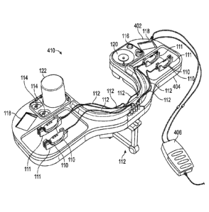
illustrate another embodiment of a modular surgical tray
system 610. In this embodiment, the modular surgical tray system 610 comprises
a
reusable or base portion 650 having a plurality of locations or interfaces
configured for
acceptance of or coupling to one or more modules. In this embodiment, the
system 610
comprises a motor and pump module 622, a fluid reservoir receiver module 620,
a power
adapter module 654, and a modular tool insert 652. In an embodiment, the motor
and
pump module 622 can comprise a BSS bottle holder. In an embodiment, the motor
and
pump module 622 can comprise drive electronics for the infusion pump and/or
the
pressure sensor. In an embodiment, the drive electronics and/or the pressure
sensor can
be located in the reusable portion of the tray. In an embodiment, the fluid
reservoir
receiver module 620 can comprise the aspirated fluid reservoir and the
aspiration pump.
In an embodiment, the fluid reservoir receiver module 620 can comprise the
drive
electronics for the aspiration pump and the pressure sensor (or one or both of
these may
be instead located in the reusable portion of the tray). In an embodiment, the
power
adapter module 654 can be incorporated into one of the displays. In an
embodiment, the
power adapter module 654 can be located underneath the tray (in the reusable
portion) or
elsewhere (for example, on the ground, or the like). FIG. 6B illustrates a
side view of the
motor and pump module 622. FIG. 6C illustrates a side view of the fluid
reservoir
receiver module 620. FIG. 6D illustrates a perspective view of the motor and
pump
module 622, the fluid receiver module 620, and the modular tool insert 652,
such as may
come as a sterile package or assembly ready for use in a sterile operating
environment. In
this embodiment, it can be seen that the modular tool insert 652 comprises a
folding or
-17-
CA 02926555 2016-04-05
WO 2015/081262
PCMJS2014/067717
hinged joint 653 enabling the insert to be folded upon itself to reduce an
overall package
size of the insert, for example, to reduce a size during storage or shipping.
[0049] FIG. 6E
illustrates a perspective view of the base portion 650 without
the BSS holder, fluid receiver, or tool insert modules coupled thereto. The
base portion
650 comprises a fluid receiver interface 621 shaped or configured to couple
with the fluid
receiver module 620, and a motor interface 623 configured or shaped to couple
with the
motor and pump module 622. The base portion 650 further comprises two tool
insert
interfaces 653 comprising recessed areas for locating and/or retention of the
tool insert
652. FIG. 6A further illustrates straps 412 configured to retain the base
portion 652 to a
support 112. In some embodiments, the straps 412 (or another portion of the
base 650)
may comprise a feature that helps to retain the modular tool insert 652 to the
base portion
650, such as a hook and loop fastener, a magnet, and/or the like. FIG. 6F
illustrates an
exploded view of the modular surgical tray system 610.
[0050] The tray in
some embodiments may also be designed to connect to or
otherwise mate with a separate surgical console. The tray and console may
share
electrical, mechanical, pneumatic, hydraulic, wireless, or other interfaces
with each other.
For example, in some embodiments the tray may provide a "docking station" or
hub for
the handpieces that can be conveniently located near the patient. This hub can
be
connected to the separate surgical console (electrically, pneumatically,
and/or the like)
and distribute the power (electricity, illumination, pneumatic/compressed air,
hydraulic,
mechanical, and/or the like) to the appropriate handpieces. The tray can also
in some
embodiments communicate information to the console, for example to control the
power
sources (voltage, current, pneumatic pressure, light intensity, and/or the
like) and/or to
display information on the surgical console's display.
[0051] The tray may
also be designed in some embodiments to connect,
mount, or otherwise mate to a surgical microscope or portion thereof. For
example, the
tray may be mounted to the optical head of the surgical microscope so that it
hangs
adjacent to the surgical site, or the tray may be mounted to the base or
upright section of
the microscope so that it is positioned adjacent to the surgical site. The
tray may also be
designed in some embodiments to tether power (electrical, laser, illumination,
pneumatic,
hydraulic, or other) and/or other functionality (e.g. data communication) from
the
microscope or a module connected to or mounted on the microscope.
-18-
CA 02926555 2016-04-05
WO 2015/081262
PCMJS2014/067717
Configuration of Profiles
[0052] The tray
and/or base unit may also in some embodiments comprise a
method of enabling the user to load specific settings and/or a user profile.
For example,
the tray or base may in some embodiments comprise a wireless REID reader or
near field
communication (NFC) link that reads a "tag" (e.g. located on the user's ID
badge) which
is programmed with the user's preferences such as aspiration and infusion
ranges, button
functions, handpiece settings, and/or the like. In some embodiments, the tag
comprises an
identifier associated with the user's preferences, instead of the tag itself
being
programmed with the user's preferences. In some embodiments, the system is
configured
to automatically apply a user's preferences and/or to load settings associated
with a
specific user or tag when the tag is read by the wireless reader. In some
embodiments, the
tray comprises an antenna portion of the wireless reader, and the base
comprises another
portion of the wireless reader, such as a processing unit, which can be
electrically
connected to the antenna portion when the tray is connected to the base. Such
a design
can be advantageous to enable a more expensive portion of the wireless reader,
such as
the processing unit, to be reusable. The tray and/or base unit may in some
embodiments
comprise a IJSB or memory card interface or similar means of allowing the user
to
transfer information to the tray or base to, among other things, load or set
settings and/or a
user profile.
[0053] FIG. 7
illustrates a top view of a surgical tray 710 similar in design to
the surgical tray 10 illustrated in FIG. 1A. The surgical tray 710, however,
further
comprises an antenna 702 configured to communicate wirelessly with a tag, near
field
communication device, and/or the like to enable configuration of parameters,
user
preferences, and/or the like. In some embodiments, the antenna 702 may be
electrically
coupled to a processing unit to enable the processing unit to configure the
parameters,
preferences, and/or the like.
Surgical Tray Components/Functions
[0054] A surgical
tray in various embodiments can be configured to provide
one or more of a multitude of components and/or functions for performing a
surgical
procedure. In addition to components and functions described above, the
components
and/or functions may comprise, but are not limited to: infusion, aspiration,
one or more
handpieces, illumination, laser therapy, display, audio feedback, one or more
footpedals,
and storage. These components and functions are described in greater detail
below.
-19-
CA 02926555 2016-04-05
WO 2015/081262
PCMJS2014/067717
Infusion
[0055] The tray in
some embodiments may provide infusion of fluids
(balanced saline solution aka BSS and other fluids, including silicone oil,
viscoelastic
gels, dyes/stains, and/or the like) and/or gases into the eye, either the
posterior or anterior
chamber, for example by using a handpiece, such as one of the various
handpiece
embodiments disclosed herein. The infusion source (for example, a bottle or
bag) may
include a light (for example, an LED) to illuminate till level, preferably but
not
necessarily the color red to minimize the impact on the surgeon's low light
vision. The
infusion fluid pathway may comprise in some embodiments a pressure and/or flow
sensor
to determine infusion and/or intraocular pressure and/or infusion flow rate.
The fluid
pathway and sensor may in some embodiments be separated by a filter or
membrane to
prevent contamination of the fluid and/or damage to the sensor, or in some
embodiments
non-contact measurement methods may be utilized. The tray may comprise in some
embodiments a means of holding or securing the infusion fluid bottle or bag,
such as a
cup-holder or hook and/or the like, and a spike, needle, or fluidic attachment
for
extracting the contents of the bottle or bag. The tray may comprise one or
more infusion
systems to provide infusion for different fluids or gases simultaneously, on
demand, or in
a particular order. The tray may comprise stopcocks or other valves (manual or
automated) to enable selection between different infusion sources (e.g. BSS or
oil) or
infusion locations (e.g. an infusion port next to the left eye vs. an infusion
port next to the
right eye).
[0056] In some
embodiments, the tray may comprise multiple infusion
systems, for example two separate systems located on opposite sides of the
tray, each
system designated for use with the adjacent eye. This can be advantageous to
help ensure
the tubing length from the infusion system to the patient's eye is minimized.
The tray may
also in some embodiments comprise multiple infusion systems (e.g. one for BSS
and one
for silicone oil) that are optimized for different viscosity fluids. In
preferred
embodiments, the total tubing length or fluid path length from either the
infusion source
(e.g. BSS bottle) or the infusion pump to the infusion cannula (which is
inserted into the
patient's eye) is minimized. Minimizing this fluid path length can improve the
overall
performance of the infusion system. The responsiveness of an infusion system
that is
actively maintaining an intraocular pressure level (e.g. via feedback control)
during a
surgical procedure is directly related to the length of the tubing set
connecting the infusion
-20-
CA 02926555 2016-04-05
WO 2015/081262
PCMJS2014/067717
source or infusion pump to the eye. Infusion systems with longer tubing sets,
as is typical
in commercially-available ophthalmology surgical consoles that are not located
immediately adjacent to the patient, result in an undesirable lag or delay
when measuring
or adjusting the intraocular pressure as compared to those with shorter tubing
sets. The
infusion cannula can also be primed faster (before insertion into the
patient's eye) in an
infusion system with short tubing sets. In a preferred embodiment, this length
(either
source to cannula or pump to cannula) will not exceed 24 inches, but
additional
embodiments may be utilized that allow this length to reach 36 inches or more.
[0057] In one
embodiment, the tray system comprises a separate infusion
system for injecting silicone oil and similar viscous fluids. The oil infusion
system may be
a separate module that is utilized only in surgical cases that require oil
infusion. The oil
infusion system may be connected to a handpiece connector in order to supply
power to
the oil infusion system and provide a communications interface between the oil
infusion
system and the tray or base electronics. In some embodiments, the oil infusion
system is
designed as a handpiece with an endoscopic needle or tube that is used to
infuse the oil or
fluid into the eye. In other embodiments, the oil infusion system interfaces
to the infusion
cannula already inserted in the eye for BSS infusion. The infusion of oil may
be done
manually (for example, by depressing or squeezing a plunger and/or the like),
it may be
done pneumatically or hydraulically (for example, using a separate pump,
compressor,
compressed gas source, and/or the like), or it may be done
electromechanically, for
example with a motor, solenoid, or similar actuator that can infuse the oil
(for example, a
ballscrevv/leadserew, Hamilton syringe type configuration that moves a plunger
to expel
the oil from a syringe or cartridge, and/or the like).
[0058] Some
embodiments utilize a pump or other means to provide fluid
infusion. The pump style may be a standard Venturi, peristaltic, or diaphragm
design, or
another standard or non-standard pump variety. The infusion system may in some
embodiments rely on other mechanisms of action to achieve fluid infusion, for
example a
fluid-filled syringe depressed either manually (for example, by the surgeon or
an assistant)
or automated (for example, via a syringe pump mechanism, actuator, motor,
servo,
ballscrew/leadscrew, spring, and/or the like).
[0059] Some
embodiments may be configured to use a manually or
automatically adjustable pole to raise or lower the BSS bottle or bag,
exploiting gravity to
provide a variable infusion pressure related to the height of the fluid
source.
-21-
CA 02926555 2016-04-05
WO 2015/081262
PCMJS2014/067717
[0060] Some
embodiments may be configured to pump air into or out of the
fluid bottle to control the infusion pressure and therefore intraocular
pressure (forced gas
infusion). Pumping gas into the bottle increases the infusion pressure, while
drawing air
out of the bottle via pumping, vacuum, or venting (for example, through a tube
or needle
whose intake port is located above the water level) decreases the infusion
pressure. Using
this technique not only enables precise control of the infusion pressure but
it also helps
dampen pressure spikes and dips. The pulsating flow output of a peristaltic
pump can also
be minimized when using the peristaltic pump to pump air into the fluid bottle
to increase
infusion pressure.
[0061] Some
embodiments utilize a compressed gas, e.g. a nitrogen or other
gas (preferably inert) filled cartridge, canister, or tank as a source of
pressure to enable
fluid infusion (for example, via Venturi action or forced gas infusion). The
cartridge,
canister, or tank may be reusable/refillable or disposable and intended for
single-use or
limited use.
[0062] Some
embodiments utilize a soft infusion fluid bag (as opposed to a
glass or rigid plastic bottle). The soft bag may in some embodiments be
located in a
fixture between two or more plates that can squeeze or otherwise exert
pressure on the
bag in one or more axes. The distance between the plates (and thus the squeeze
force) can
be controlled manually by the surgeon or assistant or automatically, for
example through a
mechanical system comprising one or more of an actuator, motor, servo, cam,
solenoid,
gear, ratchet, rack and pinion, band, belt, pulley, chain, and/or the like.
Increasing the
squeeze force increases the infusion pressure; decreasing the squeeze force
decreases the
infusion pressure. Likewise, a similar mechanism can be used on a smaller
container of
infusion fluid, for example a reservoir into which infusion fluid drips or
flows from the
original infusion bottle or bag. A check valve can be included in some
embodiments to
prevent backflow into the original bottle or bag. The soft bag may also be
located in an
air-tight rigid container, which can have air pumped in or out (or vented) to
increase or
decrease the pressure on the external surface of the bag. Since the bag is not
rigid, but
compressible, the infusion pressure can be adjusted by adjusting the pressure
in the rigid
container. Likewise, a similar mechanism can be used on a smaller container of
infusion
fluid, for example a reservoir into which infusion fluid drips or flows from
the original
infusion bottle or bag. A check valve can be included to prevent backflow into
the
original bottle or bag.
-22-
CA 02926555 2016-04-05
WO 2015/081262
PCMJS2014/067717
[0063] In some
embodiments the infusion fluid(s) are included in or integrated
into the tray system so that the tray and fluid are packaged, sterilized, and
shipped as a
single system that can be disposed of after the surgical procedure. This is in
contrast to a
system wherein the tray is packaged, sterilized, and shipped as a separate
component than
the infusion fluid (e.g. BSS bottle) which may be from a different
manufacturer
altogether. Such a system may also include a separate additional means of
introducing
infusion fluids into the fluidic path of the system, for example if the
included fluids are
exhausted during the surgical procedure.
Aspiration
[0064] In some
embodiments, the tray may provide aspiration functions, for
example from a vitreous cutter, soft-tip, or phaco handpiece. The aspiration
function may
be provided through the use of a pump or by another means. The pump style may
be a
standard Venturi, peristaltic, or diaphragm design, or another variety. The
aspiration
pump system may also rely on other mechanisms of action to achieve vacuum draw
at the
needle tip, for example a syringe with a depressed plunger connected to the
aspiration
needle either directly or via a tube, the plunger being drawn back to produce
a vacuum
force, the action of being drawn back accomplished either manually (for
example, by the
surgeon or an assistant to the surgeon) or through a semi-automated or fully
automated
process (for example, a syringe pump mechanism, an actuator, motor, servo,
ballscrew/leadscrew, spring, and/or the like), and/or the like. Some
embodiments may
utilize compressed gas (such as previously described), for example to generate
a vacuum
for aspiration through Venturi action. The aspiration fluid pathway may in
some
embodiments comprise a pressure or flow sensor to determine aspiration vacuum
pressure
and/or aspirated fluid flow rate. The fluid pathway and sensor may in some
embodiments
be separated by a filter or membrane to prevent contamination of the fluid and
damage to
the sensor, or non-contact measurement methods may be utilized.
[0065] In some
embodiments, the tray may also incorporate a reservoir tank to
hold the waste aspirated fluid and tissue. The tray may comprise a window
and/or a light
(e.g. LED) preferably but not necessarily the color red to minimize the impact
on the
surgeon's vision, to enable to the surgeon to visualize the fluid level in the
reservoir tank.
The reservoir tank may in some embodiments comprise a fluid level sensor to
measure the
level of aspirated fluid and remaining free volume. This may be utilized, for
example, to
alert the surgeon if the reservoir tank is near full capacity. The reservoir
tank in some
-23-
CA 02926555 2016-04-05
WO 2015/081262
PCMJS2014/067717
embodiments may be configured to expand as the volume of fluid inside
increases (for
example, as a balloon or bladder style reservoir, a reservoir with accordion-
style
collapsible walls, and/or the like).
[0066] Locating the
aspiration pump and waste reservoir in or near the tray
and in close proximity to the patient or within the sterile field can be
preferable in some
embodiments to, among other things, minimize the tubing length required, which
improves the performance and responsiveness of the aspiration system. This
reduces the
path length of the aspirated fluid, thereby reducing the requirements of the
aspiration
mechanism and eliminating long tubing sets that slow the response time (for
example,
when the surgeon changes the rate of aspiration or switches from aspiration to
reflux) and
can entangle the surgeon and assistants in the operating room.
[0067] The tray may
in some embodiments comprise stopcocks or other valves
(manual or automated) to select between different aspiration intake sources
(for example,
a vitreous cutter handpiece and a soft trip extrusion handpiece).
Handpieces
[0068] In some
embodiments, the tray system may comprise one or more
handheld probes or handpieces that may comprise a needle (for example, 18
gauge, 20
gauge, 23 gauge, 25 gauge, 27 gauge or other size) inserted into either the
anterior or
posterior chamber of the eye during a surgical procedure (such as, for
example, one or
more of the various handpieces described herein with reference to FIGS. I E,
4A, 5A. 6A,
and 8A). Handpieces in some embodiments may comprise one or more of vitreous
cutters/aspirators. endoilluminators, laser therapy/photocoagulation probes,
diathermy/eleetrocautery/ablation probes, scissors, soft-tip extrusion probes,
phacoemulsification/phacomorcellation probes, intraocular lens (IOL)
inserters, forceps,
mechanical probes, and/or other commonly used instruments. Some handpieces may
incorporate more than one function. A handpiece may in some embodiments
comprise
one or more buttons and/or other user interfacing features that allow the
surgeon to
control the functions of that specific handpiece and/or possibly other
functions as well
(such as, for example, rates of infusion or aspiration).
Vitreous Cutter Handpiece
[0069] In some
embodiments, a tray system comprises a vitreous cutter
handpiece for removal of vitreous during a vitreoretinal procedure. The
handpiece may in
some embodiments be tethered to the tray via a multi-conductor cable that
provides power
-24-
and an optional communications interface (for example to communicate with the
tray or base unit
electronics, for example the status of button presses on the handpiece). In
some embodiments, the
cutter mechanism may be powered by a motor or motor and gear assembly inside
the handpiece.
In some embodiments, the cutter mechanism may be powered pneumatically by an
external
pneumatic source (for example, a pump, compressor, compressed gas source,
and/or the like) that
is connected to the handpiece via one or more flexible pneumatic tubes. The
external pneumatic
source may be located within the tray or the non-disposable base unit (for
embodiments that
incorporate a base as previously described). The cutter mechanism may in some
embodiments be
powered by a transmission cable or torque coil that rotates, reciprocates, or
translates in one or
more axes. Using the principles of electromagnetism, the cable or coil may be
used to supply
electrical power to the handpiece as well, for example by rotating or
otherwise moving a magnet
in proximity to a wire coil and generating a current that can power the
electronics of the handpiece.
The cable or coil may be driven by a motor, solenoid, electromagnet, linear
actuator, and/or the
like that is located external to the handpiece, for example in the tray or non-
disposable base unit.
The cable or coil connected to the handpiece may be coupled to the motor or
drive actuator in the
base via a shaft coupling, spline coupling, or similar to enable ease-of-setup
by a surgeon or
assistant in the operating room. In some embodiments, a magnetic coupling may
be used to
maintain a sterile field between the motor and the cable or coil. The cut
speed, rate of aspiration
and other functions may be controlled by buttons or other user interfaces on
the handpiece itself,
or through a footpedal.
100701 Similar drive configurations may also be used for lens
removal or
phacomorcellation handpieces as well as other mechanically-driven instruments.
[0071] In some embodiments, a vitreous cutter handpiece may be a
handpiece 810
as illustrated in FIGS. 8A and 8B, as further described below. In some
embodiments, a vitreous
cutter handpiece may comprise one or more features similar to as disclosed in
U.S. Patent
Application Publication No. 2008/0208233, entitled DISPOSABLE VITRECTOMY
HANDPIECE.
Endoilluminator Handpiece
[0072] In some embodiments, a tray system may comprise an
endoilluminator
handpiece that provides illumination inside the eye. The handpiece may be
tethered to the tray in
some embodiments via a multi-conductor cable that provides power and an
optional
- 25 -
Date Recue/Date Received 2021-04-27
communications interface (for example to communicate with the tray or base
unit electronics, for
example the status of button presses on the handpiece). The endoilluminator
may in some
embodiments incorporate a light source (for example, white LED or RGB LED)
that is coupled to
a fiber or fiber bundle installed in an endoscopic needle. Alternately, the
light source may be
located in the tray or base unit and coupled (either permanently or using a
detachable interface) to
a fiber or fiber bundle that terminates in an endoscopic needle in the
handpiece.
[0073] In some embodiments, an endoillumination handpiece may
comprise one or
more features similar to as disclosed in U.S. Patent No. 8,172,834, entitled
PORTABLE
HANDHELD ILLUMINATION SYSTEM.
Soft-Tip Extrusion Handpiece
[0074] The tray system may in some embodiments comprise a soft-tip
extrusion
handpiece that incorporates a soft tubing material (for example, silicone or
the like) for aspirating
vitreous and fluids from the retina. The handpiece may in some embodiments be
tethered to the
tray via a multi-conductor cable that provides power and an optional
communications interface
(for example, to communicate with the tray or base unit electronics, for
example the status of
button presses on the handpiece). The rate of aspiration and other functions
may be controlled by
buttons or other user interface features on the handpiece itself, or through a
footpedal. The
endoillumination power output and other functions (such as infusion rate) may
in some
embodiments be controlled by buttons or other user interface features on the
handpiece itself, or
through a footpedal.
Diathermy/Electrocautery Handpiece
100751 The tray system in some embodiments may comprise a bipolar
electrocautery handpiece that is capable of controlled cauterization of
tissues. The handpiece in
some embodiments may comprise two nested needles or tubes separated by an
insulating layer
(such as, for example, polyimide tubing or the like). The exposed distal end
of the two needles or
tubes act as electrodes for the bipolar electrocautery. The handpiece may in
some embodiments be
tethered to the tray via a multi-conductor cable that provides power and an
optional
communications interface (for example to communicate with the tray or base
unit electronics, for
example the status of button presses on the handpiece). The handpiece may in
some embodiments
have integrated
- 26 -
Date Recue/Date Received 2021-04-27
CA 02926555 2016-04-05
WO 2015/081262
PCMJS2014/067717
electronics for generating the high voltage waveform required for
electrocautery, or
alternately the electrocautery circuity may be located in the tray or base
unit and supplied
to the handpiece via insulated wires. The diathermy/electocautery function and
other
functions may be controlled by buttons or other user interface features on the
handpiece
itself, and/or through a footpedal.
Laser Therapy Hand piece
[0076] The tray
system in some embodiments may comprise a fiber-based
laser handpiece that is capable of photocoagulation. The handpiece may in some
embodiments be tethered to the tray via a multi-conductor cable that provides
power and
an optional communications interface (for example to communicate button
presses and
system status with the tray electronics). The handpiccc may in some
embodiments
incorporate a light source, such as a laser diode, that has sufficient power
for
photocoagulation. The laser diode may in some embodiments be coupled to a
fiber or
fiber bundle that is mounted inside an endoscopic needle for insertion into
the eye or other
surgical site. Alternately, the light source and associated optics may in some
embodiments
be located in the tray or base unit with either a permanent or interchangeable
optical
interface to a fiber or fiber bundle that terminates in an endoscopic needle
located in the
handpiece. The laser therapy power output and other functions may be
controlled in some
embodiments by buttons or other user interface features on the handpiece
itself, or
through a footpedal.
Scissors
[0077] The tray
system in some embodiments may comprise a powered
scissors handpiece that enables the surgeon to cut tissue without requiring
manual
manipulation, for example using fingers to squeeze, slide, or otherwise
activate the
cutting mechanism of the scissors. In one embodiment, the scissors are
tethered to the tray
via a multi-conductor cable that provides power and an optional communications
interface (for example with the tray electronics). Power to the cutting
mechanism is
provided in some embodiments by the tray via the tethered cable. The cutting
mechanism
may in some embodiments comprise a motor, solenoid, linear actuator, nitinol
or shape
memory alloy wire (for example, a wire that contracts when a current is passed
through
the wire and expands when the current ceases and the wire cools), and/or the
like.
Alternate embodiments may position the actuator in the tray or base, and
mechanical
-27-
CA 02926555 2016-04-05
WO 2015/081262
PCMJS2014/067717
cutting may be provided via a linkage, such as a transmission cable or torque
coil that is
rotated, reciprocated, or translated alone one or more axes.
Handpiece Storage
[0078] In some
embodiments, a tray may comprise space configured to hold
the handpiece(s) when they are not in use (such as, for example, the spaces
111 illustrated
in FIG. 1E), and/or may provide connectivity to the handpiece(s) via one or
more of an
electrical, fluidic, pneumatic, optical, and/or mechanical interface. The
spaces to hold the
handpieces and the top surface of the tray in general may in some embodiments
comprise
features to cope with undesired fluids that may be present on the tray during
the
procedure. For example, the tray top and handpiece areas (which may be
recessed pockets
or wells) may have recessed channels or holes to drain any fluids or carry any
fluids away.
Likewise, any recessed areas may include absorbent or sponge-like materials to
absorb
any unwanted fluids. The handpieces may be mounted in the tray prior to
packaging and
sterilization to simplify the pre-op setup procedure. The tray may in some
embodiments
include clips. straps, or other locking mechanisms that enable the handpieces
to be
secured in place, for example during shipment or movement of the tray.
Additional
handpieces may be packaged and sterilized separately, for example to enable
replacement
of a failed handpiece during a procedure without having to open an entire new
tray
system.
Handpiece User Interface Features
[0079] Some
embodiments of handpieces comprise one or more means of
acquiring user input, such as one or more buttons or switches (including, for
example,
membrane, tactile, pushbutton, rotary, joystick, hall sensing, capacitive
touch, pressure
sensitive, and/or the like) located on the handpiece, and/or inertial sensors
(including
gyro(s), accelerometer(s), magnetometer(s), and/or the like). These input
methods can be
used to control one or more functions of the handpiece and/or console, such
as, for
example, activating, deactivating, and controlling the probe tip motion and
aspiration
functions. For example, in some embodiments, the surgeon may press and hold
one button
to activate em
ul s ifi cati on/m orcel 1 ati on, and release the button to stop
emulsifieation/morcellation. The surgeon may press and release another
separate button
repeatedly to cycle through aspiration rates. The inertial sensors can be used
in some
embodiments for position tracking as well as user input. For example, the
surgeon may
orient or move the handpiece in a particular manner to perform a function: an
example
-28-
CA 02926555 2016-04-05
WO 2015/081262
PCMJS2014/067717
would be deactivating the system when the handpiece is placed upside down on a
tray or
table (the system would recognize the upside down orientation of the handpiece
and the
lack of motion/movement); a second example would be lightly tapping on the
handpiece
with a finger to activate or deactivate a function (the accelerometer will
detect the
handpiece deflection caused by the tapping): yet another example would be
rotating the
handpiece in a clockwise fashion to increase the rate aspiration or some other
function
and rotating the handpiece in a counter-clockwise fashion to decrease the rate
of
aspiration or some other function (where the angular motion is detected by the
gyro). The
user input may in some embodiments be processed or otherwise acted upon
internally
within the handpiece, or the input may in some embodiments be relayed to a
separate
console or tray via a tethered electrical connection (for example, conductive
wires/cables)
or wireless connection (for example, RF, inductive, or infrared) for example.
The
handpiece may in some embodiments comprise a microcontroller or microprocessor
for
registering user input, controlling the functions of the handpiece, and/or
communicating
with external components of the system (for example a console or tray). The
handpiece
may in some embodiments comprise wireless capabilities to transmit and receive
information to/from a separate console, tray, display, and/or other
handpieces.
[0080] Some
embodiments comprise analog buttons (such as, for example, a
pressure or deflection sensitive button) that provide finer control over the
handpiece
functions than a standard binary (on/off) or momentary switch. For example,
one or more
analog buttons sensitive to pressure or deflection may be used to provide fine
control of
functions such as, for example, cut speed, aspiration, or illumination/laser
power output.
In one embodiment, one or more pressure-sensitive buttons in the grip of the
handpiece
can be used to control rate of aspiration or cut speed; the harder the surgeon
squeezes, the
higher the cut speed or rate of aspiration for example. Likewise, other
sensors can be
incorporated that measure flexion, deflection, or translation such that the
further a surgeon
pushes, slides, or otherwise moves a button, the higher the rate of, for
example, aspiration
or cut speed. This can be achieved, for example, with button implementations
that vary a
parameter (such as resistance or capacitance) based on an applied input (such
as pressure
or deflection). For example, one button implementation may be sensitive to
pressure such
that the harder the surgeon squeezes, the lower (or higher) the resistance,
which can be
measured by the handpiece electronics (and/or remote electronics, such as
electronics
located in a tray or base). Another implementation utilizes a change in
capacitance, such
-29-
CA 02926555 2016-04-05
WO 2015/081262
PCMJS2014/067717
that the distance (and therefore capacitance) between two parallel conductive
plates varies
with the force applied to a button. Another implementation may connect
different circuits
depending on how far a button is depressed or moved. Yet another
implementation may
utilize magnetic sensors to detect the location of a magnet and/or magnetic
field strength
to determine how far a button has been depressed or moved. Yet another
implementation
utilizes capacitive touch technology to provide analog control.
[0081] Figures 8A
and 8B illustrate an embodiment of a handpiece or surgical
instrument 810 comprising a housing or body 802 having a plurality of buttons
812, 808.
FIG. 8A is a perspective view of the handpiece 810, and FIG. 8B is a
perspective view of
the handpiece 810 with the housing or body 802 removed to enable visualization
of
features positioned beneath the housing 802. The handpiece 810 comprises a
proximal
end 806 adjacent a cable interface 807, and a distal end 804 having a surgical
tool 805,
such as a needle, extending therefrom. In some embodiments, the surgical tool
805 is
permanently or semi-permanently installed. In some embodiments, the distal end
804 is
configured to enable a surgical tool 805 to be positioned in a coupled
engagement with
the distal end 804.
[0082] The
handpiece 810 comprises three buttons 808 positioned adjacent to
an exterior surface of the housing 802. In this embodiment, the buttons 808
comprise
digital, binary, or momentary buttons or switches, meaning they have two
states, namely
on or offf, for controlling of a feature. The buttons 808 may, for example,
comprise
mechanical switches that selectively open and close an electrical circuit when
an actuation
surface of the button 808 is moved relative to the housing 802.
[0083] The
handpiece 810 further comprises in this embodiment two pressure
sensitive buttons 812. In other embodiments, the handpiece may comprise fewer
or more
pressure sensitive buttons. Each of the two pressure sensitive buttons 812
comprises a
circumferential force-sensitive resistor 813 that is configured to change a
resistance value
based on a magnitude of pressure applied against a surface of the force
sensitive resistor
813. In some embodiments, the force sensitive resistor 813 comprises a thin
multilayer
polyimide sheet. In some embodiments, as can be seen in FIG. 8A, the buttons
812
comprise an actuation surface extending circumferentially around the handpiece
810
and/or housing 802 that, when depressed, presses against the force sensitive
resistor 813.
In some embodiments, the exterior surface of the buttons 812 comprise tactile
regions or
features 814, illustrated in FIG. 8A as a plurality of raised bumps spaced
circumferentially
-30-
CA 02926555 2016-04-05
WO 2015/081262
PCMJS2014/067717
around the handpiece 810 and/or housing 802. Such a configuration may be
advantageous
to, for example, enable a user or surgeon to precisely control a feature when
the handpiece
810 is in any rotational position with respect to the user's hand.
[0084] As used
herein, the word "button" may be used interchangeably with
other words or phrases, such as switch, selector, user input, and/or the like.
One of skill
in the art will recognize that a variety of user interface features, including
buttons or other
similar features, may be used with the techniques disclosed herein to detect a
user input.
[0085] In addition
to traditional on/off or momentary buttons and analog
buttons that depend on some type of electrical or electromechanical contacts,
additional
user input solutions are feasible that eliminate the need for electronics in
the handpiece
and/or the need for tethered power and communication interfaces to the
handpiece. Such
embodiments may be advantageous to, among other things, increase
manufacturability
and/or reduce cost of disposable components of the system, such as, in some
embodiments, the handpieces.
Optical Buttons
[0086] In some
embodiments, user input can be detected using buttons that
rely on principles of optics and optical fiber, instead of (or in some
embodiments, in
addition to) electrical or electromechanical features. For example, in some
embodiments,
one or more buttons may have one or more fibers, light pipes, or optical
waveguides
associated with it, the fibers extending from the handpiece to the tray or
base unit
electronics (either as a continuous fiber or one or more fiber sections
optically connected
together). The fiber may be coupled to a light source (such as a light source
located in the
tray or base unit) and may propagate light to the button location. A button
may be
designed to bend or otherwise flex the fiber when the button is depressed,
reducing or
eliminating the light propagation through the fiber and/or changing the
polarization of the
light through the fiber, both of which are detectable by the electronics and
can be used to
indicate a button press. Other embodiments instead alter or route/reroute the
light path
(e.g. a reflective surface on a button, the surgeon's fingertip, etc.) such
that the change can
be detected and identified as a button press. The principles of fiber
interferometry can also
be used, such that a button press sufficiently alters the fiber so that the
changes to phase
altering interference fringes and the location thereof can be detected and
interpreted as
button presses. In a similar fashion, fiber Bragg grating sensors, long-period
fiber grating
sensors, and similar embodiments can be integrated into the fiber(s) to
measure strain,
-3 1 -
CA 02926555 2016-04-05
WO 2015/081262
PCMJS2014/067717
thereby detecting changes in the fiber and their location(s). Some embodiments
may
utilize one of the optical techniques disclosed herein, and some embodiments
may utilize
more than one of the optical techniques and/or may utilize one or more of the
optical
techniques and one or more non-optical techniques.
[0087] In some
embodiments, one or more buttons each have a single
continuous fiber associated with it. The fiber is routed from the electronics
in the tray or
base to the handpiece button and back to the tray or base (either as a
continuous fiber or as
two or more sections optically coupled together). When the button is pressed,
the fiber is
bent in such a manner as to decrease or eliminate the light propagation,
change the
polarization of the light in a detectable manner, or induce strain in the
fiber that alters the
interference fringes in a detectable manner. In other embodiments, one or more
buttons
each have two fibers associated with it (each either as a continuous fiber or
as two or
more sections optically coupled together), one that carries the light from the
tray to the
button and another that acts as a return for the light back to the tray. The
button is
designed such that when it is depressed, light is allowed to propagate from
the source
fiber to the return fiber. This can be accomplished, for example, with a
reflective surface
or a transmissive or light pipe material that is angled or positioned properly
when
depressed, or even using the surgeon's fingertip to redirect the light from
the source fiber
to the return fiber.
[0088] Another
embodiment comprises a single fiber for both the source and
return path of the light, since fiber can simultaneously propagate light in
both directions.
For example, an optical circulator can be configured to allow light to be
injected into the
fiber at the source (such as the tray or base unit) while simultaneously
separating any
reflected light along the same fiber to be detected by a photodetector or
other sensor in the
electronics. Such reflections could be caused by a reflective surface (or even
fingertip) at
the end of the fiber, or even a bend, flex, or twist of the fiber.
[0089] In another
embodiment, a single fiber or optical waveguide provides
the source light to each button, and each button has an additional individual
return fiber to
indicate button presses.
[0090] In other
embodiments, instead of individual fibers for each button, a
single fiber is routed from the tray or base to each button in series and back
to the tray or
base. Different buttons are designed to alter the polarization or light
propagation different
amounts, such that each button can be distinguished from each other.
Alternately, the
-32-
CA 02926555 2016-04-05
WO 2015/081262
PCMJS2014/067717
optical path distance (such as the fiber length) between each button can be
adjusted such
that the principles of optical time domain reflectometry or similar can be
used to
determine the location along the fiber of the button press, and hence which
button was
pressed.
[0091] FIGS. 9A-9D
illustrate example embodiments of handheld medical
instruments or handpieces comprising one or more optical buttons. FIG. 9A
illustrates
schematically a handpiece comprising a housing or enclosure 802 having two
optical
fibers 906 passing therethrough. In this embodiment, an actuating member 904,
when
placed close to tips or ends 907 of the optical fibers 906 is configured to
reflect light from
one optical fiber 906 to another optical fiber 906, the reflection of which
may be detected
by hardware, for example, in a surgical tray or console. In some embodiments,
the
actuating member 904 is a portion of a physical button that is configured to
reflect light.
In some embodiments, the actuating member 904 is a user's finger, in some
embodiments
a gloved finger.
[0092] In some
embodiments, the two separate optical fibers 906 may also be
implemented as, for example, a single fiber with two or more waveguides, for
example
the source light would propagate down the core and the return light down one
or more
claddings of the fiber, or vice versa. Also note that a single source fiber
may in some
embodiments supply light to multiple buttons, with each button paired with an
independent return fiber or independent waveguide, for example, multiple
claddings, in a
custom-designed return fiber. Alternatively, each button may have a separate
source fiber
providing light modulated at a different frequency for each button, with a
common shared
return fiber, with the modulated signal allowing the processing hardware to
distinguish
between different signals.
[0093] In some
embodiments, a technique for enabling multiple buttons
incorporates a filter or attenuator that attenuates the light propagation
through each button
differently (for example, 100%, 50%, 25%, and/or the like) or filters the
wavelength
(assuming a broadband light source) through one or more or each button so that
the
remaining wavelengths or the power attenuation could be measured by the
processing
hardware, such as, for example, in the surgical tray or console. In some
embodiments, the
wavelength of the light may be broadband (for example, white light or multi-
wavelength
light) or it may be single wavelength (for example infrared) and may be
modulated to
minimize interference issues with other ambient sources of light.
-33-
CA 02926555 2016-04-05
WO 2015/081262
PCMJS2014/067717
[0094] FIG. 9B
illustrates another embodiment of a surgical handpiece
comprising an optical button. The embodiment illustrated in FIG. 9B comprises
an
optical fiber 906 terminating at an end or tip 907. An actuating member 904
may have
light reflective properties such that, when the actuating member 904 is
positioned
adjacent the tip 907, light is reflected back into the fiber 906, which can
then be detected
by, for example, tray or console hardware. Sensitivity in some embodiments can
be
adjusted to require contact by the actuating member 904 with the tip 907
and/or handpiece
enclosure 802, or just a proximity to the tip 907 and/or handpiece enclosure
802.
[0095] FIG. 9C
depicts another embodiment of an optical button. The
embodiment illustrated in FIG. 9C comprises two optical fibers 906 and an
actuating
member 904 which, when moved relative to the enclosure 802 and/or fibers 906,
moves a
reflective member 908. When the reflective member 908 is positioned in front
of tips or
ends 907 of the optical fibers 906, light is reflected from one optical fiber
906 to the other
optical fiber 906, enabling detection of the button press by processing
hardware located
at, for example, the surgical tray or console.
[0096] FIG. 9D
depicts another embodiment of an optical button, wherein a
deflectable member or portion 910 connected to or part of one or more optical
fibers 906
is positioned to be deformed when an actuating member 904 is moved relative to
the
handpiece enclosure 802. For example, when the actuating member 904 contacts
and/or
presses against the deflectable member 910, the deflectable member 910 may
bend or
otherwise deformed in a manner that may be detectable by processing hardware,
such as
processing hardware located in or as a part of the surgical tray or console.
In this
embodiment, if the button is depressed, the actuating member 904 will bend the
deflectable member 910. If the deflectable member or portion 910 is deflected
sufficiently, the propagation of light through the fiber will be significantly
decreased or
eliminated, which can be detected by the processing hardware and interpreted
as a button
press. In an alternate embodiment, more light instead of less light is allowed
to propagate
the harder or further the button is pressed.
[0097] In some
embodiments, the optical button configuration illustrated in
FIG. 9D can also be configured to provide linear or pressure sensitive
feedback through
one or more of several techniques. First, the decrease in light throughput
will correlate to
how much deflection or bending the fiber is experiencing. The amount of light
detected
can be used to determine the overall position of the button. So, for example,
the harder or
-34-
CA 02926555 2016-04-05
WO 2015/081262
PCMJS2014/067717
further the user pushes the button, the greater the measured change in light
throughput.
Similarly, the bending of the fiber will change the polarization of the light
through the
fiber. This can also be detected by processing hardware and used to determine
the
magnitude of deflection. More advanced optical theory can also be used. For
example,
the fiber button can be one arm of a fiber-based interferometer (michelson,
common path,
mach zehnder, and/or the like) such that the change in optical path length and
the phase
changes induced in the fiber due to stress or bending can be detected in the
processing
hardware and measured to determine how hard or far the button was depressed.
Another
technique is to include gratings in the fiber during manufacturing to produce
a strain
sensor, such as a Fiber Bragg Grating Sensor or long-period fiber grating,
that can be used
to provide linear or pressure sensitive button functionality due to the
correlated change in
wavelength resulting from strain on the fiber (for example, caused by pressing
or bending
the fiber). These grating sensors can also be used for multiple buttons with
only a single
fiber by designing a grating along the fiber with different properties for
each button such
that the properties of each grating are distinguishable from each other by the
processing
hardware.
[0098] Yet another
embodiment can use a single fiber for multiple linear or
pressure sensitive buttons by detecting where along the fiber a strain or bend
was induced,
and correlating this to the position of the one or more buttons. This
detection can be
implemented using principles of time domain reflectometry, wherein the time
for a light
pulse to propagate through the fiber and reflect back to the source is
measured and the
distance determined, with the reflection caused by the button inducing a
strain or
impedance in the fiber. This is more difficult over a shorter fiber length and
would
benefit in some embodiments from the addition of fiber between the buttons to
increase
an optical path length between buttons.
[0099] In some
embodiments, an optical button configuration may comprise
one or more optical vvaveguides in addition to or in lieu of one or more
optical fibers,
such as a plastic light pipe or the like. Further, in various embodiments
disclosed herein
that refer to optical fibers, it should be understood that those optical
fibers may be a
continuous optical fiber and/or may comprise one or more sections coupled
together, and
or may comprise one or more optical waveguides in series with a fiber.
[0100] In some
embodiments, an optical button is configured to enable,
disable, or attenuate the propagation of light from a source waveguide (or
fiber) to a
-35-
CA 02926555 2016-04-05
WO 2015/081262
PCMJS2014/067717
return waveguide (or fiber), for example, with a reflective or absorptive
surface, including
a finger, to enable detection of a button press.
[0101] In some
embodiments, an optical button uses a single fiber or
waveguide in a loop configuration and alters a property of light in a
detectable fashion, for
example, by bending the fiber or waveguide. This property can be the power or
magnitude (for example, attenuating or increasing the light throughput), the
polarization
of the light, the wavelength of the light (for example, by filtering out some
wavelengths of
a broadband light source and detecting the remaining wavelengths; or by
shifting or
filtering the wavelength for example through a orating design), the optical
path length or
phase of the light (for example, by bending or straining the fiber which can
be detected in
an interferometer setup). A related embodiment relies on the principles of
total internal
reflection and "evanescent waves." For example, by touching the outer surface
of the fiber
(stripped of any outer protective coating, if present), the refractive indices
at the fiber
interface are changed (for example, fiber to finger instead of fiber to air)
which can alter
the propagation of light through the fiber. This change may be detectable with
sufficiently
sensitive amplification and processing equipment.
[0102] In some
embodiments, an optical button uses one fiber for multiple
buttons, for example by using custom fibers with multiple waveguides (and
possibly
modulating the signal to or from different buttons); by filtering out
different wavelengths
depending on which button is pressed (for example, using multi-wavelength or
broadband
light); by attenuating the light by different amounts depending on which
button is pressed;
by using different fiber grating parameters for each button; by measuring the
time for
reflected pulses to propagate (time domain reflectometry) where the reflection
is caused
by a deformation or strain in the fiber caused by the activation of a button.
Pneumatic/Hydraulic Buttons
[0103] In some
embodiments, buttons that utilize pneumatic and/or hydraulic
principles are incorporated into the handpieee, which can in some embodiments
(similarly
to as with the optical button embodiments) eliminate the need for any
electronics or
electrical interfacing between the handpiece and the tray or base unit.
[0104] In some
embodiments, one or more buttons of the handpiece may be
attached to or fluidly coupled with a flexible pneumatic tube (for example,
flexible
silicone, vinyl or PVC tubing or the like) that is connected to the tray or
base unit
electronics (either using a continuous section of tubing or two or more
sections in fluid
-36-
CA 02926555 2016-04-05
WO 2015/081262
PCMJS2014/067717
communication). The electronics may comprise, for example, a pressure sensor
that can
measure the pressure inside the tube. This can be used to determine whether or
not a
button is pressed. For example, a depressed button may seal or pinch the tube
that is
otherwise open, unobstructed, or patent, in such a way that is detectable by
the pressure
sensor. Similarly, in some embodiments, the tubing may terminate in the
handpiece such
that the surgeon may cover the hole in the end of the tubing (such as with his
or her
finger) to indicate a button press. A bladder or balloon, for example at the
end of the
tubing, may be included such that when the bladder or balloon is depressed or
otherwise
modified (such as by an external force, for example, from a finger or from a
movable
component of the handpiece), the change in internal air/fluid pressure can be
detected and
interpreted as a button press. In some embodiments, instead of relying on
ambient
pressure, the tubing may be fluidly connected to an air or vacuum source such
that the
measured pressure will be different depending on whether or not the tubing is
patent or
sealed. In this embodiment, each button would have its own independent tubing.
In
another embodiment. multiple buttons can share a single tube, for example if
each button
restricted the flow through the tube to a different magnitude, such that each
button would
be distinguishable from the rest. For example, the first button may restrict
the tubing
completely, while the second button restricts the inner lumen of the tubing
75%, the third
button 50%, the fourth button 25%, and so on, which may be distinguishable by
a fluidly-
coupled sensor.
101051 Figures 10A-
10C illustrate example embodiments of handpieces
comprising pneumatic or hydraulic buttons. Fig. 10A illustrates schematically
an
example wherein an actuating member 904, such as a finger or a portion of a
button,
deforms a deformable member 1002 fluidly connected with tubing 1004 to enable
detection of a change in pressure in the deformable member 1002 and/or the
tubing 1004.
In some embodiments, a flexible tube (e.g. vinyl or silicone tubing) 1004 with
a balloon
or bladder 1002 attached to the end of the tube that has some gas or fluid
within it (e.g.
air) can be used as a linear button. The harder the balloon or bladder 1002 is
squeezed,
pushed, or compressed, the higher the pressure inside the tube, which can be
measured
and processed by a pressure sensor and processing electronics located, for
example, in the
remote console or tray.
-37-
CA 02926555 2016-04-05
WO 2015/081262
PCMJS2014/067717
[0106] In some
embodiments, the balloon or bladder 1002 may be directly
pressed by the user's finger, or a button, lever, or other feature activated
by the user's
finger(s) or grip may apply the force to the balloon or bladder.
[0107] In some
embodiments, the balloon or bladder 1002 does not have to be
a separate component but can be integrated into the tubing 1004 itself The
tubing 1004
can be designed to have a ballooned area (for example, a segment with a larger
diameter).
A similar result can also be achieved (for example, the change in pressure can
be detected
and interpreted as a button press) by squeezing or compressing a tube that is
simply sealed
on the end. The advantage of a balloon or bladder with a larger diameter than
the rest of
the tubing is that the squeeze or compressive force will be amplified and
therefore easier
to measure by the pressure sensor and processing electronics and less
susceptible to noise
or interference.
[0108] In some
embodiments, multiple buttons can be included in the
handpiece by repeating the design of FIG. 10A; however it is also possible to
incorporate
multiple buttons using a single flexible tube with multiple balloons or
bladders. Each
balloon or bladder could, for example, in one embodiment, have a different
diameter/volume, such that when pressed the processing hardware could
determine which
button was pressed (for example, which balloon was compressed) based on the
amplitude
of the signal. This permutation may be desirable to use with, for example,
on/off or
momentary buttons as opposed to linear buttons, although it could also be used
with
pressure-sensitive or linear buttons.
[0109] FIG. 10B
illustrates another embodiment of a pneumatic or hydraulic
button wherein a flexible tube 1004 (for example, vinyl or silicone tubing)
that is open
(for example unsealed) on the button end 1006 can be used to detect button
presses. If the
user presses their finger (or an another actuating surface) on the open end
1006 of the tube
1004, a pressure sensor and sensitive electronics that amplify and process the
pressure
signal can detect the changes in pressure and register a button press (for
example,
momentary or on/off).
[0110] FIG. IOC
illustrates another embodiment of a pneumatic or hydraulic
button wherein a flexible tube 1004 (for example, vinyl or silicone) can
provide pressure
sensitive, linear, or digital (for example, on/off or momentary)
functionality. The tube
1004 is routed from the console or tray to the handpiece and back to the
console or tray.
There is an air source pumping air into one end of the tube and a pressure or
flow sensor
-38-
CA 02926555 2016-04-05
WO 2015/081262
PCMJS2014/067717
on the other end of the tube. When the user's finger (or another actuation
surface) 904
presses against the tube (or against a button, lever, or similar, for example
comprising
opposing members 1008, 1010) the tube is constricted. The more the user
squeezes, the
more the tube is constricted, potentially to the point that no air can flow.
Alternately,
instead of squeezing the tube, the tube may be bent, which would likewise
cause a
reduction in air flow due to the collapsing wall of the pliable tubing. This
change in air
flow can be detected by the sensor and correlated to the amount of compression
or
deflection of the tube, thereby providing a linear style button output.
[0111] Embodiments
of this and other designs can even use the pneumatic
source that is often used to drive certain instruments such as aspirators and
vitreous
cutters.
Piezo Buttons
[0112] In some
embodiments, buttons comprising piezo material (for example
piezoelectric quartz or the like) provide user input functionality. Piezo
crystals when bent,
flexed, or otherwise deflected generate a voltage spike that can be used as an
input
mechanism. For example, a mechanical button designed to deflect a piezo
material will
generate a voltage when the button is pressed. If the piezo material is
coupled via
electrical wires to electronics in the tray or base unit (or in some
embodiments in the
handpiece), this voltage spike can be detected and interpreted as a button
press. While the
handpiece is tethered to the tray or base unit electronics with electrical
wires in this
embodiment, there are no active electronics required in the handpiece itself
in this
embodiment and it is not necessary to supply power to the handpiece via any
wires or
cables. Multiple buttons can be incorporated into the handpiece, each
completing a
separate circuit (for example, two independent wires per button or one
independent wire
and one shared ground wire); alternately, multiple buttons can share a single
circuit or
pair of wires to the tray or base unit by designing each piezo element to
generate a
different range of voltages such that each piezo element is distinguishable
from the others
based on the magnitude of the voltage generated when the button is pressed.
[0113] FIG. 11
illustrates an embodiment of a handpiece having a
piezoelectric button comprising electrical wires 1102 coupled to piezoelectric
material,
such as a piezo crystal 1104 to detect deformation of the crystal via a
voltage differential
measured across the wires 1102. In some embodiments, the crystal 1104 is
configured to
be deformed by one or more of the actuating members 904, 904'.
-39-
CA 02926555 2016-04-05
WO 2015/081262
PCMJS2014/067717
[0114] In some
embodiments, the piezo button incorporates one or more piezo
crystals or elements that are connected electrically to the tray or console.
No power is
applied to the piezo crystal through the wires; instead the piezo element will
produce a
voltage when it is bent or deflected which can be detected by the tray or
console
electronics. The amplitude of the voltage that is generated is proportional to
the amount of
deflection of the piezo element¨so the further the piezo is deflected, the
higher the
voltage spike. This property can be used to provide a pressure sensitive or
linear-style
output.
[0115] To
incorporate multiple buttons without simply repeating the design
for each button, multiple piezo elements can in some embodiments share a
single set of
wires if, for example, each piezo element is designed to provide a different
voltage at a
given deflection, such that the different voltage ranges are distinguishable
from each other
and can be correlated to a particular button. Alternately, each button can be
designed to
deflect its respective piezo element (assuming all piezo elements have
approximately the
same specifications) a different amount, thereby producing a different voltage
amplitude
range depending on which button is pressed.
[0116] In any of
the illustrative embodiments disclosed in FIGS. 9A-11, the
buttons can be binary on/off switches, or variable switches that produce
responses or
outputs that are linear, non-linear, or a combination of linear and non-linear
with respect
to the input received by the button.
Power Sources
[0117] Embodiments
disclosed herein may employ one or more of a variety of
power sources to perform the intended functions, including but not limited to
actuation of
the probe tip and aspiration as well as receiving and/or processing user
input. The
handpiece may in some embodiments be tethered to a console or tray that
provides power
(for example DC or AC voltage or current) via electrical wires. The handpiece
may in
some embodiments be powered by a rechargeable (secondary) internal battery
(such as
lithium ion/lithium polymer, NiMH, NiCd, or other chemistry), a non-
rechargeable
(primary) internal battery (such as alkaline, lithium manganese, or other
chemistry),
and/or an internal capacitor of sufficient capacity (such as a "super-
capacitor" or "ultra-
capacitor"). The handpiece can be in some embodiments powered wirelessly via a
wireless power coupling system. For example, the handpiece may incorporate a
"secondary" coil that can be inductively powered from a "primary" coil that is
-40-
CA 02926555 2016-04-05
WO 2015/081262
PCMJS2014/067717
strategically located in proximity to the handpiece and driven by a power
amplifier. For
example, the primary coil can be mounted on the microscope and used to power a
handpiece containing a secondary coil and positioned by the surgeon underneath
the
microscope during the surgical procedure. This inductive link (or a different
inductive
link) can also in some embodiments be used for bi-directional communication
between
the handpiece and the tray or console. The handpiece may also in some
embodiments be
powered pneumatically or hydraulically (tethered with tubing set to console or
tray, for
example) to provide emulsification and aspiration. The handpiece may in some
embodiments be powered by a moving, reciprocating, or rotating cable
transmission or
torque transmission coil. The handpiece may in some embodiments be powered via
a
wound-up spring. The handpicce may in some embodiment comprise a turbine or
other
means of converting the cable, pneumatic or hydraulic power to electricity for
powering,
for example, internal microcontroller(s), sensor(s), actuator(s), and/or
button(s). The
handpiece may in some embodiments be powered by converting a squeezing,
gripping,
rotating, or sliding motion made by the surgeon on the handpiece grip into a
useful
motion (for example reciprocating or rotary motion to activate the probe tip).
The
handpiece may in some embodiments be powered by compressed air, for example a
canister or cartridge inserted into the handpiece, or an external source.
Additional power
sources may be used to provide the desired functionality, and in some
embodiments more
than one power source may be used to power a handpiece (for example, an AC
voltage
driving a piezoelectric crystal mounted in a phaco handpiece for
phacoemulsification and
pneumatic power to provide the aspiration of the phaco handpiece).
Pressure-Sensitive Handpiece Tip
[0118] Some
embodiments incorporate a pressure sensor in a distal tip of the
handpiece to provide intra-ocular pressure readings from the anterior chamber
or posterior
chamber. The pressure sensor readings (and/or information derived from the
pressure
sensor readings) can be visually displayed (for example, on a stand-alone
display, heads-
up display, in-microscope display, or a display integrated into the tray or
console) and/or
audibly announced. In some embodiments, alarms and/or safety measures may be
activated based on the pressure sensor readings. Furthermore the pressure
sensor readings
may be used in some embodiments in a feedback control loop to control the rate
of
infusion and/or aspiration during anterior segment or posterior segment
procedures. The
pressure sensor may in some embodiments be of the MEMS variety. The pressure
sensor
-41 -
CA 02926555 2016-04-05
WO 2015/081262
PCMJS2014/067717
may in some embodiments be a fiber-based design. In some embodiments, the
pressure
sensor is incorporated into a separate component (for example, instead of
distal tip of the
handpiece) that is also inserted into or located adjacent to the anterior
chamber or
posterior chamber of the eye. For example, the pressure sensor may be
incorporated into
an infusion cannula or chandelier light source and the pressure measurements
used to
control the rate of infusion. Other embodiments may be configured to rely on
external IOP
measurements taken through established measurement techniques and processed by
the
handpiece or tray electronics.
Illumination
[0119] In some
embodiments, the tray may comprise one or more light sources
for providing illumination at the surgical site. Endoseope-based illuminators
(endoilluminators) and other illumination devices, such as chandelier
illuminators, can be
coupled to the light source(s) via a single fiber or bundle of multiple
fibers. The fiber(s)
may in some embodiments have large numerical apertures to maximize coupling
efficiency. The fiber(s) may in some embodiments be butt-coupled to the LED
source or
interfaced through a lensing system. The endoilluminator and chandelier
illuminator,
and/or the like can be permanently coupled to the light source via the
fiber(s) or they may
be coupled via an optical connector configuration that efficiently couples
light from the
light source into the fiber and allows the fiber of the handpiece to be
attached and
detached from the light source at will or on demand by the surgeon. The tray
may in some
embodiments comprise high brightness phosphor-based white LED(s) and/or RGB
LED(s). More than one source may be provided to simultaneously accommodate,
for
example, one handpiece endoilluminator and one chandelier illumination device
through
an optical coupling; alternately, a single light source can be shared among
two or more
illumination components, for example through a free-space or fiber splitter.
The
constituent colors of the RGB LED(s) may in some embodiments be adjusted
individually
to provide improved visualization under different conditions, for example in
the presence
of a dye, stain, or indicator. Other embodiments comprise a xenon, mercury
vapor,
halogen, and/or other light source located in the tray and optical coupled to
the handpiece
via fiber. Alternate embodiments include a light source (for example, LED or
laser) in the
handpiece itself; the light source may be coupled (butt-coupled or otherwise)
to a needle
containing a light pipe, fiber, or fiber bundle that propagates the light to
the distal tip of
the endoilluminator probe. The fiber or fiber bundle may in some embodiments
have a
-42-
CA 02926555 2016-04-05
WO 2015/081262
PCMJS2014/067717
large numerical aperture to maximize coupling efficiency between the LED and
the fiber.
Alternately, the LED may be located at the distal tip of the needle,
preferably sealed from
the external environment (for example, behind a transparent window at the
distal tip of
the needle or potted in epoxy).
Laser Therapy
[0120] In some
embodiments, the tray may comprise one or more laser sources
for providing photocoagulation, ablation, cutting, and/or other laser therapy
at the surgical
site. For example, a laser therapy probe handpiece may comprise a fiber or
fiber bundle
mounted in an endoscopic needle inserted into the eye. The fiber probe may be
configured
to focus or collimate the therapeutic laser for use during surgery. The laser
therapy probe
can in some embodiments be permanently coupled to the light source via the
fiber(s) or
they may in some embodiments be coupled via a removable optical connector that
couples
light from the light source into the fiber. Alternate embodiments include a
laser source in
the handpiece itself; the laser source may be coupled (butt-coupled or
otherwise) to a
needle containing a light pipe, fiber, or fiber bundle that propagates the
light to the distal
tip of the laser therapy probe.
Display
[0121] In some
embodiments, the tray may comprise one or more display(s)
(for example, indicator(s), interface(s), LCD(s), and/or LED(s)) for
displaying system
information. The tray may also include audio feedback. The display(s) may also
be
located separate from the tray. Display(s) may be mounted in a heads-up
configuration
(for example, on the microscope) or projected into the optical path of the
microscope for
display within the visual field of the microscope. Display(s) may in some
embodiments be
located on or above the tray in the left and/or right periphery or the surgeon
to enable
viewing without turning the head. Display(s) may in some embodiments be
located on the
tray directly in front of the surgeon and viewable when looking downwards and
provided
with a shade or cover that prevents light pollution from the display from
entering the
microscope's objective lens or affecting the surgeon's vision. Alternately the
display(s)
may have a film, window, or other transparent cover that is polarized or
contains
lenticular grooves, parallax barriers, or other features that enable viewing
from limited
perspectives or are transparent from only a certain angle to prevent light
pollution.
-43-
CA 02926555 2016-04-05
WO 2015/081262
PCMJS2014/067717
Audio Feedback
[0122] In some
embodiments, the tray may comprise audio capabilities to
provide feedback to the surgeon. The audio feedback may comprise in some
embodiments
a variety of tones with different frequencies, amplitudes, durations, and/or
the like. The
audio feedback may in some embodiments comprise voice prompts that are capable
of
providing more useful and thorough feedback information to the surgeon. The
voice
prompts may be digitized audio recordings/samples or synthesized speech, and
the voice
prompts may be stored in non-volatile memory (for example, flash memory or a
hard
drive). The tray electronics may in some embodiments comprise a
microcontroller or
microprocessor that controls the voice prompts (and/or tones, and/or other
audio
feedback), activating the proper audio feedback based on input from the
surgeon, a
handpiece's hardware or software, the tray's hardware/software, and/or the
like.
Foot Pedal
[0123] In some
embodiments, the tray may comprise or be connected to one or
more foot pedals (tethered or wireless) that enable control of the handpiece
and/or tray
functions (including, for example, infusion and aspiration rates, cutter
speed, illumination
power, and/or the like).
Storage
[0124] In some
embodiments, the tray may comprise one or more areas (for
example, holes, cavities, containers, voids, pockets, hooks, fasteners,
magnets, and/or the
like) configured to hold, store, or secure items used during the surgical
procedure,
including, for example, the handpieces, sutures, syringes/needles, trocars,
and/or other
instruments or supplies. The tray may also in some embodiments comprise an
integrated
sharps container to safely secure or dispose of sharps. The tray may in some
embodiments
comprise a magnet or magnetic surface to hold needles, sharps, and other metal
items/instruments in place.
Example Disposable Tray Embodiments
[0125] In one
preferred embodiment, a tray system comprises a disposable tray
that comprises disposable electronics and disposable pumps (for infusion
and/or
aspiration). The tray system also comprises disposable handpieces with
electronics and
functional components integrated into one or more of the handpieces (for
example, motor
for vitreous cutter, LED for illumination, and/or the like). In some
embodiments, the
-44-
CA 02926555 2016-04-05
WO 2015/081262
PCMJS2014/067717
entire tray system is intended to be disposed of after a single use or limited
number of
uses.
[0126] In a second preferred embodiment, a tray system comprises a
disposable tray with disposable pumps but with little or no active
electronics, The
electronics are integrated into a reusable base that interfaces with the
disposable tray and
handpieces. The handpieces may comprise integrated electronics and functional
components.
[0127] In a third preferred embodiment, the tray itself comprises only
disposable fluidic components. The reusable base unit incorporates the
electronics and
pump drivers (for example, motors) while the disposable pump heads or a
portion thereof
are located in the disposable tray. The handpieces may include integrated
electronics and
functional components.
[0128] In a related preferred embodiment, the tray itself comprises
only
disposable fluidic components. The reusable base unit incorporates the
electronics and
pump drivers (for example, motors) while the disposable pump heads or a
portion thereof
are located in the disposable tray. Other functional components are also
incorporated into
the base instead of the handpieces. These include: a mechanical source for the
cutter and
similar instruments, which may be a pneumatic or hydraulic source or a motor
source to
drive a transmission cable or torque coil; a light source for
endoillumination: a laser
source for photocoaadation. In some cases, the handpieces have no integrated
electronics
and rely on fiber-based, pneumatic, piezo, or similar non-electronic methods
of acquiring
user input. Alternately the handpieces may not incorporate any buttons and all
control is
done via a footpedal or buttons on the tray or reusable base unit.
[0129] Anterior Chamber Surgical System
[0130] In an embodiment, the surgical systems illustrated herein can
be
configured for anterior chamber surgical procedures, for example lens or
cataract removal
(commonly known as phacoemulsification or phacomorcellation). The surgical
system
can include one or more of the following: a handpiece, a console, a surgical
tray, a
display, a foot pedal.
[0131] Handpiece
[0132] In an embodiment, the system includes a handpiece held by the
surgeon, the distal end of which is inserted into the anterior chamber of the
eye through a
small incision. The distal tip of the handpiece can be inserted into the eye
and can be, for
-45-
CA 02926555 2016-04-05
WO 2015/081262
PCMJS2014/067717
example, a hypodermic needle, tube, cannula, or trocar of size 17, 18, 19, 20,
21, 22, 23,
24, 25, 26, or 27 gauge, or in some cases larger or smaller gauge and made of
any of a
variety of materials, including stainless steel, titanium, plastic, polyimide,
or the like.
[0133] In an
embodiment, one function of the distal tip that is inserted into the
eye is to break up, emulsify, and/or morcellate the cataract or lens, through
ultrasonic
vibrations, mechanical cutting and/or agitation, ablation, laser, and/or other
techniques.
In an embodiment, the system can include a hollow channel inside the probe tip
for the
aspiration or infusion of fluids and tissues. In an embodiment, the system can
include a
separate hollow channel for infusion and aspiration that is located adjacent
to or
positioned near the probe tip that breaks up the cataract or lens.
[0134] In an
embodiment, the system can include mechanisms for vibrating,
oscillating, reciprocating, rotating, cantilevering, and/or otherwise
translating the position
of the needle in one or more axes, for example, at a frequency in the
kilohertz or higher
range. This motion can be generated through the use of piezoelectric materials
(such as
Lead-Zirconate-Titanate, aka PZT or a commonly available piezo bender element)
that
vibrate when driven by a time varying voltage signal; it can be generated
through the use
of electromagnetics, for example in a voice-coil actuator, solenoid, or motor
configuration; it can be generated through pneumatics or hydraulics; it can be
generated
through transmission drive systems, such as a rotating or reciprocating cable,
drive belt,
geared transmission, or push-pull mechanism.
[0135] In an
embodiment, the system can comprise mechanisms for
mechanically cutting or agitating a tissue sample (e.g. lens, cataract). This
can include a
guillotine or rotating (360 degrees or a portion thereof, for example 180
degrees
reciprocating) cutting mechanism such as those used for vitreous removal or
tissue
debridement. Other mechanisms can include a rotating or otherwise
moving/translating a
whisk or whisks at the distal end of the probe that break up the tissue of
interest through
the mechanical movement of the whisk.
[0136] In an
embodiment, the system can be configured to utilize a
monochromatic or narrow-band light source (for example, laser or LED or the
like) or a
broadband light source to prepare the cataract, lens, or other tissue of
interest for removal
via photochemical, photomechanical, and/or photothermal means. The light
source may
be located in the handpiece itself or located elsewhere (for example, in the
console or
-46-
CA 02926555 2016-04-05
WO 2015/081262
PCMJS2014/067717
tray) and optically routed to the handpiece via a single mode or multi-mode
fiber or fiber
bundle (as previously described).
[0137] In an
embodiment, the system can be configured to utilize heat or RF
energy to cauterize or ablate tissue of interest (including but not limited to
cataracts and
lens material).
[0138] In an
embodiment, a second function of the distal tip of the handpiece
that is inserted into the eye can be to aspirate tissue and fluid, including
the lens and
cataract fragments generated by the action of the probe tip. The probe tip can
be
connected to a pump system that creates vacuum pressure at the needle tip to
aspirate
fragments smaller than the inner diameter of the needle tip and to hold
fragments larger
than the inner diameter until they are emulsified, morcellated, or broken up
by the action
of the needle tip to a size small enough for aspiration. The aspiration can be
provided by
a pump or other means as described earlier. In an embodiment. the locate the
aspiration
mechanism (pump or otherwise) can be in a console or tray separate from the
handpiece
and tethered to the handpiece via tubing suitable for the aspiration of
fluids, for example,
flexible vinyl or PVC tubing. The close proximity of the tray reduces the
length
requirements of the tubing set, improving the performance and responsiveness
of the
aspiration. In an embodiment, the locate the aspiration mechanism (pump or
otherwise)
can be inside the handpiece or adjacent to the handpiece. This can be
advantageous
because such a design reduces the path length of the aspirated fluid, thereby
reducing the
requirements of the aspiration mechanism and eliminating long tubing sets that
slow the
response time (for example, when the surgeon changes the rate of aspiration)
and can
entangle the surgeon and assistants in the operating room.
[0139] Embodiments
of the invention incorporate a pressure sensor in the
distal tip of the handpiece as previously described, wherein the pressure
sensor readings
are used to control the rate of infusion (and/or aspiration) during a
procedure. The control
can be in the form of a feedback control loop (e.g. proportional-integral-
derivative aka
PID, a subset thereof, or similar). A simpler embodiment displays the pressure
information to the surgeon, who can then manually control the rate of
infusion. The
system can alert the surgeon when the pressure falls outside of a preset range
of pressures.
[0140] In an
embodiment, the system can be configured to include some or all
of the required functionality for anterior segment procedures, and in
particular lens and
cataract removal, in a single handpiece. The handpiece can include a mechanism
for
-47-
CA 02926555 2016-04-05
WO 2015/081262
PCMJS2014/067717
emulsification or morcellation (using one or more of the mechanisms previously
described above). The handpiece can also comprise a mechanism for aspiration
(such as a
pump or one or more of the mechanisms previously described). The handpiece may
include a mechanism for providing infusion into the eye (including any of the
means
previously described), or the infusion may be provided by a separate infusion
cannula and
infusion fluid source and controlled/driven by the handpiece or a separate
nearby tray or
console.
[0141] In the self-
contained embodiments, the handpiece can include a
reservoir that contains the infusion solution(s) (including BSS,
viscoelastics, silicone oil,
or the like) used during the procedure. The handpiece can also include a
reservoir for the
aspirated waste. The infusion and aspiration reservoirs can be contained
within or fully
integrated into the handpiece or they may be located directly adjacent to it
(for example, a
bag or bottle hanging from the handpiece or secured to the surgeon's hand,
wrist, or arm).
The reservoirs can also be located in, mounted on, or hanging from a nearby
tray, surgeon
arm support or patient headrest, or from the microscope.
[0142] In an
embodiment, the system can be configured to use of a filter (for
example, a porous membrane filter) in-line with the aspiration and infusions
systems so
that the aspirated fluid can be filtered and re-infused, significantly
reducing the amount of
required infusion solution and decreasing the size and weight requirements of
a handheld
system. The filter (and aspiration pump) can be located in the handpiece, or
in some
embodiments one or both may be located in a separate nearby tray or console
with a short
tubing set connecting the handpiece to the filter and/or pump. The filtered
system can also
be used for posterior segment and vitreo-retinal procedures.
[0143] In an
embodiment, the self-contained handpiece can comprise the
integrated pressure sensor previously described above, either in the distal
tip of the needle
or along the fluid path, utilized in a feedback control loop to control the
rate of infusion
(and/or aspiration).
[0144] In an
embodiment, the system can be configured to be used with valved
cannula(s) and/or valved trocar(s) to reduce leakage of infused fluid from the
anterior
chamber. These devices are commonly used in vitreoretinal surgery and
orthopedic
surgery but not typically in anterior segment procedures. However,
incorporating them
into the anterior procedure will reduce the required volume of infusion
fluids, resulting in
a smaller, lighter, and more compact system.
-48-
101451 Conditional language, such as, among others, "can," "could,"
"might," or
"may," unless specifically stated otherwise, or otherwise understood within
the context as used, is
generally intended to convey that certain embodiments include, while other
embodiments do not
include, certain features, elements and/or steps. Thus, such conditional
language is not generally
intended to imply that features, elements and/or steps are in any way required
for one or more
embodiments or that one or more embodiments necessarily include logic for
deciding, with or
without user input or prompting, whether these features, elements and/or steps
are included or are
to be performed in any particular embodiment. The headings used herein are for
the convenience
of the reader only and are not meant to limit the scope of the inventions.
[0146] Although this invention has been disclosed in the context of
certain
preferred embodiments and examples, it will be understood by those skilled in
the art that the
present invention extends beyond the specifically disclosed embodiments to
other alternative
embodiments and/or uses of the invention and obvious modifications and
equivalents thereof
Additionally, the skilled artisan will recognize that any of the above-
described methods can be
carried out using any appropriate apparatus. Further, the disclosure herein of
any particular feature,
aspect, method, property, characteristic, quality, attribute, element, or the
like in connection with
an embodiment can be used in all other embodiments set forth herein. For all
of the embodiments
described herein the steps of the methods need not be performed sequentially.
Thus, it is intended
that the scope of the present invention herein disclosed should not be limited
by the particular
disclosed embodiments described above.
- 49 -
Date Recue/Date Received 2021-04-27