Note: Descriptions are shown in the official language in which they were submitted.
CA 02929473 2016-05-02
WO 2015/069631
PCT/US2014/063840
MEDICAL CONNECTOR HAVING LOCKING ENGAGEMENT
BACKGROUND OF THE INVENTION
Field of the Invention
[0001] The present invention relates to a connector system for a medical
device. More
specifically, the present invention relates to a connector system for enabling
fluid transfer
between a first medical device for receiving and/or dispensing fluids and a
second medical
device for receiving and/or dispensing fluids.
Description of Related Art
[0002] A problem in connection with drug preparation, drug administration, and
other
similar handling is the risk that medical and pharmacological staff are
exposed to drugs or
solvents which might escape into the ambient air. This problem is particularly
serious when
cytotoxins, antiviral drugs, antibiotics, and radiopharmaceuticals are
concerned. Other
hazardous areas may be sample taking, such as samples concerning virus
infections or the like.
When performing infusions, it is often necessary to inject a drug or other
medical substance
into the infusion fluid inside an infusion bag or other infusion fluid
container. This is often
done by means of penetrating a septum or other fluid barrier of an injection
port on the infusion
bag or on the infusion fluid line with a needle of a syringe filled with the
medical fluid in
question. However, even before this it may be necessary to transfer the
medical fluid from a
vial to a syringe and then from the syringe to a secondary container. In each
of these steps, staff
may be exposed to the medical fluid by means of contamination. Such
contamination may be
vaporized medical fluid or aerosol in the air. The contaminations may
contaminate the staff
through their lungs or vaporized medical fluid or aerosol in the air which
condensates on the
skin to thereafter penetrate the skin of the staff. Some medicaments are even
known to penetrate
protection gloves and thereby contaminate the staff.
[0003] Exposure to contaminations like this may, on a long term basis, give
rise to
alarmingly high concentrations of medicaments in the blood or the human body
of the staff
described above. It has been understood that due to the many transferring
steps between e.g.
vials, syringes, infusion systems etc., the risk for contamination during the
actual insertion and
retraction of a needle from the container, e.g. a vial, needs to be contained.
Closed system
transfer devices have been developed to ensure that the medicament is
contained the transfer
device during transfer of the medicament.
1
CA 02929473 2016-05-02
WO 2015/069631
PCT/US2014/063840
SUMMARY OF THE INVENTION
[0004] In one aspect, a medical connector system includes: a first connector
having a
proximal end and a distal end and including a housing, a biasing member, and
at least one
projection and a second connector having a proximal end and a distal end and
including at least
one groove for receiving the at least one projection. The biasing member may
be a spring. The
proximal end of the second connector is configured to be at least partially
disposed within distal
end of the housing of the first connector. Upon application and release of a
first set of opposing
axial forces applied to the proximal end of the first connector and the distal
end of the second
connector, the first connector is locked to the second connector and, upon
application and
release of a second set of opposing axial forces to the proximal end of the
first connector and
the distal end of the second connector, the first connector is released from
the second connector.
[0005] When the first connector is locked to the second connector, the at
least one projection
of the first connector engages the at least one groove of the second connector
and, when the
first connector is released from the second connector, the at least one
projection of the first
connector is released from engagement with the at least one groove of the
second connector.
[0006] Upon application and release of the first set of opposing axial forces
applied to the
proximal end of the first connector and the distal end of the second
connector, the biasing
member biases the first connector in a proximal direction with respect to the
second connector
such that the at least one projection of the first connector engages the at
least one groove of the
second connector and locks the first connector onto the second connector.
[0007] Upon application and release of the second set of opposing axial forces
to the
proximal end of the first connector and the distal end of the second
connector, the biasing
member biases the first connector in a proximal direction with respect to the
second connector
releasing the engagement between the at least one projection of the first
connector and the at
least one groove of the second connector.
[0008] The at least one groove may include: a first section extending axially
in the distal
direction; a second section extending from the distal end of the first section
and sloping in the
distal direction away from the distal end of the first section; a third
section extending axially
in the proximal direction from the distal end of the second section; a fourth
section extending
from the proximal end of the third section and sloping in the proximal
direction away from the
proximal end of the third section; a fifth section extending axially in the
distal direction from
the proximal end of the fourth section; a sixth section extending from the
distal end of the fifth
section and sloping in the distal direction away from the distal end of the
fifth section; a seventh
section extending axially in the proximal direction from the distal end of the
sixth section; and
2
CA 02929473 2016-05-02
WO 2015/069631
PCT/US2014/063840
an eighth section extending from the proximal end of the seventh section and
sloping in the
proximal direction away from the proximal end of the seventh section.
[0009] Alternatively, the at least one groove may further include an
additional section
extending axially in a proximal direction from the proximal end of the fourth
section and the
fifth section extends from a distal end of the additional section.
[0010] The second connector may further include a distally sloping ledge on an
exterior
surface extending to a proximal end of the first section of the at least one
groove.
[0011] The first connector is locked to the second connector when the at least
one projection
of the first connector is disposed within a proximal end of the fourth section
of the at least one
groove of the second connector or within the proximal end of the additional
section of the at
least one groove of the second connector.
[0012] Upon application of the first set of opposing axial forces to the
proximal end of the
first connector and the distal end of the second connector, the at least one
projection travels
through the first and second sections of the at least one groove of the second
connector. Upon
release of the first set of opposing axial forces, the biasing member biases
the first connector
in a proximal direction with respect to the second connector such that the at
least one projection
travels through the third and fourth sections of the at least one groove of
the second connector
and is disposed within the proximal end of the fourth section of the second
connector.
[0013] Upon application of the second set of opposing axial forces to the
proximal end of
the first connector and the distal end of the second connector, the at least
one projection travels
through the fifth and sixth sections of the at least one groove of the second
connector. Upon
release of the second set of opposing axial forces, the biasing member biases
the first connector
in a proximal direction with respect to the second connector such that the at
least one projection
travels through the seventh and eighth sections of the at least one groove of
the second
connector releasing engagement between the at least one projection and the at
least one groove
of the second connector.
[0014] The first connector may also include a cam member having the at least
one projection
and the cam member may be rotatably attached to the housing.
[0015] The first connector may also include a carrier that is slidably
attached to the housing
and a needle cannula and the second connector may also include an axial
central passageway
that extends from the proximal end to the distal end of the second connector.
The biasing
member may be disposed between a proximal end of the housing and the carrier.
[0016] Upon application of the first set of opposing axial forces applied to
the proximal end
of the first connector and the distal end of the second connector, the carrier
contacts the
3
CA 02929473 2016-05-02
WO 2015/069631
PCT/US2014/063840
proximal end of the second connector and energy is stored in the biasing
member. In addition,
when the first connector is locked to the second connector, the needle cannula
is received in
the central passageway and the distal end of the needle cannula extends from
the distal end of
the second connector. When the first connector is released from the second
connector, the distal
end of the needle cannula is contained within the housing of the first
connector.
[0017] The carrier of the first connector may also include at least one
sealing member. When
the first connector is locked to the second connector, the needle cannula
extends through the
sealing member and is received within the central passageway of the second
connector and a
seal is created between the proximal end of the second connector and the
sealing member. The
seal is created between the proximal end of the second connector and the
sealing member due
to a distally directed force provided by the biasing member on the carrier and
a proximally
directed force provided on the second connector by the projection of the first
connector.
[0018] The first connector may also include an attachment portion at the
proximal end for
attaching the first connector to first medical device.
[0019] The first connector and the second connector may also include
indicators to show the
user when the medical connector system is in the locked position. Such
indicators may include
axial bands or a bullseye configuration having a dot and a circle.
[0020] The present invention is also directed to a method of transferring a
fluid from a first
medical device for receiving or dispensing fluids to a second medical device
for receiving or
dispensing fluids. A first connector having a proximal end and a distal end,
wherein the
proximal end is connected to the first medical device and the distal end is
open is provided.
The first connector includes a housing and a needle cannula. A second
connector having a
proximal end and a distal end, wherein the distal end is connected to the
second medical device,
is also provided. The second connector includes a central passageway extending
from the
proximal end to the distal end. The proximal end of the second connector is at
least partially
inserted into the open distal end of the first connector. A first set of
opposing axial forces is
applied to and released from the proximal end of the first connector and the
distal end of the
second connector. The first connector is locked to the second connector upon
release of the
first set of opposing axial forces and the needle cannula extends into the
central passageway
beyond the distal end of the second connector and into the second medical
device. The fluid
is then transferred from the first medical device to the second medical device
through the needle
cannula.
[0021] The method may further include a step of applying and releasing a
second set of
opposing axial forces to the proximal end of the first connector and the
distal end of the second
4
CA 02929473 2016-05-02
WO 2015/069631
PCT/US2014/063840
connector. The first connector is released from the locking engagement with
the second
connector and the needle cannula is disposed within the housing of the first
connector upon
release of the second set of opposing axial forces.
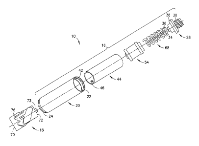
BRIEF DESCRIPTION OF THE DRAWING(S)
[0022] FIG. 1 is a perspective view of a medical connector system shown with a
first
medical device that is a syringe and a second medical device that is a
pressure regulator (a), an
IV bag adaptor (b), or a patient connector (c).
[0023] FIG. 2 is an exploded perspective view of the medical connector system
shown in
FIG. 1 according to one aspect of the present invention.
[0024] FIG. 3A is a perspective view of a first connector of the medical
connector system
shown in FIG. 1 according to one aspect of the present invention.
[0025] FIG. 3B is a cross-sectional view of the first connector of FIG. 3A
along line A-A
according to one aspect of the present invention, showing the first connector
in an actuated
position with the second connector omitted for clarity.
[0026] FIG. 4A is a perspective view of a second connector of the medical
connector system
shown in FIG. 1 according to one aspect of the present invention.
[0027] FIG. 4B is a cross-sectional view of the second connector of FIG. 4A
along line A-
A according to one aspect of the present invention.
[0028] FIG. 4C is a schematic of a groove of the second connector of FIG. 4A
according to
one aspect of the present invention.
[0029] FIG. 4D is a schematic of a groove of the second connector according to
an
alternative aspect of the present invention.
[0030] FIG. 5A is a perspective view of a patient connector provided with the
second
connector of the medical connector system of FIG. 1 according to one aspect of
the present
invention.
[0031] FIG. 5B is a cross-sectional view of the second connector of FIG. 5A
along line A-
A according to one aspect of the present application.
[0032] FIG. 6 is a cross-sectional view of the second connector of FIG. 5B
according to one
aspect of the present invention, showing the second connector inserted into
but not locked to
the first connector of FIG. 3A.
[0033] FIG. 7A is a partially transparent perspective view of a medical
connector system
according to a further aspect of the present invention.
CA 02929473 2016-05-02
WO 2015/069631
PCT/US2014/063840
[0034] FIG. 7B is a partially transparent perspective view of the medical
connector system
of FIG. 7A according to one aspect of the present invention, showing the
system upon insertion
of the second connector into the housing of the first connector.
[0035] FIG. 7C is a partially transparent perspective view of the medical
connector system
of FIG. 7A according to one aspect of the present invention, showing the
system after
application of a first set of opposing forces applied to the first connector
and the second
connector.
[0036] FIG. 7D is a transparent perspective view of the medical connector
system of FIG.
7A according to one aspect of the present invention, showing the system in a
locked state after
release of the first set of opposing forces applied to the first connector and
the second
connector.
[0037] FIG. 7E is a transparent perspective view of the medical connector
system of FIG.
7A according to one aspect of the present invention, showing the system at the
beginning of
the application of a second set of opposing forces applied to the first
connector and the second
connector.
[0038] FIG. 7F is a transparent perspective view of the medical connector
system of FIG.
7A according to one aspect of the present invention, showing the system at the
completion of
the application of a second set of opposing forces applied to the first
connector and the second
connector.
[0039] FIG. 7G is a transparent perspective view of the medical connector
system of FIG.
7A according to one aspect of the present invention, showing the system in a
released state
after release of the second set of opposing forces applied to the first
connector and the second
connector.
[0040] FIG. 8 is a perspective view of a first connector of a medical
connector system shown
according to a further aspect of the present invention, showing the connector
with a transparent
housing and indicator band.
[0041] FIG. 9 is a perspective view of a second connector of a medical
connector system
shown according to a further aspect of the present invention, showing the
connector with an
indicator band in conjunction with a vial adapter.
[0042] FIG. 10 is a perspective view of a second connector of a medical
connector system
shown according to a further aspect of the present invention, showing the
connector with an
indicator band in conjunction with an IV bag spike.
6
CA 02929473 2016-05-02
WO 2015/069631
PCT/US2014/063840
[0043] FIG. 11 is a perspective view of a second connector of a medical
connector system
shown according to a further aspect of the present invention, showing the
connector with an
indicator band in conjunction with a patient connector.
[0044] FIG. 12 is a perspective view of a medical connector system according
to one aspect
of the present invention, showing the first connector of FIG. 8 locked with
the second
connector of FIG. 9.
[0045] FIG. 13 is a perspective view of a medical connector system according
to one aspect
of the present invention, showing the first connector of FIG. 8 locked with
the second
connector of FIG. 10.
[0046] FIG. 14 is a perspective view of a medical connector system according
to one aspect
of the present invention, showing the first connector of FIG. 8 locked with
the second
connector of FIG. 11.
[0047] FIG. 15 is a perspective view of a first connector of a medical
connector system
shown according to another aspect of the present invention, showing the
connector with a
transparent housing and indicator band.
[0048] FIG. 16 is a top perspective view of a second connector of a medical
connector
system shown according to another aspect of the present invention, showing the
connector with
an indicator band in conjunction with a patient connector.
[0049] FIG. 17 is a bottom perspective view of the second connector of FIG. 16
according
to one aspect of the present invention.
[0050] FIG. 18 is a perspective view of a medical connector system according
to one aspect
of the present invention, showing the first connector of FIG. 15 locked with
the second
connector of FIG. 16.
[0051] FIG. 19 is a perspective view of a first connector of a medical
connector system
shown according to yet another aspect of the present invention, showing the
connector with a
transparent housing and indicator band.
[0052] FIG. 20 is a perspective view of a second connector of a medical
connector system
shown according to yet another aspect of the present invention, showing the
connector with an
indicator band in conjunction with a vial adapter.
[0053] FIG. 21 is a perspective view of a second connector of a medical
connector system
shown according to yet another aspect of the present invention, showing the
connector with an
indicator band in conjunction with a patient connector.
7
CA 02929473 2016-05-02
WO 2015/069631
PCT/US2014/063840
[0054] FIG. 22 is a perspective view of a first connector of a medical
connector system
shown according to a further aspect of the present invention, showing the
connector with an
opaque housing and indicator band.
[0055] FIG. 23 is a cross-sectional view of the first connector of FIG. 22
taken along line
23-23 in FIG. 22.
[0056] FIG. 24 is a perspective view of a second connector of a medical
connector system
shown according to a further aspect of the present invention, showing the
connector with an
indicator band in conjunction with a patient connector.
[0057] FIG. 25 is a perspective view of a medical connector system according
to one aspect
of the present invention, showing the first connector of FIG. 22 locked with
the second
connector of FIG. 24.
[0058] FIG. 26 is a perspective view of a first connector of a medical
connector system
shown according to another aspect of the present invention, showing the
connector with a
transparent housing and an indicator.
[0059] FIG. 27 is a perspective view of a second connector of a medical
connector system
shown according to another aspect of the present invention, showing the
connector with an
indicator.
[0060] FIG. 28 is a perspective view of a medical connector system according
to one aspect
of the present invention, showing the first connector of FIG. 26 locked with
the second
connector of FIG. 27.
[0061] FIG. 29 is a perspective view of a first connector of a medical
connector system
shown according to yet another aspect of the present invention, showing the
connector with a
transparent housing and a window.
[0062] FIG. 30 is a perspective view of a second connector of a medical
connector system
shown according to yet another aspect of the present invention, showing the
connector with an
indicator band.
[0063] FIG. 31 is a perspective view of a medical connector system according
to one aspect
of the present invention, showing the first connector of FIG. 29 locked with
the second
connector of FIG. 30.
[0064] FIG. 32 is a perspective view of a first connector of a medical
connector system
shown according to a further aspect of the present invention, showing the
connector with a
transparent housing and a window.
8
CA 02929473 2016-05-02
WO 2015/069631
PCT/US2014/063840
[0065] FIG. 33 is a perspective view of a second connector of a medical
connector system
shown according to a further aspect of the present invention, showing the
connector with an
indicator mark.
[0066] FIG. 34 is a perspective view of a medical connector system according
to one aspect
of the present invention, showing the first connector of FIG. 32 locked with
the second
connector of FIG. 33.
[0067] FIG. 35 is a perspective view of the first connector of medical
connector system of
FIG. 1, showing a grip configuration according to an alternative aspect of the
present invention.
[0068] FIG. 36 is a perspective view of the first connector of medical
connector system of
FIG. 1, showing a grip configuration according to a second alternative aspect
of the present
invention.
[0069] FIG. 37 is a perspective view of the first connector of medical
connector system of
FIG. 1, showing a grip configuration according to third alternative aspect of
the present
invention.
[0070] FIG. 38 is a perspective view of the first connector of medical
connector system of
FIG. 1, showing a grip configuration according to a fourth alternative aspect
of the present
invention.
[0071] FIG. 39 is a schematic of a groove of the second connector according to
a first
alternative aspect.
[0072] FIG. 40 is a schematic of a groove of the second connector according to
a second
alternative aspect.
[0073] FIG. 41 is a schematic of a groove of the second connector according to
a third
alternative aspect.
[0074] FIG. 42 is a schematic of a groove of the second connector according to
a fourth
alternative aspect.
[0075] FIG. 43 is a schematic of a groove of the second connector according to
a fifth
alternative aspect.
[0076] FIG. 44 is a schematic of a groove of the second connector according to
a sixth
alternative aspect.
[0077] FIG. 45 is a schematic of a groove of the second connector according to
a seventh
alternative aspect.
[0078] FIG. 46 is a schematic of a groove of the second connector according to
an eighth
alternative aspect.
9
CA 02929473 2016-05-02
WO 2015/069631
PCT/US2014/063840
DESCRIPTION OF THE INVENTION
[0079] For purposes of the description hereinafter, the terms such as "end",
"upper",
"lower", "right", "left", "vertical", "horizontal", "top", "bottom",
"lateral", "longitudinal" and
derivatives thereof shall relate to the invention as it is oriented in the
drawing figures. However,
it is to be understood that the invention may assume various alternative
variations and step
sequences, except where expressly specified to the contrary. It is also to be
understood that the
specific devices and processes illustrated in the attached drawings, and
described in the
following specification, are simply exemplary aspects of the invention. Hence,
specific
dimensions and other physical characteristics related to the aspects disclosed
herein are not to
be considered as limiting. Further, it is to be understood that the invention
may assume various
alternative variations and step sequences, except where expressly specified to
the contrary.
[0080] The present invention is directed to a connector system 10 for a
medical device. In
one aspect, the connector system 10 may be utilized for connecting and
enabling fluid transfer
between a first medical device 12 for receiving and/or dispensing fluids such
as a syringe
(FIG. 1) and a second medical device 14 for receiving and/or dispensing fluids
such as a
pressure equalization device (FIG. 1(a)), a vial adaptor, a patient connector
(FIG. 1(b)), an IV
bag adaptor (FIG. 1(c)), or a similar device used for receiving or dispensing
fluids. The overall
system is used to transfer a drug from an original container, such as a vial,
to a patient. The
medical connector 10 is the tool that is used to facilitate this closed system
transfer. The
medical connector system 10 includes a first connector 16 and a second
connector 18. FIG. 2
shows an exploded view of one aspect of the medical connector system 10.
[0081] Referring to FIGS. 2-3B, the first connector 16 is embodied as a
syringe adapter that
is configured to receive a syringe or IV line at one end and mate with the
second connector at
the other end to facilitate the sealed transfer from a first container to a
second container. The
first connector 16, however, may be provided in connection with any other
suitable medical
devices. The first connector includes a housing 20 having a proximal end 22
and a distal end
24. The housing 20 has a generally cylindrical shape that defines a central
opening 26.
[0082] A cap 28 is attached to the proximal end 22 of the housing 20. The cap
28 includes
an attachment 30 to connect the first connector 16 to the first medical device
12. The
attachment may be of any suitable configuration that allows the first
connector 16 to be securely
and sealing attached to the first medical device 12. Possible attachments
include, but are not
limited to, a luer connector or a snap-fit connector. The cap 28 has a central
passageway 32
therethrough and a proximal end 34 of a needle cannula 36 is received in the
central passageway
CA 02929473 2016-05-02
WO 2015/069631
PCT/US2014/063840
32 such that there is a fluid connection between the first medical device 12
and the needle
cannula 36 allowing the fluid in the first medical device 12 to flow into the
needle cannula 36.
[0083] In the aspect shown in FIGS. 2, 3A, 3B, and 6, the cap 28 is snap-fit
onto the housing
20. A ledge 38 extends circumferentially around the perimeter of the cap 28
and is received in
a corresponding circumferential recess 40 in the housing 20. At least one
protrusion 42 having
an upper surface that is sloped towards the distal end of the first connector
16 engages the upper
surface of the ledge 38 and holds the cap 28 within the recess 40 of the
housing 20. The sloped
upper surface of the at least one protrusion 42 allows the cap 28 to be snap
fit onto the housing
20 during assembly. Alternatively, any suitable means may be used to attach
the cap 28 to the
housing 20 as long at the cap 28 is securely attached to the housing 20.
Possible alternative
attachment means include, but are not limited to, alternative snap-fit
configurations, welding
in permanent connection, or a threaded connection. When the cap 28 is attached
to the housing
20 the needle cannula 36 extends into the central opening 26 of the housing
20, but does not
extend beyond the distal end 24 of the housing 20. The distal end of the
housing 20 is open to
allow at least a portion of the second connector 18 to be received within the
central opening 26
as will be discussed in more detail below.
[0084] The first connector 16 also includes a cam member 44. The cam member 44
may be
cylindrical in shape and includes at least one projection 46 extending into
the central opening
26 of the housing 20 as shown in FIGS. 2, 3B, 6, 8A, and 8B. The cam member 44
is rotatably
disposed within the housing 20. While the cam member 44 and the housing 20 are
shown and
described as cylindrical, they may take any suitable shape the allows for
rotation of the cam
member 44 in the housing 20, including but not limited to, a cone or a shape
having an outer
surface. The distal end 48 of the cam member 44 rests on a ledge 50 at the
distal end 24 of the
housing 20 and the proximal end of the cam member 44 is adjacent the cap 28
such that the
cam member 44 is held within housing 20 by the ledge 50 and the cap 28.
[0085] Alternatively, as shown in FIGS. 7A-7G, 9A, and 9B, the cam member 44
may be
ring shaped and may be held in a recess near the distal end 24 of the housing
20. The ring
shaped cam member 44 may be disposed within the housing 20 as shown in FIGS.
7A-7G, 9A,
and 9B or may be external to the housing 20 as shown in FIG. 8D, for example.
[0086] A carrier 54 is disposed within the central opening 26 of the housing
20. The carrier
54 has a generally cylindrical shape. The outermost surface of the carrier 54
is in sliding
contact with either the inner surface of the cam member 44 if the cam member
44 has a
cylindrical shape (FIGS. 2, 3B, 6, 8A, and 8B) or the inner surface of the
housing 20 if the cam
member 44 has a ring shape (FIGS. 7A-7G, 9A, and 9B) such that the carrier 54
may move in
11
CA 02929473 2016-05-02
WO 2015/069631
PCT/US2014/063840
an axial direction within the housing 20. The carrier 54 includes an axial
central opening 56
and at least one sealing member 58. The needle cannula 36 extends through both
the axial
central opening 56 of the carrier 54 and the at least one sealing member 58.
[0087] In the aspect shown in FIGS. 3A and 6, the two sealing members 58 are
provided
such that a first sealing member is at the proximal end 60 of the carrier 54
and a second sealing
member is at the distal end 62 of the carrier 54. Preferably, at least one
sealing member 58 is
disposed at the distal end 62 of the carrier 54. A larger ledge portion of
each sealing member
58 is received in a recess 66 in the carrier 54 to attach the sealing member
58 to the carrier 54.
In an actuated or connected position, shown in FIG. 6, the carrier 54 is moved
upward such
that the needle cannula 36 extends through the carrier 54 and the sealing
member(s) 58 to place
the first medical device 12 in fluid communication with the second medical
device 14. In a
non-actuated, or unconnected position, as shown in FIG. 3B, the distal end of
the needle
cannula 36 will be positioned in the central opening 56 of the carrier 54
between the sealing
members 58 to protect the sharpened end of the needle cannula 36 and contain
any medicament
that may be positioned within the lumen of the needle cannula 36.
Alternatively, the needle
cannula 36 could be contained in any structure that protects the sharpened end
of the needle
cannula 36 and contains any medicament that may be positioned within the lumen
of the needle
cannula 36, including, but not limited to, a single large membrane. The system
10 may also be
utilized in connection with any other suitable drug delivery mechanism or
arrangement.
[0088] A biasing member 68 is disposed between the cap 28 and the proximal end
of the
carrier 54. The biasing member 68 may be a spring, although other suitable
biasing members
may be utilized, including, but not limited to, a built in plastic spring or
an elastic material such
as rubber, TPE, or silicone. The elastic material could be placed in a grid
format with multiple
elastic strands or could be a single elastic strand. The biasing member 68
biases the carrier 54
towards the distal end 24 of the first connector 16 to ensure that the distal
end of the needle
cannula 36 is positioned within the carrier 54 when disconnected from the
second connector
18 as described above.
[0089] The second connector 18 is generally cylindrical having a distal end 70
and a
proximal end 72 and defining an axial central passageway 74, although other
suitable shapes
for the second connector 18 may be utilized. The distal end 70 of the second
connector 18 may
be integral with the second medical device 14 such as a pressure equalization
device, a vial
adaptor, a patient connector, an IV bag adaptor, or a similar fluid delivery
device (FIGS. 4A,
4B, and 7A-9B) or may include an attachment for making a connection with such
devices. For
example, referring to FIGS. 5A, 5B, and 6, the second connector 18 is provided
on a patient
12
CA 02929473 2016-05-02
WO 2015/069631
PCT/US2014/063840
connector that includes an attachment 75 to connect the second connector 18 to
a patient IV
line or other suitable connection. The attachment 75 is shown as male locking
luer connector,
although other suitable attachment arrangements may be provided. The second
connector 18
also includes a sealing member 73 at its proximal end 72.
[0090] The second connector 18 defines a groove 76 on its outer surface. The
groove 76 has
a zigzag shape extending in a number of directions (FIGS. 4A, 4C, 4D, and 5A).
The groove
76 defines a first section 78 that extends substantially axially in a distal
direction, i.e., extends
generally toward the bottom end of the second connector 18. A second section
80 of the
groove 76 extends from the distal end 78a of the first section 78 and slopes
in a distal direction
away from the distal end 78a of the first section 78, i.e., slopes downward
and away from the
bottom end of the first section 78. A third section 82 of the groove 76
extends substantially
axially in a proximal direction from the distal end 80a of the second section
80, i.e., extends
generally upward from the bottom end of the second section 80. A fourth
section 84 of the
groove 76 extends from the proximal end 82a of the third section 82 and slopes
in a proximal
direction away from proximal end 82a of the third section 82, i.e., slopes
upward and away
from the top end of the third section 82. A fifth section 86 of the groove 76
extends
substantially axially in a distal direction from the proximal end 84a of the
fourth section 84,
i.e., extends downward from the top end of the fourth section 84. A sixth
section 88 of the
groove 76 extends from the distal end 86a of the fifth section 86 and slopes
in a distal direction
away from distal end 86a of the fifth section 86, i.e., slopes downward and
away from the
bottom end of the fifth section 86. A seventh section 90 of the groove 76
extends substantially
axially in a proximal direction from the distal end 88a of the sixth section
88, i.e., extends
upward from the bottom end of the sixth section 88. An eighth section 92 of
the groove 76
extends from the proximal end of the seventh section 90 and slopes in a
proximal direction
away from proximal end of the seventh section 90, i.e., slopes upward and away
from the top
end of the seventh section 90.
[0091] The first 78, third 82, fifth 86, and seventh 90 sections of the groove
76 have been
described as extending in a substantially axial direction which includes
vertically or parallel to
the longitudinal axis of the second connector 18 and in a slightly sloping
direction as long as
they are directed in a proximal direction or distal direction overall.
[0092] In an alternative aspect, as shown in FIGS. 4D and 7A-9B, an additional
section 94
may extend axially in a proximal direction from the proximal end 84a of the
fourth section 84
and fifth section 86 may extend from the distal end 84a of the additional
section 94, i.e., the
additional section 94 may extend from the top end of the fourth section 84 and
the fifth section
13
CA 02929473 2016-05-02
WO 2015/069631
PCT/US2014/063840
86 may extend from the bottom end of the additional section 94. The additional
section 94
helps to provide additional security in the locked position but is not
required to form a locking
connection.
[0093] A proximally sloping ledge 96, i.e., a ledge that slopes towards the
bottom of the
second connector 18, extends along the exterior of the second connector 18.
The distal end of
the sloping ledge 96 extends to the proximal end of the first section 78 of
the groove 76, i.e.,
the bottom end of the sloping ledge 96 extends to the top end of the first
section 78 of the
groove 76.
[0094] The first connector 16 may be provided with two projections 46 on
opposite sides of
the cam member 44 (FIG. 3B) and the second connector 18 may be provided with
two grooves
76 on opposite sides of the second connector 18 (FIG. 5A).
[0095] In use, the proximal end 72 of the second connector 18 is inserted into
the open distal
end 24 of the housing 20 of the first connector 16 (FIG. 7B). The projection
46 on the cam
member 44 of the first connector 16 either contacts the sloping ledge 96 on
the second
connector 18 or the projection 46 is received in the first section 78 of
groove 76 depending on
the orientation of the first connector 16 with respect to the second connector
18. The proximal
end 72 of the second connector 18 contacts the distal end 62 of the carrier
54. The second
connector 18 may include a sealing member 73 positioned adjacent to the
proximal end 72 of
the second connector 18 that engages and forms a seal with the sealing member
58 positioned
at the distal end 62 of the carrier 54. After insertion, opposing axial forces
are placed on the
proximal end 77 of the first connector 16 and the distal end 70 of the second
connector 18
(FIG. 7C). The carrier 54 is forced in a proximal direction with respect to
the housing 20 by
the proximal end 72 of the second connector 18. As a result, energy is stored
in the biasing
member 68. In the case of a spring, the energy is stored by compression of the
spring. At the
same time, the second connector 18 is further received in the central opening
26 of the housing
20 of the first connector 16. As the second connector 18 is further received
in the central
opening 26 of the housing 20 of the first connector 16, the projection 46 on
the cam member
44, which is rotatably disposed in the housing 20 of the first connector 16,
either follows the
sloping ledge 96 of the second connector 18 to the groove 76 and proceeds
through the first
section 78 and second section 80 of the groove 76 or directly proceeds through
the first section
78 and second section 80 of the groove 76.
[0096] When the opposing axial forces are released, the energy stored in the
biasing member
68 forces the housing 20 of the first connector 16 in a proximal direction
with respect to the
second connector 18 (FIG. 7D). As a result, the projection 46 on the cam
member 44 proceeds
14
CA 02929473 2016-05-02
WO 2015/069631
PCT/US2014/063840
through the third section 82 and fourth section 84 of the groove 76 and, if
the additional section
94 is provided between the fourth section 84 and fifth section 86 of the
groove 76 into the
additional section 94. The proximal force provided by the biasing member 68 on
the housing
20 and, thus, the projection 46 on the cam member 44 holds the projection 46
in the groove 76
such that the first connector 16 is now locked onto the second connector 18.
In this locked
state, the housing 20 can still be rotated with respect to the second
connector 18 without
disengaging the first connector 16 from the second connector 18.
[0097] Preferably, when the first connector 16 and the second connector 18 are
in this locked
engagement, the proximal end 72 of the second connector 18 is in sealing
engagement with a
sealing member 58 on the distal end 62 of the carrier 54. The distal force
provided by the
biasing member 68 on the carrier 54 and the proximal force provided on the
second connector
18 by the projection 46 on the cam member 44 help to assure a good seal
between the sealing
member 58 and the proximal end 72 of the second connector 18.
[0098] When the first connector 16 and the second connector 18 are in this
locked
engagement, the needle cannula 36 extends into the axial central passageway 74
of the second
connector 18 and into the second medical device 14. This provides a fluid path
from the first
medical device 12 through the needle cannula 36 into the second medical device
14.
[0099] When it is desired to release the locking connection between the first
connector 16
and the second connector 18, opposing axial forces are again placed on the
proximal end 77 of
the first connector 16 and the distal end 70 of the second connector 18 (FIGS.
7E and 7F).
The carrier 54 is forced in a proximal direction with respect to the housing
20 by the proximal
end 72 of the second connector 18. As a result, energy is stored in the
biasing member 68. In
the case of a spring, the energy is stored by compression of the spring. At
the same time, the
second connector 18 is further received in the central opening 26 of the
housing 20 of the first
connector 16. As the second connector 18 is further received in the central
opening 26 of the
housing 20 of the first connector 16, the projection 46 on the cam member 44
proceeds through
the fifth section 86 and sixth section 88 of the groove 76.
[00100] When the opposing axial forces are released, the energy stored in the
biasing
member 68 forces the housing 20 of the first connector 16 in a proximal
direction with respect
to the second connector 18 (FIG 7G). As a result of the proximal force on the
housing 20, the
projection 46 on the cam member 44 proceeds through the seventh section 90 and
eighth
section 92 of the groove 76 and the first connector 16 is released from
engagement with the
second connector 18. The distal end of the needle cannula 36 is once again
contained in carrier
54 of the first connector 16.
CA 02929473 2016-05-02
WO 2015/069631
PCT/US2014/063840
[00101] In the released position the tip of the needle cannula 36 is contained
in the axial
central opening 56 of the carrier 54 between the two sealing members 58. Thus,
not only is the
user protected from an accidental needle stick, but any fluid that may remain
in the needle
cannula 36 is contained in the needle cannula 36 and or the carrier 54 by the
sealing members
58.
[00102] While the connector system 10 has been described and shown as having a
biasing
member 68, another aspect does not include a biasing member. In this aspect,
the user applies
opposing axial forces on the proximal end 77 of the first connector 16 and the
distal end 70 of
the second connector 18 pushing the first connector 16 onto the second
connector 18 until the
protrusion 46 has traveled through the first 78 and second 80 sections of the
groove 76 and the
first connector 16 cannot be advanced on the second connector 18 any further.
Then the user
applies opposing axial forces on the proximal end 77 of the first connector 16
and the distal
end 70 of the second connector 18 pulling the first connector 16 away from the
second
connector 18 until the protrusion 46 has traveled through the third 82 and
fourth 84 sections of
the groove 76 and into the additional section 94. The additional section 94 is
in the form of
detent to provide a locking engagement with the protrusion 46. When the
protrusion 46 is
locked in the additional section 94, the first connector 16 is locked to the
second connector 18.
[00103] When it is desired to release the locking connection between the first
connector 16
and the second connector 18, the user again applies opposing axial forces on
the proximal end
77 of the first connector 16 and the distal end 70 of the second connector 18
pushing the first
connector 16 onto the second connector 18 until the protrusion 46 is released
from the
additional section 94 and travels through the fifth 86 and sixth 88 sections
of the groove 76 and
the first connector 16 cannot be advanced on the second connector 18 any
further. Then the
user applies opposing axial forces on the proximal end 77 of the first
connector 16 and the
distal end 70 of the second connector 18 pulling the first connector 16 away
from the second
connector 18 until the protrusion 46 has traveled through the seventh 90 and
eighth 92 sections
of the groove 76 and the first connector 16 is released from the second
connector 18.
[00104] As shown in FIGS. 8-25, the first connector 16 may have an indicator
band 98
extending axially on the outer surface of the cam member 44. In this case, the
housing 20 of
the first connector 16 is transparent. The second connector 18 may have an
indicator band 100
extending axially on its outer surface. The indicator bands 98, 100 are placed
on the cam
member 44 and the second connector 18 such that, when the first connector 16
is in locking
engagement with the second connector 18, the indicator band 98 on the cam
member 44 which
will be visible through the transparent housing 20 will be aligned with the
indicator band 100
16
CA 02929473 2016-05-02
WO 2015/069631
PCT/US2014/063840
on the second connector 18 to give a visual indication to the user that the
connector system is
locked. Preferably, the indicator band 98 on the cam member 44 is located 90
around the
circumference of the cam member 44 from the protrusion 46. Alternatively, if
the second
connector 18 is integral with the second medical device 14, the indicator band
100 may be
included on the exterior surface of the second medical device 14.
[00105] The indicator line 98 may extend the full length of the cam member 44
and the
second connector 18 as shown in FIGS. 8 and 11 or may only extend for part of
the length of
the cam member 44 and the second connector 18 as shown in FIG. 19.
Alternatively, if the
cam member 44 is external from the housing 20, the housing 20 need not be
transparent.
[00106] In another aspect, shown in FIGS. 26-28, with a connector system
having a cam
member 44, the first connector 16 may have a dot 102 on the outer surface of
the housing 20.
In this case, the housing 20 of the first connector 16 is transparent. The
second connector 18
may have a circle 104. When the first connector 16 is in locking engagement
with the second
connector 18, the dot 104 on the housing 102 will be visible through the
transparent housing
20 and will be located in the circle 104 on the second connector 18 to give a
visual indication
to the user that the connector system is locked.
[00107] In another aspect, shown in FIGS. 29-34, with a connector system
having a cam
member 44, the cam member 44 may include a window 105 and the second connector
may
include an indicator band 100 (FIGS. 30 and 31) or an indicator mark 107
(FIGS. 33 and 34).
In this case, the housing 20 of the first connector 16 is transparent. When
the first connector 16
is in locking engagement with the second connector 18, the indicator band 100
or indicator
mark 107 will be visible through the transparent housing 20 and will be
located in the window
105 of the cam member 44 to give a visual indication to the user that the
connector system is
locked. In yet another aspect, instead of the indicator band 100 or indicator
mark 107 being on
the second connector 18, the colored portion may be provided on the carrier
54. In this case,
the indicator could be visible through a window in the opaque housing when the
carrier 54 is
moved within the housing 20.
[00108] Referring to FIGS. 35-38, although the housing 20 of the first
connector 16 is shown
to be generally cylindrical in FIG. 2, for example, the housing 20 of the
first connector 16 may
also include features to enhance the ability of a user to grip the housing 20.
[00109] Referring to FIGS. 35 and 36, the housing 20 of the first connector 16
may include
grip portions 106 that are generally planar regions compared to the
cylindrical surface of the
remaining portion of the housing 20. The housing 20 is generally cylindrical
in FIG. 35 with
a recessed, planar grip portion 106 that has an hourglass-shaped
circumference. The housing
17
CA 02929473 2016-05-02
WO 2015/069631
PCT/US2014/063840
is generally cylindrical in FIG. 36 with a recessed, planar grip portion 106
that has a generally
rectangle-shaped circumference with rounded ends. The grip portions 106
provide a contact
surface to allow the housing 20 to be more readily gripped by a user of the
connector 16.
[00110] Referring to FIG. 37, the housing 20 of the first connector 16 may
include a plurality
of annular ribs 108 that extend circumferentially around the outer surface of
the housing 20.
The housing 20 may include a plurality of the annular ribs 108 that extend the
full length or
only a portion of the length of the housing 20. The annular ribs 108 provide a
surface for a
user to more readily grip the connector 16.
[00111] Referring to FIG. 38, the housing 20 of the first connector 16 may
also define a
concave grip portion 110 including nubs or projections. The concave grip
portion 110 is a
portion of the housing 20 that extends radially inwardly around the
circumference of the
housing 20 to provide a contact surface that allows the connector 16 to be
more readily gripped
by a user.
[00112] Although the projection 46 of the first connector 16 extends radially
inward and the
groove 76 of the second connector 18 is positioned on the outer surface of the
second
connector 18, the projection 46 may extend radially outward and provided on
the outer surface
of the second connector 18 with the groove 76 of the second connector 18
provided on an
interior surface of the second connector 18.
[00113] As shown in FIGS. 39-46, the groove 76 of the second connector 18 can
take any
of a number of paths as long as, upon application and release of a first set
of opposing axial
forces applied to the proximal end 77 of the first connector 16 and the distal
end 70 of the
second connector 18, the first connector 16 is locked to the second connector
18 and, upon
application and release of a second set of opposing axial forces to the
proximal end 77 of the
first connector 16 and the distal end 70 of the second connector 18, the first
connector 16 is
released from the second connector 18. These include: pathways that can be
linked and
repeated (FIG. 39), pathways that follow the same trail during application and
release of the
first set of opposing axial forces and application and release of the second
set of axial forces
(FIGS. 44 and 45), pathways that have curved sections (FIG. 43), and pathways
that are looped
such that they have a common entry and exit point but follow a loop through
the majority of
the locking and releasing steps (FIG. 46). All paths may include at least five
positions: (1) a
starting position, (2) an initial base position, (3) an intermediate (or
locked) position, (4) a
secondary base position, and (5) an ending position. For some paths, the
initial base position
(2) and the secondary base position (4) are the same (FIGS. 44 and 45).
Similarly, the starting
position (1) and the ending position (5) may also be the same depending on the
path (FIG.
18
CA 02929473 2016-05-02
WO 2015/069631
PCT/US2014/063840
46).While the aspects shown in the figures have two projections 46 on the cam
member 44 and
two grooves 76 on the second connector 18, any number of projections 46 may be
used. The
corresponding groove or grooves 76 may be altered or scaled to account for the
number and/or
position of the projections 46 as long as the shape of the groove 76 allows
for locking of the
first connector 16 to the second connector 18 upon application and release of
a first set of
opposing axial forces and release of the first connector 16 from the second
connector 18 upon
application and release of a second set of opposing axial forces. In addition,
the number of
projections 46 and grooves 76 need not be equal. For example, one projection
46 could be
used with two or more repeated groove 76 patterns or two projections 46 could
be used with
four repeated groove 76 patterns.
[00114] The connector system has been previously described as having a cam
member 44
with at least one protrusion 46 that is rotatably disposed in the housing 20
of the first connector
16 with the second connector 18 being stationary. Alternatively, the
protrusion 46 may be
fixed directly to the housing 20 or keyed to the housing 20. In this case, the
second connector
18 would then be placed on a secondary component (similar to a cylinder)
allowing the groove
76 to rotate relative to the housing 20 and the protrusion 46.
[00115] The connector system has also been previously described as having the
groove 76
on the exterior of the second connector 18 and the cam member 44 as part of
the first connector
16. Alternatively, the groove 76 could be placed on the inner wall of the
housing 20 of the first
connector 16 and the cam member 44 could be placed on the exterior of the
second connector
18. There are two variations of this aspect. First, the cam member 44 with at
least one
protrusion 46 could be a rotating washer on the exterior of the second
connector 18 and the
groove 76 could be fixed in the inner wall of the housing 20 of the first
connector 16. Second,
the protrusion 46 could be fixed to the exterior of the second connector 18
and the groove could
be placed on the inside of a secondary component (like a cylinder) that could
rotate freely
within the inner walls of the housing 20 of the first connector 16 allowing
the groove 76 to
rotate with respect to the protrusion 46.
[00116] While this disclosure has been described as having exemplary designs,
the present
disclosure can be further modified within the spirit and scope of this
disclosure. This
application is therefore intended to cover any variations, uses, or
adaptations of the disclosure
using its general principles.
19
CA 2 92 9473 2 01 8-01-1 9