Note: Descriptions are shown in the official language in which they were submitted.
CA 02936453 2016-07-08
WO 2015/106118 PCT/US2015/010843
1
SYSTEMS AND METHODS USING ULTRASOUND FOR TREATMENT
BACKGROUND
[0001] The field of the disclosure relates generally to systems and methods
for using
ultrasound for treatment in the healthcare field.
[0002] Generally, it has been shown that some infections are resistant to
conventional
treatments, such as antibiotics alone. For example, biofilm and Methicillin-
resistant
Staphylococcus aureus are resistant to antibiotic treatment alone. It is also
known that some
treatments in the healthcare field need improvements.
BRIEF DESCRIPTION
[0003] In one aspect, a device for treating infection within a subject
comprises an
ultrasound transducer for applying ultrasound to a treatment site of the
subject.
[0004] The features, functions, and advantages that have been discussed can be
achieved independently in various embodiments or may be combined in yet other
embodiments, further details of which can be seen with reference to the
following description
and drawings.
BRIEF DESCRIPTION OF THE DRAWINGS
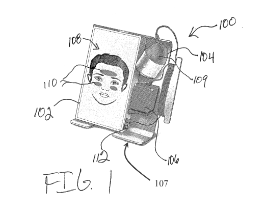
[0005] FIG. 1 is a perspective view of an exemplary high frequency ultrasound
(HFUS) device for affecting bacteria, biofilm, and/or infection within the
body.
[0006] FIG. 2 is a block diagram of the circuitry positioned in the device
housing
shown in FIG. 1.
[0007] FIG. 3 is a cross section of the treatment applicator shown in FIG. 1.
[0008] FIG. 4 is a perspective view of an alternative HFUS device having the
components of the device shown in FIG. 1.
[0009] FIG. 5 is schematic diagram of potential treatment regions of a patient
that
may be used with the device shown in FIG. 1.
[0010] FIG. 6 is a graphical representation of the results of Test 1 using the
device
shown in FIG. 1.
[0011] FIG. 7 displays microscope images of the results of Test 2 using the
device
shown in FIG. 1.
[0012] FIG. 8 displays microscope images of the results of Test 3 using the
device
shown in FIG. 1.
CA 02936453 2016-07-08
WO 2015/106118 PCT/US2015/010843
2
[0013] FIG. 9 is an exemplary flowchart of a method for use with the device
shown
in FIG. 1.
[0014] FIG. 10 is an exemplary flowchart of a method for use with the device
shown
in FIG. 1.
[0015] FIG. 11 is an exemplary drive waveform that may be used with the
applicator
shown in FIG. 1.
[0016] FIG. 12 is an exemplary drive section that may be used with the
applicator
shown in FIG. 1.
[0017] FIG. 13 is an alternative drive section that may be used with the
applicator
shown in FIG. 1.
[0018] FIG. 14 is an alternative drive section that may be used with the
applicator
shown in FIG. 1.
[0019] FIG. 15 is an alternative drive section that may be used with the
applicator
shown in FIG. 1.
[0020] FIG. 16 is a perspective view of an alternative HFUS device having the
components of the device shown in FIG. 1.
[0021] FIG. 17 is a perspective view of HFUS device with a display showing a
graphical representation of a human body.
[0022] FIG. 18 is a perspective view of HFUS device with a display showing a
graphical representation of a human knee.
[0023] FIG. 19 is a schematic showing HFUS device treating a human knee.
[0024] FIG. 20 is a schematic showing HFUS device treating a stent in a human
body.
[0025] FIG. 21 is a schematic showing HFUS device treating a screw in a human
body.
[0026] FIG. 22 is a schematic showing HFUS device treating mesh in a human
body.
[0027] FIG. 23 is a cross section of another embodiment of a treatment
applicator
similar to the treatment applicator of FIG. 1.
[0028] FIG. 24 is a perspective of another embodiment of a high frequency
ultrasound (HFUS) device for affecting bacteria, biofilm, and/or infection
within the body,
similar to the embodiment illustrated in FIG. 1.
[0029] FIG. 25 is a front elevational view of FIG. 24.
CA 02936453 2016-07-08
WO 2015/106118 PCT/US2015/010843
3
DETAILED DESCRIPTION
[0030] In one embodiment, the systems and methods described herein enable
treatment of infection of a living subject (i.e., a human or other animal)
using high frequency
ultrasound (HFUS). As used herein, the term "infection" refers to an invasion
of the living
subject by an infectious agent, regardless of whether the infectious agent
causes a disease.
Non-limiting examples of infectious agents causing infection include bacteria,
viruses, fungi,
parasites, and prions. The infectious agent(s) causing the infection may exist
in the living
subject in a planktonic state or as biofilm. As used herein, infectious agents
causing the
infection are in a planktonic state (i.e., a planktonic infection) if the
infectious agents are free-
floating within the subject, and the infectious agents are in a biofilm (i.e.,
a biofilm infection) if
the infectious agents are microorganisms adhered to each other on a surface
within the subject
and are enclosed by a self-produced matrix of a secreted extracellular
polymeric substance.
The biofilm extracellular polymeric substance excreted by the biofilm
infection may comprise
polysaccharides (e.g., exopolysaccharides), proteins, DNA, lipids and humic
substances.
Examples of infectious agents forming biofilms described herein include, but
are not limited
to, bacteria, archaea, protozoa, fungi, and algae.
[0031] FIG. 1 is a perspective view of an exemplary high frequency ultrasound
(HFUS) device, generally indicated at 100, for treating infection of a living
subject. In general,
the device is configured to deliver ultrasonic energy (e.g., high frequency
ultrasonic energy) to
a site of infection of the living subject to treat (i.e., combat, ameliorate,
inhibit, and/or prevent)
the infection. Device 100 comprises a device housing 102, a treatment
applicator 104
including an ultrasonic transducer 310, and a control circuit 200 contained
within the housing
for controlling the output of the ultrasonic transducer. In one embodiment,
device 100 is
powered by an AC power adapter 106 (e.g., an external or internal AC power
adapter)
configured to receive AC power from a power source (e.g., mains power) and
convert the AC
power to DC power used by device 100.. In the illustrated embodiment, the HFUS
device 100
also includes a DC power source within the housing 102. As a non-limiting
example, DC
power source may be a battery, including but not limited to, a rechargeable
lithium-ion battery
(e.g., battery and charger circuit 202). The HFUS device 100 may be powered in
other ways
without departing from the scope of the present invention.
[0032] In one embodiment, applicator 104 and/or housing 102 are configured to
be
hand-held and portable such that a user can utilize applicator 104 and/or
housing 102 with one
hand. In some embodiments, applicator 104 and/or housing 102 are configured to
have an
CA 02936453 2016-07-08
WO 2015/106118 PCT/US2015/010843
4
ergonomic design when held by a user. For example, in the illustrated
embodiment applicator
104 includes a recess 109 that contours to one or two fingers that aid in
stabilization of
applicator 104. Recess 109 also enables a user to hold applicator 104 with a
pinch grip for
ease of use. Housing 102 and applicator 104 are storable on a base 107 (e.g.,
a stand).
Housing 102 and/or applicator 104 may be removably coupled to base 107, such
as by magnets
(not shown)
[0033] A user interface 108 is provided on housing 102 to allow communication
between the user and device 100, in particular between the user and the
control circuit 200.
User interface 108 has a presentation function configured to present
information, such as
treatment information and/or execution events, to a user. For example, user
interface 108 may
include a display device, as illustrated, for presenting information to a
user. The display device
may include a cathode ray tube (CRT), a liquid crystal display (LCD), LED, an
organic LED
(OLED) display, a vacuum fluorescent display (VFD), and/or an "electronic ink"
display. In
some embodiments, user interface 108 may include one or more display devices.
In the
illustrated embodiment, user interface 108 displays the intended application
area and/or
configuration of device 100 for treating infection to a user. For example, as
illustrated in FIG.
1, user interface 108 comprises a display generating a graphical
representation of a human face
to which treatment of infection using device 100 is to be applied. In other
examples, device
100 may be configured to generate a graphic representation of another
portion(s) of a human
body or the entire human body on the display. For example, as shown in FIG. 18
a graphical
representation of a knee or other joint of the body may be generated on the
display to indicate
the desired treatment site. Data may be stored in a remote database, such as
cloud storage.
[0034] In the exemplary embodiment, user interface 108 also has an input
function to
allow a user to communicate with device 100, in particular control circuit
200. As an example,
to allow a user to communicate with device 100, user interface 108 may include
keys, a
pointing device, a mouse, a stylus, a membrane switch, a touch sensitive panel
(e.g., a touch
pad or a touch screen), a gyroscope, an accelerometer, a position detector,
and/or an audio user
input interface. In the illustrated embodiment, user interface 108 comprises a
touch screen
having both presentation and input functions.. In one example, user interface
108 may be
configured to receive input from user as to a desired treatment area and/or
treatment protocol.
In the illustrated embodiment user interface 108 (i.e., touch screen) may be
configured to allow
user to select a body portion for treatment and/or a specific area of a body
portion for
treatment. For example, as illustrated user interface 108 allows a user to
select a sinus area for
CA 02936453 2016-07-08
WO 2015/106118 PCT/US2015/010843
treatment by touching the desired sinus area on the display. This selection is
communicated to
control circuit 200, as explained in more detail below. .
[0035] In the exemplary embodiment, a communication interface 112 coupled to
control circuit 200 is provided on housing 102. Communication interface 112
communicates
with control circuit 200 to allow transfer of treatment and/or session
information stored by
device 100. To communicate with control circuit 200, communication interface
112 may
include, for example, a wired network adapter, a wireless network adapter,
and/or a mobile
telecommunications adapter. In some embodiments, communication interface 112
is a direct
link interface for linking two computing devices, the direct link interface
including, but not
being limited to, a serial port, a firewire port, a USB port, and an Ethernet
port. In one
embodiment, communication interface 108 includes a Bluetooth adapter capable
of
communication with a Bluetooth receiver positioned in a separate computing
device (e.g.,
tablet, pc, smartphone, and smartwatch). In some embodiments, communication
interface 112
receives information such as executable instructions and/or other data that
can be stored and/or
executed by control circuit 200.
[0036] FIG. 2 is a block diagram of device 100 illustrated in FIG. 1. As shown
in
FIG. 2, power source 106 is electrically connected to a battery and charger
circuit 202.
Electrical power (e.g., DC current) is delivered from power source 106 (e.g.,
AC adapter) to
battery and charger circuit 202. Battery and charger circuit 202 is
electrically connected to
boost converter 204. Battery and charger circuit 202 transmits electrical
power (e.g., DC
power) to boost converter 204. Boost converter 204 is electrically connected
to drive circuit
206. Boost converter 204 outputs DC power to drive circuit having a DC voltage
that is
greater than DC voltage of the power received from battery and charger circuit
202.
[0037] A processor 208 of control circuit 200 is electrically connected to
user
interface 110, communication interface 112, and drive circuit 206. In the
illustrated
embodiment, processor 208 is configured to execute instructions provided on
non-transitory
computer readable medium, such as memory device 209. Instructions provided on
memory
device 209 include instructions for operating device 100 to treat infection of
subject using
applicator 104, as explained below. Processor 208 communicates with user
interface 110 to
receive commands from user and output information to user, as described below.
In the
illustrated embodiment, processor 208 operates as a waveform generator,
whereby an electrical
signal is delivered to drive signal in accordance with the desired frequency
output of
ultrasound transducer 310. In particular, drive circuit 206 is configured to
receive DC power
CA 02936453 2016-07-08
WO 2015/106118 PCT/US2015/010843
6
from boost converter 204 and a waveform electrical signal from processor 208.
The drive
circuit delivers an AC drive signal to transducer 310 of applicator 104 based
on the waveform
electrical signal and the DC power received from the boost converter.
Transducer 310 outputs
desired HFUS energy (i.e., having a desired frequency and intensity) in
accordance with the
received drive signal. The outputted HFUS energy is suitable for treating
infection of the
subject. The output of transducer 104 may be monitored by a feedback circuit
210 in
communication with processor 208.
[0038] FIG. 3 is a side cut-away view of applicator 104 shown in FIG. 1. In
the
exemplary embodiment, applicator 104 includes shell 306 having a body portion,
generally
indicated at 300, and an applicator head portion, generally indicated at 302,
configured to
provide the HFUS energy to a treatment site in the body. A similar embodiment
of a head
portion 302' is shown in FIG. 23, with differences between the embodiment
discussed below.
In some embodiments, a retention groove 304 is formed on shell 306. Retention
groove 304 is
configured to enable applicator 104 be held by a system retention apparatus,
such as a clip or
stand provided on base 107. For example, as shown in FIGS. 24 and 25, in
another
embodiment the base 107 incudes a U-shaped cutout 305 in which the applicator
104 is
retained. A bottom edges defining the U-shaped cutout 305 is received in the
retention groove
304 of the applicator 104 to hold the applicator in the U-shaped cutout. In
one embodiment,
shell 306 of applicator 104 is fabricated from a polymer, including but not
limited to,
acrylonitrile butadiene styrene, polyether ether ketone, Polyoxymethylene.
Alternatively, shell
306 can be fabricated from any material that facilitates transmitting energy
(e.g. ultrasonic
vibrations) from applicator 104 to a treatment site of the subject, including
but not limited to,
titanium, aluminum, and stainless steel. In another embodiment, body portion
300 of outer
shell 306 may be fabricated from a polymer and head portion 302 may be
fabricated from a
metallic substance (e.g. titanium).
[0039] As described above, transducer 310 is configured to convert drive
signal into
ultrasonic vibratory energy that will be utilized to infection at a treatment
site of the subject. In
the exemplary embodiment, transducer 310 is configured to output ultrasound
energy having a
frequency selected to be above 20 kHz, such as from about 1 MHz to about 5
MHz, and an
intensity from about 0.20 W/cm2 to about 3 W/cm2, such as from about 1 W/cm2
to about 0.5
W/cm2, in accordance with the drive signal received from drive circuit 206.
Transducer 310
may comprise a piezoelectric crystal having a square shape or any other
suitable shape,
including but not limited to, round, circular, oval, and rectangular. In at
least some
CA 02936453 2016-07-08
WO 2015/106118 PCT/US2015/010843
7
embodiments, applicator 104 includes a plurality or array of transducers 310
for transmitting
ultrasonic vibratory energy. In such embodiments, transducers 310 are arranged
to focus at a
treatment site with two or more transducers 310 outputting different
frequencies such that the
intersection of the ultrasound beams will create a different frequency at the
desired treatment
signal. Alternatively, discrete transducers 310 are configured to provide
different beams that
are configured to affect particular portions (e.g. proximal, distal, etc.) of
a treatment site. In
some embodiments, discrete transducers 310 are configured to simultaneously
provide multiple
beams that are configured to treat different types of infections (e.g. MRSA
infection, bacterial
biofilm infection, and fungal biofilm infection).
[0040] In some embodiments, the frequency of the output of transducer 310 is 1
MHz. An output of ultrasonic vibratory energy of 1 MHz has a beneficial effect
on pain and
swelling. Additionally, it is believed the output of ultrasonic vibratory
energy at a frequency
of 1 MHz has an effect on infectious agents and/or biofilms by turning
infectious agents, such
as bacteria, and/or biofilm into planktonic state, causing delamination of
biofilm, creating a
physical disruption, and/or breakdown of polysaccharides present in biofilm
matrix. Further,
with respect to treating infection of sinus cavity, the application of the
high frequency
ultrasound can have an effect on the viscosity of mucus in the sinus cavity
which can enhance
drainage.
[0041] In the exemplary embodiment (FIG. 1), applicator 104 includes a
vibratory
device 312, such as but not limited to a piezoelectric transducer or an
eccentric vibrator motor,
that provides tactile vibrations to the user. Such an embodiment enables a
user to feel that
applicator 104 is functioning and/or in a treatment mode. Vibratory device 312
may be
configured to operate, thereby vibrate, when the ultrasound transducer 310 is
outputting the
ultrasound signal during treatment. In one embodiment, vibratory device 312
may be powered
by the drive signal from drive circuit 206 simultaneously with the drive
signal powering
ultrasound transducer 310. In the exemplary embodiment, ultrasonic transducer
310 and
vibratory transducer 312 are configured to power simultaneously from
electrical 202.
Alternately, transducers 310 and 312 can be configured to power individually.
In the
embodiment illustrated in FIG. 23, the head portion 302' includes a vibratory
device 313
comprising an eccentric vibrator motor 313. Other types of vibratory devices
do not depart
from the scope of the present invention.
[0042] In the exemplary embodiment (FIG. 1), applicator 104 includes an
imaging
transducer 311 (e.g., a transceiver) for sending and receiving ultrasound
suitable for imaging
CA 02936453 2016-07-08
WO 2015/106118 PCT/US2015/010843
8
the treatment site. Processor 208 may be configured to process the ultrasound
for imaging, as
explained in more detail below.
[0043] In some embodiments, applicator 104 is configured to provide multiple
treatment modalities to treat infection. In addition to providing ultrasound
energy by operation
of ultrasonic transducer 310, device 100 may be configured to apply additional
energy, other
than HFUS energy, to the treatment site. For example, applicator 104 may
include additional
treatment components 316 and/or 318. In one embodiment, treatment component
316
comprises a transducer configured to provide extracorporeal shockwaves to the
treatment site
and/or pulsed electromagnetic frequency (PEMF) energy to a treatment site. In
one
embodiment, component 318 is a conductor configured to provide electrical AC
current in the
radio frequency range or DC current. In yet another embodiment, the treatment
component 316
may include a source of ultraviolet light for using in treating in conjunction
with the ultrasonic
transducer. The source of ultraviolet light may emit light in the C range from
about 270
nanometers to approximately 320.
[0044] To inhibit overheating of applicator 104, a temperature sensor 320 is
sensor is
coupled within applicator 104 to provide temperature feedback to processor
208. If the
temperature sensed by temperature sensor 320 is greater than a threshold
temperature,
processor 208 may be configured to reduce intensity of the drive signal or
discontinue
treatment using device 100 until the temperature falls within an acceptable
range. In the
embodiment illustrated in FIG. 23, the head portion 302 includes a temperature
sensor 320' and
a heat siffl( 321 to reduce overheating of the applicator 104. The heat siffl(
321 is in thermal
contact with the transducer 310' to transfer heat from the transducer to the
heat siffl( to inhibit
overheating of the shell 306'. The heat siffl( 321 may comprise any suitable
thermally
conductive material having a thermal conductivity greater than the shell 306',
for example.
[0045] In the illustrated embodiment, a transmission component 330 is coupled
to
head portion 302 of applicator 104. Transmission component 330 is fabricated
to enable
transmission of ultrasound energy from applicator 104 into the body of a
subject without a
coupling gel. In one embodiment, transmission component 330 is an overmold
coupled on
head portion 302. In the exemplary embodiment transmission component 330 is
fabricated
from silicone. Alternatively, transmission component 330 can be fabricated
from any material
that enables the transmission of energy from applicator 104 to a treatment
site within the body
of the subject including, but not limited to, an ultra-high-molecular-weight
polyethylene, a
thermoplastic elastomer, and polytetrafluoroethylene. In some embodiments,
transmission
CA 02936453 2016-07-08
WO 2015/106118 PCT/US2015/010843
9
component 330 is a reservoir that includes an aperture for inserting and
extracting material into
the reservoir. In such an embodiment, gels and/or other substances capable of
transmitting
energy from applicator 104 to a treatment site within the body can be heated
or cooled before
inserting into the reservoir to provide heating or cooling to tissue that
contacts transmission
component 330. A specific drain or aspirate can be provided in addition to the
vibratory
circuit.
[0046] In one embodiment, head portion 302 includes at least one aperture 340
in a
portion of head portion 302 that contacts the skin of the user (e.g.
transmission component
330). In such an embodiment, a suction component 342 may be coupled to the at
least one
aperture 340 to provide suction pressure and create a partial vacuum at the
skin of a user to aid
in retaining applicator 104 against the skin of a user during a treatment
session.
[0047] FIG. 4 is a perspective view of an alternative HFUS device 400 having
the
components of device 100 shown in FIG. 1. In the exemplary embodiment, the
features of
device 100 are integrated into a single handheld unit that is configured to
treat infection within
the subject. For example, device 400 includes a device housing 402, a
treatment applicator
404, and a power source 406. Device housing 402 includes a user interface 408
similar to the
first embodiment. In one embodiment, device 400 is configured to be hand-held
and portable
such that a user can utilize device 400 with one hand. In some embodiments,
applicator 104
and/or housing 102 are configured to have an ergonomic design when held by a
user such as
including one or more recesses 412 that aids in stabilization.
[0048] It should be noted that devices 100 and/or 400 are shown to be
configured for
treating infection in the sinus of a subject. Devices 100 and/or 400 are shown
have treatment
sites being the frontal 502 and maxillary sinus 508 (shown in FIG. 5) with the
transducer
delivering ultrasound energy through the skin and into the sinus cavity to
treat infection.
[0049] High frequency ultrasound (HFUS)
[0050] To validate the effectiveness of treating infection with HFUS energy,
testing
of the output of device 100 shown in FIG. 1 was performed in a medical
biofilms laboratory.
As shown in more detail below, the output of device 100 was tested against (1)
a methicillin-
sensitive (MSSA) Staphylococcus aureus strain isolated from a sinus of a
subject with chronic
rhinosinusitis and (2) a methicillin-resistant (MRSA) Staphylococcus aureus
strain isolated
from a chronic wound of a subject. A CDC biofilm reactor (CDC-BR) was used for
growth of
MSSA Staphylococcus aureus and MRSA Staphylococcus aureus biofilms on
polycarbonate
coupons that were subjected to testing.
CA 02936453 2016-07-08
WO 2015/106118 PCT/US2015/010843
[0051] Test 1 ¨ HFUS energy from device 100 was tested on MSSA Staphylococcus
aureus coupons. The mean log density (MLD, standard deviation) of the
control biofilms
was 7.95 0.05 log CFU/cm2. Using device 100, coupons were exposed to five
minutes of
HFUS energy from applicator 104 at a frequency of 1 MHz and an intensity of
approximately 1
W/cm2. As shown by graph 600 in FIG. 6, treatment of the coupons with device
100 resulted
in a mean log reduction (MLR) of (1.08 0.13) yielding a 91.41% reduction of
MSSA
Staphylococcus aureus biofilm on the coupons.
[0052] To calculate the elimination of bacteria and/or biofilm in Test 1, the
treatments were assessed relative to untreated controls using viable plate
count methods. The
coupons were placed in tubes containing 10 ml phosphate-buffered saline (PBS).
A sequence
of vortex, sonicate, and vortex were then used to remove bacteria from the
coupons and
produce a bacterial suspension. The suspension was serially-diluted in PBS and
plated on
Tryptic Soy Agar (TSA). The plates were incubated at 37 C for 24-48 hours and
the number
of colony forming units (CFU) were counted. Based on the dilution and the
dimensions of the
coupons, the CFU per unit area (CFU/cm2) was calculated. The CFU/cm2 counts
were
logarithmically transformed (base 10) to determine log density (LD) and a mean
log density
(MLD) was calculated from replicate coupons.
[0053] Test 2 - FIG. 7 displays microscope images 640 of the results of Test 2
using
device 100 shown in FIG. 1. For Test 2, Staphylococcus aureus MSSA biofilms
were grown
in the CDC-BR, as described above, and the coupons were subjected to HFUS
output from
device 100 with a power level of 1 W/cm2 (100%) at 1 MHz for 5 minutes. Two
coupons were
treated and one coupon served as an untreated controls. After treatment, the
coupons were
treated with the LIVE/DEADO BacLightTM Viability Kit which includes two
nucleic acid
stains, SYTO-9 and propidium iodide. SYTO-9 stains live bacterial cells green
and propidium
iodide stains bacterial cells with damaged membranes (dead) red. After
treatment of the
viability kit, coupons were imaged with a Leica 5P5 confocal scanning laser
microscope. As is
shown by pictures 640, the coupon subjected to HFUS output 644 from device 100
had less
infectious agents (e.g. bacteria and/or biofilm) than the control coupon 642.
[0054] Test 3 - FIG. 8 displays microscope images 660 of the results of Test 3
using
device 100 shown in FIG. 1. For Test 3, MRSA Staphylococcus aureus biofilms
were grown
in the CDC-BR, as described above, and the coupons were subjected to HFUS
output from
device 100 with a power level of 1 W/cm2 (100%) at 1 MHz for 5 minutes. Two
coupons
were treated and one coupon served as an untreated controls. After treatment,
the coupons were
CA 02936453 2016-07-08
WO 2015/106118 PCT/US2015/010843
11
treated with the LIVE/DEADO BacLightTM Viability Kit which includes two
nucleic acid
stains, SYTO-9 and propidium iodide. SYTO-9 stains live bacterial cells green
and propidium
iodide stains bacterial cells with damaged membranes (dead) red. After
treatment of the
viability kit, coupons were imaged with a Leica SP5 confocal scanning laser
microscope. As is
shown by pictures 660, the coupon subjected to HFUS output 664 from device 100
had less
infectious material (e.g. bacteria and/or biofilm) than the control coupon
662. As shown by the
results of Tests 1-3, shown in FIGS. 6-8, device 100 is configured to
negatively affect
infectious agents inside the body. As described above and illustrated in FIGS
6-8, the systems
and methods described herein enable a user to treat biofilm. , It is believed
the output of
ultrasonic vibratory energy at a frequency of 1 MHz has an effect on
infectious agents and/or
biofilms by turning infectious agents, such as bacteria, and/or biofilm into
planktonic state,
causing delamination of biofilm, creating a physical disruption, and/or
breakdown of
polysaccharides present in biofilm matrix.
[0055] FIG. 9 is an exemplary flowchart of a method 700 for use with device
100
shown in FIG. 1. To initiate method 700, device 100 receives 702 instructions
to start a
treatment session, from input interface 110 and/or presentation interface 108.
Alternatively,
treatment information can be transmitted in any known manner including through
communication interface 108. In some embodiments, treatment information
relates to a
particular body area and/or region of the body to be treated. Alternatively,
treatment
information relates to the specific infection to be treated. After receiving
702 instructions,
device 100 performs a self-test 704. It should be noted that power and error
checking
algorithms performed during self-test 704 can be implemented at any time
throughout method
700. In the exemplary embodiment, processor 208 communicates with all hardware
components to determine if any errors occur.
[0056] After performing a self-test 704, processor 208 calibrates 706 the
output of
applicator 104. In the exemplary embodiment, processor 208 tunes 708 device
100 based on
the received 702 treatment information. In one embodiment, tuning of device
100 is performed
by determining a resonance (e.g. parallel or series) and locking into a
frequency at the
resonance selected. In one embodiment, tuning 708 of device 100 is performed
when
temperature sensor 320 detects a temperature that exceeds a predetermined
threshold for
applicator 104. Once device 100 is tuned 708, device 100 determines 710 if
applicator 104 is
coupled the skin of a user. To determine 710 coupling of applicator 104,
device 100 compares
the impedance feedback received to a threshold that is correlated to
applicator 104. In one
CA 02936453 2016-07-08
WO 2015/106118 PCT/US2015/010843
12
embodiment, the impedance of applicator 104 being coupled to skin is in the
range of 100-500
ohms. Alternatively, the impedance range can be any range that correlates to
the properties of
the applicator 104.
[0057] After determining applicator 104 is coupled to the skin 710, device 100
starts
712 a treatment session and outputs energy (e.g. HFUS and tactile vibratory
energy) through
applicator 104 based on the received 702 treatment information. During the
treatment session,
processor 208 monitors device 100 to determine if device 100 has timed out
714, completed a
treatment time 716, and/or determine 718 that applicator 104 is continuously
coupled to the
skin. It should be noted that processor 108 continuously determines if
applicator 104 is
coupled to the skin in the same manner as described in determination 710. If
during a
treatment session processor 108 determines that applicator 104 is not coupled
to the skin, the
treatment time is reset 719. The transducer may be held in position or signals
could be applied
for a few milliseconds, few seconds, or a few minutes and then moved to a new
location. This
treatment protocol could be done robotically. One could move with timers so
one position will
hold it for a period of time and then move to another position. It could be
simply isolated and
rotated so that the ultrasonic frequencies or energy would be dispersed over a
larger surface
area rotationally through movement. The output could be variable or continuous
pulse. The
output could be mobile, robotically positioned, or sequentially positioned for
a time, distance,
or specific angle location. This could be varied either remotely, via robot,
or via control.
[0058] If processor 208 determines 714 a timeout has occurred, an error is
provided
720 to a user and device 100 is shutdown 722 and output through applicator 104
is stopped.
Additionally, if processor 208 determines 716 that a treatment session is
complete, the user is
alerted 722 and device 100 is shutdown 722. In the exemplary embodiment, an
alerts 720 and
722 are provided to a user visually (e.g. blinking light or changing light
color) and/or audible.
Alternatively, alerts 720 and 722 can be provided to the user in any manner
that facilitates
notification to a user. In some embodiments, before device 100 is shutdown
722, all treatment
session data is stored 724 in memory device 209 for transferring to another
device.
[0059] FIG. 10 is an exemplary flowchart of a method 800 for determining a
location
of device 100, shown in FIG. 1, in relation to a treatment site. To determine
a location of
device 100, a user selects a treatment site, such as touching a graphical
representation of the
treatment site on display, and the selection is received 802. In the exemplary
embodiment, a
treatment site is selected through user interface 108 and transmitted to
processor 208. The
treatment site can be anywhere inside body including, but not limited to,
sinuses 502 and 504.
CA 02936453 2016-07-08
WO 2015/106118 PCT/US2015/010843
13
Once the treatment site selection is received 802, imaging signals from
imaging transducer 311
are transmitted 804 through applicator 104. In the exemplary embodiment, the
imaging signals
are pulsed ultrasound. Alternatively, the imaging signals can be any imaging
signal that
enables locating device 100 as described herein.
[0060] When imaging signals are transmitted 804 in the body, they are
reflected from
objects (e.g. tissue) in the body, and are received 806 by applicator 104. In
one embodiment,
imaging transducer in applicator 104 transmits and receives the imaging
signals described
herein. Alternatively, the imaging signals are transmitted and received by a
transducer in an
array of transducers positioned in applicator 104.
[0061] When the transmitted imaging signals have been received 806, the
signals are
processed 808 by processor 208. In the exemplary embodiment, the processed 808
signals
determine patterns of objects in the body and/or distances of the objects
inside the body. The
processed 808 signals are then compared 810 to known patterns and/or distances
of the
received 802 treatment site to determine if applicator 104 is over the
treatment site. In some
embodiments, processor 208 performs the comparison 810 by determines if the
processed
signals are within a predetermined threshold of known and/or stored
information of the
treatment site. As shown in FIG. 17, processor 208 may be configured to
generate a graphical
image of a body and indicate on the graphical image the location of applicator
104.
[0062] If processor determines 810 that applicator 104 is not over the
selected
treatment site, the user is alerted 812 and image signals are transmitted 804
again. If processor
determines 810 that applicator 104 is over the selected treatment site, the
user is alerted 814
and treatment modalities of device 100 are enabled 816 to be output. In the
exemplary
embodiment, alerts 812 and 814 are provided to the user through user interface
108. In the
exemplary embodiment, alerts 812 and 814 are visual. Alternatively, alerts 812
and 814 can be
communicated to user in manner that facilitates notification as described
herein including, but
not limited to, through auditory signals and/or tactile feedback sent from
device 100. In one
embodiment, alert 812 includes providing the user the determined location of
applicator 104.
For example, if a user selects the knee as a treatment site and the applicator
is positioned over
the tibia, the user would visually see that the applicator is not over the
selected treatment site
and that it is on the lower leg. It should be noted that method 800 could be
utilized anywhere
throughout method 700 shown in FIG. 10. In another embodiment, a marker (i.e.,
a detectable
device) may be provided on an implant or within a desired treatment site. The
marker may be
detectable by device 100. For example, the marker may be an RFID tag or
magnetic tag or
CA 02936453 2016-07-08
WO 2015/106118 PCT/US2015/010843
14
other component detectable by a sensor. Device 100 may include a detector for
detecting the
marker to determine if applicator 104 is correctly positioned for treating the
treatment site. In
one example, the marker may be biodegradable and/or degradable based on use
and treatment.
For example, the marker may be degradable by ultrasound such that after the
marker is
subjected to a certain amount of ultrasonic treatment using device 100, the
marker is no longer
detectable by device. In this example, the degradation of the marker signifies
that treatment
has been completed.
[0063] The methods and systems described herein can be utilized to treat
infections
anywhere inside the body. In one embodiment, the methods and systems described
herein are
utilized to treat sinusitis. In one example, device 100 provides treatment of
infection in the
sinus using applicator 104 with method 800 by treating the frontal sinus 502
with ultrasound
energy having a frequency of 1 MHz and an intensity of 0.5 w/cm2 for 2
minutes, and the
maxillary sinus 504 with ultrasound energy having a frequency of 1 MHz and an
intensity of
1.0 w/cm2 for 2 minutes. The treatment time, frequency and power of the
applied ultrasound
energy can vary depending on specific types of infections.
[0064] In one embodiment, method 700 and/or 800 can be utilized to determine
if a
buildup of fluid in and/or around a portion the body, such as sinus 500 shown
in FIG. 5. For
example, during acute and chronic sinusitis there is a buildup of fluid in
sinus cavity 502
and/or 504. When a sinus cavity is void of fluid, the imaging ultrasound
signal will reflect off
of an anterior side 506 of the cavity and not propagate due to the air in the
cavity. When there
is infection (e.g. sinusitis) which often leads to the presence of fluid (e.g.
mucus), the fluid will
allow the imaging signal to propagate to a posterior wall 508 of the sinus
cavity and reflect an
echo. In such an embodiment, device 100 can be optimized to compensate for the
fluid in the
sinus cavity 500 and provide output energy that will be transmitted to both
sides 506 and 508
of the sinus cavity 500. To accomplish this, as described above, separate
transducers with
varying signals or components of a transducer array could be utilized.
[0065] In some embodiments, the HFUS energy from applicator 104 is modulated.
In
such an embodiment, a narrow beam of ultrasound (carrier) is amplitude
modulated (AM) with
an audio signal which creates a narrow beam that can only be heard along the
path of the beam,
or from objects in the path of the beam. Air has non-linear acoustic
properties that cause the
signal to self-demodulate over the path of the beam (shown in FIG. 11).
[0066] The concept of self-demodulating AM signals from non-linearities in the
transmission media can be applied to the problem of getting optimal LFUS
frequencies for
CA 02936453 2016-07-08
WO 2015/106118 PCT/US2015/010843
biofilm and bacteria reduction to the sinus cavities via HFUS waveforms. As
ultrasound
travels through different materials, the wavelength (A) is determined by the
speed of sound (c)
through the media the waveform is traveling divided by the frequency of the
waveform (f), as
shown by the following equation:
A. = cif
[0067] The speed of sound (c) for various materials is shown in the table
below.
Material Velocity (m/s)
air 331
fat 1450
water (50 C) 1540
human soft
1540
tissue
brain 1541
liver 1549
kidney 1561
blood 1570
muscle 1585
lens of eye 1620
skull-bone 4080
brass 4490
aluminum 6400
[0068] By using a waveform that is optimized for bacteria and biofilm removal
and
taking advantage of the non-linearities caused by the change in the speed of
sound at the
interface of different biologic materials, device 100, and more specifically
transducer 310, is
configured to treat infections with optimal waveforms, as well as manage the
pain and
discomfort associated with the condition while ensuring patient safety during
the treatment,
using the information below. To ensure patient safety and comfort, the carrier
signal is
selected to be above 20 kHz, with the optimal frequencies being between 1MHz ¨
3 MHz.
[0069] In one embodiment, the algorithm utilized for treatment is to modulate
the
treatment signal over 1 second. If pulsed ultrasound treatment (PUS) is used
to minimize
CA 02936453 2016-07-08
WO 2015/106118 PCT/US2015/010843
16
heating at typical pulse radio is 1:9, meaning the ultrasound output would be
active for 1 ms
and off for 9 ms. Each pulsed cycle would take 10 ms.
1000 mS
mS ________________________ = 100 frequency steps
[0070] Since the desired frequency range for the treatment signal is 20 kHz ¨
80 kHz,
the frequency step would be calculated as follows:
80 kHz-20 kHz
____________________ ¨ 600 Hz/Step
100 steps
[0071] In this example, the carrier frequency would be 1Mhz, and the treatment
signal would start at 20 kHz. After every pulse cycle of 10 ms, the frequency
of the treatment
signal would be increased by 600 Hz. Once the upper limit of the treatment
signal frequency is
reached, in this example 80 kHz, the algorithm would be repeated starting at
the start
frequency until the desired treatment total time was reached. In one
embodiment, the acoustic
output will have a resonant frequency at 1 MHz and the AM envelope at a
frequency
=20 kHz + n * 600 Hz. Where n = loop count, and N < 100.
[0072] With this algorithm, each of the selected treatment envelope frequency
is
output for the same length time, but the actual amount of periodic waveforms
of the treatment
envelope frequency would vary by frequency being used. At the lowest treatment
frequency,
kHz, each envelope period is 0.05 ms, this gives 20 periods of this modulated
treatment
signal over this frequency step. At this highest selected treatment frequency,
80 kHz, each
period is 0.0125 ms, giving 80 period of the modulated treatment signal over
this frequency
step. The algorithm could be adjusted so that each desired treatment frequency
is active for
the same amount of period of the modulated signal, instead of the same amount
of time. It is
contemplated that the output could be continuous ultrasound (CUS) instead of
PUS.
[0073] In one embodiment, the algorithm is modified to allow for the total
energy
delivered being the termination condition instead of total treatment time. In
cases were the user
is applying a coupling gel to the surface of the skin prior to treatment, it
is contemplated that
the once applicator 104 is in contact with the treatment site, the amplitude
of the carrier
frequency could be gradually increased prior to treatment to allow bubble in
the coupling gel to
degas or dissipate. It is also considered that suction, or negative pressure,
could be used to
achieve better coupling from applicator 104 to the skin.
CA 02936453 2016-07-08
WO 2015/106118 PCT/US2015/010843
17
[0074] In one embodiment, a simple graphic on the presentation interface 104
indicates the area to be treated. For example, a picture of face could be
shown with sinus areas
capable of illumination. If the setting for the frontal sinuses is selected,
LEDs behind the
frontal sinus area would illuminate. If the maxillary sinus was selected, LEDs
behind the
maxillary sinus area would illuminate. The optimal time, power, and modulation
schemes
would be selected based on the area to be treated. The display could also be
implemented with
an LED, VFD, or other methods known in the art.
[0075] One method of implementing the modulation scheme would be with a
traditional center tapped transformer in a push pull configuration (shown in
FIG. 12). The
voltage to the center tap could be configured to oscillate between the at the
target treatment
frequency while the carrier frequency would be pulsed to the push-pull FETs.
Other analog
and digital methods of creating the modulated signal which are known in the
art could be
implemented as well.
[0076] Another method of implementing the modulation scheme would be with a
waveform generator and high frequency power amplifier configuration (shown in
FIG. 13).
The modulated signal, possibly supplied by a processor, would be fed into the
pre-amplifier
with a predetermined gain of Av = 1+ (R2/R1). The amplified signal would then
feed into the
high frequency power amplifier which would amplify the signal again with the
predetermined
gain of Av = 1 + (R4/R3). When driving a piezoelectric transducer, a matching
inductor is
added in series to the load to compensate for the capacitance of the load.
[0077] Another method of implementing the modulation scheme would be with a
pulse width modulated signal and high frequency power amplifier configuration
(shown in
FIG. 14). The pulse width modulated signal, possibly supplied by a processor,
would be fed
into a RC filter. The RC filter will act as a cheap digital to analog
convertor and convert the
pulse width modulated signal into an analog signal. The cut-off frequency of
the RC filter is
determined by the equation Fc = 1/ (2*Pi*R*C). The converted analog signal is
then fed into
the high frequency power amplifier with a predetermined gain of Av = 1 +
(R4/R3). When
driving a piezoelectric transducer, a matching inductor is added in series to
the load to
compensate for the capacitance of the load.
[0078] Another method of implementing the modulation scheme would be with a
Class E amplifier. The Class E amplifier is designed specifically for the
drive frequency and N-
Channel Mosfet defined. The drive frequency will be determined by the desired
frequency to
CA 02936453 2016-07-08
WO 2015/106118 PCT/US2015/010843
18
drive the load. The values of R Load, Ll, Cl, C2 and L2 are determined using
the following
equations:
(vcc-vo)2 0.414395 0.577501 0.205967)
[0079] Rimaci = __________ * 0.576801 (1.0000086 ______
QL QL2 QL3
10.91424 1.03175 0.6
c1= ______________________________ * (0.99866 +
34.2219 * f * R QL QL2 __ ) (2 __ * pi * f)2 * L1
1 1 1.01468 0.2
C2 = ____________
2 * Pi* f * R* (QL ¨ 0.104823)(1.00121 + QL ¨ 1.7879 (2 * Pi * f)2 * L1)
QL * R
L2 = ___
2 *Pi *f
[0080] When driving a piezoelectric transducer, a matching inductor is added
in
series to the load to compensate for the capacitance of the load.
[0081] While some methods and systems have been described in treating
infection in
the sinus, it is also contemplated that the methods and systems described
herein could be used
to treat infection in other sub-dermal and dermal treatment sites including,
but being limited to,
post-operative sites, joint replacements, metallic implants, polymeric
implants, cystic fibrosis,
skin ulcers, on and/or around the ear (e.g. otitis), infected nails,
endothelium, vascular, or any
passageway (i.e. bronchi, joint space synovium or intraarticular space,
peritoneum, pleura, or
prostate). For example, as shown in FIG. 19, device 100 can be used to treat
infection for a
knee replacement 1000 implanted in a subject's body. The components of knee
replacement
1000 for treatment by device 100 may include metal components, polymeric
components,
biologic components, and mesh components, among other types of components. In
another
example, shown in FIG. 20, device 100 can be used to treat infection for a
stent 1002 (e.g.,
peripheral stent, as shown, or coronary stent, or neurovascular stent)
implanted in a subject's
body. The components of stent 1002 for treatment by device 100 may include
metal
components, polymeric components, biologic components, and mesh components,
among
other types of components. In yet another example, shown in FIG. 21, device
100 can be used
to treat infection for a screw 1004 (e.g., spinal screw, as illustrated, or
other fastener)
implanted in a subject's body. The components of screw 1004 for treatment by
device 100 may
include metal components, polymeric components, biologic components, and mesh
components, among other types of components. In yet another example, shown in
FIG. 22,
device 100 can be used to treat infection for a mesh 1006 implanted in a
subject's body. The
components of mesh 1006 for treatment by device 100 may include metal
components,
CA 02936453 2016-07-08
WO 2015/106118 PCT/US2015/010843
19
polymeric components, biologic components, and mesh components, among other
types of
components.
[0082] In one embodiment, device 100 is utilized to prevent capsular
contracture.
Capsular contractures occur when the collagen-fiber capsule formed around a
foreign material
implanted in the human body tightens and squeeze together. The foreign
material can include,
but is not limited to, breast implants, artificial pacemakers, orthopedic
prostheses. It has been
shown that bacterial biofilms on foreign material implanted in the body may
cause chronic
inflammation and contribute to this condition. Device 100 can be utilized to
prevent the
capsular contracture by treating biofilm, treating scar tissue, and increasing
blood flow.
[0083] Included within the treatment of biofilms are methods to prevent the
formation
biofilms. This could be done by using device 100 in the hand held form, or by
integrating the
device into existing medical devices such as surgical dressings, wraps,
continuous passive
motion devices, wound vacs, and adhesive bandages. In such an embodiment,
applicator can
be fabricated to have at least a portion that is flexible.
[0084] In one embodiment, local pressure is combined with the output of device
100
to enhance the effect. In such an embodiment, device 100 is configured to be
inserted and/or
integrating a portion of device 100 (e.g. applicator 104 or transducers) into
a device that
applies manual pressure or a compressive force (e.g. blood pressure cuff). In
the exemplary
embodiment, a device that applies manual pressure is an orthosis for joint
rehabilitation,
including but not limited to, an orthosis from Joint Active Systems of
Effingham, Illinois.
Devices may include a pressure sensor which would guarantee that a correct
pressure is applied
to external or internal applicators or transducers. The manual or compressive
force would
enable to the device to stabilize against dynamic or pulsatile movement as
body tissue does
move which could affect location or position of the applicator or transducer.
[0085] The non-linearities and impedance mismatches can also be introduced
through
to the body by the injecting contrast agents, micro bubbles, fluids, and/or
air. Device 100 can
be utilized to apply ultrasound and micro bubbles to prevent the formation of
scar tissues. In
one embodiment, device 100 is configured to enhance and/or eliminate the use
of micro-
bubbles. By taking advantage of the acoustic mismatch or non-linearity at the
interface of the
bubbles, the signal could be modulated to enhance the therapeutic action of
the micro
bubbles. If there was enough of a mismatch at the interface at the area of
targeted scar
prevention the use of micro-bubbles might not be required.
CA 02936453 2016-07-08
WO 2015/106118 PCT/US2015/010843
[0086] The acoustic mismatch described above is useful at enhancing the
delivery of
pharmaceuticals. In one embodiment, the methods and systems described herein
are optimized
for the selective rupture of cell membranes to enhance pharmaceutical
efficacy. Pharmaceuticals could also be designed to have a specific resonance
which could be
excited to enhance delivery or cell rupture. The pharmaceuticals might be
engineered to
provide a large acoustic impedance mismatch. The signal could also be
optimized to enhance
the movement of pharmaceuticals across the blood brain barrier.
[0087] To aid in the disbursement and/or absorption of pharmaceuticals to
increase a
pharmaceutical's efficacy, the ultrasound output of device 100 can enable
tissue to heat as a
result of vibratory energy. This heating can allow cellular absorption of
pharmaceuticals to
increase. Additionally, removal and/or elimination of a portion or all of
bacteria and/or biofilm
in a location will affect the pH of the tissue in and around the treatment
site. As such, the
changing the pH of the tissue can enable increased absorption of
pharmaceuticals. This could
be used when delivering chemotherapy, vasodilators, bronchodilators, and
hyaluronic acid.
Accordingly, the device 100 may include one or more modalities for specific
use in
combination with pharmaceutical treatment to enhance the pharmaceutical
treatment. The
modality is configured to output desired ultrasound to increase a
pharmaceutical's efficacy. As
an example, the cell membrane and cell wall are rigid structures which prevent
certain
pharmaceuticals from passing into the cell (e.g., virus, parasites, and/or
bacteria). For
example, methicillin-resistant staph may be bacteria which affectively
function by altering
their cell wall/membranes to prevent a drug from getting through the membrane.
The present
device 100 may include one or more modalities for delivering ultrasound
configured to make
the cell wall/membrane more porous so that drugs can easily enter into the
cell to enhance
treatment. The ultrasound may be applied in conjunction with adjusting the
local pH to make a
more alkaline environment. For example, a more alkaline environment may make
the
pharmaceutical agent more effective, especially in combination with ultrasound
treatment.
[0088] In yet other embodiment, the device may include modalities for
enhancing the
effectiveness of other treatments, including but not limited to chemotherapy,
stem cell therapy,
biologic cell therapy, enzyme therapy, grafts (e.g., allograft and autograft),
and
biopharmaceuticals. Each modality may be specific to a certain additional
treatment to
produce a synergistic effect. That is, each modality is configured to output
desired ultrasound
that enhances that effectiveness of the particular treatment. For example,
device 100 may be
configured to deliver ultrasound to enhance tissue ingrowth or biological
response to a graft
CA 02936453 2016-07-08
WO 2015/106118 PCT/US2015/010843
21
(e.g., cartilage graft or cell graft). In a particular example, ultrasound
from device 100 may be
delivered to a scaffold made in accordance with U.S. Patent No. 7,299,805, the
entirety of
which is incorporated by reference herein, to enhance the scaffold ingrowth
fixation or
stability. Ultrasound from device 100 could be used during the time of
implantation to inhibit
infection or enhance ingrowth due to its improved vascular function, and it
could also enhance
the pharmaceutical effects locally.
[0089] Device 100 may include one or more modalities for improving fluid flow
of
medicinal agents (e.g., pharmaceuticals, biologics, enzymes, etc.) into cells.
For example, a
modality of device 100 may be configured to make viscous fluid less viscous,
thereby affecting
cell walls or cell membranes where the cell walls can become porous to
medicinal agents
which are previously resistant. The agents may become more sensitive due to
the fact the cell
walls or cell membranes may be porous.
[0090] The systems and methods described herein can also be utilized with
modalities
to clean and/or eradicate residual debris resulting from the effects of the
output of device 100.
For example, a pulsed lavage can be used after treatment with device 100 to
clean debris. In
the example of sinus treatment, a user could utilize a nasal spray before,
during, or after
treatment of device 100 on a sinus treatment site. The spray would allow mucus
positioned in
the nasal cavities to escape quickly.
[0091] It is also thought that the parasitic diseases such as trichinosis,
scabies, and
toxoplasmosis could be treated with the methods and systems described herein.
These
parasites create spores, which are very resilient and difficult to treat, may
be treated by
targeting the resonance of the spore and/or the acoustical impedance mismatch
from the spore
to the surrounding materials the waveform can be optimized to break up the
spore so they can
be treated with pharmaceutical agents.
[0092] The methods and systems described herein may be used to optimize flow
of
fluid media by selecting a modulation frequency that is optimized to the media
targeted for
manipulation. This could be used for in-vivo applications such as blood flow
in vessels or
arteries. In one example, methods and systems could also be used for
myocardial infarction to
improve blood flow through an artery, for example, and/or produce more laminar
blood flow.
If there is disruption or spasticity, one would see where the heart attack or
when one has
vasospasm. In another example, with respect to blood flow, the device 100 may
include one or
more modalities for increasing 02, white blood cells, and/or nutrients to
tissue. Ultrasound
delivered by device 100 may enhance the body's normal response. A modality of
device 100
CA 02936453 2016-07-08
WO 2015/106118 PCT/US2015/010843
22
may also make greater vascular permeability to allow greater oxygen tension,
greater white cell
delivery, and blood flow to specific area. It can dilate the blood vessel as
well. For example, a
modality of device 100 causes delivery of ultrasound that increases blood flow
or vascular
dilatation or vascular permeability to deliver more nutrients, white blood
cells, and oxygen,
which enhances the normal body response and its efficacy. Depending on the
configuration of
the waveform, it could be us to cause or prevent constriction or spasms in the
vessels. In one
embodiment, device 100 is utilized to heat mucus in the body enabling the
mucus to flow.
[0093] It could also be used in medical devices and instrumentation to enhance
the
flow of fluids and pharmaceuticals. Also industrial applications such as
injection molding, oil
pipelines, gasoline and biofuel production and transport could benefit. This
could be done
with injection molding for fracking of oil/gas with or without agents to
enhance the viscosity
of gas, oil, etc. as well as to be able to move, suction, or break up debris
at the very tip of the
fracking tip so that fluid would flow rather than be clotted up by material,
sand, etc. and allow
flow. The methods and systems described herein could be used in conjunction
with the drag
modulation techniques described in U.S. Patent No. 6,842,108 to Peter M.
Bonutti, the
contents of which are incorporated herein in their entirety. Combining
elements of the
techniques enables a more efficient reduction of drag, fluid flow, and
acoustic disturbance.
The device 100 could be used to eliminate bubbles, separate particles from
liquid, further pack
or compress materials as needed. This could be used to separate materials of
different
viscosities (e.g., oil, water, polymer metal or different types of metals).
The device could be
used for molding and manufacturing as well as inside the human body.
[0094] The systems and methods described herein can be used with mechanical
agents, such as pseudoephedrine or other vasodilators or water. Moreover, the
systems and
methods may be used to enhance delivery of Cannabidiol (CBD) and
Tetrahydrocannabinol
(THC) for pain via sonophoresis. The device 100 may increase the transdermal
absorption of
semisolid topical compounds. This method of delivery will allow localized
delivery of the
medication, potentially eliminating the need for systemic delivery. This
method could
potentially maximize the effectiveness of pain treatment while minimizing the
psychoactive
effects. The method may include the use of vasodilators, solvents, thermal,
and/or pH
optimization to enhance efficacy.
[0095] The acoustic mismatch between biologics in the body may allow for the
separation of (e.g., delamination of) biologic and/or non-biologic materials
throughout the
body based on the modulation scheme that is selected. This could allow for
acoustical
CA 02936453 2016-07-08
WO 2015/106118 PCT/US2015/010843
23
separation of dissimilar materials including but not limited to mucus, calcium
deposits from
vascular system or articular cartilage, earwax, or the treatment of
chondrocalcinosis. As such,
varying frequencies can be transmitted into a treatment site to affect
attachment to different
surfaces. For example, for use after a knee arthroplasty, device 100 could
transmit a first
frequency to affect bacteria or biofilm attachment to a native surface (i.e.
bone or soft tissue)
and a second frequency to affect bacteria or biofilm attachment to a foreign
surface (e.g. metal
or plastic). This could be done with pulsatile fluid suction and irrigation.
This could be done
subsonic, EMR, radiation, or electroshock with therapy.
[0096] This technology could be a part of a multimodal treatment with any
combination of ultrasound, vibratory, pulsed electromagnetic fields (PEMF),
electroshock,
pharmaceutical, phototherapy, or thermal treatments.
[0097] The systems and methods can also be used to dilate the cell membrane
coatings. The vibratory frequency could optimize cell wall/membrane
permeability to enhance
local effect. This could also be used for autoimmune disease to enhance the
effect. For
example, if a joint has rheumatoid arthritis, one could use ultrasound and
thermal effect could
decrease symptomatology and decrease fluid flow as well as enhance and
localize the effect of
the pharmaceutical agents from inflammatory diseases at specific locations
i.e. skin, joint,
lungs, sclarea, derma, etc. This could also be done in combination with the
treatments to
enhance flow. For example, sclarea/derma if one has pulmonary hypertension in
later stages
because of pulmonary fibrosis, ultrasound treatment could enhance airflow
through the lungs
as well as vascular permeability or vascular flow so one would decrease the
pulmonary
hypertension. Areas of pulmonary sclerosis could be used as an adjutant for
pharmaceutical
management to decrease the degree of pulmonary hypertension.
[0098] It should be noted that the systems and method described herein are not
limited to transdermal use. FIG. 16 is a perspective view of an alternative
HFUS device 900
having the components of device 100 shown in FIG. 1. In the exemplary
embodiment, the
features of device 100 are integrated into a single handheld unit that is
designed for an
intracorporeal and/or percutaneous use to affect bacteria, biofilm, and/or
infection within the
body. In such an embodiment, at least a portion of applicator 104 is located
and/or positioned
in the body to provide HFUS. However, it should be noted that any portion of
applicator can
be inserted in any body portion to provide HFUS. The applicator 104 could be
placed in the
body orifices or through an incision through cannula or expanding cannula.
CA 02936453 2016-07-08
WO 2015/106118 PCT/US2015/010843
24
[0099] In one embodiment, device 100 may include a light device, an endoscope,
a
fiber optic device, and/or other medical viewing devices to aid in viewing the
treatment site
inside the subject's body. In one example, the medical viewing device may be
associated with
applicator 104. In one particular example, applicator 104 may be provided on a
distal end of a
catheter, which includes the medical view device, for insertion into the
subject's body. In other
examples, the medical viewing device may be separate from applicator 104.
Device 100 may
include a specific modality for viewing of the treatment site using the
viewing device.
Ultrasound could also mark tissue to allow visualization of the cells or
tissue during
endoscopic procedure. In addition, additional imaging may be used in
conjunction with device
100, such as but not limited to MRI, CT, and PET scans of the treatment site.
[0100] In one embodiment, device 100 may include a fluid delivery
device for
delivering fluid to the treatment site. For example, the fluid delivery device
may be configured
for irrigating the treatment site and/or delivering pharmaceuticals or other
substances to the
treatment site. In one example, the medical viewing device may be associated
with applicator
104 such that the applicator and the fluid delivery device can be used
simultaneously or during
the same treatment. In one particular example, applicator 104 may be provided
on a distal end
of a catheter, which includes the fluid delivery device, for insertion into
the subject's body. For
example, where the modality of device 100 causes delamination of biofilm from
an implant or
other surface, the fluid delivery device can be used to remove the delaminated
biofilm from the
body. In other examples, the fluid delivery device may be separate from
applicator 104.
Device 100 may include a specific modality for delivery fluid to the treatment
site using the
fluid delivery device.
[0101] In one embodiment, output from device 900 is configured to be
utilized in
port sites within the body. For example, device 900 can be utilized with port
sites including,
but not limited to, catheter infusion sites, dialysis ports, and insulin
ports, to treat and prevent
infection. Such an embodiment can aid in ensuring the port or infusion site
does not become
infected or can be treated if it does get infected without actually removing
the device that is
inserted in the site. In some embodiments, output from device 900 is utilized
to after body
piercing and/or tattooing to prevent or reduce the chance of infection. It
could be used for
diagnostics or could be used with a Smart Phone which could allow transmission
to other sites.
The device 900 could be wireless with endpiece that could be plus/minus with a
sensor which
is implantable to skin or inside the body to actuate or sense for different
body function or fluid
function, such as, chemistries, diabetes, blood sugar, oxygen tension, etc.
This could be
CA 02936453 2016-07-08
WO 2015/106118 PCT/US2015/010843
partially biodegradable. It could be used for suction adherence with sensors
or to be held in
position with the endpiece.
[0102] In one embodiment, device 100 may be configured to apply
ultrasound
topically or transdermally through small cannula with transducer 310 closer to
the treatment
site. In such an embodiment, there would be fluid inflow and fluid egress
which would flush
out the treatment area. During the flushing process, pharmaceuticals or other
medicinal agent
may be delivered locally or intravenously and the ultrasound would enhance the
effect of the
medicinal agent locally or systemic pharmaceutical delivery. For example, stem
cells may be
delivered for chemotherapy. This could also be utilized for treatment or
management of
cancer. This could be used under image guidance. It could be transduce of skin
or through the
skin to touch implant or tissue. This could be time varied based on the
material
property/thickness or biofilm materials could be separated. This could be done
with using two
or more transducers on the surface locations.
[0103] As set forth above, applicator 104 may include imaging
transducer 311.
Ultrasound from imaging transducer 311 can be used purely diagnostically as
the ultrasound
may be able to have a different echogenic effect. For example, if there is
biofilm attached to
an implant, the ultrasound can detect the biofilm and device would inform the
surgeon the
implant has an infection and should be treated in a different way. For
example, an implant
surface that is normal, without biofilm, would have one type of echogenicity,
while an implant
that has biofilm on it, it would have a different echogenicity because the
biofilm would
dampen the reflected signal received by imaging transducer 311. Thus, device
100 would
inform the user that 1) the treatment site is infected and 2) biofilm
treatment is required. The
user then uses device 100 in the modality for treating biofilm so that biofilm
can be removed
from the implant, tissue or graft, without having to remove the implant. This
may require one
treatment or may require multiple treatments.
[0104] A specific area in which ultrasonic imaging and ultrasonic
treatment of
biofilm using device 100 is appropriate is total knee replacement. Typically,
in total knee
replacement if infection is more than 2-4 weeks, there is an assumption that
biofilm can grow
on the implant and the entire implant needs to be removed. However, in one
example using
device 100, one can leave the implant in position, treat the implant with
ultrasound to remove
the biofilm and all infectious agents, and then flush/irrigate the treatment
site with antibiotics
or other treatment agents. This procedure could be done either endoscopically
or
arthroscopically without having to remove the implant. For example, in
diabetics, device 100
CA 02936453 2016-07-08
WO 2015/106118 PCT/US2015/010843
26
could be placed on CPM and moved through flexion and extension. The ultrasound
may be
positioned superficially and may be placed different positions around the
joint. Treatment time
may be for several minutes in one location and then the position may be
changed 90 degrees
and then another 90 degrees going 3-4 positions around the knee so that the
biofilm could be
treated around the entire surface. This could also be done during endoscopic
or arthroscopic
surgery where ultrasound would be positioned percutaneously with open surgery
actually
closer to the implant in a fluent environment and then the joint would be
taken through range
of motion so the ultrasonic vibration would help multiple surfaces of the
implant to remove
biofilm wherever it is attached to the implant and not simply in one location.
It may also
require one simple ultrasonic transducer or the transducer could be placed at
3 or 4 positions
around the joint circumferentially around the knee with multiple transducers
for a period of
time (e.g., 3, 5, or 10 minutes). It would be treated with one or multiple
treatments to remove
the biofilm. The treatment site may also be imaged using device 100 to
determine if the
treatment is effective and the biofilm is being removed. This procedure could
also be used for
stents, cardiac valves, grafts, bone grafts, tissue grafts, cages, etc.
[0105] Moreover, if bacteria is also adherent or if there is bacteria
in the fluid of the
joint, the fluid changes and its viscosity becomes thicker, sludgy, more
opaque rather than
simple bleeding, hematoma, or fluid around the joint which is usually clear
yellowish fluid.
Device 100 may be configured for determining if the fluid is denser or thicker
and more
viscous, indicating there is an infection. Ultrasound can be used to pick up
more infectious
agents and these echogenicity patterns could be cataloged and stored. If there
is a difference in
echogenicity of a fluid area, this would also be an indicator for infection
and then device 100
may be configured to initiate treatments. For example, device 100 may be
configured to be
capable of changing the ultrasonic frequency from megahertz to kilohertz or
from megahertz to
different megahertz frequency to begin treatment of infection. As set forth
above, the
ultrasonic treatment may be pulsed or constant. It could be varied through
several different
frequencies to enhance the removal of biofilm but not only in just one
location. Since the
tissue may be in a three-dimensional location, treatment with ultrasound may
be made at
varying angles. In the sinuses, for example, one location may be treated, but
it may require
multiple treatments at multiple locations. Each of the separate sinuses may
need to be treated,
and ultrasound may be applied at each different sinus location. Two
ultrasounds or multiple
ultrasounds simultaneously or staggering but stabilizing may be applied at one
location. There
may be two separate locations to stabilize to enhance the treatment.
CA 02936453 2016-07-08
WO 2015/106118 PCT/US2015/010843
27
[0106] As an example, a jig or fixture may be used on the surface of
the body or
within the body to ensure the various angles are reproducibly performed for a
period of time so
the applicator 104 is stabilized against the surface for a period of time and
then moved to the
next position so the three-dimensional construct is fully treated for biofilm.
The jig (or simply
landmarks) may be used to position and hold applicator 104 for necessary
intervals of time
whether it is 1 minute, 5 minutes, or 10 minutes. Treatment may need to be
applied hourly or
2-3 times a day for 5-7 days for complete treatment. Device 100 or just
applicator 104 can be
incorporated with external brace, external jig or fixator. Transducer(s) 310
could be implanted
internally and then external energy could be applied to the internal
transducer that is implanted.
This would act as an energy directing device to direct the ultrasonic field to
the specific
location.
[0107] As disclosed above, in addition to bacterial infection, device
100 may be
used for infections caused by virus, fungus, and/or mycoplasma, for example.
Device 100 may
also be used for chemotherapy for rapidly multiplying cells like tumor cells.
With the tumor
cells ability to adhere, one could selectively modulate the frequency. When
tumor cells grow
very rapidly, they can become resistant to certain chemotherapy agents.
Ultrasound and/or
combination therapy as described may make these cancer cells more sensitive
especially if
ultrasound is applied local at specific location and specific depth.
[0108] Ultrasound can be applied in two or more specific locations,
e.g., wrapped
around the body that has ultrasonic frequencies in specific locations.
Ultrasound can also be
applied internally via probe so it is closer to the affected site or with
percutaneous ultrasound
in specific locations. For example, the probes may be offset 90 degrees to
each other or a
predetermined distance from one another (e.g., 1 centimeter) depending on
drive frequency and
location so two or more probes can have specific or more focused effect. This
could also be
done with ultrasound and/or resistive heating, pH sensitivity, pH deposits
with pH releases.
This can be performed with pulsed electron therapy, such as cold plasma. This
could be done
in combination with ultrasound.
[0109] In one embodiment, device 100 may be used to treat a subject
with an acute
cardiac event (e.g., heart attack). During a heart attack, the cardiac vessels
go into spasm and
the blood sludges through the area of the heart. Using device 100, ultrasound
can be applied to
the heart area to cause vascular dilatation. The blood vessel would dilate
rather than contract
and spasm allowing relaxation of the blood vessel and then it could enhance
the blood flow
through the coronary artery or veins to decrease the risk of damage during a
cardiac event (e.g.,
CA 02936453 2016-07-08
WO 2015/106118 PCT/US2015/010843
28
heart attack). Ultrasound from device 100 can also be used to treat deep vein
thrombosis or
pulmonary emboli. If a blood clot goes to the heart and then to the lungs,
this area could clot
off. If the area is acutely treated, the vascular spasm would relax and may
enhance breaking
up with or without thrombolytic treatments like Coumadin, Streptokinase,
Lovenox,
fractionated heparins, etc. One could relax the vessels so it would decrease
the spasticity,
dilate the vessels so there is less tissue damage from the acute vascular
event. This could be
used for peripheral vascular treatments or acute cardiovascular events. It
could also be used
for strokes. If it is in the cranium of the skull, one may need to have an
implantable device and
the ultrasonic could be percutaneously introduced to treat the local area. If
it is directly
accessible percutaneously without a bone in the way, metal or other artifact
in the way, then
one could directly treat this topically or percutaneously. If there is bone in
the way or metallic
device in the way, one may use percutaneous ultrasound using device 100 which
could be
threaded close to the area. The ultrasonic energy could be applied to treat
the spasticity to 1)
dilate the vessel, 2) enhance blood flow, 3) potentially breakup the clot.
Note, we can
reference our previous DVT patent. This could also be used with pulsatile
treatments, other
chemotherapeutic agents, or anti-thrombolytic treatments.
[0110] Device 100 could also be used as previously discussed to
potentially prevent
blood clots by keeping vessels and flow moving. This could be used topically
around the
extremities for example to prevent DVT after surgery or even during the
surgical procedure.
The ultrasound could be applied for periods of time either constantly or
intermittently every
hour or two to a specific area targeting veins to decrease risks of deep vein
thrombosis by
dilating and causing fluid flow. This could be done in addition to pulsatile
stockings or other
thrombolytic agents. The device 100 can also be used to break up clots, knots,
and mucus. It
could liquefy such and improve fluid flow. This could be done in conjunction
with suction or
pressure. This could also be used for stent with balloon dilatation via wire.
The device 100
could be used transcutaneous or percutaneous.
[0111] It is also considered that a properly configured waveform could
selectively
disrupt or change the rate of the DNA to RNA to protein sequence. This could
be used for
therapeutic purposes, but also as an adjunct to diagnosis by allowing the
technician to
selectively modify the growth rate of certain specimens.
[0112] Although the device 100 has been described above as outputting
a HFUS
waveform optimized to treat infection in a body, device 100 can be configured
to output a
LFUS waveform optimized to treat infection in a body.
CA 02936453 2016-07-08
WO 2015/106118 PCT/US2015/010843
29
[0113] The embodiments described herein may utilize executable
instructions
embodied in a non-transitory computer readable medium, including, without
limitation, a
storage device or a memory area of a computing device. Such instructions, when
executed by
one or more processors, cause the processor(s) to perform at least a portion
of the methods
described herein. As used herein, a "storage device" or "memory" is a tangible
article, such as
a hard drive, a solid state memory device, and/or an optical disk that is
operable to store data.
[0114] Although specific features of various embodiments of the
invention may be
shown in some drawings and not in others, this is for convenience only. In
accordance with
the principles of the invention, any feature of a drawing may be referenced
and/or claimed in
combination with any feature of any other drawing.
[0115] This written description uses examples to disclose various
embodiments,
which include the best mode, to enable any person skilled in the art to
practice those
embodiments, including making and using any devices or systems and performing
any
incorporated methods.