Note: Descriptions are shown in the official language in which they were submitted.
CA 02947978 2016-11-03
WO 2015/175638 PCT/US2015/030531
SELECTIVE ANALYSIS OF MODIFIED BIOLOGICAL MOLECULES WITH SOLID-
STATE NANOPORES
RELATED APPLICATION DATA
The present application claims priority pursuant to 35 U.S.C. 119(e) to
United States
Provisional Patent Application Serial Number 61/992,814 filed May 13, 2014 and
United States
Provisional Patent Application Serial Number 62/132,989 filed March 13, 2015,
each of which is
incorporated herein by reference in its entirety.
FIELD
The present invention relates to the analysis of biological molecule
compositions and, in
particular, to the selective detection and quantification of biological
molecule compositions with
solid state nanoporcs.
BACKGROUND
Immunoprecipitation and pull-down assays are workhorses in biochemistry. With
the
ability to discriminate specific substrates in heterogeneous mixtures, they
play important roles in
a wide range of fields, including proteomics, epigenomics and transcriptomics.
However, despite
their broad utility, these well-established strategies have limitations.
Besides requiring large
sample sizes, they are labor-intensive and are not inherently quantitative,
typically requiring
subsequent PCR or enrichment for downstream analysis. For these reasons,
quantitative
technologies with single-molecule sensitivity may offer important advantages.
SUMMARY
In one aspect, methods are described herein for the selective detection and
quantitative
analysis of biological molecule compositions. A method described herein
comprises providing a
mixture comprising biological molecules complexed with a translocating agent
and non-
complexed biological molecules. The mixture is contacted with a membrane
comprising at least
one nanopore and an electric field is applied across the nanopore to
translocate the biological
molecules complexed with the translocating agent through the at least one
nanopore, wherein
translocation of the complexed biological molecules is selectively detected.
In some
embodiments, a method described herein further comprises measuring change in
current through
CA 02947978 2016-11-03
WO 2015/175638 PCT/US2015/030531
the nanopore during one or more translocation events of the complexed
biological molecules.
Moreover, the method can further comprise measuring the rate of translocation
events of the
complexed biological molecules and determining concentration of the complexed
biological
molecules from the rate of translocation events. Importantly, the translocated
complexed
biological molecules can be recovered from solution and are thereby separated
from the non-
complexed biological molecules of the initial mixture. Biological molecules
suitable for analysis
according to methods described herein include nucleic acids and proteins. In
some
embodiments, for example, the biological molecules include single-stranded and
double stranded
dexoxyribonucleic acid (DNA) as well as ribonucleic acid (RNA) and RNA having
intra-strand
double helices.
These and other embodiments are described in greater detail in the detailed
description
wthich follows.
BRIEF DESCRIPTION OF THE DRAWINGS
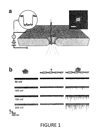
Figure 1 illustrates a method of biological molecule composition analysis
according to
one embodiment described herein.
Figure 2 illustrates complexed biological molecule translocation event rates
at various
electric field voltages in comparison to an electromobility shift assay over a
common
stoichiometric range according to some embodiments described herein.
Figure 3 illustrates complexed biological molecule event rates up to a molar
ratio of 1:1
of translocating agent and complexed biological molecule according to one
embodiment
described herein.
Figure 4 illustrates quantification of complexed biological molecule
concentration
relative to translocation event rate according to one embodiment described
herein.
Figure 5 illustrates translocation event rate versus applied voltage for
complexed
translocation agent and non-complexed translocation agent according to one
embodiment
described herein.
Figure 6 illustrates translocation event rate versus applied voltage for
complexed
translocation agent, non-complexed translocation and uncomplexed single strand
nucleic acid
according to one embodiment described herein.
2
CA 02947978 2016-11-03
WO 2015/175638 PCT/US2015/030531
Figure 7 illustrates translocation event rate versus applied voltage for
complexed ds-DNA
and non-complexed ds-DNA according to one embodiment described herein.
Figure 8 illustrates translocation event rate relative to complexed biological
molecule
concentration according to one embodiment described herein.
Figure 9 illustrates translocation event rate relative to ds-DNA length
according to one
embodiment described herein.
DETAILED DESCRIPTION
Embodiments described herein can be understood more readily by reference to
the
following detailed description and examples and their previous and following
descriptions.
Elements, apparatus and methods described herein, however, are not limited to
the specific
embodiments presented in the detailed description and examples. It should be
recognized that
these embodiments are merely illustrative of the principles of the present
invention. Numerous
modifications and adaptations will be readily apparent to those of skill in
the art without
departing from the spirit and scope of the invention.
In one aspect, methods are described herein for the selective detection and
quantitative
analysis of biological molecule compositions. A method described herein
comprises providing a
mixture comprising biological molecules complexed with a translocating agent
and non-
complexed biological molecules. The mixture is contacted with a membrane
comprising at least
one nanopore and an electric field is applied across the nanopore to
translocate the biological
molecules complexed with the translocating agent through the at least one
nanopore, wherein
translocation of the complexed biological molecules is selectively detected.
In some
embodiments, a method described herein further comprises measuring change in
current through
the nanopore during one or more translocation events of the complexed
biological molecules.
Moreover, the method can further comprise measuring the rate of translocation
events of the
complexed biological molecules and determining concentration of the complexed
biological
molecules from the rate of translocation events. Importantly, the translocated
complexed
biological molecules can be recovered from solution and arc thereby separated
from the non-
complexed biological molecules of the initial mixture.
Turning now to specific steps, a method described herein comprises providing a
mixture
including biological molecules complexed with a translocating agent and non-
complexed
3
CA 02947978 2016-11-03
WO 2015/175638 PCT/US2015/030531
biological molecules. Any biological molecule not inconsistent with the
objectives of the present
invention can be selectively detected and quantified according to methods
described herein.
Biological molecules suitable for analysis according to methods described
herein include nucleic
acids. In some embodiments, for example, biological molecules include ss-DNA,
ds-DNA as
well as RNA and RNA having intra-strand double helices. RNA can include mRNA,
miRNA,
rRNA, tRNA, tmRNA, aRNA or mixtures thereof. Nucleic acids can have any
desired number
of nucleotides not inconsistent with the objectives of the present invention.
In some
embodiments, for example, a nucleic acid for analysis has 25 to 1000
nucleotides. Additionally,
a nucleic acid can have a number of nucleotides selected from Table I.
Table 1 - Nucleic Acid Length
Nucleotides
100-600
200-500
250-400
50-300
100-200
1-500
Nucleic acids of the mixture can be derived from cukaryotic, prokaryotic
and/or viral sources.
Further, biological molecules can include nucleic acid fragments or
oligonucleotides.
Oligonucleotides of any length not inconsistent with the objectives of the
present invention can
be selectively detected and quantified according to methods described herein.
Further, a
biological molecule for analysis can include single nucleotides and/or
derivatives thereof.
In addition to nucleic acids, biological molecules suitable for analysis
according to
methods described herein include proteins. Any protein not inconsistent with
the objectives of
the present invention can be employed in methods described herein.
As described herein, biological molecules of the mixture arc selectively
complexed with
a translocating agent. A translocating agent is chemical species of sufficient
charge and/or
structure to permit selective detection of translocation of the complexed
biological molecule with
the applied electric field set by an applied voltage. In some embodiments, for
example, the
translocating agent is a biological molecule, including a protein, nucleic
acid or nucleic acid
fragment. The biological molecule can be in a naturally occurring state.
Alternatively, the
biological molecule can be modified to demonstrate specific binding, suitable
electric charge
4
CA 02947978 2016-11-03
WO 2015/175638 PCT/US2015/030531
and/or structure to permit selective detection of complexed biological
molecule translocation. In
some embodiments, a translocating agent includes a single strand nucleic acid
modified with an
affinity tag for binding one or more molecular species prior translocation of
the complexed
biological molecule. For example, a translocating agent can comprise a single
strand nucleic
acid of known specific sequence, wherein the single strand nucleic acid is
modified with a
protein tag. The protein tag can bind suitable protein in the mixture of
biological molecules prior
to translocation of the biological molecule complexed with the translocation
agent. In one
embodiment, a translocating agent of single strand nucleic acid is
biotinylated for binding
streptavidin prior to translocation of the target nucleic acid of
complimentary sequence.
In other embodiments, a translocating agent is a non-biological chemical
species, such as
a nanoparticle. Any nanoparticle not inconsistent with the objectives of the
present invention
can be employed. Nanoparticles, for example, can comprise organic
nanoparticles including
carbon nanoparticles (carbon nanotubes, graphene, fullerenes, etc.).
Nanoparticles can also
include inorganic nanoparticles such as semiconductor nanoparticles, ceramic
nanoparticles
and/or metal nanoparticles.
Translocating agent is added to the biological molecule mixture, and the
translocating
agent selectively binds biological molecules to provide the complexed
biological molecules.
Biological molecules not selectively bound by the translocating agent form the
non-complexed
biological molecules of the mixture.
For complexing the biological molecule for selective translocation detection,
the
translocating agent can comprise a site specific binding region. Depending on
the identity of the
biological molecule to be complexed, the site specific binding region can be a
DNA or RNA
binding domain. For example, in some embodiments, a translocating agent can
bind directly to a
nucleic acid warranting use of a nucleic acid binding domain.
Single strand nucleic acid translocating agent can selectively hybridize with
a target
single strand nucleic acid of complimentary sequence in the mixture of
biological molecules.
Selective hybridization followed by translocation can permit quantification of
the target single
strand nucleic acid in the mixture. In some embodiments, multiple single
strand nucleic acid
translocating agents of differing sequences can be used to identify the
presence and/or quantify
several target single strand nucleic acids in the mixture. Highly conserved
sequences, for
example, can be employed in translocating agents permitting identification of
specific species in
5
CA 02947978 2016-11-03
WO 2015/175638 PCT/US2015/030531
the mixture, such as various bacterial species. Homology could also be
monitored where
mismatch of one or more base pairs are registered in the analytical results.
Further, DNA
melting and/or annealing characteristics can be elucidated by employing single
strand nucleic
acid translocating agents. Transition between the single strand form
(uncomplexed with
translocating agent) and double stranded form (complexed with translocating
agent) can be
temporally correlated with translocation events.
Alternatively, the site specific binding region can be a protein binding
domain.
In other embodiments, the nucleic acid can be provided an affinity tag for
binding the
translocating agent, thereby warranting a protein binding domain.
A translocating agent can permit selective detection of complexed biological
molecule
translocation by several mechanisms depending on identity of the complexed
biological
molecule. In some embodiments, for example, the biological molecule of
interest is not of
sufficient charge to translocate through the nanopore at the selected
conditions of applied electric
field and/or other solution conditions. In such embodiments, the translocating
agent provides the
biological molecule of interest sufficient charge to undergo translocation at
the selected applied
electric field and/or other solution conditions. Therefore, translocation of
the complexed
biological molecule can be selectively detected as the non-complexed
biological molecules of the
mixture do not translocate under the selected conditions.
Alternatively, the biological molecule of interest is of high charge and
undergoes
translocation at a rate undetectable with conventional electronics employed in
nanopore analysis.
Proteins, for example, often translocate at rates undetectable by conventional
electronics, thereby
rendering nanopore apparatus unsuitable for protein detection and
quantification. In such
embodiments, the translocating agent can be of sufficient opposite charge
and/or size to retard
the protein translocation rate for detection and quantification by
conventional nanopore systems
and electronics. Similarly, single strand nucleic acids often translocate at
rates undetectable by
conventional electronics, thereby rendering the nanopore apparatus unsuitable
for nucleic acid
detection and quantification. In such embodiments, single strand nucleic acid
translocating agent
having sequence complimentary to a single strand nucleic acid target in the
mixture of biological
molecules is employed. The single strand nucleic acid translocating agent
exhibits stntcture
permitting detection of the complex formed with the target single strand
nucleic acid. As
described herein, the single strand nucleic acid translocating agent can be
modified with an
6
CA 02947978 2016-11-03
WO 2015/175638 PCT/US201.5/030531
affinity tag, such as a protein tag, for binding one or more molecular species
prior translocation
of the complexed target single strand nucleic acid. Further, the translocation
agent may provide
a translocation rate or nanoporc dwell time that can be sufficiently
differentiated from other
species in the mixture.
As described herein, the mixture comprising biological molecules complexed
with the
translocating agent and non-complexed biological molecules is contacted with a
membrane
comprising at least one nanopore. In some embodiments, the membrane comprises
an array of
nanopores. The membrane can have any thickness and be formed from any material
not
inconsistent with the objectives of the present invention. In some
embodiments, a membrane is
non-metallic. A non-metallic membrane can comprise one or more electrically
insulating
materials, including ceramic materials. Suitable ceramics include metal
oxides, metal nitrides,
metal carbides or metal carbonitrides or combinations thereof. In some
embodiments, a ceramic
suitable for use as a membrane is silicon nitride (SiN, SiN4). Additionally, a
membrane ceramic
can comprise silicon oxide, silicon carbide, aluminum oxide or a transition
metal oxide.
In some embodiments, a ceramic membrane is polycrystalline in nature. In some
embodiments, a ceramic membrane is single crystalline in nature. Moreover, a
ceramic
membrane can be multilayered. Individual layers of a multilayered membrane can
comprise the
same material or divergent materials. In some embodiments, individual layers
of a ceramic
membrane are independently selected from the group consisting of transition
metal carbide,
transition metal nitride, transition metal carbonitride, transition metal
oxide, alumina, silica and
silicon nitride.
Further, a membrane can comprise one or more semiconducting materials. In some
embodiments, suitable semiconducting materials include I INI semiconductors,
Group IV
semiconductors or I IIN semiconductors. In some embodiments, a semiconductor
material
comprises a ternary semiconductor or a quaternary semiconductor. Suitable
semiconductor
materials can have an amorphous structure, crystalline structure or mixture
thereof. Crystalline
semiconductor materials can be polycrystalline or single crystalline.
In some embodiments, a membrane is metallic. In such embodiments, a membrane
can
be a metal or various alloys of metals. In some embodiments, for example,
suitable metals are
transition metals, including noble metals such as gold. Alternatively, a
membrane, in some
7
CA 02947978 2016-11-03
WO 2015/175638 PCT/1JS2015/03(1531
embodiments, is not gold. Metallic membranes can be coated with dielectric or
electrically
insulating materials for use in methods described herein.
In some embodiments, a membrane comprises an organic material. For example, a
membrane can comprise one or more polymeric materials. Suitable polymeric
materials include
thermoplastics, thermosets or elastomers. A polymeric material, in some
embodiments,
comprises one or more of polyethylene, polypropylene, and polycarbonate.
Membranes suitable for use methods described herein can have any desired
thickness. In
some embodiments, a membrane has a thickness suitable for detecting and/or
conducting
analysis of one or more nucleic acid segments, including single-strand nucleic
acid segments. In
some embodiments, a membrane has an average thickness less than about 200 nm
or less than
about 100 nm. In some embodiments, a membrane has an average thickness
according to Table
Table II Nanopore Membrane Thicknesses (nin)
Membrane Thickness (nm)
<50
1-30
10-20
20-50
50-100
100-500
250-750
Further, a membrane can have a thickness on the atomic scale. In some
embodiments, a
membrane has a thickness less than 1 nm, such as 0.1 nm to 0.9 nm. In some
embodiments, the
thickness of a membrane is measured prior to nanopore formation according to a
method
described herein.
In addition, a nanopore of a membrane described herein can have any size and
shape not
inconsistent with the objectives of the present invention. In some
embodiments, for example, at
least one nanopore has a diameter greater than about 1 nm or greater than
about 5 nm. A
nanopore of a membrane described herein can have a diameter according to Table
III.
Table III Nanoporc Diameter (um)
8
CA 02947978 2016-11-03
WO 2015/175638
PCT/US2015/030531
Nanopore Diameter
> 10
1-20
1-10
1-5
5-10
10-15
10-20
1.5-4
Further, a nanopore can have a thickness commensurate with the average
thickness of the
membrane. Therefore, in some embodiments, a nanopore can have a thickness
selected from
Table II herein. Alternatively, a nanopore has a thickness less than the
average thickness of the
membrane.
Moreover, the diameter and/or thickness of a nanopore can be selected based on
a desired
signal to noise ratio (SNR) of a measurement described herein, such as a
current measurement
associated with a translocation event. The SNR of a translocation event, in
some embodiments,
is higher for larger diameter nanopores and lower for smaller diameter
nanoporcs. Additionally,
in some embodiments, the diameter and/or thickness of a nanopore are selected
based on a
desired dwell time of a translocated species in the nanopore or a desired
duration of a
translocation event. In some embodiments, the dwell time and/or the duration
of a translocation
event is longer for a thicker nanopore than for a thinner nanopore. Dwell
time, in some
embodiments, is the time elapsed from an initial conductance drop in the
nanopore until its return
to the baseline value.
A membrane described herein can be formed in any manner not inconsistent with
the
objectives of the present invention. In some embodiments, for instance, a
membrane is formed
according to a method described in Patent Cooperation Treaty (PCT) Application
Publication
WO 2012/170499, the entirety of which is hereby incorporated by reference.
10 As described herein, an electric field is applied across the
nanopore to transtocate the
biological molecules complexed with the translocating agent through the at
least one nanopore,
wherein the translocation events are selectively detected. The electric field
can be set according
to any applied voltage not inconsistent with the objectives of the present
invention. Suitable
applied voltages, for example can range from 1 mV to 5 V. In some embodiments,
the applied
voltage ranges from 10 mV to 1 V or 50 mV to 500 mV.
9
CA 02947978 2016-11-03
WO 2015/175638 PCT/US2015/03053t
In some embodiments, a method described herein further comprises measuring
change in
current through the nanopore during one or more translocation events of the
complexed
biological molecules. Moreover, the method can further comprise measuring the
rate of
translocation events of the complexed biological molecules and determining
concentration of the
complexed biological molecules from the rate of translocation events. In some
embodiments, for
example, concentration of the complexed biological molecules
Importantly, the translocated complexed biological molecules can be recovered
from
solution and are thereby separated from the non-complexed biological molecules
of the initial
mixture.
These and other embodiments arc further illustrated in the following non-
limiting
examples.
EXAMPLE 1 ¨ Selective Detection and Quantification Double-Stranded DNA
Selective detection and quantification of ds-DNA modified with biotin for
binding a
monovalent strcptavidin (MS) translocating agent is detailed in this example.
SS-nanopore
discrimination of monobiotinylated ds-DNA employed in this example is
illustrated in Figure 1.
In Figure la, an electrical bias is applied across a thin-film membrane with a
single nanopore
immersed in electrolyte solution. This facilitates the electrokinctic
translocation of molecules (or
molecular complexes) through the pore, each of which can produce an ionic
current event. This
technique was used to measure MS (Figure lb left) and monobiotinylated 90 bp
ds-DNA (bio90,
Figure lb center) individually at concentrations of 8 uM and 1 gM,
respectively. Over a range of
50-200 mV, few events were identified for either molecule. However, when MS
and bio90 are
incubated together at a molar ratio of 8:1 (MS:bio90) prior to measurements, a
remarkable
increase in the number of translocation events per unit time (Figure lb right)
was observed. The
event rate of the admixture was consistently more than an order of magnitude
greater than that of
either constituent molecule alone; at 200 mV applied voltage, for example, the
MS-bio90
complex yielded a rate of 23.3 0.9 s-1, while the event rates of MS and bio90
individually were
0.09 0.04 s-1 and 1.1 0.2 s-1, respectively. In order to verify that the
MS-bio90 events
corresponded to actual translocations rather than stochastic interactions
between the complex and
the nanoporc, polarity of the applied voltage was reversed during measurement
and recapture
events were observed.
CA 02947978 2016-11-03
WO 2015/175638
PCT/US2015/030531
In order to investigate the system further, a series of SS-nanopore
measurements were
performed in which MS was titrated against a constant amount (1 nM) of bio90.
Over all
investigated voltages, the measured event rate rose dramatically up to a molar
ratio of 1: I (Figure
2a). However, from unity up to a molar ratio of 8:1 (MS:bio90), additional MS
did not increase
the event rate further. This was a result of the limited supply of ds-DNA
needed to form
nucleoprotein complexes; the protein had an extremely low off rate (-10-5 s-1)
and each
oligonucleotide contained only a single biotin moiety, so it was expected that
nearly all bio90 in
solution was bound at or above an equimolar concentration. Comparing the
translocation results
to an electromobility shift assay (EMSA) performed with MS and bio90 over the
same
stoichiometric range, a strikingly similar trend was observed (Figure 2b).
These data supported
the conclusion that virtually all observed translocation events for the
admixture corresponded to
MS-bio90 complexes. Additional evidence of the high specificity of this
approach was provided
by control measurements in which non-biotinylated dsDNA incubated with MS
yielded a
negligible event rate, equivalent to bio90 alone.
Selective quantification of modlfied oligonucleotides: In Figure 3, event
rates up to a
molar ratio of 1:1 were examined and a linear dependence on applied voltage
was found. This
implied that the capture process for the MS-bio90 complex was governed by
diffusion rather
than by interactions with the pore, in agreement with previous studies.
Importantly, the observed
trend offered a route to quantification of MS-bio90 complexes in solution as
event frequency can
vary with molecular concentration. Because nearly all events observed in the
present system
were attributed exclusively to the translocation of complexes, the linear fits
in Figure 3 link the
concentration of MS-bio90 in solution to specific event rates produced at a
given voltage. The
measurements described thus far have been performed in a protein-limited
regime (MS:bio90 <
1:1), and so the measured event rate facilitated quantification of MS-bio90
complexes in a
background of unconjugated bio90. However, the same approach could in
principal be used to
quantify biotinylated oligonucleotides in a heterogeneous solution with non-
biotinylated DNA as
well.
To investigate this possibility, a blind test was conducted on two samples
prepared by a
third party. Each of these samples contained a different mixture of
biotinylated and non-
biotinylated 90 bp ds-DNA mixed to a total concentration of 1 uM (equivalent
to that of the
measurements described above). To ensure that all bio90 was complexed, both
solutions are
11
CA 02947978 2016-11-03
WO 2015/175638 PCT/US2015/030531
incubated with MS at a concentration of 4 p.M. As described in the previous
sections, MS alone
produced a negligible number of measurable events, and so excess protein did
not perturb the
measurements. SS-nanopore analysis revealed a linear relationship between
applied voltage and
event rate for both samples, as expected. Comparing the event rates obtained
from the two blind
samples to prior measurements (Figure 4) derived a value for the bio90
concentration in each:
850 35 nM in Sample 1 and 520 20 nM in Sample 2. Remarkably, these
experimentally-
determined concentrations were in excellent agreement with the prepared values
of 800 + 20 and
480 20 nM, respectively. These results demonstrate that our 55-nanopore
approach is uniquely
capable of quantifying DNA having single nucleotide biotin modifications
selectively, even
within a mixed sample.
Methods
SS-nanopore device jabrication and electrical measurement
Nanopores were fabricated using a technique described elsewhere as in WO
2012/170499.
Briefly, the beam of a scanning helium ion microscope (Carl Zeiss Orion Plus)
was focused on a
suspended silicon nitride thin film membrane (thickness 30 nm) in a silicon
support chip.
Calibrated exposure times were used to mill nanopores with diameters ranging
from 7.3-7.7 nm.
The support chip containing an individual pore was then positioned in a custom
flow cell with
fluid access to both sides of the membrane. Measurement solution (900 mM NaC1
and 6 mM
PBS buffer) was introduced on either side of the flow cell, and Ag/AgC1
electrodes were
immersed in the solution. Electrical measurements (Axopatch 200B) were used to
verify that the
device exhibited low RMS noise (typically <20 pA) and linear current-voltage
characteristics
that matched the calibrated nanopore diameter. Translocation measurements were
performed by
replacing the solution on one side of the device with measurement solution
containing
biomolecules. Conductance was recorded at a bandwidth of 200 kHz and filtered
at 100 kHz
with a four-pole Bessel filter. Analysis was performed with custom software
with which we
applied an additional low-pass filter of 25 kHz to all measurements. The event
threshold for
analysis was set at 4 standard deviations and only events with durations from
12-2000 tis were
considered.
12
CA 02947978 2016-11-03
WO 2015/175638 PCT/US2015/030531
Bioniolecules
Bio90 oligonucleotides were purchased (Integrated DNA Technologies,
Coralvillc, IA) with the
sequence: TGT ATA CCA TGG CCA GGA TCC TGG GCC ATC TGG TATB GTA ATT CAT
AAA GAA TTC TCA TTC TGC AGG TGC ACA TGT TAA CAC TAG TCG TGA. The TB
represents a single internal biotinylated dT. The opposing strand (forming the
dsDNA) contained
no modified nucleotides. The non-biotinylated oligonucleotide used in the
mixture (blind
measurements) had the same sequence but with no biotin moiety. The streptav-
idin variant
employed (SAelD3) contained one active biotin-binding site and was supplied by
the Howarth
lab (Oxford University). This mutant protein (54.5 kDa) retains binding
affinity and stability
similar to wild-type streptavidin and contains a hexaglutamate tag used for
isolation that imparts
a negative charge of -17.1e under comparable pH conditions.
Electrophoretic Mobility Shift Assay
MS was incubated with bio90 for 20 minutes at room temperature at molar ratios
ranging from
0:1 to 8:1 (MS:bio90). The mixtures were then loaded onto a 1.5% agarose gel
with ethidium
bromide for visualization. The buffer reservoir of the electrophoresis unit
was submerged in an
ice bath to minimize dissociation of the protein-DNA complex.
EXAMPLE 2 - Selective Detection and QuantOcation Double-Stranded DNA
The ability to differentiate biotinylated forms of dsDNA and ssDNA due to
variations in
drag force imparted by the two types of DNA molecules was demonstrated. This
effect was
demonstrated by analyzing both single strand (ss) and double strand (ds)
versions of 34 base pair
(bp) monobiotinylated oligonucleotide by nanopore in the presence MS. A
significant increase
in event rate was observed for the dsDNA-MS, and no enhancement was observed
for the
ssDNA-MS as illustrated in Figure 5. Based on this result, biotinylated ssDNA
can be employed
as a sequence specific translocating agent that can bind to its complimentary
target sequence in a
mixture of biological molecules. Sequence specific translocating capability of
the 34bp
biotinylated ssDNA was tested on a mixture comprising single strand nucleic
acid of
homologous sequence and a background of four non-homologous ss-DNA sequences.
MS was
introduced into the mixture and thermal cycling was conducted to promote
annealing. Figure 6
illustrates the results where translocation of a hybridized complex of
biotinylated dsDNA-MS
13
CA 02947978 2016-11-03
WO 2015/175638 PCT/US2015/030531
was observed. Importantly, translocation of uncomplexed ss-DNA and uncomplexed
biotinylated ss-DNA-MS translocating agent was not observed.
EXAMPLE 3 ¨ Translocation Characterization of ds-DNA
A series of SS-nanopore measurements were performed in which translocation
event rate
of a 150 bp ds-DNA strand having a single hydroxymethylcytosine enzymatically
labeled with
biotin and subsequently bound to MS was measured against nanopore applied
voltage and
concentration of the 150 bp ds-DNA strand. Nanopore construction and
operational parameters
were consistent with those provided in Example 1 with the applied voltage
ranging from 50-600
mV. Figure 7 illustrates results of the testing where ds-DNA-biotin-MS
translocation event rate
increased with increasing voltage. For comparison, 150 bp ds-DNA strand not
employing the
biotin-MS translocating agent architecture was tested and failed to register a
translocation
response. Translocation event rate dependency on ds-DNA-biotin-MS
concentration was also
explored. As illustrated in Figure 8, translocation event rate varied
generally linearly with ds-
DNA-biotin-MS concentration at a constant applied voltage of 200 mV. The
shaded portion of
Figure 8 represents the noise floor. Translocation event rate dependency on ds-
DNA length was
also investigated. As provided in Figure 9, translocation rate exhibited
substantially linear
dependence on ds-DNA length. With these relationships established, methods
described herein
provide a powerful tool for characterization and quantification of biological
molecules, including
nucleic acids.
Various embodiments of the invention have been described in fulfillment of the
various
objects of the invention. It should be recognized that these embodiments are
merely illustrative
of the principles of the present invention. Numerous modifications and
adaptations will be
readily apparent to those skilled in the art without departing from the spirit
and scope of the
invention.
14