Note: Descriptions are shown in the official language in which they were submitted.
SYSTEMS AND METHODS FOR ANCHORING AN IMPLANT
[0001] This application claims priority to U.S. Prov. Patent Application No.
62/016582, titled "Systems and Methods for Anchoring a Cardiac Implant" and
filed June
24, 2014.
[0002] This application is related to U.S. Patent Application No. 14/742199,
titled
"Mitral Valve Implants for the Treatment of Valvular Regurgitation" and filed
June 17,
2015.
BACKGROUND
Field
[0003] The present invention generally provides, in some embodiments, improved
medical devices, systems, and methods, typically for treatment of heart valve
disease
and/or for altering characteristics of one or more valves of the body.
Embodiments of the
invention include implants for treatment of mitral valve regurgitation.
[0004] The human heart receives blood from the organs and tissues via the
veins, pumps
that blood through the lungs where the blood becomes enriched with oxygen, and
propels
the oxygenated blood out of the heart to the arteries so that the organ
systems of the body
can extract the oxygen for proper function. Deoxygenated blood flows back to
the heart
where it is once again pumped to the lungs.
[0005] The heart includes four chambers: the right atrium (RA), the right
ventricle
(RV), the left atrium (LA) and the left ventricle (LV). The pumping action of
the left and
right sides of the heart occurs generally in synchrony during the overall
cardiac cycle.
[0006] The heart has four valves generally configured to selectively transmit
blood
flow in the correct direction during the cardiac cycle. The valves that
separate the atria
from the ventricles are referred to as the atrioventricular (or AV) valves.
The AV
-1-
Date Recue/Date received 2020-05-25
CA 02958065 2017-02-1.3
WO 2015/200497
PCT/US2015/037451
valve between the left atrium and the left ventricle is the mitral valve. The
AV valve
between the right atrium and the right ventricle is the tricuspid valve. The
pulmonary
valve directs blood flow to the pulmonary artery and thence to the lungs;
blood returns to
the left atrium via the pulmonary veins. The aortic valve directs flow through
the aorta
and thence to the periphery. There are normally no direct connections between
the
ventricles or between the atria.
[0007] The
mechanical heartbeat is triggered by an electrical impulse which
spreads throughout the cardiac tissue. Opening and closing of heart valves may
occur
primarily as a result of pressure differences between chambers, those
pressures resulting
from either passive filling or chamber contraction. For example, the opening
and closing
of the mitral valve may occur as a result of the pressure differences between
the left
atrium and the left ventricle.
[0008] At the
beginning of ventricular filling (diastole) the aortic and
pulmonary valves are closed to prevent back flow from the arteries into the
ventricles.
Shortly thereafter, the AV valves open to allow unimpeded flow from the atria
into the
corresponding ventricles. Shortly after ventricular systole (i.e., ventricular
emptying)
begins, the tricuspid and mitral valves normally shut, forming a seal which
prevents flow
from the ventricles back into the corresponding atria.
100091
Unfortunately, the AV valves may become damaged or may otherwise
fail to function properly, resulting in improper closing. The AV valves are
complex
structures that generally include an annulus, leaflets, chordae and a support
structure.
Each atrium interfaces with its valve via an atrial vestibule. The mitral
valve has two
leaflets; the analogous structure of the tricuspid valve has three leaflets,
and opposition or
engagement of corresponding surfaces of leaflets against each other helps
provide closure
or sealing of the valve to prevent blood flowing in the wrong direction.
Failure of the
leaflets to seal during ventricular systole is known as malcoaptation, and may
allow blood
to flow backward through the valve (regurgitation). Heart valve regurgitation
can have
serious consequences to a patient, often resulting in cardiac failure,
decreased blood flow,
lower blood pressure, and/or a diminished flow of oxygen to the tissues of the
body.
Mitral regurgitation can also cause blood to flow back from the left atrium to
the
pulmonary veins, causing congestion. Severe valvular regurgitation, if
untreated, can
result in permanent disability or death.
-2-
CA 02958065 2017-02-1.3
WO 2015/200497
PCT/US2015/037451
Description of the Related Art
[0010] A variety of
therapies have been applied for treatment of mitral valve
regurgitation, and still other therapies may have been proposed but not yet
actually used to
treat patients. While several of the known therapies have been found to
provide benefits
for at least some patients, still further options would be desirable. For
example,
pharmacologic agents (such as diuretics and vasodilators) can be used with
patients
having mild mitral valve regurgitation to help reduce the amount of blood
flowing back
into the left atrium. However, medications can suffer from lack of patient
compliance. A
significant number of patients may occasionally (or even regularly) fail to
take
medications, despite the potential seriousness of chronic and/or progressively
deteriorating mitral valve regurgitation. Pharmacological therapies of mitral
valve
regurgitation may also be inconvenient, are often ineffective (especially as
the condition
worsens), and can be associated with significant side effects (such as low
blood pressure).
[0011] A variety of
surgical options have also been proposed and/or employed
for treatment of mitral valve regurgitation. For example, open-heart surgery
can replace
or repair a dysfunctional mitral valve. In annuloplasty ring repair, the
posterior mitral
annulus can be reduced in size along its circumference, optionally using
sutures passed
through a mechanical surgical annuloplasty sewing ring to provide coaptation.
Open
surgery might also seek to reshape the leaflets and/or otherwise modify the
support
structure. Regardless, open mitral valve surgery is generally a very invasive
treatment
carried out with the patient under general anesthesia while on a heart-lung
machine and
with the chest cut open. Complications can be common, and in light of the
morbidity
(and potentially mortality) of open-heart surgery, the timing becomes a
challenge sicker
patients may be in greater need of the surgery, but less able to withstand the
surgery.
Successful open mitral valve surgical outcomes can also be quite dependent on
surgical
skill and experience.
[0012] Given the
morbidity and mortality of open-heart surgery, innovators
have sought less invasive surgical therapies. Procedures that are done with
robots or
through endoscopes are often still quite invasive, and can also be time
consuming,
expensive, and in at least some cases, quite dependent on the surgeon's skill.
Imposing
even less trauma on these sometimes frail patients would be desirable, as
would be
providing therapies that could be successfully implemented by a significant
number of
physicians using widely distributed skills. Toward that end, a number of
purportedly less
-3-
CA 02958065 2017-02-1.3
WO 2015/200497
PCT/US2015/037451
invasive technologies and approaches have been proposed. These include devices
which
seek to re-shape the mitral annulus from within the coronary sinus; devices
that attempt to
reshape the annulus by cinching either above to below the native annulus;
devices to fuse
the leaflets (imitating the Alfieri stitch); devices to re-shape the left
ventricle, and the like.
[0013] Perhaps most
widely known, a variety of mitral valve replacement
implants have been developed, with these implants generally replacing (or
displacing) the
native leaflets and relying on surgically implanted structures to control the
blood flow
paths between the chambers of the heart. While these various approaches and
tools have
met with differing levels of acceptance, none has yet gained widespread
recognition as an
ideal therapy for most or all patients suffering from mitral valve
regurgitation.
[0014] Because of
the challenges and disadvantages of known minimally
invasive mitral valve regurgitation therapies and implants, still further
alternative
treatments have been proposed. Some of the alternative proposals have called
for an
implanted structure to remain within the valve annulus throughout the heart
beat cycle.
One group of these proposals includes a cylindrical balloon or the like to
remain
implanted on a tether or rigid rod extending between the atrium and the
ventricle through
the valve opening. Another group relies on an arcuate ring structure or the
like, often in
combination with a buttress or structural cross-member extending across the
valve so as
to anchor the implant. Unfortunately, sealing between the native leaflets and
the full
perimeter of a balloon or other coaxial body may prove challenging, while the
significant
contraction around the native valve annulus during each heart beat may result
in
significant fatigue failure issues during long-term implantation if a buttress
or anchor
interconnecting cross member is allowed to flex. Moreover, the significant
movement of
the tissues of the valve may make accurate positioning of the implant
challenging
regardless of whether the implant is rigid or flexible.
[0015] In light of
the above, it would be desirable to provide improved
medical devices, systems, and methods. It would be particularly desirable to
provide new
techniques for treatment of mitral valve regurgitation and other heart valve
diseases,
and/or for altering characteristics of one or more of the other valves of the
body. The
need remains for a device which can directly enhance leaflet coaptation
(rather than
indirectly via annular or ventricular re-shaping) and which does not disrupt
leaflet
anatomy via fusion or otherwise. but which can be deployed simply and
reliably, and
without excessive cost or surgical time. It would be particularly beneficial
if these new
-4-
CA 02958065 2017-02-1.3
WO 2015/200497
PCT/US2015/037451
techniques could be implemented using a less-invasive approach, without
stopping the
heart or relying on a heart-lung machine for deployment, and without relying
on
exceptional skills of the surgeon to provide improved valve and/or heart
function.
SUMMARY
[0016] In some
embodiments, a system is provided. The system can include an
anchor comprising a proximal end and a distal end, the distal end configured
to engage
tissue. The system can include a suture coupled to the proximal end of the
anchor. The
system can include an implantable medical device, wherein the suture is
configured to
pass through at least a portion of the implantable medical device. The system
can include
a clip comprising at least two strands twisted together. In some embodiments,
the suture is
configured to pass through at least a portion of the clip after passing
through the
implantable medical device. In some embodiments, the suture is configured to
pass
through at least a portion of the clip as the clip slides toward the
implantable medical
device and the anchor.
[0017] The system
can include a needle on an end of the suture. In some
embodiments, the at least two strands are nitinol. In some embodiments, the at
least two
strands comprise a shape memory material. In some embodiments, the clip is
linear. In
some embodiments, the clip is non-linear. In some embodiments, the anchor
comprises a
helical portion. In some embodiments, the anchor comprises a needle radially
surrounded
by the anchor. In some embodiments, the implantable medical device is a
coaptation
assistance device configured to improve leaflet coaptation of a cardiac valve.
In some
embodiments, the suture passes from one surface of the implantable medical
device to a
second, opposed surface of the implantable medical device. In some
embodiments, the
suture forms a loop on one side of the clip.
[0018] In some
embodiments, a system is provided. The system can include a
delivery tool comprising an outer sleeve and an inner shaft. In some
embodiments, the
inner shaft is operably coupled to a control handle configured to move the
inner shaft
relative to the outer sleeve. The system can include a lasso extending through
the inner
shaft, the lasso configured to engage a suture. The system can include a clip
disposed on
the inner shaft. In some embodiments, the control handle is configured to pull
the lasso
and the suture inside the inner shaft. In some embodiments, after the lasso
and the suture
-5-
CA 02958065 2017-02-13
WO 2015/200497
PCT/US2015/037451
are pulled inside the inner shaft, the outer sleeve is configured to push the
clip off the
inner shaft.
100191 In some
embodiments, the clip comprises at least two strands twisted
together. In some embodiments, the at least two strands are nitinol. In some
embodiments,
the at least two strands comprise a shape memory material. In some
embodiments, the clip
is linear. In some embodiments, the clip is non-linear. The system can include
the suture
and an anchor, wherein the suture extends from the anchor. In some
embodiments, the
anchor comprises a helical portion. In some embodiments, the anchor comprises
a central
needle. The system can include the suture and the implantable medical device,
wherein
the suture extends through the implantable medical device. In some
embodiments, the
implantable medical device is a coaptation assistance device configured to
improve leaflet
coaptation of a cardiac valve. In some embodiments, the suture passes from one
surface of
the implantable medical device to a second, opposed surface of the implantable
medical
device. In some embodiments, the suture forms a loop on one side of the clip
after the
lasso and the suture is pulled inside the inner shaft. In some embodiments,
the suture
forms a loop on one side of the clip after the clip is pushed off the inner
shaft.
100201 In some
embodiments, a method of anchoring is provided. The method
can include the step of extending a suture from an anchor and through an
implantable
medical device. The method can include the step of extending the suture
through a clip
after extending the suture through the implantable medical device, wherein the
suture
passes through at least a portion of the clip as the clip slides toward the
implantable
medical device and the anchor.
100211 In some
embodiments, the extending a suture from an anchor and
through an implantable medical device comprises extending a needle on an end
of the
suture through the implantable medical device. In some embodiments, extending
the
suture through a clip comprises extending the suture between two strands of
wire twisted
together. In some embodiments, the clip comprises at least two strands twisted
together.
In some embodiments, the clip comprises two or more nitinol wires. In some
embodiments, the clip is linear. ln some embodiments, the clip is non-linear.
In some
embodiments, the anchor comprises a helical portion. The method can include
the step of
driving a helical portion of the anchor into tissue. The method can include
the step of
engaging the anchor with tissue. The method can include the step of engaging
the anchor
with cardiac tissue. In some embodiments, the anchor comprises a central
needle. The
-6-
method can include the step of driving a central needle into tissue, the
anchor surrounding
the central needle. In some embodiments, the implantable medical device is a
coaptation
assistance device. The method can include the step of passing the suture from
one surface
of the implantable medical device to another, opposed surface of the
implantable medical
device. The method can include the step of forming a loop of the suture on one
side of the
clip. The method can include the step of sliding the clip toward the
implantable medical
device. In some embodiments, the clip is initially disposed on the inner shaft
of a tool,
further comprising pushing the clip from the inner shaft of the tool. In some
embodiments,
the clip is initially disposed on the inner shaft of a tool, the inner shaft
having a lasso
extending therethrough, further comprising threading the suture through the
lasso. The
method can include the step of pulling the lasso and the suture into the inner
shaft. The
method can include the step of pulling a loop of the suture through the clip.
The method
can include the step of lowering the inner shaft and the clip toward the
implantable medical
device and the anchor. The method can include the step of pushing the clip off
the inner
shaft. The method can include the step of pushing the clip off the inner shaft
with an outer
sleeve of the tool. The method can include the step of placing the clip
adjacent to the
implantable medical device. The method can include the step of placing the
implantable
medical device adjacent to the tissue. The method can include the step of
placing the
implantable medical device adjacent to the anchor.
[0021a] In
accordance with an aspect of the invention is a system for
anchoring an implantable medical device within tissue of a patient,
comprising:
an anchor comprising a proximal end and a distal end, the distal end
configured to engage tissue,
a suture coupled to the proximal end of the anchor,
an implantable medical device, wherein the suture is configured to pass
through at least a portion of the implantable medical device,
a clip comprising a longitudinal axis and at least two strands twisted
together, wherein the suture is configured to pass through the at least two
strands
in a direction transverse to the longitudinal axis as the clip slides toward
the
implantable medical device and the anchor.
-7-
Date Recue/Date Received 2022-07-04
10021b1 In accordance with an aspect of the invention is a system
for
anchoring an implantable medical device within tissue of a patient,
comprising:
an anchor comprising a proximal end and a distal end, the distal end
configured to engage tissue,
a suture coupled to the anchor,
an implantable medical device, and
a clip comprising a longitudinal axis and at least two strands twisted
together, wherein the
suture is configured to pass through the at least two strands in a direction
transverse to the
longitudinal axis as the clip slides toward the implantable medical device and
the anchor.
[0021c] In accordance with an aspect of the invention is a system
for
anchoring an implantable medical device within tissue of a patient,
comprising:
an anchor comprising a proximal end and a distal end, the distal end
configured to engage tissue,
a suture coupled to the proximal end of the anchor,
an implantable medical device, wherein the suture is configured to pass
through at least a portion of the implantable medical device,
a clip comprising at least two strands twisted together, wherein the clip is
configured to allow the user to lock the implantable medical device to tissue
without
applying knots to the suture, wherein the strands apply a force on the surface
of the
suture and lock onto the suture;
wherein the suture is configured to pass transversely through the at least two
strands twisted together after passing through the implantable medical device,
wherein the suture is configured to pass through at least a portion of the
clip as the
clip is configured to slide toward the implantable medical device and the
anchor.
[0021 d] In accordance with an aspect of the invention is a system
for
anchoring an implantable medical device within tissue of a patient,
comprising:
an anchor comprising a proximal end and a distal end, the distal end
configured to engage tissue,
a suture coupled to the proximal end of the anchor,
-7a-
Date Recue/Date Received 2022-07-04
an implantable medical device, wherein the suture is configured to pass
through at least a portion of the implantable medical device,
a clip comprising a longitudinal axis and at least two strands twisted
together, wherein the suture is configured to pass through at least a portion
of the
clip after passing through the implantable medical device, wherein the suture
is
configured to pass through at least a portion of the clip in a direction
transverse to
the longitudinal axis as the clip slides toward the implantable medical device
and
the anchor.
10021e11 In accordance with an aspect of the invention is a system
for
anchoring an implantable medical device within tissue of a patient,
comprising:
an anchor comprising a proximal end and a distal end, the distal end
configured to engage tissue,
a suture coupled to the proximal end of the anchor,
an implantable medical device, wherein the suture is configured to pass
through at least a portion of the implantable medical device,
a clip comprising at least two strands twisted together, wherein the suture is
configured to pass transversely through the at least two strands twisted
together
after passing through the implantable medical device,
a delivery tool comprising an outer sleeve, an inner shaft, and a loop tip
extending through the inner shaft, the loop tip configured to engage the
suture.
1002111 In accordance with an aspect of the invention is a system
for
anchoring an implantable medical device within tissue of a patient,
comprising:
an anchor comprising a proximal end and a distal end, the distal end
configured to engage tissue,
a suture coupled to the proximal end of the anchor,
an implantable medical device, wherein the suture is configured to pass
through at least a portion of the implantable medical device,
a clip comprising a longitudinal axis and at least two strands twisted
together,
wherein the suture is configured to pass through at least a portion of the
clip after passing
through the implantable medical device, wherein the suture is configured to
pass through
-7b-
Date Recue/Date Received 2022-07-04
at least a portion of the clip in a direction transverse to the longitudinal
axis as the
clip slides toward the implantable medical device and the anchor.
[0021g] In
accordance with an aspect of the invention is use of an
implantable medical device for anchoring within tissue, wherein the use
comprises a suture
for extending from an anchor and through the implantable medical device,
thereafter the
suture is for extending through a clip, wherein the suture is passable through
at least a
portion of the clip as the clip slides toward the implantable medical device
and the anchor.
BRIEF DESCRIPTION OF THE DRAWINGS
[0022] FIGS. 1 A-1 F schematically illustrate some of the tissues of the heart
and
mitral valve, as described in the Background section and below, and which may
interact
with the implants and systems described herein.
[0023] FIG. 2A illustrates a simplified cross-section of a heart,
schematically
showing mitral valve function during diastole.
[0024] FIG. 2B illustrates a simplified cross-section of a heart,
schematically
showing mitral valve function during systole
[0025] FIGS. 3A-3B illustrate a simplified cross-section of a heart,
schematically
showing mitral valve regurgitation during systole in the setting of mal-
coaptation of the
mitral valve leaflets.
[0026] FIG. 4A illustrates a stylized cross section of a heart, showing mitral
valve
mal-coaptation in the settings of functional mitral valve regurgitation.
-7c-
Date Recue/Date Received 2022-07-04
CA 02958065 2017-02-1.3
WO 2015/200497
PCT/US2015/037451
100271 FIG. 4B illustrates a stylized cross section of a heart,
showing mitral
valve mal-coaptation in the settings of degenerative mitral valve
regurgitation.
[0028] FIGS. 5A-5E illustrates embodiments of annular anchoring.
[0029] FIGS. 6A-6G illustrates embodiments of secondary anchoring.
[0030] FIG. 7 illustrates an embodiment of an anchor with a linked
suture
mechanism.
[0031] FIG. S illustrates an embodiment of an anchor with a releasable
L-lock
feature.
[0032] FIG. 9 illustrates an embodiment of an anchor with a clip.
[0033] FIG. 10 illustrates an embodiment of an anchor with a clip.
[0034] FIGS. II A-11B illustrate an embodiment of an anchor with a
clip.
[0035] FIG. 12 illustrates an embodiment of an anchor with a clip.
[0036] FIG. 13 illustrates an embodiment of an anchor with a clip.
100371 FIG. 14 illustrates an embodiment of an anchor.
[0038] FIG. 15 illustrates an embodiment of an anchor.
[0039] FIG. 16 illustrates an embodiment of an anchor with a clip.
[0040] FIG. 17 illustrates an embodiment of an anchor with a clip.
[0041] FIGS. 18A-18D illustrates an embodiment of an anchor.
[0042] FIGS. 19A-19B illustrates an embodiment of an anchor.
[0043] FIGS. 20A-20B illustrates an embodiment of an anchor.
[0044] FIG. 21 illustrates an embodiment of an anchor.
[0045] FIGS. 22A-22B illustrates an embodiment of an anchor.
[0046] FIGS. 23A-23E illustrates an embodiment of an arrangement of
clips
according to some methods of use.
[0047] FIGS. 24A-24E illustrates embodiments of anchors.
[0048] FIGS. 25A-25C illustrates embodiments of an anchor and a
suture.
[0049] FIGS. 26A-26D illustrates an embodiment a tool for use with the
anchor and a suture of FIG. 25A.
[0050] FIG. 27 illustrates a method step in some methods of use of the
anchor
and a suture of FIG. 25A.
100511 FIG. 28 illustrates a method step in some methods of use of the
anchor
and a suture of FIG. 25A.
-8-
CA 02958065 2017-02-13
WO 2015/200497
PCT/US2015/037451
100521 FIG. 29 illustrates a method step in some methods of use of the
anchor
and a suture of FIG. 25A.
100531 FIG. 30 illustrates a method step in some methods of use of the
anchor
and a suture of FIG. 25A.
100541 FIG. 31 illustrates a method step in some methods of use of the
anchor
and a suture of FIG. 25A.
[0055] FIG. 32 illustrates an embodiment of a clip.
100561 FIG. 33 illustrates embodiments of a clip.
100571 FIG. 34 illustrates an embodiment a tool for use with the
suture clip of
FIG. 32.
100581 FIG. 35 illustrates a method step in some methods of use of the
suture
clip of FIG. 32.
[0059] FIG. 36 illustrates a method step in some methods of use of the
suture
clip of FIG. 32.
[0060] FIG. 37 illustrates a method step in some methods of use of the
suture
clip of FIG. 32.
100611 FIG. 38 illustrates a method step in some methods of use of the
suture
clip of FIG. 32.
100621 FIG. 39 illustrates a method step in some methods of use of the
suture
clip of FIG. 32.
[0063] FIG. 40 illustrates a method step in some methods of use of the
suture
clip of FIG. 32.
100641 FIG. 41 illustrates a method step in some methods of use of the
suture
clip of FIG. 33.
100651 FIG. 42 illustrates a method step in some methods of use of the
suture
clip of FIG. 33.
100661 FIGS. 43A-43G illustrates embodiments of an anchor.
100671 FIGS. 44A-44C illustrates embodiments of an anchor driver.
[0068] FIGS. 45A-45B illustrates an embodiment of the dimensions of
the
anchor driver of FIGS. 44A-44C.
100691 FIGS. 46A-46E illustrates an embodiment of a suture clip.
-9-
CA 02958065 2017-02-13
WO 2015/200497
PCT/US2015/037451
DETAILED DESCRIPTION
[0070] The devices,
systems and methods described within this disclosure, in
some embodiments, are generally for the treatment of mitral valve
regurgitation (MR).
However, devices, systems, and methods as disclosed herein can also be
utilized for other
cardiac as well as non-cardiac indications, including those involving the
mitral, aortic,
tricuspid, and/or pulmonic valves. Mitral valve regurgitation occurs when the
mitral
valve does not prevent the backtlow of blood from the left ventricle to the
left atrium
during the systolic phase. The mitral valve is composed of two leaflets, the
anterior leaflet
and the posterior leaflet, which coapt or come together during the systolic
phase to
prevent backflovv. There are generally two types of mitral valve
regumitations, functional
and degenerative regurgitations. Functional MR is caused by multiple
mechanisms
including abnormal or impaired left ventricular (LV) wall motion, left
ventricular dilation
and papillary muscle disorders. Degenerative MR is caused by structural
abnormalities of
the valve leaflets and the sub-valvular tissue including stretching or rupture
of the
chordae. Damaged chordae may lead to prolapsing of the leaflets which means
that the
leaflets bulge out (generally into the atrium), or become flail if the chordae
become torn,
leading to backflows of blood. As will be described below, the devices, system
and
methods in this disclosure provide a new coaptation surface over the native
posterior
valve such that the backward flow of blood is minimized or eliminated.
100711 Referring to
FIGS. 1A-1D, the four chambers of the heart are shown,
the left atrium 10, right atrium 20, left ventricle 30, and right ventricle
40. The mitral
valve 60 is disposed between the left atrium 10 and left ventricle 30. Also
shown are the
tricuspid valve 50 which separates the right atrium 20 and right ventricle 40.
the aortic
valve 80, and the pulmonary valve 70. The mitral valve 60 is composed of two
leaflets,
the anterior leaflet 12 and posterior leaflet 14. In a healthy heart, the
edges of the two
leaflets oppose during systole at the coaptation zone 16.
[0072] The fibrous
annulus 120, part of the cardiac skeleton, provides
attachment for the two leaflets of the mitral valve, referred to as the
anterior leaflet 12 and
the posterior leaflet 14. The leaflets are axially supported by attachment to
the chordae
tendinae 32. The chordae, in turn, attach to one or both of the papillary
muscles 34, 36 of
the left ventricle. In a healthy heart, the chordae support structures tether
the mitral valve
leaflets, allowing the leaflets to open easily during diastole but to resist
the high pressure
developed during ventricular systole. In addition to the tethering effect of
the support
-10-
CA 02958065 2017-02-1.3
WO 2015/200497
PCT/US2015/037451
structure, the shape and tissue consistency of the leaflets helps promote an
effective seal
or coaptation. The leading edges of the anterior and posterior leaflet come
together along
the zone of coaptation 16, with a lateral cross-section 160 of the three-
dimensional
coaptation zone (CZ) being shown schematically in FIG. 1E.
[0073] The anterior
and posterior mitral leaflets are dissimilarly shaped. The
anterior leaflet is more firmly attached to the annulus overlying the central
fibrous body
(cardiac skeleton), and is somewhat stiffer than the posterior leaflet, which
is attached to
the more mobile posterior mitral annulus. Approximately 80 percent of the
closing area is
the anterior leaflet. Adjacent to the commissures 110, 114, on or anterior to
the annulus
120, lie the left (lateral) 124 and right (septal) 126 fibrous trigones which
are formed
where the mitral annulus is fused with the base of the non-coronary cusp of
the aorta
(FIG. IF) . The fibrous trigones 124, 126 form the septal and lateral extents
of the central
fibrous body 128. The fibrous trigones 124, 126 may have an advantage, in some
embodiments, as providing a firm zone for stable engagement with one or more
annular or
atrial anchors. The coaptation zone CL between the leaflets 12, 14 is not a
simple line, but
rather a curved funnel-shaped surface interface. The first 110 (lateral or
left) and second
114 (septal or right) commissures are where the anterior leaflet 12 meets the
posterior
leaflet 14 at the annulus 120. As seen most clearly in the axial views from
the atrium of
FIG. IC, ID, and 1F, an axial cross-section of the coaptation zone generally
shows the
curved line CL that is separated from a centroid of the annulus CA as well as
from the
opening through the valve during diastole CO. In addition, the leaflet edges
are scalloped,
more so for the posterior versus the anterior leaflet. Mal-coaptation can
occur between
one or more of these A-P (anterior-posterior) segment pairs Al/P1, A2/P2, and
A3/P3, so
that mal-coaptation characteristics may vary along the curve of the coaptation
zone CL.
100741 Referring
now to FIG. 2A, a properly functioning mitral valve 60 of a
heart is open during diastole to allow blood to flow along a flow path FP from
the left
atrium toward the left ventricle 30 and thereby fill the left ventricle. As
shown in FIG.
2B, the functioning mitral valve 60 closes and effectively seals the left
ventricle 30 from
the left atrium 10 during systole, first passively then actively by increase
in ventricular
pressure, thereby allowing contraction of the heart tissue surrounding the
left ventricle to
advance blood throughout the vasculaturc.
[0075] Referring to
FIG. 3A-3B and 4A-4B, there are several conditions or
disease states in which the leaflet edges of the mitral valve fail to oppose
sufficiently and
-11-
CA 02958065 2017-02-1.3
WO 2015/200497
PCT/US2015/037451
thereby allow blood to regurgitate in systole from the ventricle into the
atrium.
Regardless of the specific etiology of a particular patient, failure of the
leaflets to seal
during ventricular systole is known as mal-coaptation and gives rise to mitral
regurgitation.
[0076] Generally,
mal-coaptation can result from either excessive tethering by
the support structures of one or both leaflets, or from excessive stretching
or tearing of the
support structures. Other, less common causes include infection of the heart
valve,
congenital abnormalities, and trauma. Valve malfunction can result from the
chordae
tendinae becoming stretched, known as mitral valve prolapse, and in some cases
tearing
of the chordae 215 or papillary muscle, known as a flail leaflet 220, as shown
in FIG. 3A.
Or if the leaflet tissue itself is redundant, the valves may prolapse so that
the level of
coaptation occurs higher into the atrium, opening the valve higher in the
atrium during
ventricular systole 230. Either one of the leaflets can undergo prolapse or
become flail.
This condition is sometimes known as degenerative mitral valve regurgitation.
[0077] In excessive
tethering, as shown in FIG. 3B, the leaflets of a normally
structured valve may not function properly because of enlargement of or shape
change in
the valve annulus: so-called annular dilation 240. Such functional mitral
regurgitation
generally results from heart muscle failure and concomitant ventricular
dilation. And the
excessive volume load resulting from functional mitral regurgitation can
itself exacerbate
heart failure, ventricular and annular dilation, thus worsening mitral
regurgitation.
[0078] FIGS. 4A-4B
illustrate the backflow BF of blood during systole in
functional mitral valve regurgitation (FIG. 4A) and degenerative mitral valve
regurgitation (FIG. 4B). The increased size of the annulus in FIG. 4A, coupled
with
increased tethering due to hypertrophy of the ventricle 320 and papillary
muscle 330,
prevents the anterior leaflet 312 and posterior leaflet 314 from opposing,
thereby
preventing coaptation. In FIG. 4B, the tearing of the chordae 215 causes
prolapse of the
posterior leaflet 344 upward into the left atrium, which prevents opposition
against the
anterior leaflet 342. In either situation, the result is backflow of blood
into the atrium,
which decreases the effectiveness of left ventricle compression.
[0079] Disclosed
herein are systems and methods to secure intracardiac
implants, such as replacement heart valves, annuloplasty rings, cardiac
patches, left atrial
appendage devices, patent foramen ovalc, ASD, or VSD closure devices, sensors,
pacemakers, AICDs, ventricular assist devices, drug delivery devices, and
coaptation
-12-
assist devices, for example, in place within the heart. The implant can be any
device known
in the art, including those disclosed in U.S. Patent Application No.
14/742,199. In some
embodiments, disclosed herein are tissue anchoring mechanisms for such
implants. In some
embodiments, disclosed herein are clip mechanisms to secure implants to the
anchors that
can be already embedded in tissue. In some embodiments, systems and methods as
disclosed herein can be utilized with those disclosed in U.S. Pat. Nos.
8,845,717,
8,888,843, or U.S. Pat. App. No. 14/742,199. These anchors, which can be
described in
some embodiments as annular, atrial, and/or ventricular anchors, or
generically as
"anchors". The anchors, in some embodiments, may take various forms or
combinations of
forms and include, for example, screws, treble hooks, grappling hooks, barbs,
staples,
umbrellalike elements, T-bars, and the like, as described herein. A suture
clip can be used
as described herein. The suture clip can advantageously allow rapid attachment
of an
implant to an anchor, as described herein. A suture clip can include a clip
structured out of
a shape memory material, such as nitinol. The suture clip can include ends
capped by a
crimped hypotube as described herein.
[0080] FIGS. 5A-5E illustrates various embodiments of annular anchors. In some
methods of use, the anchors can be part of the initial implant deployment
process. The
anchors can hold the implant while secondary anchors are placed. In some
embodiments,
the secondary anchors pierce the implant and underlying tissue, securing the
implant to the
tissue. In some embodiments, the primary and/or secondary anchors could
include
grappling hooks (as illustrated in FIG. 5A), stacked helical coils (as
illustrated in FIG. 5B),
spiral umbrella coils, and the like. Fig. 5 A shows an anchor 200 with
grappling hooks. Fig.
5B shows an anchor 202 that resembles an umbrella. The anchor 202 can include
a spiral
204 that increases in diameter distally or proximally. The anchor 202 can
include a
sharpened point 206. In both embodiments, the anchors 200, 202 may be made of
a shape
memory material, stainless steel, or other biocompatible materials.
[0081] In both embodiments, the anchors may be loaded into a delivery catheter
such as the delivery catheter 208 illustrated in Fig. 5C. Locking mechanisms
such as those
described herein may be used to reversibly lock the anchors to the delivery
catheter. The
delivery catheter 208 may have a pointed end 210 so that the delivery catheter
208 may be
guided to an appropriate location and initially pierce the tissue. After
-13 -
Date Recue/Date received 2020-05-25
CA 02958065 2017-02-1.3
WO 2015/200497
PCT/US2015/037451
the delivery catheter 208 is placed at an appropriate location and the initial
piercing is
accomplished, one or more of the anchors may be advanced and set in place.
This step is
followed by unlocking and retracting the delivery catheter 208. In some
embodiments, the
initial delivery of the anchors could be, for example, from a straight tube
having a
sidewall with sufficient column strength to keep the anchors in a first
constrained
configuration (as illustrated in FIG. 5C), the anchors transformable to a
second deployed
configuration (as illustrated in FIG. 5D).
100821 Fig. 5D is
an illustration of how the umbrella anchor 202 of Fig. 5B
may look after it has been set into the tissue to anchor the coaptation
assistance device
212 or other implant. Due to the natural unstressed shape of the anchor 202,
when
deployed in the tissue over the coaptation assistance device 212, the deformed
shape
would have an effective spring-force on the face of the coaptation assistance
device 212,
ensuring a good foothold and surface area to interface with the tissue to be
anchored.
100831 Also shown
herein in FIG. 5E are different drive shaft apertures to
accommodate various geometries of drive shafts. The drive shaft apertures 214
could have
a circular, square, triangular, or hexagonal cross-section as shown, although
other
geometries, including rectangular, pentagonal, and others are also possible.
100841 FIGS. 6A-6F
illustrate embodiments of secondary anchoring. FIG. 6A
shows an embodiment for anchoring the coaptation assistance device 212. In
FIG. 6A, a
suture or tape 216 is used to "sew" the coaptation assistance device 212 to
the tissue. The
suture or tape 216 may be made of one of several materials including, but not
limited to,
polypropylene or nylon. FIG. 6A illustrates a coaptation assist device 212
having the tap
218 interwoven along its superior edge 220. In some embodiments, the MR is
assessed
while securing the coaptation assistance device 212 and the pitch and/or the
location of
the sewing action is determined according to the presence or absence of the
MR.
100851 FIG. 6B-6D
shows another embodiment of an anchor catheter 222 that
delivers multiple anchors. The anchor catheter 222 can have a hollow shaft.
The hollow
shaft can be pointed at the distal end which may be used to pierce the
coaptation
assistance device 212 and tissue. Multiple anchors 224, 226, 228 may be
arranged within
the hollow shaft of the anchor catheter 222. The anchors 224, 226, 228 can be
hollow
barrels or other configurations.
100861 A suture 230
may be threaded through the anchors 224, 226, 228 as
shown. The suture 230 may be secured to the first anchor 224 by arranging the
suture 230
-14-
to exit the second anchor 226 and enter the first anchor 224 through a side
aperture. The suture
230 may then be secured by means of a knot as depicted in dotted lines within
the first anchor
224. The suture 230 in the other anchors 226, 228, except the first anchor
224, may appear as
illustrated for the anchor 226. The anchors 226, 228, except the first anchor
224 have a portion
of their walls cut out. The cut outs can aid in better trapping the anchors
within the tissue,
similar to a toggle-bolt. At the proximal end of the anchor catheter 222, a
feature such as a
pusher tube 232 may be present to cause the anchors 224, 226, 228 to exit the
anchor catheter
222 at the distal end. The pusher 232 may be attached to a handle (not shown)
so as to enable
an operator to deposit one or more anchors 224, 226, 228 when appropriate.
[0087] Fig. 6B-C illustrates how the anchor catheter 222 of Fig. 6D may
operate. In
Fig. 6B, the anchor catheter 222 can be advanced through the coaptation
assistance device 212
through a slot. The anchor catheter 222 then pierces the tissue. The operator
pushes the first
anchor 224 out of the anchor catheter 222, depositing the anchor 224 within
the tissue. Once
the first anchor 224 is deposited, the rest of the anchors 226, 228 are
deposited as illustrated in
Fig. 6C. In Fig. 6C, the anchor catheter 222 is pulled out of the tissue after
depositing the first
anchor 224 in order to enter a second location. At the second location, the
anchor catheter 222
can deposit the second anchor 226. This process is continued until desired to
secure the
coaptation assistance device 212 to the tissue. After the last anchor 228 is
delivered, a cutter
(not shown) can be advanced inside the anchor catheter 222 to cut the suture
230, leaving
behind the anchors 224, 226, 228.
[0088] In some embodiments, the anchors 224, 226, 228 may be radio opaque or
they
may be covered by a radio graphic marker. During the process of delivery of
the anchors 224,
226, 228, the radio opaque markers may be visualized if a fluoroscope is used.
This may help
in spacing the anchors 224, 226, 228 around the annulus of the coaptation
assistance device
1200.
[0089] As illustrated in FIG. 6B-6D, the anchors 224, 226, 228 could include
suture-
linked 230 toggle bolts. The anchors could include a spiral continuous anchor
as shown in
FIGS. 6A, 6E, and 6F. Other anchor designs are shown and described in U.S.
App. No.
14/742,199.
[0090] FIG. 6E-6F shows an embodiment for anchoring the coaptation assistance
device 232 or other implant. In FIGS. 6E and 6F, a suture or tape 234 is used
to "sew" the
coaptation assistance device 232 to the tissue. The suture or tape 234 may be
Date Recue/Date received 2020-05-25
-15-
CA 02958065 2017-02-1.3
WO 2015/200497
PCT/US2015/037451
made of one of several materials including, but not limited to, polypropylene
or nylon.
FIG. 6E illustrates a coaptation assist device 232 having the tape 234
interwoven along its
superior edge 236.
[0091] Fig. 6G
illustrates an embodiment of a coaptation assistance device
500. The coaptation assistance device 500 can include a coaptation assistance
body 515.
The coaptation assist body 515 can include a first coaptation surface 535. The
first
coaptation surface 535 can be disposed toward a mal-coapting native leaflet,
in the
instance of a mitral valve, the posterior leaflet when implanted. The
coaptation assist body
515 can include a second coaptation surface 540. The second coaptation surface
540 can
be opposed to the first coaptation surface 535 as shown in Fig. 6G. The second
coaptation
surface 540 can be disposed toward a mal-coapting native leaflet, in the
instance of a
mitral valve, the anterior leaflet when implanted. The first coaptation
surface 535 and the
second coaptation surface 540 can be bounded by a first lateral edge and a
second lateral
edge. The first coaptation surface 535 and the second coaptation surface 540
can be
bounded by an inferior edge and a superior edge 545.
[0092] The first
coaptation surface 535 and the second coaptation surface 540
are two sides of the same implant structure forming the coaptation assistance
body 515.
The shape of the coaptation assistance body 515 may be characterized
generally, in some
embodiments, by the shape of the superior edge 545, the shape of the first
coaptation
surface 535, and the second coaptation surface 540.
[0093] The
coaptation assistance device 500 can include a ventricular
projection 525 as shown in Fig. 6G. The ventricular projection 525 can extend
from the
inferior edge of the coaptation assistance body 515. The ventricular
projection 525 can be
placed within the left ventricle when implanted. The ventricular projection
525 can
provide an anchoring mechanism. The distal end 530 of the ventricular
projection 525
generally provides the anchoring mechanism. The distal end 530 of the
ventricular
projection 525 may have different shapes.
[0094] The
coaptation assistance device 500 can include a support structure
505. The support structure 505 can be referred to as a spine. The support
structure 505 can
define, at least in part, the shape of the coaptation assistance device 500.
[0095] In Fig. 6G,
the support structure 505 is shown by dotted lines. In some
cmbodimcnts, the support structure 505 is made of a shape memory material such
as but
not limited to nitinol (NiTi), polyether ether ketone (PEEK) or other stiff
polymer or
-16-
CA 02958065 2017-02-13
WO 2015/200497
PCT/US2015/037451
fatigue resistant metal. The use of shape memory materials enables advantages
described
herein. For example, one advantage of a shape memory material is that its
superelastic
properties helps the coaptation assistance device 500 maintain its shape and
functionality
as a coaptation assistance device as the heart contracts and dilates and
exerts pressure on
the coaptation assistance device 500. Another example of an advantage is that
a shape
memory material lends itself to percutaneous delivery methods which will be
described
herein.
100961 The support
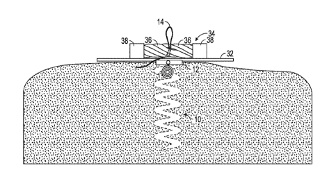
structure 505 can include one or more section. In some
embodiments, the support structure 505 includes one section. In some
embodiments, the
support structure 505 includes two sections. In some embodiments, the support
structure
505 includes three or more sections. In some embodiments, one or more sections
of the
support structure 505 can include one or more subsection. In the embodiment
shown in
Fig. 5A, the support structure 505 includes two sections: a first section
505.2 and a second
section 505.1.
100971 The first
section 505.2 can extend through at least a portion of the
coaptation assistance device 500 between the superior edge 545 and the
ventricular
projection 525. In some embodiments, the first section 505.2 can extend
through the
entire length between of the coaptation assistance device 500 between the
superior edge
545 and the ventricular projection 525. In some embodiments, the first section
505.2
extends from a location between the superior edge 545 and the inferior edge of
the
coaptation assistance body 515. In some embodiments, the first section 505.2
extends
from a location between the inferior edge of the coaptation assistance body
515 and the
ventricular projection 525. In some embodiment, the first section 505.2
extends along the
coaptation assistance body 515 and continues on to support the ventricular
projection 525.
100981 The second
section 505.1 can extend through at least a portion of the
coaptation assist body 515 between the first lateral edge and the second
lateral edge. In
some embodiments, the second section 505.1 can extend through the entire
length
between of the first lateral edge and the second lateral edge. In some
embodiments, the
second section 505.1 extends from a location between the superior edge 545 and
the
inferior edge of the coaptation assistance body 515. In some embodiments, the
second
section 505.1 extends from a location closer to the superior edge 545 than the
inferior
edge of the coaptation assistance body 515. In some embodiments, the second
section
505.1 extends from the first lateral edge toward the second lateral edge. In
some
-17-
CA 02958065 2017-02-1.3
WO 2015/200497
PCT/US2015/037451
embodiments, the second section 505.1 extends from the second lateral edge
toward the
first lateral edge. In some embodiments, the second section 505.1 extends
along a section
between the first lateral edge and the second lateral edge. In some
embodiments, the
second section 505.1 extends along the edge of the coaptation assistance
device 500.
[0099] In some
embodiments, the first section 505.2 and the second section
505.1 of the support structure 505 may be one integral piece or unitary
structure. In some
embodiments, the first section 505.2 and the second section 505.1 of the
support structure
505 are separate components. In some embodiments, the first section 505.2 and
the
second section 505.1 may be two separate sections joined together by methods
such as but
not limited to crimping and laser welding.
101001 In some
embodiments, the first section 505.2 is integrated within the
coaptation assistance body 515 as described herein. In some embodiments, the
first
section 505.2 in integrated within the ventricular projection 525 as described
herein. In
some embodiments, the first section 505.2 is removable from the coaptation
assistance
body 515 as described herein. In some embodiments, the first section 505.2 is
removable
from the ventricular projection 525 as described herein. In some embodiments,
the second
section 505.1 is integrated within the coaptation assistance body 515 as
described herein.
In some embodiments, the second section 505.1 is removable from the coaptation
assistance body 515 as described herein. In some embodiments, the first
section 505.2 can
have a first zone that is generally oriented substantially parallel to a
longitudinal axis of
the body 515, and a second zone that is generally oriented substantially
perpendicular to
the longitudinal axis of the body 515 as illustrated.
101011 When the
coaptation assistance device 500 is placed within the heart,
the coaptation assistance device 500 is such that, in some embodiments, the
ventricular
projection 525 will generally be placed within the left ventricle as shown in
Figure 5G.
The ventricular projection 525 provides a mechanism to anchor the coaptation
assistance
device 500 using the structure of the ventricles. An example of positioning of
the
coaptation assistance device 500 over the posterior leaflet is illustrated in
Fig. 5G.
[0102] Bearing in
mind that other examples of positioning are possible and are
discussed elsewhere within this disclosure, in this particular example, the
coaptation
assistance device 500 is illustrated with a ventricular projection 525 that
has a curved
shape. The ventricular projection 525 and/or the first support 505.2 may be
composed of
shape memory materials, in which case the curved shape is retained after
implantation.
-18-
CA 02958065 2017-02-13
WO 2015/200497
PCT/US2015/037451
The curved shape may enable the coaptation assistance device 500 to stay in
position as
engages to the native posterior leaflet 14.
101031 FIG. 7
illustrates an embodiment of an anchor with a linked suture
mechanism. FIG. 7 illustrates a screw-type anchor, but other anchor designs
are
contemplated. The anchor is operably coupled to a linked suture mechanism. The
anchor
can embed itself in the tissue of a patient, for example, the endocardium
and/or
myocardium. The linked suture mechanism can couple one anchor to another
anchor as
described in U.S. Prov. App. No. 62/014,060.
101041 FIG. 7 shows
an embodiment of an anchor catheter 238 that delivers
multiple anchors. Several anchors 240 can be stacked within the anchor
catheter 238.
Each anchor 240 may include a coil section 242. The coil section 242 can
include a
pointed end 244. The anchor 2240 may include an anchor head 246. The anchor
head 246
may have one of several cross sections shown by Fig. 5E. Other cross sections
are
possible.
101051 FIG. 7
illustrates a central suture 248 configured to be housed within
the central lumen of a coil. The central suture 248 can include a ball 250
coupled to the
end of the central suture 248. FIG. 7 illustrate how the central suture 248
and ball 250
may be used. The ball 250 can sit in a pocket inside the first anchor 240. The
central
suture 248 can connect the first anchor 240 to another anchor (not shown in
the figure).
This arrangement may provide the ability to use the central suture 248 as a
guide wire to
return back to an anchor 240 after the anchor 240 has been screwed into the
tissue. The
operator may wish to return to the anchor 240 to reposition or adjust the
anchor 240. In
addition, if one or more anchors 240 came loose, the central suture 248 may
provide a
tether for the loose anchors 240, therefore preventing embolic events.
101061 FIG. 8
illustrates an embodiment of an anchor with a releasable L-lock
feature. Referring to FIG. 8, the delivery catheter 252 and an anchor 254 can
have
matching or complementary features that enable them to be locked temporarily.
In some
embodiments, the delivery catheter 252 includes one or more distal locking
tabs 256. The
anchor 254 can include the hub 258. The distal locking tabs 256 of the
delivery catheter
252 may couple with features in the hub 258. Distal locking tabs 256 may be
made, for
example, of some shape memory material such as nitinol. The natural position
of the
locking tabs 256 is set such that they bend inwards and towards each other as
illustrated in
-19-
CA 02958065 2017-02-13
WO 2015/200497
PCT/US2015/037451
FIG. 8. In some methods, a guidewire or a catheter such as steerable catheter
can be
inserted between the distal locking tabs 256, and the distal locking tabs 256
can be pushed
out against the hub 258. The hub 258 is designed with matching pockets 260
such that the
distal locking tabs 256 fit into these pockets 260. As long as the steerable
catheter is
present to force the distal locking tabs 256 outwards into the pockets 260,
the tip of the
delivery catheter 252 remains locked to the hub 258. Other locking mechanisms
are
possible. The anchor 254 can be configured to complement other tools. The
tools can
include locking tabs 256 as shown. The tool can include one or more locking
tabs 256.
The anchor 254 can include complementary releasable connector as shown which
engages
the locking tabs 256. The hub 258 can be proximal to a helical screw-type
anchor. The
hub 258 may have features such as windows which can lock the locking tabs 256
of the
tool.
[0107] In some
embodiments, the tissue can be welded, heat treated, or
otherwise adapted to change the tissue properties. In some methods, the tissue
is altered to
firm up the tissue. In some methods of use, the tissue is altered to prevent
undesired
anchor pull-out effects. In some embodiments, tissue fixation mechanisms can
include
magnets, adhesives (e.g., cyanoacrylates or UV light activated adhesives, for
example), or
fixation features akin to a gecko/lizard's foot.
101081 FIG. 9
illustrates an embodiment of an anchor 262 and a clip 264. The
anchor 262 can be coupled to a suture 266 as described herein. The clip 264
can be
disposed on the suture 266. In some embodiments, the clip 264 can be movable
along the
suture 266. The clip 264 can be moved from a proximal direction to a distal
direction,
toward the anchor 262. The clip 264 can comprise a shape memory material. The
shape
memory material can be nitinol. The clip 264 can have a pre-formed shape
similar to a
paper clip. The clip 264 can inhibit movement of an implant such as a
coaptation assist
device 212. The implant (not shown) can be placed between the clip 264 and the
anchor
262. The position of the clip 264 can prevent further proximal movement of the
implant.
The clip 264 can comprise wires formed to hold onto a suture. Shown distally
is an
anchor 262, which can be a helical-type or other anchor configured to engage
tissue.
[0109] FIG. 10
illustrates an embodiment of an anchor 268 and a clip 270. The
anchor 268 can be coupled to a suture 272 as described herein. The clip 270
can be
disposed on the suture 272. In some embodiments, the clip 270 can be movable
along the
suture 272. The clip 270 can be moved from a proximal direction to a distal
direction,
-20-
CA 02958065 2017-02-1.3
WO 2015/200497
PCT/US2015/037451
toward the anchor 268. The clip 270 can inhibit movement of an implant such as
a
coaptation assist device 212. The implant (not shown) can be placed between
the clip 270
and the anchor 268. The position or the clip 270 can prevent further proximal
movement
of the implant. The clip 264 can comprise an eyelet 274. The suture 272 is
threaded
through an eyelet 274. In some methods of use, the clip 264 is slid down the
suture 272.
In some methods of use, the clip 264 is designed to couple an implant with the
anchor
268. The clip 264 is slid distally to hold the implant to the anchor 268. The
anchor 268
can be a helical-type or other anchor configured to engage tissue, as
described herein. The
anchor 268 can be operably connected to a tether, such as the suture 272
shown. The clip
270 can be movable, such as slidable, along at least part of the length of the
suture 272.
The movement of the clip 270 can lock fabric or other feature of the implant
to the anchor
268.
[0110] FIGS. 11A-
11B illustrate an embodiment of an anchor 276 and a clip
278. The anchor 276 can be coupled to a suture 280 as described herein. The
clip 278 can
be disposed within the suture 280. In some embodiments, the clip 278 can be
movable
within the suture 280. The clip 278 can be moved from a proximal direction to
a distal
direction, toward the anchor 276. The clip 278 can comprise a shape memory
material.
The shape memory material can be nitinol. The clip 278 can inhibit movement of
an
implant such as a coaptation assist device 212. The position of the clip 278
can prevent
further proximal movement of the implant. The clip 278 can comprise shape
memory
material, such as nitinol for example. The clip 278 can be configured to slide
as shown in
FIGS. 11A-11B. FIG. 11A shows the clip 278 is a first configuration. The
anchor 276 can
be coupled with a tether, such as a hollow braided suture 280, with a lumen
282
therethrough. The lumen 282 can be configured to fit the clip 278 in the first
configuration, which is shown to be in the shape of a wire. The clip 278 can
be slid into
place relative to the anchor 276. In some methods of use, application of
energy, such as
electrical current, electromagnetic energy, thermal energy, or the like can
transform the
clip 278 from the first linear configuration to the second non-linear (e.g.,
curved, or knot-
like) configuration. In the second configuration, the clip 278 can secure the
implant such
as the coaptation assist device 212 to the anchor 276 as illustrated in FIG.
11B.
[0111] FIG. 12
illustrates another embodiment of an anchor 284 and a clip
286. The anchor 284 can be coupled to a suture 288 as described herein. The
clip 286 can
be disposed on the suture 288. In some embodiments, the clip 286 can be
movable along
-21-
CA 02958065 2017-02-1.3
WO 2015/200497
PCT/US2015/037451
the suture 288. In some embodiments, the clip 286 is moveable in only one
direction. The
clip 286 can be moved from a proximal direction to a distal direction, toward
the anchor
284. The clip 286 can inhibit movement of an implant such as a coaptation
assist device
212. The implant (not shown) can be placed between the clip 286 and the anchor
284. The
position of the clip 286 can prevent further proximal movement of the implant.
In some
embodiments, the suture 288 could be another structure such as a plurality of
tie-like
arms, a threaded rod, hook-and-loop fastener arms, and the like. The clip 286
can include
leaf springs 290 operably connected to the suture 288 and configured to grab
onto the
suture 288. In some embodiments, the clip 286 can be integrated into the
implant, such as
the coaptation assist device 212. In other embodiments, the clip 286 is a
separate
component.
101121 FIG. 13
illustrates yet an embodiment of an anchor 292 and a clip 294.
The anchor 292 can be coupled to a suture 296 as described herein. The clip
294 can be
disposed within the suture 296. In some embodiments, the clip 294 can be
movable within
the suture 296. The clip 294 can be moved from a proximal direction to a
distal direction,
toward the anchor 292. The clip 294 can comprise a ball 298. The clip 294 can
inhibit
movement of an implant such as a coaptation assist device 212. The position of
the clip
294 can prevent further proximal movement of the implant. The clip 294 can be
configured to slide as shown by the arrow. The anchor 292 can be coupled with
a tether,
such as a hollow braided suture 296, with a lumen 300 therethrough. The lumen
300 can
be configured to fit the clip 294. The hollow tube 269 can have at least a
portion with a
flexible sidewall 302 as shown. The lumen 300 can be sized to accept or house
the ball
298. The ball 298 can be moved within the lumen 300 to a position in which the
sidewall
302 radially expands by virtue of the diameter of the ball 298, such that the
ball 298 and
suture 296 combination form a lock.
101.131 FIG. 14
illustrates an embodiment of an anchor 304. The anchor 304
can be an activated biopsy-type clamp. The anchor 304 can include a plurality
of lever
arms, such as two lever arms 306, 308. The lever arms 306, 308 can intersect
at a fulcrum
point 310 as shown. The anchor 304 can include a spring element 312. The
anchor 304
can grab onto tissue in between the two lever arms 306, 308. The anchor 304
can grab
onto an implant such as the coaptation assist device 212 (not shown) in
between the two
lever arms 306, 308.
-22-
CA 02958065 2017-02-1.3
WO 2015/200497
PCT/US2015/037451
[0114] FIG. 15
illustrates an embodiment of an anchor 310. The anchor 310
can be a multi-stage grapple or treble hook anchoring device. The device can
include a
tubular body 312 configured to be inserted into tissue. The tubular body can
include a
lumen 314. The lumen 314 can include with one, two, three, four, or more
secondary arms
316. The secondary arms 316 can be in a first linear configuration within the
tubular body
312. The secondary arms 316 can be moved distally outside of the tubular body
312 such
that they spread out (e.g., radially outwardly) to grip tissue as illustrated.
The secondary
arms 316 can include sharpened points. The secondary arms 316 can be coupled
to each
other.
[0115] FIG. 16
illustrates an embodiment of an anchor 318 and a clip 320. The
anchor 318 can be coupled to a suture 322 as described herein. The clip 320
can be
disposed on the suture 322. In some embodiments, the clip 320 can be movable
along the
suture 322. The clip 320 can be moved from a proximal direction to a distal
direction,
toward the anchor 318. The clip 320 can inhibit movement of an implant such as
a
coaptation assist device 212. The implant (not shown) can be placed between
the clip 320
and the anchor 318. The position of the clip 320 can prevent further proximal
movement
of the implant. The clip 320 can comprise a threaded lumen 324. The anchor 318
can
include a hub 326. The hub 326 can be threaded to engage the threaded lumen
324. In
some methods of use, the clip 320 is rotated to engage the hub 326. The clip
320 is
rotated distally to hold the implant to the anchor 318. The anchor 318 can be
a helical-
type or other anchor configured to engage tissue, as described herein. The
suture 322 can
be a tether, guidewire, or rail. FIG. 16 illustrates an embodiment of a clip
320 that can be
placed over a suture 322. The clip 320 can be operably connected to an implant
(not
shown).
[0116] FIG. 17
illustrates an embodiment of an anchor 328 and a clip 330. The
anchor 328 can include a knob 332. The clip 330 can be disposed over the knob
332. The
clip 330 can be moved from a proximal direction to a distal direction, toward
the anchor
328. The clip 330 can inhibit movement of an implant such as a coaptation
assist device
212. The coaptation assist device 212 can he placed between the clip 330 and
the knob
332. The position of the clip 330 can prevent further proximal movement of the
coaptation assist device 212. FIG. 17 illustrates a clip 330 including a
grommet, having an
outer rim and an inner eyelet as shown. The grommet can snap over the knob 332
or other
-23-
CA 02958065 2017-02-13
WO 2015/200497
PCT/US2015/037451
structure of the anchor 328. The clip 330 can be used in order to secure the
implant to the
anchor 328 as shown.
101171 FIGS. 18A-
18D illustrates an embodiment of an anchor 334. The
anchor 334 is a combination of an anchor and a clip. The combination can be
formed of a
shape memory material such as nitinol. As illustrated, a suture 336 can be
threaded
through a slot 338 in the anchor 334 such that a loop is formed as shown in
FIG. 18A. In
some embodiments, the anchor 334 is twisted as shown in FIG. 18B. In some
embodiments, the anchor 334 has a first configuration shown in FIG. 18A. In
some
embodiments, the anchor 334 has a second configuration shown in FIG. 18B. When
the
anchor 334 is twisted, the suture 336 can be more tightly held by the anchor
334. The
anchor 334 can be loaded into a needle-like introducer 340 as shown in FIG.
18C. The
introducer can have features of other tools described herein. In some
embodiments,
another suture 342 is threaded through a loop. Using a pushing mechanism, for
example,
the anchor 334 can be released into tissue as shown in FIG. 18D. The anchor
combination
can further engage in tissue if it is untwisted, rotated, or the like.
[0118] FIGS. 19A-
19B illustrate an embodiment of an anchor 344. The anchor
344 can include a helical shape, as shown. A needle 346 with a sharpened
distal end 348
can be used to more easily insert the helix portion of the anchor 344 into
tissue. The
anchor 344 does not necessarily need to be rotated, such as helical screw
anchors. Rather,
the embodiment shown in FIGS. 19A-19B can be pushed or driven straight into
tissue.
The anchor 344 can be removably connected to the needle 346. In the
illustrated
embodiment, the needle 346 passes through the center of, and be radially
surrounded by
the anchor 344. The anchor 344 can be coupled with a tether, such as a suture
350. The
anchor 344 can include a proximal shoulder 352 configured to spread the load
over a
larger surface area. The anchor 344 need not necessarily helical and can be
another
interfering geometry sufficient to maintain its position within tissue
following needle
retraction.
[0119] FIGS. 20A-
20B illustrate an embodiment of an anchor 354. The anchor
354 can comprise a shape-set shape memory material. The material can be
nitinol. In
some embodiments, the anchor 354 is deliverable from an inner lumen of a
needle or
introducer 356 as shown in FIG. 20A. The needle 356 can be advanced distally
below the
tissue surface, as shown. In some embodiments, a pusher can be used to expose
the
anchor 354. The anchor 354 can assume the shape-set (e.g., assumes a curved
-24-
CA 02958065 2017-02-13
WO 2015/200497
PCT/US2015/037451
configuration) to be retained within the tissue as shown in FIG. 20B. The
proximal end of
the anchor 354 can be operably connected to a tether, such as a suture 358.
The pusher
and needle 356 can then be retracted once the anchor 354 is retained within
the tissue.
[0120] FIG. 21
illustrates another embodiment of an anchor 360. The anchor
360 of FIG. 21 can he similar to the anchor of FIG. 5A. The anchor 360 can
take the
configuration of a barbed structure, such as basket or umbrella when delivered
into the
tissue. The anchor 360 can include a plurality of distal barbs or hooks 362.
The anchor
360 can have a first configuration within an introducer 364. The anchor 360
can have a
second configuration when deployed from the introducer 364. The anchor 364 can
be
coupled to a suture 366. The anchor 360 can comprise a shape memory material
or other
suitable material. The material can be nitinol.
[0121] FIGS. 22A-
22B illustrate an embodiment of an anchor 368. The anchor
368 can include a toggle bolt. The anchor 368 can include a proximal eyelet
370. The
eyelet 370 can be operably connected to a suture 378. The anchor 368 can
include barbs
374 that can radially expand when within the tissue. The anchor 368 includes a
toggle bolt
376 longitudinal axis. The anchor 368 is inserted within the tissue as shown
in FIG. 22A.
After the anchor 368 is inserted into tissue, the toggle bolt 376 and/or the
eyelet 370 can
be rotated. The barbs 374 are pushed outward, further securing the anchor 368
in the
tissue as shown in FIG. 22B.
101221 FIGS. 23A-
23E illustrate an embodiment of an arrangement of clips
378, 380 according to some methods of use. In some arrangements, two clips
378, 380 are
placed next to each other to increase the surface area of the lock. This can
reduce the
number of clips 378, 380. In some embodiments, increasing the surface area of
the clip
378, 380 can potentially reduce the numbers of anchors 382 needed. In some
embodiments, 2, 3, 4, 5, or more clips can be placed in close proximity to
each other, such
as directly adjacent to each other as illustrated. In some embodiments, one
clip 378 spans
two or more anchors 382, 384 as shown in FIG. 23E.
[0123] FIG. 24A
illustrates an embodiment of an anchor 386. The anchor 386
can be a helical-type or other anchor configured to engage tissue. The anchor
386 can
include a reinforcement portion 390. The reinforcement portion 390 can
advantageously
reduce the risk of anchor pull-out and embolization. In some embodiments, the
anchor
386 includes a helical or other anchor configured to reside within the tissue
as described
herein. The anchor 386 can include a hub 392. The hub 392 can be in the shape
of a disc.
-25-
CA 02958065 2017-02-1.3
WO 2015/200497
PCT/US2015/037451
The hub 392 can be operably attached to the anchor 386 as shown. In the
embodiment
shown in FIG. 24A, operably connected to the hub 392 can be one, two, or more
shape
memory clips 394. The shape memory material can be nitinol. The clips 394 can
increase
the surface area that the anchor 386 engages.
[0124] FIG. 24B
illustrates an embodiment of an anchor 396. The anchor 396
can be a helical-type or other anchor configured to engage tissue. The anchor
396 can
include a reinforcement portion 398. The reinforcement portion 398 can
advantageously
reduce the risk of anchor pull-out and embolization. In some embodiments, the
anchor
396 includes a helical or other anchor configured to reside within the tissue
as described
herein. The anchor 396 can include a hub 400. The hub 400 can be in the shape
of a disc.
The hub 400 can be operably attached to the anchor 396 as shown. As
illustrated in FIG.
24B, the anchor 396 can include a spring 402. The spring 402 can be located in
between a
surface of the hub 400 and a proximal cap 404. The proximal cap 404 can
include a T or
treble hook 406 designed to engage tissue. The hook can include a barb 408 to
increase
engagement with the tissue.
[0125] FIG. 24C
illustrates an embodiment of an anchor 410. The anchor 410
can be a helical-type or other anchor configured to engage tissue. The anchor
410 can
include a reinforcement portion 412. The reinforcement portion 412 can
advantageously
reduce the risk of anchor pull-out and embolization. In some embodiments, the
anchor
410 includes a helical or other anchor configured to reside within the tissue
as described
herein. The anchor 410 can include a hub 414. The hub 414 can be in the shape
of a disc.
The hub 414 can be operably attached to the anchor 410 as shown. In FIG. 24C,
the hub
414 can have a barbed feature 416. The barbed feature 416 can be located in
between the
hub 414 and a proximal cap 418. The barbed feature 418 can be located
centrally. The
anchor 410 can press against tissue with either the T/treble hook feature 420
extending
from the cap 418.
[0126] FIG. 24D
illustrates an embodiment of an anchor 422. The anchor 422
can be a helical-type or other anchor configured to engage tissue. The anchor
422 can
include a reinforcement portion 424. The reinforcement portion 424 can
advantageously
reduce the risk of anchor pull-out and embolization. In some embodiments, the
anchor
422 includes a helical or other anchor configured to reside within the tissue
as described
herein. The anchor 422 can include a hub 424. The hub 424 can be in the shape
of a disc.
The hub 424 can be operably attached to the anchor 422 as shown. the method of
use can
-26-
CA 02958065 2017-02-1.3
WO 2015/200497
PCT/US2015/037451
include the application of energy. Energy, such as RF, microwave, or other
energy is
utilized to create a bond between the tissue and the hub. In some embodiments,
a
biocompatible adhesive, such as a cyanoacrylate for example, is utilized to
bond the hub
and the tissue.
[0127] FIG. 24E
illustrates all embodiment of an anchor 426. The anchor 426
can be a helical-type or other anchor configured to engage tissue. The anchor
426 can
include a reinforcement portion 428. The reinforcement portion 428 can
advantageously
reduce the risk of anchor pull-out and embolization. In some embodiments, the
anchor
426 includes a helical or other anchor configured to reside within the tissue
as described
herein. The anchor 426 can include a hub 428. The hub 428 can be in the shape
of a disc.
The hub 428 can be operably attached to the anchor 426 as shown. In FIG. 24E,
the hub
428 includes a suture 430 or wire extending proximally. The anchor 426 can
include a
crimp feature 432 to promote stabilization of the anchor.
[0128] FIGS. 25A-
25C illustrate embodiments of an anchor and a suture. In
some embodiments, the anchor 10 includes a helical or other anchor configured
to reside
within the tissue. In other embodiments, the anchor can take the form of any
anchor as
described herein. The anchor can include a proximal hub 12. The hub 12 can be
connected
to a tether, such as an end of the tether. The tether can be a suture 14
having a needle 16,
as shown. In other embodiment, the tether is a tube, rail, or guidewire. The
anchor can
include a cross pin 18. The cross-section 25B-25B illustrates a cross pin 18
in the hub 12.
The suture 14 can be operably attached to the cross pin 18.
[0129] Also
illustrated is an alternative embodiment shown in FIG. 25C. In
some embodiments, the anchor 434 includes a grappling hook. The grappling hook
has a
plurality of arms 436. In the illustrated embodiment, three arms 436 are shown
but one or
more arms are contemplated. The arms 436 can serve as a tissue anchor instead
of the
helical structure shown in other embodiments herein.
[0130] FIGS. 26A-
26D illustrate an embodiment of an anchor driver 20. The
anchor driver 20 can include a proximal control handle 22. The proximal
control handle
22 can include a torque knob 24 and a suture locking knob 26. The torque knob
24 can
control a torque shaft 28. The anchor driver 20 call include a distal tubular
body 30 sized
to accept the torque shaft 28 therein. The torque shaft 28 can be configured
to rotate an
anchor, as described herein. Rotation of the anchor may be necessary to lodge
the anchor
-27-
CA 02958065 2017-02-13
WO 2015/200497
PCT/US2015/037451
within tissue. In other embodiments, a pusher described herein is used to
expel the anchor
from the anchor driver 20.
101311 Also
illustrated is the anchor 10. In some embodiments, the suture 14
can be attached to the anchor 10 via a knot. In some embodiments, an end of
the suture 14
can be heat-formed like a ball. The anchor 10 can be loaded into the anchor
driver 20 as
illustrated. In some embodiments, the torque driver 20 engages a feature of
the anchor 10.
As described herein, the torque driver 20 can engage the crossbar 18 of the
anchor 10. The
anchor 10 can be initially retracted inside the tubular body 30 as shown in
FIG. 26C.
101321 Rotating the
torque knob 24 in an appropriate direction can rotate the
torque driver 28. Rotating the torque driver 28 can rotate the anchor 10 in
order to engage
or disengage the anchor 10 from the tissue. The suture locking knob 28 can
help to
maintain the anchor 10 in place during anchor delivery. In some methods, the
suture 14 is
pulled through the suture locking knob 28. Tension on the suture 14 can hold
the anchor
against the torque driver 28. Tension on the suture 14 can engage the crossbar
18 with
the torque driver 28. Other configurations of coupling the anchor 10 to the
torque driver
are contemplated.
101331 FIGS. 27-31
illustrate method steps for some methods of use of the
anchor 10 and the anchor driver 20. FIG. 27 illustrates the anchor driver 20
approaching
the tissue. In some embodiments, the tissue is cardiac tissue having
endocardial,
myocardial, and epicardial layers. In other embodiments, the tissue is
associated with
other organs or anatomical structures such as the bladder, intestine, lungs,
stomach,
kidneys, liver, skin, gall bladder, pancreas, brain, spinal cord, bone,
muscle, fascia,
ligaments, tendons, cartilage, etc. The anchor 10 is initially retracted
within the distal
tubular body 30 as shown in FIG. 27..
101341 FIG. 28
illustrates the anchor driver 20 at the specific tissue location
for anchoring. The distal tip of the anchor driver 20 is placed over the
tissue. In some
embodiments, the distal tip touches the tissue. The torque knob 24 is rotated
in an
appropriate direction. The anchor 10 is coupled to the torque shaft 28 such
that rotation of
the torque knob 24 causes rotation of the anchor 10. The anchor 10 is rotated
to engage
the anchor 10 with the tissue. Further rotation causes the anchor 10 to travel
distally into
the tissue. In other embodiments, the anchor 10 is pushed into the tissue. For
instance,
FIGS. 19A-19B illustrate an embodiment of an anchor that can be pushed into
tissue
rather than rotated to engage tissue. In some embodiments, the anchor driver
20 includes a
-28-
CA 02958065 2017-02-1.3
WO 2015/200497
PCT/US2015/037451
pusher (not shown) instead of a torque driver 28. The pusher can cause distal
movement
of an anchor without rotation.
101351 FIG. 29
illustrates the anchor 10 fully deployed within the tissue. The
anchor driver 20 is removed. The hub 12 of the anchor 10 can be adjacent to
the tissue as
shown. The hub 12 of the anchor 10 can be embedded within the tissue. The
suture 14 and
the needle 16 can extend from the hub 12. The suture 14 can be attached to the
anchor 10
by a knot. In some embodiments, the end of the suture 14 is heat formed into
the shape of
a ball.
101361 FIG. 30
illustrates that once the anchor 10 is fully deployed into tissue,
the needle 16 on the suture 14 can be utilized to attach an implant 32
(illustrated
schematically) to the anchor 10. Non-limiting examples of the implant 32
include, for
example, a coaptation assistance device, an annuloplasty ring, an artificial
valve, cardiac
patch, sensor, pacemaker, or other implants. The implant 32 could be a mitral
valve ring
or artificial mitral or aortic valve in some embodiments. The implant can be
any implant
described herein. The implant 32 can be lowered toward the needle 16 as shown.
The
needle 16 can engage the implant 32. In some embodiments, the needle 16 can
pass
through the implant 32, as shown in FIG. 31. The needle 16 and the suture 14
are initially
in position 1. The needle and the suture can pass through the implant to be in
position 2.
Other suture paths are contemplated. FIG. 31 illustrates the needle 16 being
used to
engage the suture 14 with the implant 32. The needle 16 and the suture 14 are
shown
passing through the implant 32.
101371 FIG. 32
illustrates an embodiment of a clip 34. The clip 34 can be
considered a knotless suture clip. The clip 34 can include a cable. The cable
can be made
of nitinol or superelastic nitinol, for example. The cable can include at
least two wires or
strands 36. In the illustrated embodiment, the cable includes seven strands,
but one two or
more strands are contemplated. The two strands can be twisted, and heat set to
maintain
the shape of the cable. In some embodiments, the cable can be hollow to have a
central
lumen. In some embodiments, the cable can be solid as illustrated. In some
embodiments,
the cable is comprised of nitinol wires or strands 36 twisted together to form
a cable. In
some embodiments, the clip 34 can include one, or a pair of end crimps 38 as
shown. The
end crimp 38 can be in the form of a tube that surrounds the end of the cable.
101381 In some
embodiments, the wires or strands 36 may have a rough
surface finish to increase the friction between the clip 34 and the suture 14
to improve the
-29-
CA 02958065 2017-02-13
WO 2015/200497
PCT/US2015/037451
locking force, or a smooth surface finish in other embodiments. In some
embodiments,
the suture 14 can be comprised of materials with a rough surface to increase
the friction
between the clip 34 and the suture 14 to improve the locking force. In some
embodiments,
the suture 14 can include barbs to increase the friction between the clip 34
and the suture
14 to improve the locking force.
101391 The clip 34
can serve the same or a similar function as other clips
described herein. The clip 34 can allow a user to lock the implant 32 to the
tissue without
applying knots to the suture 14. The clip 34 therefore, can result in
advantageous rapid
attachment. In some methods of use, the rapid attachment can be accomplished
by
inserting the suture 14 through the wires or strands 36 of the cable. The
wires or strands
36 of the cable can apply a force on the surface of the suture 14 and lock
onto the suture
14, advantageously preventing loosening.
101401 FIG. 33
illustrates embodiments of a clip. The clips 34A, 3413 can be
considered a knotless suture clip. The clips 34A, 34B can include a cable. The
cable can
be made of nitinol or superelastic nitinol, for example. The cable can include
at least two
wires or strands 36. The two strands 36 can be twisted, and heat set to
maintain the shape
of the cable. In some embodiments, the clips 34A, 34B can include one end
crimp 38, or a
pair of end crimps 38. The end crimp 38 can be in the form of a tube that
surrounds the
end of the cable. The cable need not be substantially linear in geometry. For
example, the
clip 34A can be V shaped as shown in FIG. 33. The clip 3413 can be a circular
design
having a single end crimp 38 as shown in FIG. 33. Depending on the desired
clinical
result, U shapes, other arcuate shapes including oval shapes, triangular,
square,
rectangular, or other shapes can also be utilized.
101411 FIG. 34
illustrates an embodiment of a clip driver 40. The clip driver
40 can be used for deploying clips. While clip 34 is shown, other clips
described herein
can be used with the clip driver 40. The clip driver 40 can include a proximal
control
handle 42 including a control 44, such as a trigger mechanism as shown. The
clip driver
40 can include an elongate body 46. The elongate body 46 can include an outer
sleeve 48
and an inner shaft 50. The clip 34 (or other clips described herein) can be
removably
carried on the inner shaft 50 as shown. In some embodiments, the inner shaft
50 passes
through a slot formed by the two strands 36. The inner shaft 50 passes from
one side of
the clip 34 to the other side of the clip 34. The strands 36 can provide
tension to maintain
the clip 34 on the inner shaft 50.
-30-
CA 02958065 2017-02-1.3
WO 2015/200497
PCT/US2015/037451
101421 The distal
end of the clip driver 40 can include a snare 52. The snare 52
can be a lasso. The snare can extend through a lumen of the inner shaft 50. In
some
embodiments, the inner shall 50 has a single lumen. In other embodiments, the
inner shaft
50 has two lumens, one for each end of the snare 52. The snare 52 can be
coupled to the
proximal control handle 42. The proximal control handle 42 can retract the
snare 52
within the outer sleeve 48. The proximal control handle 42 can retract the
snare 52 within
the inner shaft 50. The driver can also include an end crimp as illustrated.
101431 HG. 35
illustrates the clip driver 40 during use. The anchor 10 can be a
helical or other anchor as previously described. The anchor 10 can be anchored
into
tissue. The tissue can be cardiac tissue. The suture 14 can extend from the
anchor 10. The
implant 32 can be positioned relative to the anchor. The implant 32 can be a
cardiac
implant or other body implant. The suture 14 and the needle 16 from the anchor
10 can be
threaded through the implant 32 as previously described. The driver 40 can be
moved
toward the implant 32. The driver 40 can be moved in position relative to the
suture 14.
101441 FIG. 36
illustrates another method step in some methods, where the
suture 14 is threaded through the snare 52. The needle 16 can be threaded
through the
snare 52. The snare 52 can be looped around the suture 14. The snare 52 can be
looped
around the needle 16. The suture 14 and/or the needle 16 can be threaded
through the
snare 52 before the clip 34 is deployed. As illustrated in FIG. 37, the suture
14 is then
pulled gently in the direction of the arrow. The clip driver 40 can be moved
toward the
implant 32. The inner shaft 50 can be positioned adjacent to the implant 32.
The clip is
placed over the implant 32 before the clip 34 is deployed.
101451 By
activating the control 44 (e.g., the trigger mechanism) to a first
position, the clip 34 can be deployed. FIG. 38 illustrates the step where the
control 44 is
pulled proximally. This action can retract the inner shaft 50. The inner shaft
50 can be
retracted into the outer sleeve 48. The outer sleeve 48 can be moved toward
the implant
32. The outer sleeve 48 can be positioned adjacent to the implant.
101461 This action
can retract the snare 52 with the inner shaft. The snare 52
can be retracted into the outer sleeve 48. The snare 52 retracts the suture 14
with the snare
52. The snare 52 passes through the clip 34. The snare 52 brings the suture 14
through the
clip. After the suture 14 is pulled into the inner shaft 50, the clip 34 is
advanced by the
outer sleeve 48. The clip 34 is deployed over the suture 34. In some
embodiments,
separate mechanism or movements retract the inner shaft 50 and the snare 52.
In some
-31-
CA 02958065 2017-02-13
WO 2015/200497
PCT/US2015/037451
embodiments, only the inner shaft 50 is retracted. In some embodiments, both
the inner
shall 50 and the snare 52 are retracted.
101471 As
illustrated in FIG. 39, the clip 34 is deployed over the suture 14,
tension can be applied to the suture 14 to firmly attach the implant 32 to
tissue. Excess
suture 14 can then be trimmed. Following deployment of the clip 34 at a
desired location,
the suture 14 can be cut by activating the control 44 (e.g., the trigger
mechanism) to a
second position. The second position can be associated with cutting the suture
14. As
illustrated in FIG. 40, the clip driver 40 is then removed. In some
embodiments, the suture
14 forms a loop through the cable of the clip 34. In some embodiment, the
suture 14
forms a suture path from the anchor 10, through the implant 32, through the
clip 34, and
curving and extending back through the clip 34. Other suture paths are
contemplated. The
method steps described herein can be repeated several times, including 2, 3,
4, 5, 6, 7, 8,
9, 10, 15, 20, 25, or more times (e.g., in a replacement mitral valve
procedure) at selected
locations. The clips 34 can be used for anchors 10 on the annulus. The clips
34 can be
used for every anchor or selected anchors. The clips 34 can be used to attach
the artificial
mitral valve to the annulus. Other uses for the clips 34 and anchors 10 are
contemplated.
101481 FIG. 41
illustrates the clip 34A of FIG. 33 following deployment. FIG.
42 illustrates the clip 34B of FIG. 33 following deployment. The clips 34A.
3413 operably
connected to the implant 32 and the anchors 10. The clips 34A, 3413 are
described herein
in connection with FIG. 33.
101491 FIGS. 43A-
43C illustrates embodiments of an anchor 10. The anchor
can have a helical shaft as shown or other geometry configured to engage
tissue. The
anchor 10 can be made of an appropriate material, such as nitinol for example.
The
proximal end of the helical shaft can include a central cross pin 18. The
central cross pin
18 can be created by laser welding for example. The helical shaft can include
any desired
number of coil turns or revolutions appropriate for tissue anchoring, such as
1, 2, 3, 4, 5,
6, or more turns. The distal tip of the anchor 10 can have a sharp point grind
tip as
illustrated. The tip can be the distal most feature of the anchor 10. In some
embodiments,
the helix can have an outer diameter of between about 3-5mm, such as about
4.3mm. The
helical shaft can be made of a wire having a diameter, in some embodiments, of
between
about .010" and about .030", such as about .020" in some cases. The pitch can
be, in some
embodiments, between about 1mm and about 2mm, such as about 1.5mm in some
-32-
CA 02958065 2017-02-1.3
WO 2015/200497
PCT/US2015/037451
embodiments. The gap between coils can be, for example, between about 0.5mm
and
about 2mm, such as about lmm in some embodiments.
[0150] FIGS. 43D-
43G illustrate method steps of using a loading suture 54.
The loading suture 54 can be used to transfer a slip knot of a tether. The
tether can include
suture 14 and needle 16. The suture 14 can be 2-0, 3-0, 4-0, or 5-0 suture in
some
embodiments. The loading suture 54 forms a loop and is passed under the cross
pin 18 of
the proximal end of the anchor 10. The suture 14 is passed around the loop of
the loading
suture 54. '1'he suture 14 can have one or more needles 16 attached to the
suture 14, as
previously described. The loading suture 54 is pulled therefore passing the
loop of the
suture 14 to the other side of the cross pin 18. The ends of the suture 14 can
be passed
through the loop of the suture 14, therefore forming a girth hitch knot around
the cross pin
18 as shown.
[0151] FIGS. 44A-
44C illustrates an embodiment of an anchor driver 56. The
anchor driver 56 can, for example, include features illustrated and described
in connection
with FIG. 26-28. The distal tip of the anchor driver 56 can be positioned
proximate the
tissue. With forward pressure on the anchor 10, the torque shaft control can
be rotated in
an appropriate direction, such as clockwise, to rotate a torque shaft 58. The
anchor 10 is
coupled to the torque shaft 58 such that rotation of the toque shaft control
causes rotation
of the anchor 10. Further rotation of the torque shaft control drives the
anchor 10 into the
tissue. The anchor driver 56 can include a distal housing 60. The distal
housing 60 can be
retracted in order to visualize the anchor engagement. If needed, the distal
housing 60 can
be advanced back over the proximal end of the anchor 10 and additional torque
can be
applied to further drive and secure the anchor 10 into tissue. Further
inspection can be
done following the additional torqueing, and repeated as necessary. In some
embodiments, the cross pin 18 of the anchor 10 can fit into a cutout at the
tip of the torque
shaft 58.
[0152] The anchor
driver 56 can include a Tuohy-Borst or similar leak-
resistant valve or adapter. The adapter can be located on the proximal end of
a handle of
the anchor driver 56. The adapter can secure the suture 14 or loading suture
54 while the
anchor 10 is being driven into the tissue. Once satisfied with anchor
engagement, the
adapter can be undone to loosen the suture 14 or loading suture 54. The anchor
driver 56
can be removed. If a loading suture 54 is coupled to the anchor 10, then the
method steps
described previously can be used. The suture 14 (e.g.. 3-0 or 4-0 suture in
some
-33-
CA 02958065 2017-02-1.3
WO 2015/200497
PCT/US2015/037451
embodiments) can be loaded through the loop of the loading suture 54. The
suture 14 can
be pulled through the anchor cross pin 18 as illusirated and described in
connection wiih
FIG. 43. In some methods, the loading suture 54 can be fully retracted to
"dock" the cross
pin 18 of the anchor 10 into the cutout at the tip of the torque shaft 58. The
suture 14 can
be secured via the Tuohy-Borst or other adapter.
[0153] FIGS. 45A-
45B illustrate non-limiting dimensions for embodiments of
the anchor driver 56, including the torque shaft 58. In some embodiments, the
dimensions
are approximately the dimensions listed. Schematic and cross-sectional views
are also
shown.
[0154] FIGS. 46A-
46E illustrate an embodiment of a clip. The clip can be
shape-set. The clip can be made of a shape memory material such as nitinol.
The clip can
include two lever arms, a fulcrum cross-over point, and a loop portion as
illustrated. In
some methods of use, the clip can be loaded onto a thin-walled hypotube of an
inserter. In
some methods of use, the clip can be pushed off the hypotube using a pusher
tube. The
clip can cinch down onto the suture 14, immobilizing the implant 32 against
the anchor
10. The clip can have a loaded state in which the hypotube and the suture 14
are at least
partially surrounded by the lever arms of the clip. The clip can have a
clipped state in
which the suture is compressed as the lever arms close in together. Also
illustrated is the
clip removably attached over the sidewall of the hypotube as part of the
delivery system.
Distal movement of the pusher can move the clip distally off the hypotube and
onto the
suture 14. The suture 14 can be attached, such as pre-attached to the anchor
10, such as a
helical anchor as previously described. In some embodiments, the implant
surface and
implant grommet is sown over the tissue. The anchor 10 shown below the tissue
in dashed
lines. Excess suture can be removed as previously described.
[0155] It is
contemplated that various combinations or subcombinations of the
specific features and aspects of the embodiments disclosed above may be made
and still
fall within one or more of the inventions. Further, the disclosure herein of
any particular
feature, aspect, method, property, characteristic, quality, attribute,
element, or the like in
connection with an embodiment can be used in all other embodiments set forth
herein.
Accordingly, it should be understood that various features and aspects of the
disclosed
embodiments can be combined with or substituted for one another in order to
form
varying modes of the disclosed inventions. Thus, it is intended that the scope
of the
present inventions herein disclosed should not be limited by the particular
disclosed
-34-
CA 02958065 2017-02-1.3
WO 2015/200497
PCT/US2015/037451
embodiments described above. Moreover, while the invention is susceptible to
various
modifications, and alternative forms, specific examples thereof have been
shown in the
drawings and are herein described in detail. It should be understood, however,
that the
invention is not to be limited to the particular forms or methods disclosed,
but to the
contrary, the invention is to cover all modifications, equivalents, and
alternatives falling
within the spirit and scope of the various embodiments described and the
appended
claims. Any methods disclosed herein need not be performed in the order
recited. The
methods disclosed herein include certain actions taken by a practitioner;
however, they
can also include any third-party instruction of those actions, either
expressly or by
implication. For example, actions such as "inserting a coaptation assist body
proximate
the mitral valve" includes "instructing the inserting of a coaptation assist
body proximate
the mitral valve." The ranges disclosed herein also encompass any and all
overlap, sub-
ranges, and combinations thereof. Language such as "up to," "at least,"
"greater than,"
"less than," "between," and the like includes the number recited. Numbers
preceded by a
term such as "approximately", "about", and "substantially" as used herein
include the
recited numbers, and also represent an amount close to the stated amount that
still
performs a desired function or achieves a desired result. For example, the
terms
"approximately", "about", and "substantially" may refer to an amount that is
within less
than 10% of, within less than 5% of, within less than 1% of, within less than
0.1% of, and
within less than 0.01% of the stated amount.
-35-