Note: Descriptions are shown in the official language in which they were submitted.
8401 8 9 5 1
BIOMATERIALS FOR TRACK AND PUNCTURE CLOSURE
This is a divisional application of Canadian Patent Application No. 2,760,704
filed May 4,2010.
Technical Field
The technical field relates to surgical methods and closure of punctures, for
instance,
percutaneous closure of femoral access punctures.
Background
Clinicians perform many medical procedures .by puncturing a blood vessel and
introducing a small tube through the blood vessel that is guided to other
parts of the body. A
common point of entry is the. femoral artery. Once the medical procedure is
completed, the
artery or other blood vessel has to be adequately closed so the patient can
leave the operation
site, and the puncture needs to heal.
Many devices have been created to facilitate closure after iatropic punctures
have been
made in the femoral artery. Examples include devices described in US patents
5,108,421 to
Fowler, 5,192,302 or 5,222,974 to =Kensey, and US Pub 2006/0100664 to Pal. The
PERCLOSE system, introduced in 1994, was the first suture-mediated device to
be approved
by the Food and Drug Administration. PERCLOSE PROGLIDE is the latest
generation,
introduced in 2004. It offers improvements in the ease of knot delivery
and'stre.ngth and of
the suture material. The system is composed of a sheath, a guide, a knot
pusher accessory,
and a suture trimmer. The ANGIO-SEAL device is made up of three components: a
specially
designed polymer anchor, an absorbable collagen sponge, and an absorbable self-
tightening
suture. The sponge is positioned in the puncture track outside the artery wall
by a pulley
systen. created by the anchor and suture. The device seals and sandwiches the
arteriotomy
between the anchor and the collagen plug. The STARCLOSE is a clip-mediated
closure
device approved by the Food and Drug Administration in 2005. The STARCLOSE
introduces a small, circumferential, flexible clip that mechanically binds the
surface of the
femoral Artery together. The clip is Made of nitinol, a nickel-iitanium alloy
with elastic
properties that allow it to return to its original shape once released from
the device. Its use
1
CA 2977830 2017-08-30
WO 2010/129510 PCT/1JS2010/033488
involves a multi-step deployment process with a specialized application. Thc
clip is left on
the outside of the artery. The MYNX is a rolled-up biodegradable polymer sheet
that is
pushed into the puncture track and allowed to swell. The swelling secures the
device and
prevents blood flow.
Summary
A percutaneous puncture of a blood vessel involves creating a track through
the skin
and puncturing a blood vessel. The need to close the blood vessel is widely
recognized
because patients have traditionally been required to stay for long times with
manual
compression. Accordingly, devices have been made to shorten this time. There
is another
aspect to the closure and healing process, however, which is the sealing of
the track that leads
to the puncture. Blood from the tissue walls of the track can ooze into the
track.
Conventional approaches involving a plug inside or at the blood vessel do not
address
blood seepage from the track. Described herein, however, are devices that
address both
puncture closure and track sealing.
Further, a vascular closure system that can efficiently close large bore
punctures will
enable advancement and adoption of additional percutaneous medical tools that
would benefit
from large access sites. Unfortunately, the force of blood pressure that tends
to displace a
plug in a blood vessel is proportional to the surface area of the plug so that
the forces tending
to push a plug out of a puncture increase by a power of two as the plug area
is increased.
Accordingly, many conventional approaches to plugging a small bore puncture do
not scale-
up to medium and large bore punctures. A medium bore puncture is defined
herein as a
puncture made with a gauge between, and inclusive of, 11 F to 14 F. A large
bore puncture
has a gauge of more than 14F. A small bore puncture has a gauge of less than
11 F.
Described herein, however, are devices that provide medium and large bore
sealing.
Certain embodiments herein include techniques for sealing punctures with a
combination of a biomaterial tarnponade-and-adhesive combination. Another
embodiment
provides application of adhesives to the track lumen. Another system provides
a tarnponade
biomaterial with adhesive coatings to seal against the track lumen so the
material can seal
both the track and the artery. Other systems described address closure of an
access site that is
not femoral but is superficial. For instance, in a brachial access site, there
is not enough track
available to deploy a conventional vascular closure device.
SUBSTITtli SHEET (RULE 26)
CA 2977830 2017-08-30
84018951
In a first aspect, there is provided an apparatus for treatment of a track and
blood vessel puncture, comprising: an applicator comprising a distal portion
sized for
placement in the track and having a distal opening and a lumen, and a pusher
at least partially
received in the lumen for pushing the plug out of the lumen through the distal
opening; a plug
comprising a proximal and a distal end preloaded in the applicator; and a
first reactive
precursor and a second reactive precursor preloaded in the applicator,
wherein: the plug is
sized for placement in the track and disposed within the lumen, the first and
second precursors
are disposed in the lumen at a position distal to the distal end of the plug
for release into the
track prior to expulsion of the plug from the lumen, and the first reactive
precursor and the
second precursor are in a dry state and react with a physiological fluid after
placement in the
track to dissolve and form a matrix material which secures the plug within the
puncture.
In a further aspect, there is provided use of an apparatus as defined herein
for
treatment of a blood vessel puncture and puncture track.
In a further aspect, there is provided the apparatus as defined herein for use
in
the treatment of an iatropic track and blood vessel puncture.
In a further aspect, there is provided use of the apparatus as defined herein
for
the treatment of an iatropic track and blood vessel puncture.
2a
CA 2977830 2019-01-25
r
WO 29101129510 PCT/US2018/033488
Brief Description of the Figures
Fig. lA depicts an embodiment of a coated biomaterial;
Fig. 1B depicts the embodiment of Fig. lA being rolled-up;
Fig. 1C is a cross-section of the embodiment of Fig 1B after being rolled-up;
Fig. 2A depicts a partially coated biomaterial with a coating disposed as a
plurality of
stripes;
Fig. 2B depicts a partially coated biomaterial with a coating disposed as a
plurality of
blebs;
Fig. 2C depicts a partially coated biomaterial with a coating disposed as a
plurality of
rings;
Fig. 3A depicts a biomaterial coated on each of two faces;
Fig. 3B depicts a biomaterial partially coated on each of two faces, with the
coating
being a plurality of blebs;
Fig. 3C depicts a biomaterial partially coated on each of two faces, with the
coating
being a plurality of stripes;
Fig. 4A depicts a biomaterial sheet partially coated on a longitudinal half;
Fig. 4B depicts the material of Fig. 4A in a rolled configuration;
Fig. 5A depicts an elongate member attached to an occluding device positioned
in a
track and blood vessel;
Fig. 5B depicts a plug with a partial and discontinuous coating on a half of
the plug;
Fig. 5C depicts a delivery apparatus and the plug of Fig 5B deployed over the
elongate
member of Fig. 5A;
Fig. 5D depicts the apparatus of Fig. 5C in a process of delivering the plug
of Fig. 5B;
Fig. 5E depicts the apparatus of Fig. 5C in a further stage of a process of
delivering
the plug of Fig. 5B;
Fig. 5F is an alternative embodiment of the plug of Fig 5B as deployed in a
track;
Fig. 6A depicts a delivery apparatus for a biomaterial plug with a
substantially planar
shape;
Fig. 6B is a longitudinal cross-section of the apparatus of Fig. 6A;
Fig. 6C depicts the delivery apparatus of Fig. 6A and 6B with the biornaterial
sheet
removed and the radially expanding member in a storage (undeployed) position;
Fig. 6D depicts the apparatus of Fig. 6C in a deployed (radially expanded)
position;
Fig. 6E depicts an elongate member attached to an occluding device positioned
in a
track and blood vessel;
SUBSTITA SHEET (RULE 26)
CA 2977830 2017-08-30
W02010/129510 PCT/US2010/033488
Fig. 6F depicts a cross-section of the apparatus of Figs. 6A and 6B as
deployed over
the elongate member of Fig. 6E;
Fig. 6G depicts a process of expanding the radially expanding member of thc
embodiment of Fig. 6F to deploy the biomaterial in the track;
Fig. 6H depicts a process of using the embodiment of Fig. 6F to deploy the
biomaterial in the track;
Fig. 61 depicts a further process of using the embodiment of Fig. 6F to deploy
the
biomateri al in the track;
Fig. 6J depicts a further, optional, process of using the embodiment of Fig.
6F to
deploy the biomaterial in the track;
Fig 6K depicts an outcome of the process depicted at Fig. 6J;
Fig. 7 depicts an embodiment of a hand-held swab applicator;
Fig. 8A depicts a plug deployed in a vascular access site;
Fig. 8B depicts a process of using the embodiment of Fig. 7 in the site of
Fig. 8A;
Fig. 8C depicts a further process of using the embodiment of Fig. 7 in the
site of Fig.
8A;
Fig. 8D depicts an outcome of a process of Fig. 8C;
Fig. 9A depicts a coated plug with a backing and a compression device for
compressing the plug on skin of a patient;
Fig. 9B depicts the plug of Fig. 9A in place on a patient's skin;
Fig. 10A depicts an applicator;
Fig. 1013 depicts the applicator of Fig. 10A in use at a vascular access site
for delivery
of a precursor and a plug;
Fig. 10C depicts the applicator of Fig. 10B in a process of delivering the
precursor;
Fig. 10D depicts the applicator of Fig. 10C in a process of delivering the
precursor;
Fig. 10E depicts the applicator of Fig. 10B in a process of delivering the
plug; and
Fig. 11 is an embodiment of a plug system as used in Example 2.
Detailed Description of Preferred Embodiments of the Invention
An embodiment of the invention is an adhesive plug sized for placement in an
iatropic
tract. In the case of surgical access procedures, the tract is a track through
a tissue that
terminates in a blood vessel puncture. The track is a lumen defined by tissue
walls. The
adhesive plug should be small enough to pass into the track. The plug has a
portion that is
coated with a precursor or precursors that are substantially dry and react
with physiological
SUBSTITUTE SHEET (RULE 26)
CA 2 977 8 30 2 01 7-08-30
,
WO 2010/129510 PCT/US2010/033488
fluid to dissolve and form a matrix material. The material contributes to
forming an adhesive
force between the plug and a tissue, which could be walls of the track of
tissues at or near the
puncture. Coatings enhance closure of the blood vessel and also sealing of the
track itself.
These and other embodiments create new options for closing large bore or other
punctures,
for sealing tracks that otherwise tend to ooze blood, and for sealing short
tracks.
Embodiments described herein include precursors or coated matrices placed into
a track,
including plugs or sheets.
Coatings and precursors
Figure lA depicts biomaterial sheet 102 with a substantially dry adhesive
coating 104
that is non-adherent when substantially dry but is adherent when exposed to a
physiological
solution (e.g., fluids in a wound or interstitial fluids). The term sheet
refers to a generally
planar structure with a thickness that is much less than the surface area. The
sheet 102 is
depicted as rectangular but may have other shapes. Figure 1B is a conceptual
image of how
sheet 102 may be rolled-up. Figure 1C is a cross-sectional view of the same
sheet in a fully
rolled-up configuration. The sheet may thus be rolled so that it is biased to
open by uncurling
when unconstrained.
The coatings may be made with precursors that react with each other to create
a
matrix material. The matrix may be covaIently crosslinked, or not, depending
on the
precursor. Covalently crosslinked materials have a distinct chemical structure
from not-
covalently crosslinked material, and different properties including mechanical
strength and
solubility. The matrix may be a hydrogel. A hydrogel is hydrophilic but does
not dissolve in
water.
Precursors are components that undergo a chemical reaction to become part of a
material. Hydrogel precursors can be prepared that react with each other to
form covalent
bonds in solution, with the precursors forming the structure of the hydrogel
and being
crosslinked into the hydrogel. In the case of a hydrogel, the dissolution of
the precursors
accompanied by a natural separation between them in the solution contributes
to creating a
hydrogel structure. In contrast, a common epoxy or cyanoacrylate material that
merely reacts
to form a solid is not a hydrogel. Accordingly, some embodiments use a coating
made with
one or more hydrogel precursors that form a covalently crosslinked hydrogel.
The precursors
in the coating may be chosen to rapidly dissolve and crosslinIc upon exposure
to physiological
fluid. The precursors may be used to prepare coatings that are essentially
city until exposed to
SUBSTITUTE SHEET (RULE 26)
CA 2977830 2017-08-30
WO 2010/129510 PCT/US2010/033488
a physiological fluid. The fluid drives them into solution so that they can
react with each
other.
To form covalently crosslinked hydrogels, the precursors must be crosslinked
together. In general, polymeric precursors will form polymers that will be
joined to other
polymers at two or more points, with each point being a linkage to the same or
different
polymers. Precursors with at least two reactive groups can serve as
crosslinkers since each
reactive group can participate in the formation of a different growing polymer
chain, e.g., as
in free radical polymerization. ln the case of functional groups without a
reactive center,
among others, crosslinking requires three or more such functional groups on a
precursor. For
instance, many electrophilic-nucleophilic reactions consume the electrophilic
and
nucleophilic functional groups so that a third functional group is needed for
the precursor to
form a crosslink. Such precursors thus may have three or more functional
groups and may be
crosslinked by precursors with two or more functional groups. A crosslinked
molecule may
be crosslinked via an ionic or covalent bond, a physical force, or other
attraction. A covalent
crosslink, however, will typically offer stability and a chemically distinct
structure.
In some embodiments, one or more precursors are multifunctional. Precursors
may
comprise three or more electrophilic or nucleophilic functional groups, such
that a
nucleophilic functional group on one precursor may react with an electrophilic
functional
group on another precursor to form a covalent bond.
The precursors may have biologically inert and hydrophilic portions, e.g., a
core. In
the case of a branched polymer, a core refers to a contiguous portion of a
molecule joined to
arms that extend from the core, with the arms having a functional group, which
is often at the
teiiiiinus of the branch. The hydrophilic precursor or precursor portion
preferably is water
soluble, meaning that it has a solubility of at least 1 g/100 mL in an aqueous
solution. A
hydrophilic portion may be, for instance, a polyether, for example,
polyalkylene oxides such
as polyethylene glycol (PEG), polyethylene oxide (PEO), polyethylene oxide-co-
polypropylene oxide (PPO), co-polyethylene oxide block or random copolymers,
and
polyvinyl alcohol (PVA), poly (vinyl pyrrolidinone) (P'VP), poly (amino acids,
dextran, or a
protein). The precursors may have a polyalkylene glycol portion and may be
polyethylene
glycol based, with at least about 80% or 90% by weight of the polymer
comprising
polyethylene oxide repeats. The polyethers and more particularly poly
(oxyalkylenes) or poly
(ethylene glycol) or polyethylene glycol are generally hydrophilic.
A precursor may also be a macromolecule (or ti2acroiner), which is a molecule
having
a molecular weight in the range of a few thousand to many millions. In some
embodiments,
SUBSTITUTE SHEET (RULE 26)
CA 2977830 2017-08-30
84018951
however, at least one of the precursors is a small molecule of about 1000 Da
or less, The
macromolecule, when reacted in combination with a small molecule of about 1000
Da or less,
is preferably at least five to fifty times greater in molecular weight than
the small molecule
and is preferably less than about 60,000 Da; artisans will immediately
appreciate that all the
ranges and values within the explicitly stated ranges are contemplated. A more
preferred
range is a macromolecule that is about seven to about thirty times greater in
molecular weight
than the crosslinker and a most preferred range is about ten to twenty times
difference in
weight. Further, a macromolecular molecular weight of 5,000 to 50,000 is
useful, as is a
molecular weight of 7,000 to 40,000 or a molecular weight of 10,000 to 20,000;
artisans will
immediately appreciate that all the ranges and values within the explicitly
stated ranges are
contemplated.
Certain macromeric precursors are the crosslinkable, biodegradable, water-
soluble
macromers described in U.S. Pat. No. 5,410,016 to Hubbell et al.
These macromers are characterized by having at least two polymerizable
groups, separated by at least one degradable region.
Synthetic precursors may be used. Synthetic refers to a molecule not found in
nature
or not normally found in a human. Some synthetic polymers are free of amino
acids or free of
amino acid sequences that occur in nature. Some synthetic molecules are
polypeptides that
are not found in nature or are not normally found in a human body, e.g,,
tri-, or tetra-
lysine, Some synthetic molecules have amino acid residues but only have one,
two, or three
that are contiguous, with the amino acids or clusters thereof being separated
by non-natural
polymers or groups. Polysaccharides or their derivatives are thus not
synthetic.
Precursors may have, e.g., 2-100 arms, with each arm having a terminus,
bearing in
mind that some precursors may be dendrimers or other highly branched
materials. An aim on
a hydrogel precursor refers to a linear chain of chemical groups that connect
a crosslinkable
functional group to a polymer core. Some embodiments are precursors with
between 3 and
300 arms; artisans will immediately appreciate that all the ranges and values
within the
explicitly stated ranges are contemplated, e.g., 4 to 16, 8 to 100, or at
least 6 arms.
Thus hydrogels can be made, e.g., from a multi-armed precursor with a first
set of
functional groups and a low molecular-weight precursor having a second set of
functional
groups. For example, a six-armed or eight-armed precursor may have hydrophilic
arms, e.g.,
polyethylene glycol, terminated with primary amines, with the molecular weight
of the arms
being about 1,000 to about 40,000; artisans will immediately appreciate that
all ranges and
7
CA 2977830 2019-01-25
WO 2010/129510 PCT/US2010/033488
values within the explicitly stated bounds are contemplated. Such precursors
may be mixed
with relatively smaller precursors, for example, molecules with a molecular
weight of
between about 100 and about 5000, or no more than about 800, 1000, 2000, or
5000 having at
least about three functional groups, or between about 3 to about 16 functional
groups;
ordinary artisans will appreciate that all ranges and values between these
explicitly articulated
values are contemplated. Such small molecules may be polymers or non-polymers
and
natural or synthetic.
Precursors that are not dendrimers may be used. Dendritic molecules are highly
branched radially symmetrical polymers in which the atoms are arranged in many
arms and
subarnis radiating out from a central core. Dendrimers are characterized by
their degree of
structural perfection as based on the evaluation of both symmetry and
polydispersity and
require particular chemical processes to synthesize. Accordingly, an artisan
can readily
distinguish dendrimer precursors from non-dendrimer precursors. Dendrimers
have a shape
that is typically dependent on the solubility of its component polymers in a
given
environment, and can change substantially according to the solvent or solutes
around it, e.g.,
changes in temperature, pH, or ion content. Dendrimers are highly ordered,
possess high
surface area to volume ratios, and exhibit numerous end groups for potential
funetionalization. Embodiments include multifunctional precursors that are not
dendrimers.
Some embodiments include a precursor that consists essentially of an
oligopeptide
sequence of no more than five residues, e.g., amino acids comprising at least
one amine, thiol,
carboxyl, or hydroxyl side chain. A residue is an amino acid, either as
occurring in nature or
derived thereof. The backbone of such an oligopeptide may be natural or
synthetic. In some
embodiments, peptides of two or more amino acids are combined with a synthetic
backbone
to make a precursor; certain embodiments of such precursors have a molecular
weight in the
range of about 100 to about 10,000 or about 300 to about 500. Artisans will
immediately
appreciate that all ranges and values between these explicitly articulated
bounds are
contemplated.
Precursors may be prepared to be free of amino acid sequences cleavable by
enzymes
present at the site of introduction, including free of sequences susceptible
to attach by
metalloproteinases and/or collagenases. Further, precursors may be made to be
free of all
amino acids, or free of amino acid sequences of more than about 50, 30, 20,
10, 9, 8, 7, 6, 5,
4, 3, 2, or 1 amino acids. Precursors may be non-proteins, meaning that they
are not a
naturally occurring protein and can not be made by cleaving a naturally
occurring protein and
can not be made by adding synthetic materials to a protein. Precursors may be
non-collagen,
SUBSTITUTE SHEET (RULE 26)
CA 2977830 2017-08-30
WO 2010/129510 PCl/US2010/033488
non-fibrin, non-fibrinogen), and non-albumin, meaning that they are not one of
these proteins
and are not chemical derivatives of one of these proteins. The use of non-
protein precursors
and limited use of amino acid sequences can be helpful for avoiding immune
reactions,
avoiding unwanted cell recognition, and avoiding the hazards associated with
using proteins
derived from natural sources. Precursors can also be non-saccharides (free of
sactharides) or
essentially non-saccharicles (free of more than about 5% saceharides by w/w of
the precursor
molecular weight). Thus a precursor may, for example, exclude hyaluronic acid,
heparin, Or
gellan. Precursors can also be both non-proteins and non-saccharides.
Peptides may be used as precursors. In general, peptides with less than about
10
residues are preferred, although larger sequences (e.g., proteins) may be
used. Artisans will
immediately appreciate that every range and value within these explicit bounds
is included,
e.g., 1-10, 2-9, 3-10, 1, 2, 3, 4, 5, 6, or 7. Some amino acids have
nueleophilic groups (e.g.,
primary amines or thiols) or groups that can be derived as needed to
incorporate nucleophilic
groups or electrophilic groups (e.g., carboxyls or hydroxyls). Polyamino acid
polymers
generated synthetically are normally considered to be synthetic if they are
not found in nature
and are engineered not to be identical to naturally occurring biomolecules.
Some hydrogels are made with a polyethylene glycol-containing precursor.
Polyethylene glycol (PEG, also referred to as polyethylene oxide when
occurring in a high
molecular weight) refers to a polymer with a repeat group (CH2CF120)õ, with n
being at least
3. A polymeric precursor having a polyethylene glycol thus has at least three
of these repeat
groups connected to each other in a linear series. The polyethylene glycol
content of a
polymer or arm is calculated by adding up all of the polyethylene glycol
groups on the
polymer or arm, even if they are interrupted by other groups. Thus, an arm
having at least
1000 MW polyethylene glycol has enough CH2CH20 groups to total at least 1000
MW. As is
customary terminology in these arts, a polyethylene glycol polymer does not
necessarily refer
to a molecule that terminates in a hydroxyl group.
Initiating Systems
Some precursors react using initiators. An initiator group is a chemical group
capable
of initiating chain growth (e.g., a free radical) polymerization reaction. For
instance, it may
be present as a separate component, or as a pendent group on a precursor. Free
radical
initiator groups include thermal initiators, photoactivatable initiators, and
oxidation-reduction
(redox) systems. Long wave UV and visible light photoactivatable initiators
include, for
example, ethyl eosin groups, 2, 2-dimethoxy-2-phenyl acetophenone groups,
other
SUBSTITUTE SHEET (RULE 26)
CA 2 977 8 30 2 01 7-08-3 0
W02010/129510 PCT/US2010/033488
acetophenone derivatives, thioxanthone groups, benzophenone groups, and
camphorquinone
groups. Examples of thermally reactive initiators include 4, 4' azobis (4-
cyanopentanoic
acid) groups, and analogs of benzoyl peroxide groups. Several commercially
available low
temperature free radical initiators, such as V-044, available from Wako
Chemicals USA, Inc.,
Richmond, Va., may be used to initiate free radical crosslinking reactions at
body
temperatures to form hydrogels with the aforementioned monomers. .
Metal ions may be used either as an oxidizer or a reductant in redox
initiating systems.
For example, ferrous ions may be used in combination with a peroxide or
hydroperoxide to
initiate polymerization, or as parts of a polymerization system. In this case,
the ferrous ions
would serve as a reductant. Alternatively, metal ions may serve as an oxidant.
For example,
the eerie ion (4+ valence state of cerium) interacts with various organic
groups, including
carboxylic acids and urethanes, to remove an electron to the metal ion, and
leave an initiating
radical behind on the organic group. In such a system, the metal ion acts as
an oxidizer.
Potentially suitable metal ions for either role are any of the transition
metal ions, lanthanides
and actinides, which have at least two readily accessible oxidation states.
Particularly useful
metal ions have at least two states separated by only one difference in
charge. Of these, the
most commonly used are ferric/ferrous; cupric/cuprous; ceric/cerous;
cobaltic/cobaltous;
vanadate V vs. IV; permanganate; and manganic/manganous. Peroxygen containing
compounds, such as peroxides and hydroperoxides, including hydrogen peroxide,
t-butyl
hydroperoxide, t-butyl peroxide, benzoyl peroxide, cumyl peroxide may be used.
An example of an initiating system is the combination of a peroxygen compound
in
one solution, and a reactive ion, such as a transition metal, in another. In
this case, no
external initiators of polymerization are needed and polymerization proceeds
spontaneously
and without application of external energy or use of an external energy source
when two
complementary reactive functional groups containing moieties interact at the
application site.
Functional Groups
The precursors may have functional groups that react with each other to form
the
material, either outside a patient, or in situ. The functional groups
generally have
polymerizable groups for polymerization or react with each other in
electrophile-nudeophile
reactions or are configured to participate in other polymerization reactions.
Thus in some embodiments, precursors have a polymerizable group that is
activated
by photoinitiation or redox systems as used in the polymerization arts, e.g.,
or electrophilic
functional groups that are carbodiimidazole, sulfonyl chloride,
chlorocarbonates, n-
SUBSTITUTI SHEET (RULE 26)
CA 2977830 2017-08-30
=
e486-16- =
hydroxysuccinimidyl ester, succinimidyl ester or sulfasuccinimidyl esters, or
as in U.S. Pat.
Nos. 5,410,016, or 6,149,931.. The
nucleophilic functional groups may be, for example, amine, hydroxyl, carboxyl,
and thiol.
Another class of electrophiles are acyls, e.g., as in U.S. Pat. No. 6,958,212,
which describes,
among. other things, Michael addition schemes for reacting polymers. ,
Certain functional groups, such as alcohols or carboxylic acids, do not
normally react
with other functional groups, such as amines, under physiological conditions
(e.g., pH 7.2-
11.0, 37 C). However, such functional groups can be made more reactive by
using an
. 10 activating group such as N-hydroxysuceinimide. Certain activating '
groups include
earbonyldihnidazole, sulfonyl chloride, aryl halides, sulfosuccinimidyl
esters, N-
hydroxysuccinimidyl ester, succinimidyl ester, epoxide, aldehyde, maleimides,
irnidoesters
and the like. The N-hydroxysuccinituide esters or N-hyclroxysulfosue,cinitnide
(NHS) groups
. are useful groups for crosslinking of proteins or amine-containing polymers,
e.g., amino
terminated polyethylene glycol. Other functional groups are SG (suecinimidyl
glutarate), SS
(succinimidyl succinate), SC (succinimidyl carbonate), SAP (succinimidyl
adipate),
carboxymethyl hydroxybutyrie acid (CM-HBA or "CM") may be used and have
esteric
linkages that are hydrolytically labile. More hydrophobic linkages such as sub
erate linkages
may also be used, with these linkages being less degradable than succinate,
glutarate or
adipate linkages.
An advantage of an NHS-amine reaction is that the reaction kinetics are
favorable, but
the gelatiou rate may be adjusted through pH or concentration. The NHS-amine
crosslinking
reaction leads to formation of N-hydroxysuccinimide as a side product.
Sulfonated 9r
ethoxylated forms of N-hydroxYsuceinimide. have a relatively increased
solubility in water
and hence their rapid clearance from the body. An NHS-amine crosslinking
reaction may be
carried out in aqueous solutions and in the presence of buffers, e.g.,
phosphate buffer (pH 5.0-
7.5), trietbanolamine buffer (pH 7.5-9.0), or borate buffer (pH 9.0-12), or
sodium bicarbonate
buffer (pH 9.0-10.0). Aqueous solutions of NHS based crosslinkers and
functional polymers
preferably are made just before the crosslinking reaction due to reaction of
NHS groups with
water. The reaction rate of these groups may be delayed by keeping these
solutions at lower
pH (pH 4-7).
In some embodiments, each precursor comprises only nucleophilic or only
electrophilic functional groups, so long as both nucleophilic and
electrophilic precursors are
used in the crosslinking reaction. Thus, for example, if a crosslinker has
nucleophilic
11
CA 2977830 2017-08-30
9486'-16:. 4110
functional groups such as amines, the functional polymer may have
electropbilic functional
groups such as N-hydroxysuccirtimides. On the other hand, if a crosslinker has
electrophilic
functional groups .such as sulfosuccinimides, then the functional polymer may
have
nucleophilic functional groups such as amines or thiols. Thus, functional
polymers such as
proteins, poly(ally1 amine), or amine-terminated di-or multifunctional
poly(ethylene glycol)
can be used. =
One embodiment has reactive precursor species with 3 to 16 nucleophilic
functional
groups each and reactive precursor species with 2 to 12 electrophilic
functional groups each;
artisans will immediately appreciate that all the ranges and values within the
explicitly stated
ranges are contemplated.
The functional groups may be, e.g., electrophiles readable with nucleophiles,
groups
= reactable with specific nucleophiles, e.g., primary amines, groups that
form amide bonds with
materials in the biological fluids, groups that form amide bonds with
earboxyls, activated-
acid functional groups, or a combination of the same. The functional groups
may be, e.g., a
strong eleetrophilie functional group, meaning an electrophilic functional
group that
effectively fonns a covalent bond with a primary amine in aqueous solution at
pH 9.0 at room
temperature and pressure and/or an eIectrophilic group that reacts by a of
Michael-type
reaction. The strong- electrophile may be of a type thaLdoes not participate
in a Michael-type -
reaction or of a type that participates in a Michaels-type reaction.,
A Michael-.type reaction refers to the 1, 4 addition reaction of a nucleophile
on a
conjugate unsaturated. system. The addition mechanism could be purely polar,
or proceed
through a radical-like intermediate. state(s); Lewis acids or appropriately
designed hydrogen
bonding species can act as catalysts. The term conjugation can refer both to
alternation of
carbon-carbon, carbon-heteroatdm or .heteroatom-heteroatom multiple bonds with
single
bonds, or to the linking of a functional group to a macromolecule, such as a
synthetic polymer
or a protein. Michael-type reactions are discussed in detail in U.S. Pat. No.
6,958,212..
_
=
Examples of strong electrophiles that do not participate in a
Michael-type reaction are: succinbnides, suceinimidyl esters, NHS-esters, or
maleimides.
Examples of Michael-type electrophilos are aerylates, methactylates,
methylmethacrylates,
and other unsaturated poiymerizable groups.
Regarding reaction rates, buffers, e.g., borate, carbonate, or phosphate salts
can be
added to the coating, the biomaterial, or the tissue to adjust the pH to
increase the reaction
rate of electrophilie functional groups such as succimide esters or
maleimide,s.
12
CA 2977830 2017-08-30
9486-16 =
A number: of hydrogel precursors are available that could be chosen and
processed
into coatings with these characteristics. For instance, polyethylene .glycol
(PEG, a polymer
with (CH2-CH2-0) repeats, this mer also being referred to as the PEG group)
with
electrophilic and/or nueleophilic functional groups may be used. These may
also be used in
combination with non-PEGs, e.g., di-, tri-, or tetralysine, among others; in
general, see U.S.
Pat. Nos. 7,597,882 filed April 24, 2007; 6,605,294 filed August 14, 1998;
6,566,406 filed
December 3, 1999; 6,703,047 filed February 2, 2001; 7,220,270 filed January
13, 2004; and
U.S. Serial No. 11/406,791 filed April 19, 2006
= Precursors
that are non-PEG based compounds are included. Some precursors are free
of the PEG group (CH2-CH2-0), some are free of more than one PEG group, and
some are
free of all ethers. Other precursors have more than one PEG group but do not
have more than
two of them adjacent to each other. Some precursors have less than 500, 400,
300, 200, 100,
or 50 in molecular weight of PEG groups, while others have between 40-500
molecular
weight of PEG groups; artisans will immediately appreciate that all the ranges
and values
within the explicitly stated ranges are contemplated.
Precursors may be prepared in a purified preparation that has a high
concentration of
the precursors, i.e., more than abOut 75% w/w¨ Such preparations may be
prepared with a
greater purity, e.g., more than about 90%, 95%, or 99% w/w. Artisans will
immediately
appreciate that all the ranges and values within the explicitly stated ranges
arc contemplated.
More than one type of precursor may be mixed together to form the purified
preparation as
appropriate. One advantage of using such a preparation is that it may be used
directly without
dilution, e.g., when crosslinking other precursors.
Some precursors preparations may be prepared to be essentially free of water.
For
instance, dry reagents may be used, or the crosslinIcer may be purified
through precipitation or
lyophilIzation processes.
The adherence, strength and swelling of the coating can be controlled by the
amount,
pattern and type of precursor in the coating. Coating is a tenn that denotes a
layer on an
object. The term coating or layer may be used interchangeably, and a plurality
of layers being
collectively referred to as a coating when appropriate. In contrast, other
cOnstructs, e.g.,
sheaths, sleeves, membranes, and molded objects, can be manufactured
separately from a
particular device and are not coatings and are not layers. For example, a
sleeve, sheath, or
membrane requires a certain minimum of mechanical robustness so as to maintain
its identity
= before being associated with an object. Further, a process of coating
creates an intimacy of
13
=
CA 2977830 2017-08-30
WO 2010/129510 PCT/US2010/033488
contact between the coating and the device that is often desirable; for this
reason, some
processes involve coatings instead of other manufacturing procedures.
It is recognized, however, that a coating can have variable characteristics.
Thus a
coating may be discontinuous with a surface at some points and still retain
its characteristic as
a coating. Coatings may also be formed of a single layer, or a plurality of
layers. Coatings,
and layers, can have a variable thickness, variable composition, variable
chemical properties.
Coatings, and layers, may cover all or a portion of a surface. Layers may,
e.g., be
superimposed upon other layers to create a coating.
Layers may be made from a single type of precursor or a plurality of
precursors.
Some layers are useful for providing a foundational layer that contacts a
device and serves to
anchor subsequently applied layers. For example, a first layer with reactive
functional groups
may be applied to a device, and a subsequent layer may be applied to the
foundational layer.
A therapeutic agent may be associated with a foundational layer, the
subsequently
applied layer, or both. Therapeutic agents may be associated with a precursor
before the
precursor is applied to a device. The precursor may be prepared and then
exposed to a
solution containing a solvent for the agent. The agent and the precursor are
allowed to
interact, and the agent becomes associated with the precursor. Alternatively,
a therapeutic
agent may be added to a precursor melt or the therapeutic agent may be exposed
to a
precursor at essentially the same time that the agent and the precursor are
essentially
simultaneously applied to a device. The precursor and the agent may be in the
same or
different solvent, or alternatively, in the same or different non-solvents
that are carrier agents.
The application of one or both of the precursor and the agent in a non-solvent
would affect
the resultant layer. Therapeutic agents may be associated with a layer after
the layer is
applied to a device. One method is to expose the layer to a mixture containing
the agent. The
mixture may include a relatively good solvent for both the agent and the layer
so that the layer
is swelled and the agent migrates therethrough. When the solvent is removed,
the agent is left
in the layer.
Coating formation
Processes for forming a layer on an object, e.g., a backing, a biomaterial, or
a medical
device, may include applying a composition to a device by spraying, or by
dipping the device
into a composition for forming a polymeric layer. Materials taught herein may
be formed in
layers upon a medical device, including a layer that covers all of a device, a
discontinuous
layer that covers a portion of the device, and layers upon other layers.
Layers that contact
SUBSTITU __________________________ IV SHEET (RULE 26)
CA 2 977 8 30 2 01 7-08-30
' WO 2010/129510 PCT/US2010/033-188
each other may be crosslinked to each other, e.g., by covalent crosslinks
between polymers in
the layers.
Creation of dry precursor coatings may be done in any of several ways. The
components may be melted together and then a thin coating applied to the
biomaterial that is
to be adhered to the site. Melting points for such precursors would be chosen
to provide for
the material to be a solid at room temperature (about 20 C) and/or at
physiological
temperature (about 37 C). For instance, precursors may be selected so that a
thin coating of
melted PEG ester and amine precursors may be applied to one or both sides of a
lyophilized
hydrogel biomaterial and allowed to come to room temperature, at which point
the coating is
.. solidified.
Another approach to make a coating is to use a blend of two or more precursors
in a
dry powder form. This dry powder form can be generated by a dry blending
process or, if
stability does not prove to be an issue, by a solvent based blending process
(such as methanol
or water) as a co-solvent, followed by drying. The powder can be mixed with
binding agents
to prepare a coating.
One embodiment of an adhesive coating is made by mixing of polyethylene glycol
(PEG) reactive esters (e.g., succinimide esters and/or maleimides) and PEG
amines (e.g.,
equimolar amounts of esters and amines). Salt forms of PEG amines may be used
instead of
free PEG amines, for stability purposes, since they withstand storage and
sterilization better
and have a lower tendency to spontaneously pre-react. An alternative
embodiment is a
layering of the precursors. Thus a first layer of one precursor is applied to
the device and a
second layer of another precursor is applied thereupon. Or different ratios of
the precursors
could be applied in various layers. Accordingly, a first layer could comprise
or consist of
precursors with nucleophilic groups and a second layer could comprise
electrophilic groups.
Full and partial coatings, uncoated portions of materials
Figure 2A depicts hiomaterial sheet 102 with stripes of adhesive 104. Figure
2B
depicts biomaterial sheet 102 with blebs 106 of coating. A bleb is a term used
herein to
denote a deposit on a relatively much larger and continuous field, with a
plurality of blebs
being deposits not connected to each other: for example, a drop or dot, be it
rounded or
irregular in shape. Examples of bleb volumes are 0.1 to 100 ul; artisans will
immediately
appreciate that all the ranges and values within the explicitly stated ranges
are contemplated.
Figure 2C depicts a biomaterial rod 108 with a plurality of stripes 110.
SUBSTITUTig SHEET (RULE 26)
CA 2977830 2017-08-30
WO 2010/129518 PCT/US2010/033488
Figure 3A depicts biomaterial sheet 112 with two faces separated by a
thickness with
a continuous coating 114 on each of the two faces. Figure 3B depicts
biomaterial sheet 112
with two faces separated by a thickness with a discontinuous and partial
coating 116 on each
of the two faces, with the discontinuous coating comprising a plurality of
blebs 118 that are
approximately elliptical or circular. Figure 3C depicts biomaterial sheet 112
with two faces
separated by a thickness with discontinuous coating 120 on each of the two
faces, with the
discontinuous coating 120 comprising a plurality of domains that are stripes
122.
The coatings may be disposed in various patterns, e.g., dots, stripes, dashed
stripes,
checkerboard, or wavy stripes. The patterns are disposed across a surface area
with between,
e.g., 10% and 90% coverage; artisans will immediately appreciate that all the
ranges and
values within the explicitly stated ranges are contemplated. For instance in
Figure 3C the
stripes overlay about a third or a half the pattern surface area. The choice
of the pattern and
the percent coverage of the surface area can be used to tune the rate of
dissolving (and thus
bonding) of precursors, the exposure of the biomaterial to physiological
fluids, e.g., as in
accelerating or decelerating swelling of a hydratable and swellable
biomaterial.
Figure 4A depicts biomaterial 112 having a partial and continuous coating 124.
The
adhesive coating 124 is formed on one portion of biomaterial 112 that is then
rolled to form
plug 126 with a first half or other portion 128 that is free of coating and a
second half or other
portion that is coated 130, as at Figure 4B. The coating 124 in this example
extends fully
around the circumference of the coated portion.
A non-continuous (discontinuous) coating (e.g., dots, stripes) allows for the
non-
coated material to react directly with its environment. In the case of a
material that readily
absorbs fluid, an uncoated portion allows for blood or other fluid absorption
into the material.
A swellable material would thus swell more readily and rapidly when it is
uncoated.
Accordingly, a discontinuous coating (e.g., dots, stripes) can enhance fluid
absorption. In the
first place, the fluid has enhanced access to the coating itself In the second
place, the
underlying biomaterial may be chosen as a hydratable material that fills
and/or swells in
response to fluid. A discontinuous coating provides enhanced access to such a
material,
improving its swelling ability to fill the track and/or apply compressive
force to a vascular
puncture. A swelling material causes the hydrogel coating to be in close
proximity to the
tissue.
The coating and/or underlying biomaterial can be prepared with an open
structure that
facilitates rapid fluid uptake. A discontinuous coating that has channels or
spaces that allow
fluid access to the coating accelerates dissolution of the precursors therein.
The precursors
SUBSTITUT1SHEET (RULE 26)
CA 2977830 2017-08-30
'
WO 2010/129510 PCT/US2010/033488
may also be lyophilized to provide a porous and permeable structure that
facilitates fluid
uptake. For example, the precursors may be prepared as a solution that is
frozen and then
lyophilized. Alternatively, the precursors may be prepared in a solution that
is rapidly
removed by evaporation, either by use of a volatile solvent (small alcohols or
volatile organic
solvents) and/or with a low-pressure. In contrast, a drying-out process at
ambient or
comparable conditions is not rapid evaporation and can provide opportunity for
aggregation
that tends to resist rapid dissolution. Dry chemistry protocols that minimize
exposure to
water generally assist in preparing precursors and coatings.
If a coating is found to interfere with the unfurling or opening of a material
substrate
that is intended to transition from a compact to an expanded position or
shape, a thin
interfacial layer of a releasing agent may be applied. A releasing agent may
be generally
applied for these or other reasons. Such releasing agents may include finely
powdered sugars,
salts, liquid PEGs, pharmaceutically acceptable oils, or other
pharmaceutically acceptable
vehicles.
Buffers may be used in combination with a coating. The buffers may be mixed
with
the precursors in a layer or provided as a separate layer on or under the
precursor layer(s). A
buffer embodiment is a carbonate, phosphate, or borate buffer effective to
increase
concentration around the applicator above neutral to accelerate the reaction.
In some
embodiments, the pH is raised to a value between pH 8.5 and 10; artisans will
immediately
appreciate that all the ranges and values within the explicitly stated ranges
are contemplated,
e.g., more than about pH 8.5, more than about pH 9, from about pH 8 to about
pH 10. One
measure of the effectiveness of the buffer is to test the rate of the reaction
and/or quality of
the seal when the buffer is used: in the case of a track, the precursors can
be tested with and
without the buffer to determine if the pH is effectively being raised as
indicated by changes to
the precursors' activity. An embodiment of the buffer is a powder mixed with a
binding
agent. A binding agent may be chosen that dissolves in a physiological fluid.
Examples of
binding agents are polyethylene glycols (e.g., 1000 - 30,000 Mw; artisans will
iannediately
appreciate that all the ranges and values within the explicitly stated ranges
are contemplated,
e.g., 10,000) or polysaccharides. Further, in some embodiments, the buffer and
binding agent
mixture is chosen to dissolve more rapidly that the precursor reservoir on the
applicator. For
instance, a PEG binding agent may be chosen that has a lower MW than a PEG-
containing
precursor on the applicator.
SUBSTITU __________________________ IV SHEET (RULE 26)
CA 2 977 8 30 2 01 7-08-30
0486-16 .
Materials that receive a layer
Some embodiments involve a coated biotnaterial. Many biomaterials may be
adapted
for coating. Collagens are degradable and generally well-accepted in the
medical community
and may be processed into desired shapes. = Other naturally-derived
biomaterials include
gelatin, hyaluronie acid, fibrin, fibrinogen, and polysaccharides. Synthetic
materials (not
found in nature and not processed from materials found in nature)
may.alternatively be used,
as described above. In general, the material is to be processed into a shape
that is suited to
the application, so that it fits into the tissue site and satisfies the
intended use, such as
stopping flow from a blood vessel and/or from tissue around a track.
This biomaterial may be made of a biodegradable material. Lyophilized ,
biocompatible materials are suited for this purpose, since they are
biocompatible, have a
history of human use in this setting, and can also swell and aid in the
vascular closure. Such
materials can be used to simply and rapidly seal small bore sites. Such
existing technology
works well without adhesive adjuncts or coatings for the smaller holes, but
with access sites
of about 8F and larger, the potential exists for plugs that are secured by
swelling to be
dislodged and result in the consequent development of a hematoma. U.S. Serial
No.
11/465,791 filed August 18, .2006, which discloses exemplary materials and
methods for
making them..
= The biomaterial itself may be made of components that are described
herein for use as
precursors. The coating may be the same as the biomaterial but would normally
have
different characteristics that are suited to its specialized fimetion:
Accordingly, such
materials may be reacted with each other outside the body to prepare a
biomaterial and
prepared with a shape as desired.
Lyophilized hydrogels made from PEG precursors are well suited for the
biomaterials
to be used as plugs, since they are biocompatible, have a history of human use
in this setting,
= and can also swell to aid in the vascular closure. PEG hydrogel
precursors may be selected
for a thin coating. For instance, melted PEG ester and amine precursors may be
applied to
one or both sides of the lyophilized hydrogel biomaterial and allowed to come
to room
temperature, at which point the coating is solidified.
The biomaterial may be water-degradable, as measurable by the hydrogel being
dissolvable in vitro in an excess of water by degradation of water-degradable
groups. This
test is predictive of hydrolytically-driven dissolution in vivo, a process
that is in contrast to .
cell or protease-driven degradation. 'The hydrogels can be selected to be
absorbable over
18
=
CA 2977830 2017-08-30
WO 20101129510 PCT/US2010/033488
days, weeks, or months, depending on the drug selected, disease being treated,
the duration
for release that is needed, and the release profile of the specific drug
selected.
The biodegradable linkage may be water-degradable or enzymatically degradable.
Illustrative water-degradable biodegradable linkages include polymers,
copolymers and
oligomers of glycolide, dl-lactide, 1-lactide, dioxanone, esters, carbonates,
and trimethylene
carbonate. Illustrative enzymatically biodegradable linkages include peptidic
linkages
cleavable by metalloproteinases and collagenases. Examples of biodegradable
linkages
include polymers and copolymers of poly(hydroxy acid)s, poly(orthocarbonate)s,
poly(anhydride)s, poly(lactone)s, poly(aminoacid)s, poly(carbonate)s,
poly(phosphonate)s.
If it is desired that the biocompatible crosslinked polymer be bioresorbable
or
absorbable, one or more precursors having biodegradable linkages present in
between the
functional groups may be used. The biodegradable linkage optionally also may
serve as the
hydrophilic core of one or more of the precursors. For each approach,
biodegradable linkages
may be chosen such that the resulting biodegradable biocompatible crosslinked
polymer will
degrade or be absorbed in a desired period of time.
The crosslinked hydrogel degradation will generally proceed by the water-
driven
hydrolysis of the biodegradable segment when water-degradable materials are
used. If
polyglycolate is used as the biodegradable segment, for instance, the
crosslinked polymer
could be made to degrade in about 1 to about 30 days depending on the
crosslinking density
of the network. Similarly, a polycaprolactone based crosslinked network can be
made to tend
to degrade in about 1 to about 8 months. The degradation time generally varies
according to
the type of degradable segment used, in the following order: polyglycolate <
polylactate <
polytrimethylene carbonate < polycaprolactone. Polymers that include ester
linkages may
also be included to provide a desired degradation rate, with groups being
added or subtracted
near the esters to increase or decrease the rate of degradation. Thus it is
possible to construct a
hydrogel with a desired degradation profile, from a few days to many months,
using a
degradable segment.
Use of a coated material
An embodiment of a system for using a coated material is set forth in Figure
5. At
Figure 5A, an iatropic tract 200 has track 202 and puncture 204 in blood
vessel 206. A
balloon 205 has been inflated via guidewire or larger gauge introducer wire
207 using means
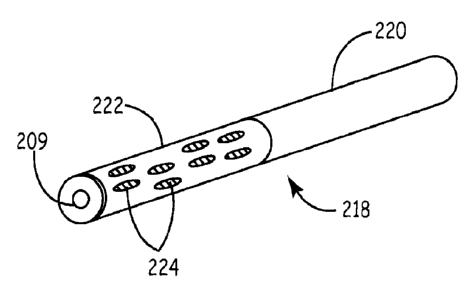
known to artisans. Figure 5B depicts plug 218 that has uncoated proximal
portion 220 and
partially coated distal portion 222. A coating comprising matrix precursors is
provided as a
SUBSTITU-MSHEET (RULE 26)
CA 2977830 2017-08-30
WO 2010/129510 PCT/US2010/033488
plurality of blebs 224. The plug has axial bore 209 for passage over a
guidewire, hollow wire,
catheter, or other elongate member. A guidewire is a hollow wire with an outer
dimension of
less than about 0.08 inches. Hollow wire is a broader teini referring to
guiclewires or larger
wires with an inner bore. Figure SC depicts applicator 208 loaded with plug
218. Applicator
208 has pusher rod 210 with handle 212 that is received by delivery sheath 214
that has
handle 216. Sheath 214 is preloaded with plug 218. Pusher rod 210 has a first
deployment
position and a second tamping position. Wire 207 passes through plug 218 and
applicator
208. With pusher rod 210 in the deployment position, sheath 214 is introduced
into track 202
to place its distal tip 223 proximate puncture 204. While pusher rod 210 is
held stationary or
forced downwardly to apply force against plug 218, a user pulls handles 216
upwardly, as at
Figure 5D and arrows D, to move distal tip 223 upwardly relative to the user
to expose coated
distal portion 222.
At Figure 5E, the user pushes pusher rod 210 downwardly to compress plug 218.
The
coated and uncoated portions of the plug are firmly held against the tissue
for a predetermined
amount of time, e.g., 10-120 seconds (artisans will immediately appreciate
that all the ranges
and values within the explicitly stated ranges are contemplated). The coating
dissolves and
physiological fluids access the uncoated plug portions. A swellable plug
swells as a result,
and contribute to hemostasis at the plug. The balloon is subsequently deflated
through the
hollow wire and withdrawn through the plug. Swelling may contribute to resist
devasation
(forcing of the plug out of the track). Adhesion of the coating to the tissue
further contribute
to resist devasation, i.e., to promote stable positioning. Some embodiments
may provide a
plug biased to open, e.g., a plug made of a sheet furled about its axis so
that it is biased to
unfurl, or a compressed and resilient material. The term plug is a broad term
that refers to a
material blocking a channel, and includes rods, hollow tubes, dumbbell shapes,
cones, and so
forth. The plug is preformed outside the body unless in situ formation is
indicated.
As is evident, the plug does not enter the blood vessel, although it could be
so placed.
The plug achieves closure proximate the blood vessel without actually entering
it. The plug
can engage the adventitia or be proximate the adventitia, i.e., about 1-5 mm
away from the
adventitia (artisans will immediately appreciate that all the ranges and
values within the
explicitly stated ranges are contemplated). The closure at such positions
allows for natural
clotting processes to take place at the blood vessel puncture.
Alternatively, Figure SF depicts an alternative embodiment, with plug 250 that
has
uncoated distal portion 252 and partially coated proximal portion 254. This
configuration
will allow for adherence of the coated plug, preventing expulsion due to blood
pressure in the
SUBSTITU120 SHEET (RULE 26)
CA 2977830 2017-08-30
WO 2010/129510 PCT/US2010/033488
'
vessel, while ensuring dissolved and polymerized coating stay away from the
vessel
arteriotomy and intravascular space. In the depicted example, track 256 has
proximal portion
258 unsealed, with blood oozing into the upper portion of the track and
congealing.
Another embodiment for using a coated material as a fully implanted device for
vascular closure is depicted at Figure 6. Figures 6A and 6B depict applicator
300 extending
from catheter 302. Applicator 300 has plug 304, inner mandrel 306, and outer
mandrel 308.
Plug 304 has a biomaterial sheet 310 with coating 312. The coating may be a
coating as
described herein, for instance one or more dried precursors that form a matrix
when exposed
to a physiological fluid. Sheet 310 has opening 314. Opening 314 may be sized
to
accommodate a guidewire or a larger gauge hollow wire. Sheet 310 is disposed
on a plurality
of struts 316. Struts 316 are connected at one distal portion to inner mandrel
306 and at
another proximal portion to outer mandrel 308. Axial bore 318 passes through
applicator 300
and, as depicted, may be coaxial with opening 314. As shown in Figures 6C-6D,
the relative
movement of inner mandrel 306 and outer mandrel 308 moves struts 316 from a
storage
position to a deployed position, arrow D, wherein the struts are moved
radially outwards,
arrow D'.
Figure 6E depicts balloon 320 that has been inflated via guidewire, larger
gauge
hollow wire, or catheter 322 using means known to artisans to place balloon
320 across
vascular puncture 324 in track 326. Applicator 300 is passed over wire 322
into track 326,
inside catheter 302, Figure 6F. Catheter 302 is positioned proximate balloon
320, and moved
upwardly as at arrows G in Figure 6G, to expose plug 304. Outer mandrel 308 is
moved
downwardly as at arrow G' relative to inner mandrel 306 to force struts 316
radially
outwards, as at arrows G". Sheet 310 and coating 312 are forced against the
surrounding
tissues and held for a predetermined time, e.g., 10 to 200 seconds (artisans
will immediately
appreciate that all the ranges and values within the explicitly stated ranges
are contemplated).
Track 326 may be thereby deformed as compression is applied. At Figure 6H, the
struts 316
are moved from the deployed position to the storage position by relative
movement of the
mandrels. Plug 304 remains in place. The applicator is optionally rotated to
help release the
struts, e.g., from 45 to 360 degrees (I turn) or several turns (artisans will
immediately
appreciate that all the ranges and values within the explicitly stated ranges
are contemplated).
At this juncture, the applicator and balloon and guidewire may be removed.
A further optional step is to move catheter 302 downwardly, as at arrow I in
Figure 61,
to compress and/or hold plug 304 in place while balloon and/or guidewire
and/or applicator
are removed, as at arrow I'. Figure 6J depicts another optional step, wherein
materials are
SUBSTITU12 SHEET (RULE 26)
CA 2 977 8 30 2 01 7-08-30
Ask_
14 8 6 -1 6 lir
=
=
introduced via axial bore 318 after the balloon and hollow wire are removed.
In this step, one =
or more matrix-forming precursors are introduced through the applicator into a
space
proximal the plug and in the track. Precursors as described herein may be
used, and may be
introduced in a solution. The plug prevents entry of the precursors into the
blood vessel. The
catheter and applicator maybe positioned as depicted or otherwise (or
altogether removed, in
the case of catheter 302). Figure 6K depicts the track after this
optional.step, with matrix 330
in place. The matrix may be a inatrix as described herein, e.g., covalently
crosslinked and/or
a hydrogel., The matrix may be positioned through all or a portion of the
track, e.g., the most
distal half, substantially throughout, or in, the proximal half. The matrix
may be created in
situ from one or more precursors.
In the context of vascular closure, the term proximal means close to the user
that is
deploying the device, and distal means relatively farther away and closer to
the blood vessel.
Radially outwards means a movement from a center of the track towards the
track periphery,
as in an axial umbrella opening-up to encounter the lumen of the track.
Downwards means
towards the blood vessel and upwards means away from the blood vessel.
The plug may thus be a sheet with a full or partial coating on one side or
both sides
(and/or, on the edges of the sheet). The coating may be in a pattern. The
coating may be
made of one or more precursors set forth herein. The sheet maybe made of a
material as ' = - described herein, and includes biodegradable and non-
degradable materials.
The applicator may employ other mechanisms to deploy the sheet or other plug
shape.
Further, various occlusive devices and deployment systems may be used to
tamponade a
puncture, with the balloons herein being described as one type of occlusion
member for
exemplary purposes. Alternatives include pledgets or temporary plugs, e.g, as
a in U.S. Pub.
Nos. 2006/0100664 or 2006/0034930.. Artisans
reading this application in its entirety will appreciate the broad
applicability of the coated
materials for use in a variety of puncture closure systems.
The biomaterials for the plug, the sheet, or other matrix materials, may be
provided
with a shape suited to the particular application. Such shapes include, for
example, rods,
cylinders (hollow rods), teardrop-shapes, a tube, a roiled-up sheet, a twisted
sheet, or a
=
braided sheet. One shape, is a planar material (square, rectangle, oval, or
other) that is rolled-
up. The rolling can contribute mechanical properties such as uncurling to
force the material
against a track.
- 22
=
CA 2977830 2017-08-30
W02010/129510 PCT/US2010/033488
For instance, an embodiment is a rod of lyophilized hydrogel with a circular
or oval
cross-section, which after coating or dusting with a reactive hydrogel
coating, may be inserted
into vascular tracks for closure. A solid rod does not need to uncurl,
resulting in improved
application consistency. Alternatively, lyophilized hydrogels can be made in a
planar shape,
rolled, and placed within a sheath and introduced percutaneously. The coating
may be on
only the exterior or a portion of the rolled-up shape or the planar shape may
receive a
continuous or discontinuous coating before rolling. Upon deployment, the
hydrogel coating
dissolves and forms a reactive thin film that can help adhere the lyophilized
hydrogels over
and around the access site.
The adherence, strength and swelling of the lyophilized hydrogel biomaterial
substrate
can be controlled by the amount, pattern and type of the hydrogel coating.
Adhesives used in
vascular access tracks have a significant mechanical advantage relative to
other bioadhesive
uses. For example, sealants used to seal blood vessel anastomoses in open
surgical
procedures depend heavily on both tissue adherence to the adventitia adjacent
to the
anastomosis, and on the cohesive strength of the adhesive itself. This
cohesive strength of
such materials is an important factor, even though the adhesive may be only 1-
2 mm thick.
One mechanical advantage is that the walls of the track provide a large
surface area for
adherence, and the resulting plug can be provided that has a high cohesive
strength due to its
greater thickness. Thus, this increased surface for adherence and longer path
length allow
these vascular access closure adhesives to function more as adherent plugs
than as patches,
allowing them to withstand higher pressures.
Another use of a coated material is a swab applicator for sealing a track.
Coatings of
precursors may be located on biomaterials delivered into a puncture tract or
placed on
applicators to wipe them onto the tissue tract lumen. One embodiment for
preparing the
precursors is a lyophilization from a frozen liquid. Figure 7 depicts swab
applicator 380 with
rod 382, swab 384, and coating 386. The coating may be a coating as described
herein, e.g., a
coating comprising one or more precursors in a dry state that form a matrix
upon exposure to
a physiological fluid. Moreover, the coating may be supplemented with
coagulation factors,
e.g., salts, calcium salts, metal salts, thrombin, collagen, fibrin(ogen), or
blood clotting factors
that participate in the intrinsic or extrinsic blood clotting cascade. The
swab 384 may be
provided with a diameter suited to percutaneous track passage, with a maximum
gauge of
about 1 to about 6 mm; artisans will immediately appreciate that all the
ranges and values
within the explicitly stated ranges are contemplated, e.g., from about .1 to
about 3, from about
1 to about 4, or from about 2 to about 3 mm, or less than about 5 mm or less
than about 3
SUBSTITUT SHEET (RULE 26)
CA 2977 8 30 2 01 7-08-30
=
WO 2010/129510 PCT/US2010/033488
mm. The maximum width, also referred to somewhat loosely as a maximum
diameter, is the
maximum length that a track is to be distended. The term "gauge" refers to the
smallest
diameter circular opening that a device can pass through. The same ranges
and/or values may
be expressed in terms of maximum circumference by using the formula
circumference = 27ER,
with R being from about 1 to about 3 mm. As depicted, the coating partially
covers the swab
but may alternatively cover all of it. Moreover, the shape of the swab may be
tear-shaped,
round, ellipsoidal, or other shapes. The rod may be plastic, metal, wood, or
other material
with a stiffness and strength suited to the swabbing use.
The swab may be used in any track, be it from biopsy of a tissue or organ or a
result of
percutaneous vascular access. The swab may be used by itself, in combination
with manual
tamponade, or in combination with a plug. The latter use is depicted in Figure
8. At Figure
8A, a blood vessel 400 with a puncture 402 has been plugged with a plug 404
placed in track
407, Blood 408 has seeped from the walls of track 406 into the track and onto
the skin. As
shown in Figure 8B, a user moves swab applicator 380 through track 407 to
brush coating
386 onto the walls of the track, with movement as at arrow B. As shown in
Figure 8C,
mechanical tamponade 420, e.g., manual pressure or pressure mediated by a
device or
adhesive, compresses track 407, with coating 386 precursors reacting to form a
matrix that
contributes to closure. After a predetermined time (e.g., 30 seconds to 10
minutes; artisans
will immediately appreciate that all the ranges and values within the
explicitly stated ranges
are contemplated), the pressure is removed, with track seepage being stopped,
as at Figure
8D.
Bleeding from the vessel track could thus be controlled with the use of a
precursor
coated applicator that is introduced into the track and moved in and out, or
spun around,
allowing the coating to dissolve and coat the track tissues. A coated
enlargement on the distal
end could be used to both clear blood from the track and to ensure intimate
contact between
the dissolved precursors and the tissue as the rod is advanced in and out. A
brief external
compression applied when the swab is pulled from the track would allow the
track to be glued
closed as the hydrogel polymerizes. Additionally, this compression may remove
precursors at
the skin level, eliminating the possibility of having a continuous length of
gel from the skin to
the vascular closure device. With the track glued shut, bleeding from the
track tissue would
be controlled, and would not be allowed to reach the skin surface. A
degradable biomaterial
could absorb within hours or 1-30 days of application (artisans will
immediately appreciate
that all the ranges and values within the explicitly stated ranges are
contemplated, e.g, 1 to 5
days), as longer persistence would not be required.
SUBSTITU12e SHEET (RULE 26)
CA 2 977 8 30 2 01 7-08-30
WO 2010/129510 PCT/US2010/033488
Figure 9 depicts device 450 for applying a mechanical pressure to skin. Sheet
452
with coating 454 is applied to a patient's skin, with base 456 being secured
thereupon with
strap 458 having an adjustable feature, as in holes 460 that receive knob 462.
Alternatively,
buckles, snaps, or other adjustment means may be used. After a predetermined
time, e.g., 10
seconds to 10 minutes, the backing is removed. The sheet may be biodegradable
or
removable. Release agents may be placed between the sheet and coating to
facilitate removal.
Coatings and materials as set forth herein may be used. The coating provides
adherence to a tissue. For instance, a puncture track on a wrist or for
brachial access, or other
locations may have a short track that is not well suited to receiving an
implant into the track.
For this or other applications, the backing material receives a coating of
precursors that react
with fluid from the tissue to form an adhesive hydrogel. The backing is left
on until the
healing process is complete or may be removed after adhesion is established. A
biomaterial
may be placed between the backing and the coating to provide further
structure. Release
agents may be included as needed to assist removal of the backing. The backing
material
and/or biomaterial may have, e.g., a planar shape, for instance, a
rectangular, square, circular,
or oval sheet. The base for applying a compressive force is optional. The base
spreads a
compressive force through the base and backing to compress an adhesive coating
against a
tissue surface.
Alternatively, a powdered mix may be applied into the track or tissue site
before
introduction of a biomaterial. For instance, a powder may be drawn up within a
sheath below
a position occupied by a lyophilized hydrogel biomaterial within the sheath.
When the
lyophilized hydrogel is ejected into the access site, the dry precursors are
also ejected and
begin dissolving and reacting. Alternately, a biomaterial may be dusted with
such powders.
Figure 10 depicts an embodiment that exemplifies introducing precursors into a
track
in combination with a plug, and optionally in the same applicator as the plug.
Applicator 500
has sheath 502 and push rod 504. Sheath 502 has handles 506 and distal tip
508. Push rod
504 has handles 510 and optionally removable stop 512. A permanent stop may
alternatively
be used, for example, an enlarged diameter portion of the push rod that
provides resistance
without preventing continued movement. Sheath 502, Figure 10B, is preloaded
with plug 516
and precursor or precursors 518. A non-reactive agent 520, e.g., a release
agent, may further
be preloaded at the distal end portion 522. The non-reactive agent 520
provides for
absorption or repulsion of fluids prior to release of precursors 518. The non-
reactive agent
does not form a matrix material but is optionally a coagulation enhancing
material as already
SUBSTITU12g SHEET (RULE 26)
CA 2977830 2017-08-30
WO 2010/129510 PCT/US2010/033-188
described. Distal tip 508 and at least an accompanying portion of sheath 502
is sized for
track entry (e.g., small, or medium, or large bore), and is introduced into
track 524.
At Figure 10C, the push rod 504 is pushed until stopped by stop 512, expelling
precursors 518 and any accompanying materials. As depicted, the precursors are
expelled in
the proximal half of the track, but may be expelled at other positions, e.g.,
the proximal (skin
side) opening of the track, the track midpoint, or the distal half or distal
tip of the track. As
shown in Figure 10C, sheath 502 is further pushed downwardly, arrow D, and
precursors 518
are distributed on the walls of the track. Plug 516 is expelled, Figure 10E,
with removable
stop having been removed, and sheath 502 being drawn upwardly as at arrows E,
and
applicator 500 is removed. Various tamponakling or swabbing steps may further
optionally be
employed.
Various embodiments have been described herein and may be directed to sealing
medium or large bore punctures although the same embodiments could also be
used to seal
smaller punctures if sized accordingly. One of the motifs is to use a coating
of matrix
precursors on, or with, an acceptably shaped article made from a biomaterial
that can be
introduced into the track. The dry precursors, which are present either as a
coating, or co-
located in the percutaneous closure system, dissolve when exposed to liquids
at the site
needing closure, and polymerize over a period of time, thus securing with
adherence the plug
to the site needing closure and effecting such closure. The entire materials
and mechanism
can be prepared with systems and devices compatible with percutaneous use.
Various
precursors and biomaterials are described herein for the same. Another motif
is that a
biomaterial for the large (or other) bore puncture is coated on a proximal
end. An applicator
is placed in the track and the biomaterial is at least partially forced into
the track to reveal at
least part of a coating on the material. The material is optionally compressed
therein by a
member of the applicator, e.g., a push-rod. The proximal end adheres to the
tissue track. The
biomaterial may be expelled from an applicator all at once or partially
exposed in stages, e.g.,
so that the proximal end with die adhesive is not exposed until immediately
prior to adhesion.
Other embodiments provide for a coating at the distal end of a biomaterial
placed into
a track. An applicator is introduced into the track and manipulated to place
at least a part of
the distal portion into the track, e.g., by a push rod that forces the
material out of a sheath
containing the material. The revealed portion's coating adheres to the tissue
and the sheath is
further withdrawn. The material is optionally compressed. The adhesive coating
will thus
secure the hydrogel at the site of the closure to better seal such larger (or
other sized)
punctures and/or tracks.
SUBSTITUT2 SHEET (RULE 26)
CA 2977830 2017-08-30
WO 2010/129510 PCT/US2010/033488
In the case of arteriotomy, a useful area for sealing is near an arteriotomy
in a distal
portion of the track. The desiccation of track blood by water uptake into the
coating and
optionally the underlying biomaterial would tend to leave high viscosity layer
of blood
between the biomaterial/coating and the tissue to further contribute to
adhesivity.
In addition to biomaterials and/or coatings being used to seal a puncture or
other
iatropic site, lyophilized hydrogels with or without coatings can be used in
the same track for
needle track (or other puncture device track) hemostasis, and to act as a
space filler
supporting the hiomaterials against vascular pressure. In one embodiment, a
first biomaterial
is placed in a distal portion of the track and a second device (same or
different biomaterial) is
placed in a proximal portion of the track.
Alternatively or additionally, a skin closure (suture, clip, glue, tape) may
bc placed at
the skin to help to hold the biomaterials in place, while also reducing the
potential of blood
pressure pushing the biomaterials from the vascular track. Such closures may
generally be
used, e.g., for one or more biomaterials placed into a track, with the
biomaterial(s) being
coated or uncoated.
For instance, in addition to lyophilized hydrogel with a coating being used to
seal the
puncture or other iatropic site, lyophilized hydrogels without coating can be
used in the same
track for needle track hemostasis, and to act as a space filler supporting the
sealed lyophilized
hydrogels against vascular pressure. The skin closure (suture, clip, glue,
tape) may be used
help to hold the lyophilized hydrogel biomaterial in place, while also
reducing the potential of
blood pressure pushing the lyophilized hydrogel(s) from the vascular track.
Some embodiments do not fill the track but instead are placed topically over
the track,
e.g., as in a brachial access site. The backing with coating and/or
biomaterial embodiments
described herein may be used for this application. Some of the coating and/or
biomaterial
may be forced into a proximal portion of the track, with the same being
biodegradable and
providing a sealing role.
The application of coatings consisting of dry PEG precursors to sheets of
backing
materials has previously been disclosed. However, the use of these coatings
were envisioned
for open surgical situations, and not for interventional purposes.
Different design
requirements exist between these coated open surgical and coated
interventional devices so
that different materials must be chosen and combined. For example, in open
surgery, it is
desirable that the backing materials be non-swelling, so as to not lose
strength and not distort
underlying tissues. In
contrast, swelling or expanding backing materials may be
advantageous in interventional applications.
SUBSTITU __________________________ .27 SHEET (RULE 26)
CA 2977830 2017-08-30
WO 2010/129510 PCT/US2010/033488
In contrast to other adhesive systems that require using a solution of
materials, the
coating-based approaches described herein could be processed to provide better
shelf life
stability by storage in an oxygen and moisture free environment (e.g., foil
pouch). Also,
placing a coated device would generally be easier than placing a device that
further requires
combination with an adhesive at the same time, i.e., the coatings are easier
to use. As no
reconstitution in solvent is required, these devices should be immediately
ready to use once
removed from their packaging.
Different coating strategies can be used for different applications.
Continuous
coating on one side can result in adherence to tissue, while minimizing or
eliminating
adherence from the other. Coating on both sides (top) can result in uniform
adherence on
both surfaces, with less substrate absorption and swelling. In contrast,
coating with dots or
lines on one or both sides (middle and bottom), or on the edges, could allow
for directional
tissue adherence while still allowing for fluid absorption and substrate
swelling.
Various embodiments with various features have been disclosed by way of
example
to illustrate the invention. The features of the various embodiments may be
mixed-and-
matched to provide further combinations and subcombinations as guided by the
need to make
functional embodiments. The headings and subheadings are merely for convenient
reference
and are not limitations as to disclosure.
An apparatus for treatment of an iatropic track and blood vessel puncture
comprising
an applicator and a plug, with the applicator comprising a distal sheath
portion sized for
placement in the track and having a distal opening and a lumen, and a pusher
received by the
lumen for pushing the plug out of the lumen through the sheath distal opening,
with the plug
being sized for placement in the track and comprising a coated portion with a
substantially
dry coating, wherein the coating comprises at least one precursor that
dissolves in
physiological fluid after placement in the track and undergoes a covalent
bonding reaction to
form a matrix material that adheres the plug to the track and/or blood vessel,
and an uncoated
portion that exposes a porous portion of the plug to blood in the track, with
the porous portion
at least partially dehydrating the blood in the track.
A method for treatment of an iatropic track and blood vessel puncture
comprising
percutaneously introducing a porous plug into the track through an applicator
having a lumen
terminating in a distal opening, the plug comprising a dry coating on a distal
portion of the
plug and being free of the coating on a proximal portion of the plug, wherein
the coating
comprises at least a first precursor that dissolves in physiological fluid
after placement in the
track and undergoes a covalent bonding reaction to form a matrix material that
adheres the
SUBSTITUT SHEET (RULE 26)
CA 2977830 2017-08-30
WO 2010/129510 PCT/US2010/033488
plug to tissue of the track and/or blood vessel, with the coating promoting
adhesion at or near
the puncture and the uncoated portion of the plug at least partially
dehydrating blood in the
track to reduce flow of blood from the lumen of the track into the track.
An apparatus for treatment of an iatropic track and blood vessel puncture
comprising
an applicator, a plug, and at least a first precursor, with the applicator
comprising a distal
portion sized for placement in the track and having a distal opening and o
lumen, and a pusher
at least partially received in the lumen for pushing the plug out of the lumen
through the distal
opening, with the plug being sized for placement in the track and disposed
within the lumen,
and with
the precursor being disposed in the lumen at a position distal to the plug for
release into the track prior to expulsion of the plug from the lumen, wherein
the precursor
dissolves in physiological fluid after placement in the track and forms a
matrix material.
A method for treatment of an iatropic track and blood vessel puncture
comprising
placing a distal portion of an applicator into the track, expelling a reactive
precursor from the
applicator into at least a portion of the track, and subsequently expelling a
plug into the track,
wherein the precursor forms a matrix material adhesive to the track lumen and
substantially
stops flow of blood from walls of the track into the track lumen.
An apparatus for treatment of an iatropic track and blood vessel puncture
comprising
an applicator, a plug, and at least a first precursor, with the applicator
comprising a distal
portion sized for placement in the track and having a distal opening and a
lumen, and a pusher
at least partially received in the lumen for pushing the plug out of the lumen
through the distal
opening, with the plug being sized for placement in the track and disposed
within the lumen,
and with the precursor being disposed in the lumen at a position distal to the
plug for release
into the track prior to expulsion of the plug from the lumen, wherein the
precursor dissolves
in physiological fluid after placement in the track and forms a matrix
material.
A method for treatment of an iatropic track and blood vessel puncture
comprising
placing a distal portion of an applicator into the track, expelling a reactive
precursor from the
applicator into at least a portion of the track, and subsequently expelling a
plug into the track,
wherein the precursor forms a matrix material adhesive to the track lumen and
substantially
stops flow of blood from walls of the track into the track lumen.
A handheld applicator comprising a proximal portion graspable by a user and a
distal
portion comprising a swab coated with at least a first precursor that, in the
presence of a
physiological fluid, reacts to form a matrix, with the coated swab having a
maximum gauge
of no more than about 5 nun.
SUBSTITU1SHEET (RULE 26)
CA 2977830 2017-08-30
WO 2010/129510 = PCT/US2010/033488
A method of preventing blood flow in a tissue track comprising wiping the
track walls
with a swab coated with a matrix precursor that dissolves in physiological
fluid in the track
and forms a matrix on walls of the track for stopping the blood flow, with the
coated swab
having a maximum gauge of no more than about 5 mm.
An applicator comprising a sheet disposed on a radially expandable member,
with the
sheet being a biodegradable biomaterial coated with one or more precursors
that form a tissue
adherent matrix upon exposure to a physiological fluid to adhere the sheet to
the tissue, with
the radially expandable member being operable to radially expand the sheet and
thereafter be
completely separated from the sheet, wherein the sheet further comprises an
opening coaxial
with a central axial bore of the applicator, with the applicator having a
maximum gauge of no
more than about 1 or 3 or 5 mm.
A method of sealing a percutaneous vascular access site comprising passing an
occlusion device through a track that leads to the site and occluding a
puncture in a blood
vessel at the site, with the occlusion device being connected to an elongate
member that
passes through the track, passing an applicator over the elongate member, with
the applicator
comprising a sheet disposed on a radially expandable member, with the sheet
being a
biodegradable biomaterial coated with one or more precursors that form a
tissue adherent
matrix upon exposure to a physiological fluid, radially expanding the radially
expandable
member to thereby radially expand the sheet and forcing the coating against a
tissue, with the
coating dissolving to folin a matrix adherent to the tissue, separating the
applicator from the
sheet, and withdrawing the applicator, elongate member, and occlusion device
from the site.
The method, apparatus, or system as in the foregoing, provided as a kit. The
kit may
be provided in a single sterile package. Precursors and make-up water and/or
buffers may be
included.
Example 1
Melted 4a2OkSG (four-anned 20,000 MW succinimidyl glutarate-terminated
polyethylene glycol polymer) and 8a20k amine (eight-armed 20,000 MW amine-
terminated
polyethylene glycol polymer) (2:1 ratio) were combined and brought to a
temperature of
50 C. The experimenters briefly wiped both sides of a lyophilized PEG-based
hydrogel
(sheet rolled end-to-end) onto a hot plate containing the melted polymer. This
was tried with
and without borate buffer coating, to raise the reaction pH and accelerate
polymerization, on
the lyophilized hydrogel prior to polymer application.
SUBSTITUT SHEET (RULE 26)
CA 2977830 2017-08-30
= WO 2010/129510
PCT/US2010/033488
Once cooled, the samples were hydrated with physiological saline and squeezed
between two fingers. The resulting adherence to the fingers was strong; in
fact, when pulled
apart, the samples failed cohesively, and not at the adhesive-skin interface.
Example 2
A lyophilized PEG-based hydrogel as used in a MYNX system was obtained in a
flat
unrolled state. The sheet was rolled and placed in the lumen of a 11 Fr sheath
that was about
6 inches long. A second sheet of the same material was placed on a melt of PEG
precursors
at a temperature of about 75 C. The melt was a 1:1 ratio of methylene blue and
4a10CM:8a20k amine (a 4-armed 101cDa polyethylene glycol precursor terminated
with
carboxymethyl hydroxybutyric acid precursor and a second precursor that is an
8 armed 20
kDa multifunctional PEG terminated with amines). The sheet was removed and
allowed to
cool, at which point it was coated on one side and was not tacky. The sheet
would tend to
curl or shrink if exposed to too high of a temperature. The coating tended to
form cracks
when thick but was free of cracks in thinner coatings. The coated sheet was
rolled and placed
into the sheath. Figure 11 depicts this arrangement. Sheath 600 contains
uncoated rolled
sheet 602 and adjacent rolled and coated sheet 604, with a small gap 606 at
the distal end 608
of the sheath, which was open.
A test apparatus was constructed with a three-way valve that connected a
digital
pressure gauge, a syringe of about 10 ml volume having a buffer solution, and
a 1-ml plastic
syringe in a vertical, upright position with the plunger removed. The valve
was turned to a
first position so that the 1-ml syringe was isolated. The lower portion of the
1-ml syringe was
filled with physiological buffer solution. The sheath was introduced into the
1-ml syringe and
a pusher rod was used to push the plugs out of the sheath and into the bottom
of the syringe.
The sheath and pusher rod were used to force the sheets down and were held in
place for a
about a minute. The precursor reacted in the buffer and formed an adhesive
matrix that
adhered both sheets to the bottom of the syringe. The sheath and the pusher
rod were
removed. The three-way valve was moved to a position that allowed
communication between
the 1-ml syringe, the 10-ml syringe, and the pressure gauge were in
communication with each
other. The 10-ml syringe was used to force fluid through the valve while the
pressure gauge
was observed. Pressures in excess of 1000 Tar were observed with no leaking or
movement
of the sheets. The test was repeated several times with different sheets. The
precursors
reacted to form both rolled sheets into a single cohesive mass. The most
distal portion of the
SUBSTITU-13 SHEET (RULE 26)
CA 2977830 2017-08-30
WO 2010/129510 PCT/US2010/033488
distal sheet assumed the shape of the syringe distal end, a male Luer-Lock
shape, showing
compressibility and moldability.
As is evident, other embodiments of this exemplary system may be made using
the
precursors and materials and devices set forth herein.
SUBSTITU13SHEET (RULE 26)
CA 2 977 8 30 2 01 7-08-30