Note: Descriptions are shown in the official language in which they were submitted.
WO 2017/044149
PCT/US2016/013855
SYSTEM AND METHOD FOR PRODUCING BLOOD PLATELETS
CROSS-REFENCE TO RELAYED APPLICATIONS
[0001] This application is based on, claims the benefit of
U.S. Provisional Application No. 61/215,369 filed September 8, 2015, and
entitled
"PLATELET B1OREACTOR."
STATEMENT REGARDING FEDERALLY SPONSORED RESEARCH
[0002] This disclosure was made with government support under R00HL114719
awarded
by the National Institutes of Health. The government has certain rights in the
disclosure.
BACKGROUND OF THE DISCLOSURE
[0003] The present disclosure generally relates to fluid systems, including
microfluidic
devices, systems that include such devices, and methods that use such devices
and systems.
More particularly, the present disclosure relates to devices, systems, and
methods for
generating biological products.
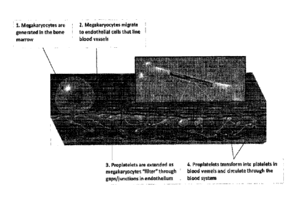
[0004] Blood platelets, or thrombocytes, are irregular, disc shaped cell
fragments that
circulate in the blood and are essential for hemostasis, angiogenesis, and
innate immunity. In
vivo, platelets are produced by cells, known as megakaryocytes. As illustrated
in FIG. 1,
megakaryocytes generated in the bone marrow migrate and contact endothelium
cells that line
blood vessels. There they extend long, branching cellular structures called
proplatelets into
the blood vessel space through gaps in the endothelium. Experiencing shear
rates due to blood
flow, proplatelets extend and release platelets into the circulation. In
general, normal platelet
counts range between 150,000 and 400,000 platelets per microliter of blood.
However, when
blood platelet numbers fall to low levels, a patient develops a condition
known as
thrombocytopenia and becomes at risk for death due to hemorrhage. Known causes
for
thrombocytopenia include malignancy and chemotherapy used to treat it, immune
disorders,
infection, and trauma.
[0005] Despite serious clinical concerns for deleterious immune system
response, risk due
to sepsis and viral contamination, treatment of thrombocytopenia generally
involves using
replacement platelets derived entirely from human donors. However, the process
of obtaining
platelets from transfusions is lengthy, costly, and often requires finding
multiple matching
-1-
Date Recue/Date Received 2022-06-30
CA 02998107 2018-03-08
WO 2017/044149
PCT/US2016/013855
donors. In addition, the usability of harvested platelets are limited due to a
short shelf-life on
account of bacterial testing and deterioration. Moreover, screening for
viruses not known to
exist is not possible. Combined with shortages created by increased demand and
near-static
pool of donors, it is becoming harder for health care professionals to provide
adequate care for
patients with thrombocytopenia, and other conditions related to low platelet
counts.
Alternatives to transfusion have included use of artificial platelet
substitutes, these have thus
far failed to replace physiological platelet products.
[0006] In some approaches, production of functional human platelets has
been attempted
using various cell culture techniques. Specifically, platelets have been
produced in the
laboratory using megakaryocytes obtained from various stem cells. Stem cells
utilized have
typically included embryonic stem cells, umbilical cord blood stem cells and
induced
pluripotent stem cells. Other stem cell sources have included stem cells found
in bone marrow,
fetal liver and peripheral blood. However, despite successful production of
functional platelets
in the laboratory, many limitations remain to use in a clinical setting.
[0007] For instance, only approximately 10% of human megakaryocytes have
been shown
to initiate proplatelets production using state-of-the art culture methods.
This has resulted in
yields of 10 to 100 platelets per CD34+ cord blood-derived or embryonic stem
cell-derived
megakaryocyte, which are themselves of limited availability. For example, the
average single
human umbilical cord blood unit can produce roughly 5.106 CD34+ stem cells.
This poses a
significant bottleneck in ex vivo platelet production. In addition, cell
cultures have been unable
to recreate physiological microenvironments, providing limited individual
control of
extracellular matrix composition, bone marrow stiffness, endothelial cell
contacts, and
vascular shear rates. Moreover, cell cultures have been unsuccessful in
synchronizing
proplatelet production, resulting in non-uniform platelet release over a
period of 6 to 8 days,
which is on the order of platelet shelf-life.
[0008] Therefore, in light of the above, there remains a need for efficient
ways to produce
clinically relevant platelet yields that can meet growing clinical demands,
and avoid the risks
and costs associated with donor harvesting and storage.
SUMMARY OF THE DISCLOSURE
[0009] The present disclosure overcomes the drawbacks of aforementioned
technologies
by providing a system and method capable of efficient and scalable production
of platelets, and
other biological products. Specifically, the disclosure describes various
bioreactor
-2-
CA 02998107 2018-03-08
WO 2017/044149
PCT/US2016/013855
embodiments that include a number of features and capabilities aimed at
generating clinically
and commercially relevant biological products. In some aspects, the system and
method
described herein may be used to generate high platelet yields usable for
platelet infusion. As
such, many significant drawbacks of present replacement therapies can be
overcome, since
these predominantly rely on transfusions from human donors.
[0010] As will be described, in some aspects, provided bioreactor
embodiments can be
configured to recreate physiological conditions and processes associated with
platelet
production in the human body. Specifically, provided bioreactor embodiments
can be
configured for selective functionalization using various materials and
substances that can
facilitate platelet production. Also, by including capabilities for uniform
biological material
trapping and controllable shear stresses, efficient production of platelets,
and other biological
products, can be achieved using the bioreactor embodiments described. In some
designs,
provided bioreactor embodiments are configured for rapid assembly and
disassembly, and
adaptable to thermoplastic molding and other large scale manufacturing
processes.
[0011] In accordance with one aspect of the disclosure, a system and method
for generating
biological products is provided. The system includes a first substrate having
formed therein a
plurality of inlet channel extending substantially along a longitudinal
direction, and a second
substrate having formed therein a plurality of outlet channel corresponding to
the plurality of
inlet channel and extending substantially along the longitudinal direction,
the second substrate
configured to releasably engage the first substrate. The system also includes
a peinieable
membrane, arranged between the substrates, forming rnicrofluidic pathways
between
respective inlet and outlet channels and configured to selectively capture
biological source
material capable of generating biological products, wherein at least one
channel is tapered
transversally to control a pressure differential profile regulating perfusion
through the
permeable membrane.
[0012] In accordance with another aspect of the disclosure, a method for
generating
biological products is provided. The method includes seeding a bioreactor
assembly with
biological source material capable of generating desired biological products,
the bioreactor
assembly a first substrate having formed therein a plurality of inlet channel
extending
substantially along a longitudinal direction, and a second substrate,
configured to releasably
engage the first substrate, and having formed therein a plurality of outlet
channel
corresponding to the plurality of inlet channel and extending substantially
along the
longitudinal direction, wherein at least one channel is tapered transversally
to control a
-3-.
CA 02998107 2018-03-08
WO 2017/044149
PCT/US2016/013855
pressure differential profile therein. The bioreactor assembly also includes,
a permeable
membrane, arranged between the substrates, forming microfluidic pathways
between
respective inlet and outlet channels and configured to selectively capture
biological source
material. The method also includes introducing fluid media into the bioreactor
assembly at
predetermined flow rates to generate the desired biological products, and
harvesting the desired
biological products from the bioreactor assembly.
[0013] The foregoing and other aspects and advantages of the disclosure
will appear from
the following description. In the description, reference is made to the
accompanying drawings
which form a part hereof, and in which there is shown by way of illustration a
preferred
embodiment of the disclosure. Such embodiment does not necessarily represent
the full scope
of the disclosure, however, and reference is made therefore to the claims and
herein for
interpreting the scope of the disclosure.
BRIEF DESCRIPTION OF THE DRAWINGS
[0014] The present disclosure will hereafter be described with reference to
the
accompanying drawings, wherein like reference numerals denote like elements.
[0015] FIG.1 is an illustration showing in vivo platelet production in bone
marrow.
[0016] FIG. 2 is a schematic diagram of a system for producing biological
products, in
accordance with aspects of the present disclosure.
[0017] FIG. 3 is an illustration showing an embodiment of a bioreactor, in
accordance with
aspects of the present disclosure.
[0018] FIG. 4A is a perspective view showing one embodiment of a bioreactor
assembly
including the bioreactor of FIG. 3.
[0019] FIG. 4B is a cross-sectional view of the embodiment shown in FIG.
4A.
[0020] FIG. 4C is another cross-section view of the embodiment shown in
FIG. 4A.
[0021] FIG. 5A shows an example system, in accordance with aspects of the
present
disclosure.
[0022] FIG. 5B shows the bioreactor included in the system of FIG. 5A.
[0023] FIG. 6 shows an embodiment of a bioreactor, in accordance with
aspects of the
present disclosure.
[0024] FIG. 7A shows an embodiment of a bioreactor assembly that is
disassembled, in
accordance with aspects of the present disclosure.
-4-
CA 02998107 2018-03-08
WO 2017/044149
PCT/US2016/013855
[0025] FIG. 713 shows the bioreactor assembly of FIG. 7A assembled.
[0026] FIG. 8 shows another embodiment of a system, in accordance with
aspects of the
present disclosure.
[0027] FIG. 9A shows yet another embodiment of a bioreactor, in accordance
with aspects
of the present disclosure.
[0028] FIG. 9B shows yet another embodiment of a bioreactor, in accordance
with aspects
of the present disclosure.
[0029] FIG. 10 is a schematic showing a system, in accordance with aspects
of the present
disclosure.
[0030] FIG. 11 shows yet another embodiment of a bioreactor, in accordance
with aspects
of the present disclosure.
[0031] FIG. 12 shows yet another embodiment of a bioreactor, in accordance
with aspects
of the present disclosure.
[0032] FIG. 13 is a flowchart setting forth steps of a process, in
accordance with aspects of
the present disclosure.
[0033] FIG. 14A is a graph showing megakaryocytes produced using a static
culture.
[0034] FIG. 14B is a graph showing proplatelets and platelets produced
using a static
culture.
[0035] FIG. 14C is a graph showing megakaryocytes produced in accordance
with aspects
of the present disclosure.
[0036] FIG. 14D is a graph showing proplatelets and platelets produced in
accordance with
aspects of the present disclosure.
[0037] FIG. 15 is a graph showing a timeline of platelet production, in
accordance with
aspects of the present disclosure.
[0038] FIG. 16 shows yet another embodiment of a bioreactor, in accordance
with aspects
of the present disclosure.
DETAILED DESCRIPTION OF THE DISCLOSURE
[0039] The present disclosure provides systems and methods capable of
efficient and
scalable production of platelets, and other biological products.
[0040] Turning
now to FIG. 2, a schematic diagram of an example system 100 for
producing platelets, and other biological products, is shown. In general, the
system 100
-5-
CA 02998107 2018-03-08
WO 2017/044149
PCT/US2016/013855
includes a biological source 102, a bioreactor assembly 104, and an output
106, where the
biological source 102 and output 106 are connectable to various inputs and
outputs of the
assembly 104, respectively.
[0041] Specifically, the biological source 102 may be configured with
various capabilities
for introducing into the assembly 104 different biological source materials,
substances, gas, or
fluid media, to efficiently produce desirable biological products, such as
platelets. For
instance, the biological source 102 may include one or more pumps for
delivering or sustaining
fluid media in the bioreactor assembly 104. Examples include microfluidic
pumps, syringe
pumps, peristaltic pumps, and the like.
[0042] As shown in FIG. 2, in some implementations, the system 100 may also
include a
controller 108 for controlling the biological source 102. Specifically, the
controller 108 may
be a programmable device or system configured to control the operation of the
bioreactor
assembly 104, including the timings, amounts, and types of biological source
material,
substances, fluid media or gas introduced therein. In some aspects, the
controller 108 may be
configured to selectively functionalize and/or operate the assembly 104 to
recreate
physiological conditions and processes associated with platelet production in
the human body.
For example, the controller 108 may be programmed to deliver a selected number
of
megakaryocytes to the assembly 104. In addition, the controller 108 may
control fluid flow
rates or fluid pressures in the bioreactor assembly 104 to facilitate
proplatelet extension and
platelet production. For instance, the controller 108 may establish flow rates
up to 150,000
microliters/hr in various channels configured in the bioreactor assembly 104.
[0043] Although the controller 108 is shown in FIG. 2 as separate from the
biological
source 102, it may be appreciated that these may be combined into a single
unit. For instance,
the biological source 102 and controller 106 may be embodied in a programmable
microfluidic
pump or injection system. In addition, in some implementations, the controller
108 and/or
biological source 102 may also include, communicate with, or received feedback
from systems
or hardware (not shown in FIG. 2) that can regulate the temperature, light
exposure, vibration,
and other conditions of the bioreactor assembly 104. By way of example, FIG. 8
shows a
bioreactor system 800 that includes a controller 802 connected to heaters 804
and a
thermocouple 806 for monitoring and controlling temperature. It may be
appreciated that the
configuration shown in FIG. 8 is non-limiting, and any number of heaters, and
heater
arrangements may be possible.
-6-
CA 02998107 2018-03-08
WO 2017/044149
PCT/US2016/013855
[0044] Referring again to FIG. 2, in general, the output 106 is configured
to receive fluid
media containing various biological products generated in the bioreactor
assembly 104.
However, in some implementations, as will be described, such effluent may be
redirected or
circulated back into the bioreactor assembly 104. In this manner, less fluid
volume may be
utilized, and the biological products generated can be more concentrated. In
some aspects, the
output 106 may also include capabilities for collecting, storing and/or
further processing
received fluid media.
[0045] It may be appreciated that the above-described system 100 has a
broad range of
functionality, and need not be limited to replicating physiological conditions
or processes, nor
producing platelets. That is, the system 100 may be used to generate a wide
variety of
biological products. For instance, the system 100 may be used to separate,
break up or dissolve
various biological source materials or substances, such as megakaryocytes and
other cells, and
collect their product or content. Specifically, by controlling fluid flow and
pressures, as well as
other conditions, various contents of captured biological source materials may
be released and
subsequently harvested. In some aspects, the system 100 may also be utilized
to differentiate
and/or culture various cells, biological substances or materials, such as
megakaryoctytes, for
obtaining biological source material needed to generate desirable biological
products.
Example biological products include growth factors, and other components found
in cells.
Produced biological products, in accordance with the present disclosure, in
addition to clinical
use, may find use in a variety of applications including as components of cell
culture medias
and cosmaceuticals, such as cosmetics, shampoos, skin additives, creams, or
cleaners, and so
forth.
[0046] Various embodiments of the above system 100 will now be described.
It may be
appreciated that these are non-limiting examples, and indeed various
modifications or
combinations are possible and considered by one of ordinary skill in the art
to be within the
intended scope of the present application.
[0047] Referring now to FIG. 3, an non-limiting microfluidic bioreactor
300, in accordance
with aspects of the present disclosure, is shown. In general, the bioreactor
300 includes a first
substrate 302, a second substrate 306, and a permeable membrane 304 arranged
therebetween.
[0048] In particular, the first substrate 302 can include a number of inlet
channels 308, or
inlet chambers, formed therein. As shown in FIG. 3, in some embodiments, the
channels can
be arranged generally parallel to one another and extending substantially
along a longitudinal
direction (for example, the x direction). However it may be appreciated that
any channel
-7-
CA 02998107 2018-03-08
WO 2017/044149
PCT/US2016/013855
arrangement capable of achieving platelet and other biological product
production, as
described in the present disclosure, may be possible. Also, the inlet channels
308 may be
connected to an inlet port 310 by way of an inlet manifold 312 formed in the
first substrate 302,
where the inlet manifold 312 is configured to provide similar or comparable
fluid pathways for
fluid media traversing therethrough. Such configuration may be advantageous
for distributing
various biological substances or materials uniformly across the bioreactor
300, or can be
manipulated to distribute various biological substances or materials
selectively or differentially
within different fluid pathways. For
instance, a concentration of platelet-producing
megakaryocyte cells dispersed in a fluid medium can be introduced in one
infusion step into the
inlet port 310 to achieve a similar density or spatial distribution across the
permeable
membrane 304. In alternative configurations, each inlet channel 308 may
include different
sized or separate inlet ports.
[0049] The
second substrate 306 can include a plurality of outlet channels 310, or outlet
chambers, each corresponding to a respective inlet channel, and also extending
substantially
parallel along the longitudinal direction. Similarly, the second substrate 304
may also include
an outlet manifold 316 formed therein, the outlet manifold 316 connected to
the outlet channels
314 and configured to direct fluid media, including various biological
products, substances or
materials, to an output via an outlet port 318.
[0050] By
way of example, the substrates described above may have lateral dimensions in
a range between 10 mm and 100 mm, and a thickness in a range between 1 and 10
mm,
although other dimensions may also be possible. Also, a longitudinal dimension
of the inlet
channels 308 and/or outlet channels 314 be in the range of 1000 to 30,000
micrometers or,
more particularly, in the range of 1000 to 3000 micrometers, while at least
one transverse
dimension may be in the range of 100 to 3,000 micrometers or, more
particularly, in the range
of 100 to 300 micrometers. Other dimensions are also possible. As will be
described, in some
embodiments, the inlet channels 308 or outlet channels 314 may also be tapered
transversally
either entirely or over a portion of the longitudinal dimension to control
shear rates or pressure
differentials between the channels, over an active contact area, regulating
perfusion through the
permeable membrane 304.
[0051]
Various implementations of the bioreactor 300 are possible depending upon
specific uses or applications. In particular, in some embodiments, the inlet
channels 308 and
outlet channels 314, along with other microfluidic elements of bioreactor 300,
may be
longitudinally (for example, along the x-direction shown in FIG. 3) and
transversally (y- and
-8-
CA 02998107 2018-03-08
WO 2017/044149
PCT/US2016/013855
z-direction) shaped and dimensioned to reproduce physiological conditions,
such as those
found in bone marrow and blood vessels. For instance, channel shapes and
dimensions may be
selected to achieve physiological flow rates, shear rates, fluid pressures or
pressure
differentials similar to those associated with in vivo platelet production, as
described with
reference to FIG. 1. In addition, configurations of the bioreactor 300 may be
chosen to allow
cooperation with other instrumentation, such as microscopes or cameras. For
instance, the
bioreactor 300 may be configured to adhere to standard microplate dimensions.
[0052] In some configurations of the bioreactor 300, the inlet channels 308
and outlet
channels 314 terminate in their respective substrates to create a single fluid
conduit 320 from
the inlet port 310 to the outlet port 318, as shown in FIG. 3. That is to say,
fluid medium
introduced into the inlet port 310 is necessarily extracted from the outlet
port 318. However, it
may appreciated that additional inlet and outlet ports may also be possible
with the bioreactor
300. For example, the first substrate 302 may also include one or more outputs
connected to
the inlet channels 308. Similarly, the second substrate 306 may also include
one or more inputs
connected to the outlet channels 314. In this manner, multiple fluid pathways
can be possible,
which would allow for selectively preparing various portions of the bioreactor
300
independently.
[0053] Although not shown in FIG. 3, the bioreactor 300 may also include a
number of
microfluidic filtration and resistive elements, connected to the channels and
arranged at various
points along the various fluid pathways extending between the inlet port 310
and outlet port
318. For instance, one or more filtration elements may be placed proximate to
the inlet port
310 to capture contaminants or undesirable substances or materials from an
inputted fluid
medium. In addition, one or more resistive elements may also be included to
control flow
forces or damp fluctuations in flow rates. In addition to resistive and
filtration elements,
elements may also be included. For example, air bubbles traps may be
configured with one or
more inputs to eliminate any air bubbles from entering the bioreactor 300.
[0054] By way of example, FIG. 6 illustrates a microfluidic connector 602
coupled to an
input 604 of an example bioreactor 600, in accordance with the above
descriptions. As shown,
the input 604 includes an expansion region 606 and a conical region 608
separated by a mesh
610. For example, the mesh 610 may have a size of approximately 140
micrometers, although
other values may be possible. As configured, the input 604 is capable of
preventing air bubbles
from entering the bioreactor 600.
-9-
CA 02998107 2018-03-08
WO 2017/044149
PCT/US2016/013855
[0055] Referring again to the bioreactor 300 of FIG. 3, each substrate is
shown to include
16 inlet channels 312 and 16 outlet channels 314. It may be readily
appreciated that more or
fewer channels or chambers may be possible. For example, as illustrated in the
bioreactor 900
shown in FIG. 9A, two inlet channels 902 and two outlet channels 904 may be
utilized. In
addition to inlet and outlet channels, additional conduits may also be formed
in the substrates
of the bioreactor 300 of FIG. 3. For instance, as shown in FIG. 9A and 9B, a
perfusion channel
906 may also be included in the bioreactor 900. Specifically, the perfusion
channel 906 would
allow the flow of gas, which can subsequently perfuse through the substrate
materials and into
the bioreactor 900 inlet/outlet channels. For example, a 5% CO2 gas mixture
may be perfused
into the channels.
[0056] In general, the permeable membrane 304 of FIG. 3 can include any
rigid or flexible
layer, film, mesh or material structure configured to connect corresponding
inlet channel 308
and outlet channel 314 via microfluidic pathways formed therein. In one
embodiment,
microfluidic pathways in the permeable membrane 304 may be formed using pores,
gaps or
microchannels, distributed with any density, either periodically or
aperiodically, about
permeable membrane 304. In another embodiment, the permeable membrane 304 can
include
a three-dimensional structure formed using interwoven micro- or nano-fibers
arranged to allow
fluid therethrough. Although shown in FIG. 3 as rectangular in shape, it may
be appreciated
that the permeable membrane 304 may have any shapes, including circular
shapes, oval shapes,
and so forth. In accordance with aspects of the disclosure, the permeable
membrane 304 may
be configured to selectively capture specific biological source materials or
substances to
produce desired biological products. For instance, the permeable membrane 304
may be
configured to selectively capture platelet-producing cells and allow
proplatelet extensions
therethrough.
[0057] By way of example, the permeable membrane 304 may include
longitudinal and
transverse dimensions in a range between 1 and 100 millimeters, and have a
thickness in a
range between 0.1 to 20 micrometers, although other dimensions are possible.
Also, the
permeable membrane 304 may include pores, gaps or microchannels sized in a
range
approximately between 3 micrometers and 10 micrometers, and more specifically
approximately between 5 and 8 micrometers. In some aspects, pore, gap or
microchannel size,
number, and density may depend on a number of factors, including desired
biological products
and product yields, as well as flow impedances, shear rates, pressure
differentials, fluid flow
rates, and other operational parameters.
-10-
CA 02998107 2018-03-08
WO 2017/044149
PCT/US2016/013855
[0058] As appreciated from FIG. 3, the inlet channels 308 and outlet
channels 314 overlap
to define an active contact area in the permeable membrane 304. For example,
an active
contact area 326 may be in a range between 1 mm2 to 20 mm2, although other
values are
possible, depending upon the dimensions and number of channels utilized. In
some
implementations, the active contact area along with permeable membrane 304
characteristics
may be optimized to obtain a desired biological product yield. For example, a
permeable
membrane 304 with 47 mm diameter, 5% active contact area, and pore density of
ii
pores/cm2 could provide 200,00 potential sites for generating a desired
biological product
yield, such as a desired platelet yield. In some applications, the active
contact area may be
configured to trap at least approximately I .104 megakaryoeytes.
[0059] The bioreactor 300 may be manufactured using any combination of
biocompatible
materials, inert materials, as well as materials that can support pressurized
gas and fluid flow,
or gas diffusion, and provide structural support. In some aspects, materials
utilized in the
bioreactor 300 may be compatible with specific manufacturing processes, such
as injection
molding. In addition, materials utilized may optically clear to allow
visualization of fluid
media, and other substances, present or flowing in various portions of the
bioreactor 300.
[0060] By way of example, the first substrate 302, or second substrate 306,
or both, or
portions thereof, may be manufactured using cell-inert silicon-based organic
polymer
materials, such as polydimethylsiloxane (''PDMS"), thermoplastic materials,
such as zeonor
cyclo olefin polymer ("COP"), glass, acrylics, and so forth. On the other
hand, the permeable
membrane 304 may be manufactured using PDMS, thermoplastics, silk, hydrogels,
extracellular matrix proteins, polycarbonate materials, polyesthersulfone
materials, polyvinyl
chloride materials, polyethyleneterephthalat materials, and other synthetic or
organic
materials.
[0061] In accordance with aspects of the present disclosure, the bioreactor
300 may be
selectively functionalized using various biological substances and materials.
Specifically, the
bioreactor 300 may be selectively functionalized by way of fluid media,
containing desired
biological substances and materials, being introduced therein. Alternatively,
or additionally,
the bioreactor 300, or components thereof, may be functionalized using various
preparation or
manufacturing processes. For example, the permeable membrane 304 may be pre-
prepared
with platelet-producing cells prior to assembly of the bioreactor 300. In some
aspects, the
bioreactor 300 may be utilized to differentiate and/or culture megakaryocytes,
as well as other
cells, biological substances or materials.
-11-.
CA 02998107 2018-03-08
WO 2017/044149
PCT/US2016/013855
[0062] As described, in some aspects, the bioreactor 300 may be
advantageously
functionalized to replicate in vivo physiological conditions in order to
produce platelets, or
other biological products. For instance, in one application, a top surface 322
of the permeable
membrane 304 may be selectively coated with extracellular matrix proteins, for
example, while
a bottom surface 322 can be left without, or can be coated with different
proteins or substances.
This can be achieved, for instance, by infusing a first fluid medium
containing extracellular
matrix proteins, using inputs and outputs in the first substrate 302. At
substantially the same
time, a second fluid medium flow can be maintained in the second substrate 306
using
respective inputs and outputs, where the second fluid medium would either
contain no proteins,
or different proteins or substances. Preferably, flow rates of the first and
second fluid media
would be configured such that little to no fluid mixing would occur. Such
selective
functionalization would ensure that introduced platelet-producing cells, for
example, coming
to rest on the top surface 322 would contact extracellular matrix proteins,
while proplatelets
extended through the permeable membrane 304, and platelets released therefrom,
would not
contact extracellular matrix proteins, or would contact different proteins or
biological
substances.
[0063] Non-limiting examples of biological substances and materials for
functionalizing
the bioreactor 300 may include human and non-human cells, such as
megakaryocytes,
endothelial cells, bone marrow cells, osteoblasts, fibroblasts, stem cells,
blood cells,
mesenchymal cells, lung cells and cells comprising basement membranes. Other
examples can
include small molecules, such as CCL5, CXCL12, CXCL10, SDF-1, FGF-4, S1PR1,
RGDS,
Methylcellulose. Yet other examples can include, extracellular matrix
proteins, such as bovine
serum albumin, collagen type I, collagen type IV, fibrinectin, fibrinogen,
laminin, vitronectin.
In particular, to replicate three-dimensional extracellular matrix
organization and physiological
bone marrow stiffness, cells may be infused in a hydrogel solution, which may
subsequently be
polymerized. The hydrogel solution may include, but is not limited to
alginate, matrigel,
agarose, collagen gel, fibrin/fibrinogen gel.
[0064] In some aspects, various portions of the bioreactor 300 may be
configured to allow
for assembly and disassembly. Specifically, as shown in FIG. 3, the first
substrate 302,
permeable membrane 304, and second substrate 306 may be configured to be
removably
coupled to one another. When engaged using fasters, clips, or other releasable
locking
mechanisms, for example, a hermetic seal can then be formed between various
surfaces of the
substrates and permeable membrane 304 to reinstate fluid pathway integrity
between the input
-12-
CA 02998107 2018-03-08
WO 2017/044149
PCT/US2016/013855
310 and output 318. This capability can facilitate preparation, as described
above, as well as
cleaning for repeated use. In addition, disassembly allows for quick exchange
of various
components, for repurposing or rapid prototyping. For instance, a permeable
membrane 304
having different pore sizes, or different preparations, may be readily
swapped.
[0065] Alternatively, the bioreactor 300 may be manufactured as a single
device, for
sample using an injection molding technique, where the permeable membrane 304
would be
molded into the substrates. Such implementations may be advantageously
integrated into large
scale manufacturing techniques. By way of example, FIG. 16 shows a bioreactor
1600
generated using a molding technique. The bioreactor 1600 may be formed using
PDMS,
thermoplastic, such as copolymer ("COP"), cyclic olefin copolymer ("COC"),
polymethylmethacrylate ("PMMA"), polycarbonate ("PC"), and other materials. As
shown in
FIG. 16, in some embodiments, the bioreactor 1600 may be include a glass
substrate 1602
either on the top, or the bottom, of the bioreactor 1600 or both.
Alternatively, the bioreactor
1600 may include an acrylic top substrate 1604, alone or in combination with a
glass or acrylic
substrate. These substrates could be used provide structural support to the
bioreactor 1600.
[0066] By way of example, FIGs. 7A-B show an example bioreactor assembly
700 both
disassembled and assembled, respectively. As shown in FIG. 7A, the bioreactor
assembly 700
in general includes a base plate 702, a clamp arm 704, a top clamp frame 706,
fastener 708, and
bioreactor 710. When assembled, as shown in FIG. 7B, a compression is provided
to the
bioreactor 710 bringing respective substrates of the bioreactor 710 to form a
hermetic seal
therebetween. In some designs, the bioreactor assembly 700 may incorporate
hard stops with
springs to be able to apply an even pressure across top and bottom surfaces of
the bioreactor
710 to prevent leaks and avoid over-compression. Other fastening mechanisms or
variations
thereof are also possible, for example, as shown in FIG. 5A, or FIG. 8.
[0067] Referring now to FIGs. 4A-4C, a non-limiting embodiment of a
microfluidic
bioreactor assembly 400, in accordance with aspects of the present disclosure,
is illustrated.
Referring specifically to the cross-sectional view of FIG. 4B, the bioreactor
assembly 400 can
include a top substrate 402, a bottom substrate 404, and a membrane 406
therebetween that
connects a number of corresponding inlet channels 408 and outlet channels 410
formed in
respective substrates. As described, the membrane 406 is configured to form
fluid pathways
between the substrates, for instance, via pores included therein. As shown in
the perspective
view of FIG. 4A, the bioreactor assembly 400 can also include a base portion
412 and top
portion 414 configured to fasten the membrane 406 and substrates together, for
example using
-13-
CA 02998107 2018-03-08
WO 2017/044149
PCT/US2016/013855
screws. For simplicity, FIG. 4A shows only a portion of the bioreactor
assembly 400. When
fastened, the base portion 412 and top portion 414 bring the substrates into
an aligned
engagement (for example using guides, grooves, or holes) capable of forming a
hermetic seal
therebetween in order to allow fluid or gas flow, or both, in the channels
without leaks.
Conversely, when unfastened, various portions of the bioreactor assembly 400
can be
individually prepared, exchanged, cleaned and reused.
[0068] As
described, in some configurations, the bioreactor assembly 400 may be
configured to allow visualization during operation. Referring specifically to
FIG. 4B, each
substrate may have a glass layer 416 arranged proximate thereto, forming a
stack that includes
glass/substrate/membrane/substrate/glass. In alternative configurations, the
glass layers may
be excluded, with the substrates and/or membrane 406 configured to have
appropriate
structural integrity and desired material characteristics, such as
transparency appropriate for
allowing visualization of fluid media therethrough. As may be appreciated, the
specific
configurations of the bioreactor assembly 400 and portions thereof, as well as
manner of
assembly, may be modified or adapted to the specifics of the particular
application.
[0069] The inlet
channels 408 and outlet channels 410 of the bioreactor assembly 400 need
not have equal dimensions. That is, as shown in FIG. 4B, the inlet channels
408 can be larger
(or smaller) in at least one transverse (or longitudinal) dimension compared
to the outlet
channels 410. By way of example, the inlet channels may have a tirst
transverse dimension, or
channel height of approximately 0.1 mm, a second transverse dimension, or
channel width of
approximately 0.7 mm, while the outlet channels may have a channel height of
approximately
0.1 mm and a channel width of approximately 0.5 mm. The channels may have a
longitudinal
dimension or length of approximately 25 mm. Various other dimensions may be
possible. In
some aspects, dimensions, including channel depths and widths, may be
configured such that
megakaryocytes experience desired shear stress in the center of the channels,
and avoid
trapping at channel walls.
[0070] In
addition, in some aspects, at least some of the inlet channels 408 or outlet
channels 410, or both, may also be tapered transversally over at least a
portion of the
longitudinal dimension forming the active contact area 422. By way of example,
a channel
depth may begin at 0.5 mm and taper to a point. As described, such
configurations may be
advantageous for controlling a pressure differential profile in the channels
in order to regulate
perfusion through the membrane 406 in the active contact area 422.
-14-
CA 02998107 2018-03-08
WO 2017/044149
PCT/US2016/013855
[0071] Referring particularly to the cross-sectional view of FIG. 4C, an
inlet channels 408
and outlet channels 410 are shown to be tapered transversally, forming a taper
angle a with the
longitudinal direction x. That is, a surface 424 of the inlet channels 408, or
outlet channel 410,
or both, forms the taper angle with the longitudinal direction, or x
direction, as shown. By way
of example, the taper angle a can have values between 0 and 5 degrees,
although other taper
angles values may be possible. In some aspects, the taper angle associated
with inlet channels
408 and outlet channels 410 need not be the same. As a result of the taper in
the channels, fluid
media introduced via the inlet 418 and extracted via the outlet 420 would
experience a uniform
differential pressure profile across the active contact area 422, as indicated
by the arrows.
Herein, the pressure differential profile refers to the various pressure
differences between the
inlet channels 408 and outlet channels 410 present different points along the
longitudinal
direction x.
[0072] By way of example, FIG. 5A-B show one embodiment of the bioreactor
system
described with reference to FIG. 2. Specifically referring to FIG. 5A, the
system 500 may
include a syringe pump 502 fluidly connected, using plastic tubing 504, to a
bioreactor chip
assembly 506, as described with reference to FIGs. 4A-4C. In particular, the
tubing 504 may
be fitted using Luer Lock connectors 508 in order to provide detachable, leak-
proof
connections to the syringe pump 502 as well as to an external output. As
shown, the bioreactor
chip assembly 506 includes a bioreactor 550 fastened to a standard microplate-
sized base 510
using a chip clamps 512. Referring particularly to FIG. 5B, the bioreactor 550
is shown to
include a plurality of channels 552, or chambers, formed in PDMS substrates
included therein,
and connected to respective inlet 554 and outlet 556 ports. As described,
inlet and outlet
channels 552 are separated by a permeable membrane 558. The bioreactor 550
also includes
glass slides 560, with or without holes, arranged on the top and bottom of the
PDMS substrates.
The bioreactor 550 further includes a PDMS molded track 562 for achieving a
hermetic seal.
[0073] Another embodiment the bioreactor system described with reference to
FIG. 2 is
shown in FIG. 10. Specifically, the system 1000 includes a biological input
source 1002, a
bioreactor 1004, an output 1006, a controller 1008, and a recirculator 1010.
As described, the
input source 1002 is fluidly connected to a plurality of inlet channels 1012,
while the output
1006 is fluidly connected to a plurality of outlet channels 1014, with the
inlet and outlet
channels being separated by a permeable membrane (not shown in FIG. 10). As
described,
effluent containing produced biological products, such as platelets, may be
received by the
output 1006.
-15-
CA 02998107 2018-03-08
WO 2017/044149
PCT/US2016/013855
[0074] In addition to the outlet port 1016 configured to direct effluent
from the outlet
channels 1014 to the output 1006 for collection, storage or further
processing, the bioreactor
1004 shown in FIG. 10 also includes another outlet port 1018 connected to the
outlet channels
1014. Such configuration allows effluent flowing out of the outlet channels
1014 to be
redirected, using the recirculator 1010, back into the outlet channels 1014 by
way of an inlet
port 1020 connected to the outlet channels 1014, as indicated by arrows in
FIG. 10. This allows
for reduced operational volumes as well as the ability to concentrate produced
biological
products. In addition, pressure differentials and shear stress profiles in the
channels can be
independently controlled by the controller 1008. By way of example, the
recirculator 1010
may be a peristaltic pump, which may be configured to achieve physiologically
relevant
conditions.
[0075] Although not shown, the bioreactor 1004 may also include a perfusion
channel for
perfusing gas, such as CO2, into the channels. In addition, the bioreactor
1004 may be
included in a bioreactor assembly capable of assembly and disassembly.
[0076] Embodiments of bioreactor systems described thus far need not be
limited to planar
geometries. For example, as shown in FIGs. 11 and 12, cylindrical geometries
may also be
implemented. Specifically referring to FIG. 11, a cross-section of an example
cylindrical
bioreactor 1100 is shown, which includes an outer chamber 1102 and inner
chamber 1104
formed in a substrate 1106. As shown, the chambers are separated a porous
membrane 1108,
which is configured to form microfluidic pathways connecting the chambers, as
described. The
chambers are each connected to various inlet and outlet ports (not shown in
FIG. 11) that may
facilitate infusion and collection of fluid media flowing therethrough, as
indicated by the
arrows. In some aspects, as shown in FIG. 11, an inner surface 1110 of the
substrate 1112
includes a curvature, or continuous taper, configured to control fluid flow
between the
chambers. In some aspects, the curvature may be configured to achieve a
uniform pressure
differential across an active area of the permeable membrane 1108.
[0077] In another example, FIG. 12 shows a cross-section of a cylindrical
bioreactor
assembly1200. In general, the bioreactor assembly 1200 includes a first
substrate 1202
separated from a second substrate 1204 by a permeable membrane 1206. As may be
appreciated, this configuration is similar to the one described with reference
to FIG. 4B, but
adapted to a cylindrical geometry. Specifically, the first substrate 1202
includes a plurality of
inlet channels 1208 formed therein. Similarly, the second substrate 1204
includes a plurality of
outlet channels 1210 formed therein, wherein the inlet channels 1208 and
outlet channels 1210
-16-
CA 02998107 2018-03-08
WO 2017/044149
PCT/US2016/013855
extend substantially along a longitudinal direction perpendicular to the cross-
section shown in
FIG. 12, and overlap over an active contact area. The channels may or may not
be tapered. A
fluid medium flowing in the inlet channels 1208 would then traverse the
permeable membrane
1206 radially into the outlet channels 1210, as indicated by the arrows.
[0078] The
channels of the bioreactor assembly 1200 may be connected to various
inlet/outlet ports, and inlet/outlet manifolds (not shown in FIG. 12), similar
to configurations
described with reference to F1G. 3. In addition, in some aspects, the channels
may be tapered
transversally over at least a portion of the longitudinal direction in order
to control a pressure
differential regulating perfusion through the permeable membrane 1206. When
the substrates
are brought into engagement, by way of a base plate 1212 and fastener 1214, an
even pressure
is applied across the permeable membrane 1206, forming a hermetic seal between
the
substrates and permeable membrane 1206.
[0079] Turning
now to FIG. 13, a flowchart setting forth steps of a process 1300, in
accordance with aspects of the present disclosure, is shown. The process 1300
may begin at
process block 1302 with providing biological source material, such as
megakaryocytes or
progenitor cells or stem cells. In some aspects, biological source material
may also be
generated at process block 1302. For instance, megakaryocytes may be generated
by first
isolating stem cells from umbilical cord blood, for example, and expanding
them using various
reagents. Such expanded cells may then be differentiated into megakaryocytic
lineage,
followed by a step of inducing polyploidization to generate mature
megakaryocytes usable
producing platelets. Alternatively, megakaryocytes may be obtained from
induced pluripotent
cells.
[0080] As
indicated by process block 1304, the provided or generated biological source
material may then be seeded in a bioreactor assembly, for instance, as
described with reference
to FIGs. 3-5. In some aspects, this step may include preparing various
components bioreactor
assembly. For example, a permeable membrane may be seeded with biological
source material
using various culture and plating techniques prior to device assembly.
Alternatively, or
additionally, a number of infusion, incubation, and other steps may also be
performed to
prepare the bioreactor assembly with the biological source material. For
instance,
megakaryocytes dispersed in a fluid medium may be selectively infused in the
bioreactor
assembly. By virtue of appropriately sized pores or microchannels,
megakaryocytes may then
be captured in a permeable membrane.
-17-
CA 02998107 2018-03-08
WO 2017/044149
PCT/US2016/013855
[0081] In some aspects, as described, the bioreactor assembly may be
functionalized with
various biological substances and compositions to optimize the production of
desired
biological product. For example, physiological conditions found in bone marrow
and blood
vessels may be reproduced to replicate in vivo platelet production. This may
be achieved by
selective infusion, or other preparation, as described steps. For instance,
the bioreactor
assembly may be functionalized with various cells including endothelial cells,
bone marrow
cells, blood cells, and cells comprising basement membranes. The bioreactor
assembly may
also be functionalized with various small molecules including CCL5, CXCL12,
CXCL 10,
SDF-1, FGF-4, S1PR1, RGDS, Methylcellulose, and exrracellular matrix proteins,
including
collagen, fibrinectin, fibrinogen, lam in, vitronectin, and combinations
thereof. Such
selective infusion of various biological compositions may be achieved
sequentially or in
parallel. In some aspects, parallel infusion may be performed, using multiple
inlets and outlets,
such that laminar flow media streams do not mix. Any of above biological
substances or
compositions may be infused using various fluid media, including cell culture
media, whole
blood, plasma, platelet additive solutions, suspension media, and so on. In
some aspects, the
above infusion and seeding processes may be visually monitored using a camera,
a microscope,
and the like, to verify adequate conditioning and coverage. In addition,
various conditions,
including temperature, light or vibration may be adjusted during performing
either process
block 1302 or process block 1304.
[0082] Referring again to FIG. 13, at process block 1306 fluid media may
then be
introduced into a seeded and functionalized bioreactor assembly in order to
produce desired
biological products, such as platelets. By controlling fluid media flow rates
and pathways in a
selected bioreactor assembly embodiment, as described, conditions can
controlled to facilitate
or optimize production of desired biological products. In particular,
perfusion rates through the
permeable membrane, along with shear stresses due to the traversing fluid can
be controlled.
By way of example, flow rates may be in a predetermined range approximately
between 5,000
and 150,000 microliters per hour, although other values are possible,
depending upon the
application, specific bioreactor assembly embodiment, and desired biological
products. For
instance, flow rates may vary depending whether the bioreactor assembly is
being prepared, or
being operated to generate desired biological products.
[0083] In some aspects, flow rates may be configured to maintain shear
rates in a
predetermined range advantageous for efficient production of desired
biological products, such
as platelets. In general, such predetermined range may be between 10 s-1 and
10,000 s-1,
-18-
CA 02998107 2018-03-08
WO 2017/044149
PCT/US2016/013855
although other values may be possible. In some aspects, physiological shear
rates consistent
with proplatelet extension and platelet production in vivo may be desirable.
For example,
physiological shear rates may be between 500 s-1 and 2500 s-1.
[0084] Then, at process block 1308, biological products generated in the
bioreactor
assembly may then be harvested. For instance, generated biological products
carried by
traversing fluid media and may be collected and separated from the effluent
for subsequent use.
In some aspects, post-collection processing may be performed. For instance,
process block
1308 may also include a process to dialyze the bioreactor-derived platelets in
an
FDA-approved storage media, such as platelet additive solution. In particular,
a dynamic
dialysis system may be used, for instance, using continuous flow at low shear
through a
0.75mm, 0.65j.i mPES lumen (Spectrum Labs). Thus, the culture media may be
replaced with a
media that can be infused into human patients. In addition, in some aspects,
the post-collection
processing at process block 1308 may also include a process to irradiate the
biological products
generated. Such step is often required by the FDA before platelets can be used
on human
patients.
[0085] In summary, the present disclosure provides a novel approach for
efficient and
scalable production of platelets, and other biological products. By way of
example, FIG.
14A-D show flow cytornetry graphs comparing animal results of mature
megakaryocyte, and
proplatelet/platelet production using previous static culture techniques with
those obtained
using the approach of the present disclosure. In particular, FIG. 14A and 14B
show mature
megakaryocytes and proplatelets/platelets produced in static cultures,
respectively, while FIG.
14C and 14D show mature megakaryocytes and proplatelets/platelets successfully
produced
using the present approach, respectively. In addition, FIG. 14C illustrates
successful
confinement of megakaryocytes. As another example, FIG. 15 shows a graph
illustrating a
timeline of cell yield using a bioreactor and method, as described. As shown,
a significant
number of platelets ("PLT") can be generated within the first hour of
operation of the
bioreactor. These results indicate that the present approach can be
successfully implemented to
produce clinically-relevant numbers of platelets.
[0086] The various configurations presented above are merely examples and
are in no way
meant to limit the scope of this disclosure. Variations of the configurations
described herein
will be apparent to persons of ordinary skill in the art, such variations
being within the intended
scope of the present application. In particular, features from one or more of
the
above-described configurations may be selected to create alternative
configurations comprised
-19-
CA 02998107 2018-03-08
WO 2017/044149
PCT/US2016/013855
of a sub-combination of features that may not be explicitly described above.
In addition,
features from one or more of the above-described configurations may be
selected and
combined to create alternative configurations comprised of a combination of
features which
may not be explicitly described above. Features suitable for such combinations
and
sub-combinations would be readily apparent to persons skilled in the art upon
review of the
present application as a whole. The subject matter described herein and in the
recited claims
intends to cover and embrace all suitable changes in technology.
-20-