Note: Descriptions are shown in the official language in which they were submitted.
CA 02998481 2018-03-12
WO 2017/049056
PCT/US2016/052087
JOINT SPACER SYSTEMS AND METHODS
CROSS-REFERENCE
[0001] This application claims the benefit of US Provisional Patent
Application No.
62/366,219, filed July 25, 2016, and US Provisional Patent Application No.
62/220,530, filed
September 18, 2015. The entire contents of both these applications are
incorporated herein
by reference.
TECHNICAL FIELD
[0002] This disclosure relates generally to devices and methods for preparing
and realigning
bones.
BACKGROUND
[0003] Bones, such as the bones of a foot, may be anatomically misaligned. In
certain
circumstances, surgical intervention is required to correctly align the bones
to reduce patient
discomfort and improve patient quality of life.
SUMMARY
[0004] In general, this disclosure is directed to devices and techniques for
preparing and
realigning one or more bones from an anatomically misaligned position to an
anatomically
aligned position. In some examples, the devices and techniques are utilized to
correct a
bunion deformity where a first metatarsal is anatomically misaligned relative
to a medial
cuneiform and/or second metatarsal. To correct such a misalignment, a system
may be
utilized that includes a bone preparing guide and a spacer. The bone preparing
guide can
provide one or more cutting surfaces and/or cutting slots along or through
which a cutting
instrument can be translated to prepare opposed ends of the first metatarsal
and/or medial
cuneiform for relative realignment. The spacer can serve as an alignment
and/or reference
tool for the bone preparing guide.
[0005] In some examples, a clinician inserts the spacer into the joint space
between the first
metatarsal and medial cuneiform. The spacer can have a variety of different
configurations,
such as a centered insertion portion, an offset insertion portion, a constant
thickness, a
tapered thickness, or the like. After suitably positioning the spacer, the
clinician may insert
the bone preparing guide across the joint space, e.g., by installing the bone
preparing guide
over a portion of the spacer projecting out of the joint space. The clinician
can then use the
CA 02998481 2018-03-12
WO 2017/049056
PCT/US2016/052087
bone preparing guide to cut the end of the first metatarsal and/or the end of
the medial
cuneiform to facilitate realignment of the bones relative to each other. In
some examples, the
clinician utilizes a tissue removing instrument location check to check the
position and/or
orientation of one or more cutting surfaces or slots relative to the bone(s)
to be cut before
making such cuts. In either case, the clinician may adjust the position of the
first metatarsal
either before or after making the cuts to achieve realignment of the
metatarsal.
[0006] In one example, a method for preparing one or more bones is described.
The method
includes inserting a spacer into a space defined between a first bone and a
second bone. The
method further involves aligning a bone preparation guide with a portion of
the first bone or
the second bone while the spacer is inserted into the space, using the spacer
as a reference. In
addition, the method includes contacting the portion of the first bone or the
second bone with
a tissue removing instrument using the bone preparation guide to guide the
tissue removing
instrument.
[0007] In another example, a bone preparation guide is described that includes
a body and a
spacer. The body has a first guide surface to define a first preparing plane
and a second guide
surface to define a second preparing plane. The first and second guide
surfaces are spaced
from each other by a distance. The example specifies that a first end extends
from the body
in a first direction and a second end extends from the body in a second
direction, the second
direction being different than the first direction, with each of the first end
and the second end
including a fixation aperture configured to receive a fixation device. The
example also
specifies that the spacer extends from the body in a third direction, the
third direction being
different than the first and second directions. The spacer is configured to be
placed into a
joint space between opposing bones.
[0008] In another example, a spacer configured to be inserted into a joint
space between first
and second opposing bones is described. The spacer includes a first portion
configured to
extend into the joint space and a second portion, opposite the first portion,
configured to
extend above the joint space. The spacer also includes an intermediate portion
disposed
between the first portion and the second portion. The example specifies that
the spacer is
configured to serve as a reference to position a tissue removing instrument
with respect to the
first and/or second bone.
[0009] The details of one or more examples are set forth in the accompanying
drawings and
the description below. Other features, objects, and advantages will be
apparent from the
description and drawings, and from the claims.
- 2 -
CA 02998481 2018-03-12
WO 2017/049056
PCT/US2016/052087
BRIEF DESCRIPTION OF THE DRAWINGS
[0010] FIG. 1 is a perspective view of a bone preparing guide and spacer in
accordance with
an embodiment of the invention.
[0011] FIG. 2A is a perspective view of a spacer in accordance with an
embodiment of the
invention.
[0012] FIG. 2B is a perspective view of the spacer of FIG. 2A on a foot in
accordance with
an embodiment of the invention.
[0013] FIGS. 3A and 3B are perspective and side views, respectively, of the
bone preparing
guide and spacer on a foot in accordance with an embodiment of the invention.
[0014] FIG. 4A is a perspective view of a spacer in accordance with another
embodiment of
the invention.
[0015] FIG. 4B is a perspective view of the spacer of FIG. 4A on a foot in
accordance with
an embodiment of the invention.
[0016] FIGS. 4C-4F illustrate an example configuration of a spacer where the
thickness of
the spacer varies across the width of the spacer.
[0017] FIGS. 4G-4J illustrate another example configuration of a spacer where
the thickness
of the spacer varies across the width of the spacer.
[0018] FIG. 4K illustrates an example configuration of a spacer that includes
at least two pins
separated from each other with a gap.
[0019] FIG. 4L illustrates an example configuration of the spacer in FIG. 4K
where the ends
of the pins are tapered to a point to facilitate insertion.
[0020] FIG. 4M illustrates an example configuration of the spacer in FIG. 4K
where the pins
have an alternative cross-sectional shape.
[0021] FIG. 5 is a top plan view of an embodiment of a bone preparing guide
and the spacer
of FIG. 4A on a foot in accordance with an embodiment of the invention.
[0022] FIGS. 6A and 6B are perspective views of a bone preparing guide and a
tissue
removing instrument location check member in accordance with an embodiment of
the
invention.
[0023] FIGS. 6C and 6D are perspective views of the bone preparing guide and
tissue
removing instrument location check member on a foot in accordance with an
embodiment of
the invention.
[0024] FIGS. 7 and 8 are perspective views of a tissue removing instrument
used in
conjunction with the bone preparing guide in accordance with an embodiment of
the
invention.
- 3 -
CA 02998481 2018-03-12
WO 2017/049056
PCT/US2016/052087
[0025] FIG. 9 is a side perspective view of a foot depicting bone plates
across a joint between
first and second bones in accordance with an embodiment of the invention.
[0026] The details of one or more examples are set forth in the accompanying
drawings and
the description below. Other features, objects, and advantages will be
apparent from the
description and drawings, and from the claims.
DETAILED DESCRIPTION
[0027] The following detailed description is exemplary in nature and is not
intended to limit
the scope, applicability, or configuration of the invention in any way.
Rather, the following
description provides some practical illustrations for implementing exemplary
embodiments of
the present invention. Examples of constructions, materials, and dimensions
are provided for
selected elements, and all other elements employ that which is known to those
of ordinary
skill in the field of the invention. Those skilled in the art will recognize
that many of the
noted examples have a variety of suitable alternatives.
[0028] In general, this disclosure is directed to surgical instruments and
techniques that can
be used in a bone correction procedure. Embodiments of the disclosure include
a spacer,
bone preparing guide, and/or tissue removing instrument location check member
along with
methods of positioning such spacers and guides in a medical procedure. Such
instruments
can be used alone or in combination to improve the efficacy of a bone
correction procedure as
compared to when the procedure is used without the instruments.
[0029] In an exemplary application, embodiments of the spacer, bone preparing
guide, and/or
tissue removing instrument location check member can be used before and/or
during a
surgical procedure, such as a bone alignment, osteotomy, fusion procedure,
and/or other
procedures where one or more bones are to be prepared (e.g., cartilage or bone
removal
and/or cut). Such a procedure can be performed, for example, on bones (e.g.,
adjacent bones
separated by a joint or different portions of a single bone) in the foot or
hand, where bones
are relatively smaller compared to bones in other parts of the human anatomy.
In one
example, a procedure utilizing one or more embodiments of the disclosure can
be performed
to correct an alignment between a metatarsal (e.g., a first metatarsal) and a
second metatarsal
and/or a cuneiform (e.g., a medial, or first, cuneiform), such as in a bunion
correction
surgery. An example of such a procedure is a Lapidus procedure (also known as
a first tarsal-
metatarsal fusion). In another example, the procedure can be performed by
modifying an
alignment of a metatarsal (e.g., a first metatarsal). An example of such a
procedure is a
basilar metatarsal osteotomy procedure.
- 4 -
CA 02998481 2018-03-12
WO 2017/049056
PCT/US2016/052087
[0030] For example, the described surgical instruments can be used in
combination during a
tarsometatarsal ("TMT") fusion procedure to achieve a multi-planar realignment
(e.g., bi-
planar, tri-planar) of a first metatarsal with respect to a medial cuneiform.
The spacer can be
used to properly position a bone preparation guide, or cut guide, with respect
to the TMT
joint and, more particularly, guide surfaces or slots of the cut guide with
respect to bone ends
to be cut. In some examples, one or more guide surfaces or slots of the cut
guide are angled.
For example, the cut guide may be configured to position a guide slot through
which a cutting
instrument is translated parallel to the end face of a first metatarsal while
another guide slot is
skewed with respect to the end face of a medial cuneiform. The guide slot
positioned over
the end of the medial cuneiform may angle proximally from the medial to the
lateral sides of
the medial cuneiform, resulting in a wedge-shaped section of bone being
removed from the
medial cuneiform. The disclosed instruments can help appropriately prepare the
ends of the
first metatarsal and medial cuneiform for repositioning in multiple planes
(e.g., a frontal
plane, a transverse plane, and/or a sagittal plane), allowing the first
metatarsal to be corrected
from an anatomically misaligned position to an anatomically aligned position.
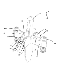
[0031] FIG. 1 shows a perspective view of an exemplary bone preparing guide
and spacer 10.
The bone preparing guide and spacer 10 can include a bone preparing guide 12
and a spacer
14. In some applications, the bone preparing guide and spacer 10 can be
provided to
facilitate the positioning and/or preparation of a bone or bones. In the
illustrated example,
the bone preparing guide 12 includes a body 16 defining a first guide surface
18 to define a
first preparing plane and a second guide surface 20 to define a second
preparing plane. A
tissue removing instrument (e.g., a saw, rotary bur, osteotome, etc., not
shown) can be
aligned with the guide surfaces to remove tissue (e.g., remove cartilage or
bone and/or make
cuts to bone). The first and second guide surfaces 18, 20 can be spaced from
each other by a
distance (e.g., between about 2 millimeters and about 10 millimeters, such as
between about 4
and about 7 millimeters). In the embodiment shown, the first and second guide
surfaces are
parallel, such that cuts to adjacent bones using the guide surfaces will be
generally parallel.
[0032] In some embodiments, as shown in FIG. 1, a first facing surface 22 is
positioned
adjacent the first guide surface 18 and/or a second facing surface 24 is
positioned adjacent the
second guide surface 20. In such embodiments, the distance between the first
guide surface
and the first facing surface defines a first guide slot 26, and the distance
between the second
guide surface and the second facing surface defines a second guide slot 28.
Each slot 26, 28
can be sized to receive a tissue removing instrument to prepare the bone ends
therethrough.
The first and second slots 26, 28 may be parallel or skewed (e.g., non-
parallel) to each other.
- 5 -
CA 02998481 2018-03-12
WO 2017/049056
PCT/US2016/052087
In the illustrated embodiment, the facing surfaces each contain a gap along
their respective
lengths, such that each of the surfaces is not a single, continuous surface.
In other
embodiments, the facing surfaces can each be a single, continuous surface
lacking any such
gap.
[0033] In some embodiments, an opening 30 can be defined by the body 16
between the first
and second facing surfaces 22, 24. The opening 30 can thus be an area between
the slots 26,
28 useful, for instance, for allowing a practitioner to have a visual path to
an osteotomy site
(e.g., cartilage, bones, and/or joint space) during bone preparation and/or to
receive
instruments, as discussed further below. In the embodiment shown, the opening
30 extends
across the body 16 a distance from a surface 32 opposite of the first facing
surface 22 to a
surface 34 opposite of the second facing surface 24.
[0034] The embodiment shown also includes a first end 40 extending from the
body 16 in a
first direction and a second end 42 extending from the body 16 in a second
direction. The
second direction can be different than the first direction (e.g., an opposite
direction). The
first and second ends 40, 42 can each extend out perpendicularly from the body
16 as shown
or, in other embodiments, the first and second ends 40, 42 can extend out from
the body at
differing angles. As shown, each of the first end 40 and the second end 42 can
include at
least one fixation aperture 44 configured to receive a fixation device (e.g.,
a pin, not shown)
to secure the guide 12 to one or more bones. Such apertures 44, as shown, may
extend
through each respective end at a vertical or skewed angle relative to a top
surface of the guide
12.
[0035] The bone preparing guide 12 can also include a first adjustable
stabilization member
46 engaged with the first end 40. In some embodiments, the bone preparing
guide 12 also
includes a second adjustable stabilization member 48 engaged with the second
end 42. Each
of the members 46, 48 can be threaded and engage a threaded aperture defined
by the ends
40, 42. The elevation of each end 40, 42 can be adjusted with respect to one
or more bones
by adjusting the member 46, 48 at the end for which an elevation adjustment is
desired. In
some embodiments, as shown, the members 46, 48 may be cannulated such that
they can
receive respective fixation devices. While bone preparing guide 12 is
illustrated with two
adjustable stabilization members, in other examples, the guide can include
fewer adjustable
stabilization members (e.g., none, one) or more adjustable stabilization
members (e.g., three,
four, or more) and the disclosure is not limited in this respect.
[0036] As noted, the bone preparing guide as shown in FIG 1 includes the
spacer 14. The
spacer can extend from the body in a third direction, the third direction
being different than
- 6 -
CA 02998481 2018-03-12
WO 2017/049056
PCT/US2016/052087
the first and second directions (e.g., perpendicular to the first and second
directions), and it
can be configured to be placed into a joint space between opposing bones. In
some
embodiments, the spacer is integral with the guide and the guide and spacer
are a single
component, for example, a unibody construction. In other embodiments, the
spacer is
physically separate from and insertable into the guide. In these embodiments,
the spacer and
bone cutting guide may be provided as part of a sterile kit (e.g., packed in
single common
container), alone or in combination with other components to facilitate the
procedure being
performed.
[0037] In the embodiment shown, the spacer 14 can be selectively engaged with
the bone
preparing guide 12, such that the spacer and guide can be attached and
detached. For
instance, the spacer 14 can be received by the body 16 of the guide 12, such
as by inserting
the spacer into opening 30. When the spacer 14 is received in the opening 30,
the spacer 14
may extend from the body in between the first guide surface 18 and the second
guide surface
20, or in between the first and second slots 26, 28, when provided. In some
instances where
the spacer 14 is received within the opening 30, no connection between the
guide 12 and
spacer 14 need be present, as the opening 30 itself may be sufficient for
engaging the guide
and spacer. The spacer 14 can engage the guide 12 such that the guide 12
and/or spacer 14
can move relative to one another while engaged (e.g., in a vertical
direction). For example, in
some embodiments, the guide 12 can move relative to the spacer 14 while the
guide 12 and
spacer 14 are engaged, such that the guide can be inserted over the spacer or
removed from
the spacer while the spacer is engaged within a joint space. For example, a
distal portion of
the spacer can be inserted into a joint space (e.g., tarsal-metatarsal joint
space) and the guide
12 positioned over the top of the spacer with a proximal portion of the spacer
projecting out
from the top of the guide. In yet further embodiments, the guide 12 and spacer
14 can be
removably attachable, such as by magnets, a snap-fit, male-female interfacing
parts, or other
temporary connections.
[0038] FIGS. 2A and 2B show an embodiment of a spacer 14. In some embodiments,
the
spacer 14 is configured to be used to guide bone preparation instruments
during a surgical
procedure. In other embodiments, the spacer 14 is configured to be inserted
into a bone
preparation guide. FIG. 2A illustrates a perspective view of the spacer 14,
while FIG. 2B
illustrates the spacer 14 on a foot. The spacer 14, whether a stand-alone
component, a
separate component selectively engageable with a guide, or integrated with a
guide, can be
configured to be inserted into a space between two bones 50 and 52 (e.g.,
adjacent bones
separated by a joint or different portions of a single bone).
- 7 -
CA 02998481 2018-03-12
WO 2017/049056
PCT/US2016/052087
[0039] In one application, the space between two bones into which the spacer
14 is
configured to be inserted can be a TMT joint space, such as the first
metatarsal-cuneiform
joint as shown in FIG. 2B where bone 50 is a first metatarsal and bone 52 is a
medial
cuneiform. The spacer 14 can be inserted into a space between two bones in a
variety of
differing directions, depending on the application. In one example, the spacer
14 is inserted
from the generally dorsal side of a foot. In another example, the spacer 14 is
inserted from a
generally dorsal-medial side of a foot or a medial surface of a foot.
[0040] As seen in the embodiment of FIG. 2A, the spacer 14 can include a first
portion 60
that is configured to extend, at least partially, into a space between two
bones (e.g., a joint
space between bones 50 and 52 as shown in FIG. 2B). In the embodiment shown in
FIG. 2A,
the first portion 60 is a generally planar member having opposite planar
surfaces. In other
embodiments, the generally planar member has one or more slots and/or
apertures. In yet
other embodiments, the first portion has at least two extending members
configured to extend
into the joint space. The extending members can include any cross-sectional
shape, such as
cylindrical, triangular, or frustoconical shape.
[0041] As shown in FIG. 2A, the first portion 60 can include a keel 62, where
the keel 62 is
configured to facilitate insertion of an end of the first portion 60 into the
space between two
bones. As shown, the tip of the keel 62 is linear (e.g., extending in a plane
parallel to the
width of the keel). In other embodiments, the tip of the keel 62 may be
rounded and/or
tapered to provide for easier insertion of the keel 62 into the space between
two bones. In
some embodiments, the keel 62 can have a width that is less than or equal to a
width of the
space between two bones (e.g., a width of a joint). In addition, the keel 62
can have a
thickness (e.g., extending in a direction perpendicular to the width of the
keel and along the
length of the space between two bones) that is equal to or less than the
length of the space
between two bones. In certain applications it may be desirable to configure
the thickness of
the keel 62 to be thicker relative to the length of the space between two
bones such that the
keel 62 fits snuggly into the space between two bones. For example, the
thickness of the keel
62 may be sized to alter, such as expand, the space between two bones when
inserted. The
keel 62 can have a uniform thickness along its length as seen in FIG. 2A or
can have a
thickness that varies, such as a thickness that tapers in a direction
proceeding toward the tip
of the keel 62 (e.g., a wedge-shaped keel).
[0042] A length of the keel 62, and first portion 60, can be configured to
allow the keel 62 to
extend vertically to a bottom base of the space between two bones (e.g., where
the spacer 14
is used in an application on the foot as seen in FIG. 2B, the keel 62 can
extend from a
- 8 -
CA 02998481 2018-03-12
WO 2017/049056
PCT/US2016/052087
generally dorsal side of the foot to a generally plantar side). In other
examples, the length of
the keel 62 can be configured such that the keel 62 extends only partially
into the space
between two bones, such as through a point in the space between two bones
where there is
present opposing interfacing surfaces (e.g., planar interfacing surfaces of
two bones) and
stops extending into the space between two bones where a bump or eccentric
interfacing
surface is present. The keel 62 as shown in the illustrated example is
generally linear along
its length. However, in other examples the keel 62 may include one or more
contours along
its length, where the one or more contours are configured to conform to an
anatomic
geometry of one or more bone ends interfacing at the space into which the keel
62 is inserted.
[0043] The keel 62, and the first portion 60, can be made of various materials
appropriate for
one or more desired applications of the spacer. In one example, the keel can
made of a rigid
material, such as metal or plastic, which does not deform or otherwise change
geometry when
inserted into the space between two bones. By preventing the keel from
deforming when
inserted into the space between two bones, the spacer can be maintained in
general alignment
with the interfacing bone surfaces at the space. In other examples, the keel
can be made of a
flexible material in order to generally maintain alignment of the spacer with
the interfacing
bones surfaces at the space while providing the keel with some give in its
geometry to
conform to one or more non-parallel portions of interfacing bones at the
space. In other
embodiments, the keel includes a combination of a rigid material and a
flexible material. For
example, a perimeter of the keel may include the flexible material to
facilitate insertion into
the joint space and a central portion can include a rigid material to prevent
deformation.
[0044] The spacer 14 can further include a second portion 64 at or near an end
of the spacer
14 opposite the keel 62. The second portion 64 can be designed to be gripped,
such as by a
hand of a surgeon during a procedure. The second portion 64 can have, in some
instances,
one or more recesses 66 (two recesses 66 are shown in FIG. 2A, with each
recess 66 disposed
opposite the other) to enhance a grip on the second portion 64. In some
embodiments, the
second portion 64 can also include a roughened texture to also enhance a grip
on the second
portion 64. The one or more recesses 66 and/or roughened texture can be
particularly
beneficial where the second portion 64 is to be gripped by a wet and/or gloved
hand of a
surgeon.
[0045] In some embodiments, the spacer 14 can have an intermediate portion 68
disposed
between the first and second portions 60, 64. The first, second, and
intermediate portions can
be provided as an integral member or can be provided as separately joined
components. In
either case, each portion can comprise a material different from the other
portions, and the
- 9 -
CA 02998481 2018-03-12
WO 2017/049056
PCT/US2016/052087
material can have different characteristics, such as rigidity and flexibility,
than the materials
of the other portions. Alternatively, all the portions of spacer 14 may be
fabricated from the
same material, such as a unitary body formed of metal or plastic.
[0046] In embodiments where the spacer is provided as a separate component
from the bone
preparation guide and configured to be engaged with the bone preparation
guide, the
intermediate portion 68 can be engageable with the body of the guide (e.g., at
the opening
defined by the body of the guide). In the example shown, the intermediate
portion 68 can
have a first region 70 and a second region 72. The first region 70 can have an
extended
thickness relative to the thickness of the first portion 60 (and thus the keel
62) and can
transition from interfacing with the guide to the second portion 64 along its
length. The
second region 72 can have an extended width relative to the width of the first
portion 60 (and
thus keel 62). The extended thickness of the first region and/or the extended
width of the
second region 72 can allow the spacer 14 to be more stably received by the
body of the guide
in examples where the spacer 14 and guide are separate components.
[0047] In embodiments where the spacer 14 is configured to be used as a stand-
alone device
without a bone guide, the intermediate portion 68 can be used to provide a
first guide surface
and an opposite second guide surface. In such embodiments, the first portion
60 (and thus the
keel 62) of the spacer can be inserted into a joint space, and a surface of
the intermediate
portion 68 can be used to provide a guide surface. For example, the surface of
the first region
70 on a first side of the spacer 14 can be configured as a first guide
surface, and the surface of
the first region 70 on an opposite side of the spacer 14 can be configured as
a second guide
surface. In some embodiments, at least the first region 70 of the intermediate
portion 68 has
a thickness greater than the thickness of the first portion 60. The difference
in thickness of
the first region 70 and the first portion 60 on each side of the spacer 14 can
define a length
and thickness of tissue to be removed by a tissue removing instrument guided
by the surface
of the intermediate portion.
[0048] In use, the first portion 60 may be inserted into a joint space and a
tissue removal
instrument placed against the intermediate portion 68 to guide a preparation
of a first bone on
a first side of the spacer 14. A tissue removal instrument can be placed
against the
intermediate portion 68 to guide a second preparation to a second bone on a
second, opposite
side of the spacer 14. In a specific example, the tissue removing instrument
can be guided by
the spacer 14 for about one-half of a thickness of the tissue to be removed.
Then the spacer
can be removed from the joint space and the tissue removing instrument could
be reinserted
to finish the tissue removal.
-10 -
CA 02998481 2018-03-12
WO 2017/049056
PCT/US2016/052087
[0049] FIGS. 3A and 3B show perspective and side views, respectively, of the
bone
preparation guide and spacer 10 on a foot. Depending on the embodiment, the
bone
preparing guide and spacer 10 can be positioned onto the foot in different
ways. In
embodiments where the spacer 14 is a separate component from the guide and
configured to
be engaged with the guide, the spacer 14 can first be inserted into the space
between two
bones (as shown in FIG. 2B). In such embodiments, after the spacer 14 is
appropriately
inserted into the space between two bones, the guide 12 can then be placed
onto the foot so as
to engage the already inserted spacer 14 (e.g., by sliding the guide 12
vertically downward
toward the foot on the spacer 14, such as via the opening defined by the body
of the guide
12). Alternatively, the guide 12 can first be positioned in proximity to the
space between two
bones and the spacer 14 then inserted through the guide 12 and into the joint
space underlying
the guide. The guide 12 can preliminarily position the spacer 14 (e.g. via the
opening defined
by the body of the guide 12) into the space between two bones. After
preliminarily being
positioned, the clinician can manipulate the location of guide 12 and/or
spacer 14 to orient the
components at a desired location relative to the two bones (e.g., by orienting
the cutting slots
of the guide 12 relative to the ends of bones 50 and 52, respectively). In
other embodiments
(e.g., where the spacer 14 and guide 12 are integral or separate components),
the bone
preparing guide and spacer 10 can be positioned on the foot with guide 12
being positioned in
the joint between the two bones as a single structure, e.g., thereby
simultaneously positioning
the guide 12 and spacer 14 on the foot.
[0050] Independent of how spacer 14 is inserted into the joint between bones
50, 52 relative
to installation of guide 12 over the bones, the spacer 14 can serve to provide
initial stability to
the guide 12 (e.g., prior to the guide 12 receiving fixation devices). For
example, the spacer
14 can engage with the guide 12 and, once seated in the space between the
bones, act to
support the guide 12. Moreover, where the guide 12 and spacer 14 are separate
components
insertable into each other, the depth that the spacer 14 is inserted within
the space between
bones can be adjusted without needing to remove the guide 12. Similarly, the
distance
between where the guide 12 is positioned vertically in relation to the space
between bones
can be adjusted while leaving the spacer 14 in place.
[0051] In operation, spacer 14 can serve as an alignment and/or reference tool
for the guide
12 with respect to the one or more bone surfaces to be prepared (e.g., cut,
morselized). Such
surfaces to be prepared can include all or a portion of an interfacing surface
of bone 50 or 52
as shown. When the spacer 14 is inserted into the space (e.g., a joint)
between the bones 50,
52, the spacer 14 can act to align the guide 12 at an appropriate position
(e.g., longitudinal
-11-
CA 02998481 2018-03-12
WO 2017/049056
PCT/US2016/052087
along the bones 50, 52) and orientation (e.g., angle relative to the bones 50,
52 in multiple
planes selected from more than one of a frontal plane, a transverse plane, and
a sagittal plane)
for the intended procedure relative to the surfaces of the bones 50, 52 to be
prepared.
[0052] For example, when the spacer 14 is inserted into the space between
bones 50, 52, the
spacer can help align and orient, in one or more planes, the first guide
surface 18 and the
second guide surface 20 (and/or slots 26, 28, when provided) of the guide 12
relative to
respective surfaces of the bones 50, 52 to be prepared. When inserted into the
space between
bones 50, 52, the spacer 14 can engage the guide 12 (e.g., physically restrict
the free range of
movement of guide 12), longitudinally aligning the guide 12 with the surface
of each of
bones 50 and 52 to be prepared. For example, spacer 14 can longitudinally
align the first
guide surface 18 with the end surface of the bone 52 and the second guide
surface 20 with the
end surface of the bone 50. Additionally, when inserted into the space between
bones, the
spacer 14 can extend out from the space and provide an indication as to the
location of the
interfacing, end surfaces of each of bones 50 and 52. In this way, orienting
the guide 12
relative to the spacer 14 serves as an angular reference relative to the end
surfaces of each of
bones 50 and 52. Thus, the spacer 14 can facilitate accurate preparation of a
desired surface
of one or more bones 50, 52.
[0053] In some applications, it can be desirable to prepare interfacing
surfaces of one or both
bones 50, 52 by cutting a slice from one or both surfaces, where the slice has
a generally
constant thickness. In such an application, the spacer 14 can be configured to
orient a first
guide surface 18 parallel to the preparation surface of bone 52 and/or a
second guide surface
20 parallel to the preparation surface of bone 50. In the case of a metatarsal-
cuneiform joint
as shown in FIG. 3A, where interfacing bone end surfaces are relatively
planar, orienting the
first guide surface 18 and the second guide surface 20, or slots 26, 28, when
provided,
parallel to the bone end surfaces to be cut using the spacer 14 as a reference
can facilitate
removal of a slice of relatively constant thickness. Depending on the
procedure being
performed, the end of bone 50 facing bone 52 and/or the end of bone 52 facing
bone 50 may
be morselized in addition to or in lieu of being cut to prepare the end of the
bone.
[0054] In other additional or alternative applications, it can be desirable to
prepare
interfacing surfaces of one or both bones 50, 52 by cutting a wedge-shaped
portion from the
interfacing surfaces of one or both bones 50, 52, where the wedge-shaped
portion does not
have a uniform thickness (e.g., in the medial to lateral direction of cut).
For instance, in one
application, a plantar-based wedge may be cut from a medial cuneiform (e.g.,
bone 52), e.g.,
to correct a misaligned first metatarsal (e.g., bone 50). To cut a wedge-
shaped portion from
- 12 -
CA 02998481 2018-03-12
WO 2017/049056
PCT/US2016/052087
the interfacing end surface of one or both bones 50, 52, the first guide
surface 18 and the
second guide surface 20, or slots 26 and 28, when provided, can be oriented at
a skewed
angle relative to respective bones 52 and/or 50 using the spacer 14 as a
reference for the
skewed angle at which the one or more bones 50, 52 are to be cut. For example,
first guide
surface 18 and/or second guide surface 20 may skew proximally back along the
length of the
medial cuneiform as the guide surface extends from the medial side of the
medial cuneiform
to the lateral side of the medial cuneiform. As a result, the bone portion cut
using a guide
surface so configured can remove more bone from the lateral side of the medial
cuneiform
than the medial side of the medial cuneiform, resulting in a wedge-shaped
portion of bone
being removed from the medial cuneiform.
[0055] In addition to serving as a reference for positioning and orientation
in bone
preparation, the spacer 14 can also serve as a reference for indicating a
thickness of tissue
(e.g., such as bone) to be removed from a surface of a bone 50 and/or 52. For
instance, a
distance between a first longitudinal surface of spacer 14 and the first guide
surface 18 may
define a thickness of tissue to be cut from the surface of the bone 52.
Similarly, a distance
between a second longitudinal surface (e.g., opposite the first longitudinal
surface) of the
spacer 14 and the second guide surface 20 may define a thickness of tissue to
be cut from the
surface of the bone 50. Thus, a clinician can utilize the spacer 14 as a
reference prior to
preparing one or more bones in order to visualize whether the proposed bone
preparation
matches the desired bone preparation, such as whether the thickness of the cut
to be made is
in accordance with a desired thickness of tissue to be removed, and make any
adjustments
prior to implementation of the bone preparation action.
[0056] In some embodiments, a guide can be designated with an identification
number
representing a cut thickness facilitated by that particular guide, which is
defined by the
distance between an adjacent surface of the spacer 14 and the respective guide
surface of the
guide. In certain embodiments, the distance between a first guide surface 18
and the spacer
14 can be different from the distance between a second guide surface 20and the
spacer 14,
such that different thickness cuts can be made on different, interfacing
bones. In some
examples, the distance between one or more guide surfaces on the guide and the
spacer 14
can be adjustable, allowing a user to vary the thickness of matter removed by
a cut.
[0057] In applications where cut guide 12 includes a guide surface (e.g.,
first guide surface
18 and/or second guide surface 20) that skews proximally from the medial to
the lateral side
of the foot to which the cut guide is applied, the angle of skew may be fixed
or may be
variable. For example, cut guide 12 may have an adjustable guide surface
(e.g., first guide
- 13 -
CA 02998481 2018-03-12
WO 2017/049056
PCT/US2016/052087
surface 18 and/or second guide surface 20) where the angle of skew can be
adjusted. In
practice, a clinician can set the angle of the guide surface(s) relative to
the bone, e.g., to
remove more on the lateral side than the medial side, more on the medial side
than the lateral
side, or the same amount on both the lateral and medial sides, depending on
the angle set. In
some configurations, the adjustable angle can be temporarily fixed, or locked,
to prevent
inadvertent movement after setting the desired angle. Depending on the design,
the guide
surface (e.g., first guide surface 18 and/or second guide surface 20) may be
adjustable to
provide an angle (e.g., between the guide surface and first and/or second
facing surfaces 22,
24) ranging from 0 degrees to 25 degrees, such as 0 degrees to 10 degrees. For
example, the
guide surface may be adjustable from 25 degrees to a non-zero degree angle
less than 25
degrees.
[0058] Although the spacer 14 is illustrated to be engageable with the guide
12, the spacer 14
can also be configured to engage with other guides. For example, spacer 14 may
be used as
part of a kit that includes a plurality of guides with different sized cut
widths. A clinician can
select one of the plurality of different sized guides and use the selected
guide in combination
with the spacer to perform a procedure. For example, a clinician may select
one guide 12 to
engage with spacer 14 and provide a designated first cut thickness. If the
clinician, using the
spacer 14 as a reference, determines that the cut thickness that would result
using the first
guide is not desirable for the particular operation, the spacer 14 can be
removed from the first
guide and engaged with a second guide having a designated second cut thickness
different
from the first cut thickness of the first guide.
[0059] While spacer 14 illustrated and described above with respect to FIGS. 1-
3 can
provide an effective tool for performing various medical procedures, such as
helping to
position a bone preparation guide during a tarsal-metatarsal fusion procedure,
the
configuration of spacer 14 may vary, e.g., depending on the characteristics of
use and
targeted clinical outcome.
[0060] As one example, the width of the spacer 14 can be sized such that the
spacer extends
into a lateral region of adjacent bone ends, when the spacer is positioned
between the bones.
For example, the width of the portion of spacer 14 configured to be positioned
between
adjacent portions of bone (e.g., first portion 60) can be wide enough (in the
medial to lateral
direction) that the spacer extends at least into the lateral half of the end
faces of the adjacent
bones between which the spacer is positioned.
[0061] For example, when spacer 14 is configured to be inserted into a tarsal-
metatarsal joint
space (e.g., between first metatarsal 50 and medial cuneiform 52), first
portion 60 of the
- 14 -
CA 02998481 2018-03-12
WO 2017/049056
PCT/US2016/052087
spacer may be project laterally a distance sufficient to position the first
portion of the spacer
between the lateral half of the end faces of first metatarsal 50 and medial
cuneiform 52. The
first portion 60 may contact the ends of first metatarsal 50 and medial
cuneiform 52 on the
lateral side of the bone ends (e.g., lateral-most half, lateral-most third,
lateral-most edges). In
some configurations, the first portion 60 of spacer 14 extends laterally a
distance sufficient to
contact a medial side of an adjacent bone (e.g., second metatarsal), when
positioned between
first metatarsal 50 and medial cuneiform 52.
[0062] In configurations where spacer 14 is symmetric, the first portion 60 of
the spacer may
also project medially to contact the ends of first metatarsal 50 and medial
cuneiform 52 on
the medial side of the bone ends (e.g., medial-most half, medial-most third,
medial-most
edges). Alternatively, as discussed in more detail below, first portion 60 may
be laterally
offset in the medial-lateral dimension. In either case, second portion 64 may
be symmetric
with respect to the first portion and have the same width as the first portion
or may be
asymmetric with respect to the first portion and/or have a different width
than the first
portion.
[0063] Configuring spacer 14 to have a portion positionable within a lateral
portion of a joint
space can be useful to help prevent misalignment of guide 12. In practice,
when bone 50 is
realigned relative to bone 52 and/or an adjacent bone (e.g., second
metatarsal), a gap between
bone 50 and bone 52 may open on the medial side of the joint space between the
ends of the
bones. This gap may occur, e.g., as an intermetatarsal angle between a first
metatarsal and
second metatarsal is closed by pivoting and/or rotating the first metatarsal
toward the second
metatarsal. In some situations, as a gap on the medial side of the joint space
opens, a spacer
positioned inside of the joint space can shift (e.g., laterally or medially)
and/or rotate (e.g.,
such that the portion of the spacer above the joint space does not project
dorsally but instead
projects in a lateral-dorsal or medial-dorsal direction). When the guide 12 is
subsequently
positioned over the shifted and/or rotated spacer 14, the cutting surface(s)
of the guide may
be misaligned with respect to when the ends of bones 50 and 52 are desirably
cut.
[0064] By configuring the spacer 14 so that at least a portion of the spacer
positioned
between the ends of bones 50 and 52 (e.g., within the tarsal-metatarsal joint
space) is on the
lateral side of the joint space, the spacer may remain properly positioned
through realignment
of bone 50. For example, as the intermetatarsal angle between a first
metatarsal and second
metatarsal is closed by pivoting and/or rotating the first metatarsal toward
the second
metatarsal, the lateral side of the ends of bones 50 and 52 may move toward
each other while
the medial side of the ends of the bones may move away from each other. This
can pinch the
- 15 -
CA 02998481 2018-03-12
WO 2017/049056
PCT/US2016/052087
lateral portion of spacer 14 between the ends of the bones (e.g., by having
the lateral sides of
the ends of the bones press against opposing sides of the spacer). As a
result, the spacer 14
may be retained in a stable position in the joint space between the bones such
that, when
guide 12 is subsequently positioned over spacer 14, the guide is appropriately
positioned to
cut the ends of bones 50 and 52 (e.g., by translating a cutting instrument
along and/or through
guide surfaces of the guide to resects the bone ends).
[0065] Spacer 14 can have a variety of different configurations that allow at
least a portion of
the spacer to be positioned on lateral sides of the end faces of the bones
between which the
spacer is inserted. As another example, the portion of spacer 14 that is
configured to project
plantarly of bone preparation guide 12 (e.g., the portion positioned in the
joint space between
opposed ends of bones 50 and 52 while guide 12 is positioned on top of the
bones) may be
asymmetrical with respect to portion of the spacer located inside of guide 12
and/or
projecting out of the top of the guide (e.g., dorsally of the guide). For
example, instead of
configuring first portion 60 of spacer 14 to be positioned substantially
centered in the joint
between the ends of bones 50 and 52, the first portion 60 of the spacer may be
offset relative
to an axis extending through a geometric center of the ends of the bones and
the joint
therebetween. When so configured, the spacer 14 may be inserted into one side
of the joint
space between the bones 50 and 52 while leaving another side of the joint
space devoid of
any spacer substrate.
[0066] As one example in accordance with this configuration, spacer 14 may be
configured
so that first portion 60 is offset relative to an axis extending through a
geometric center of
second portion 64. For example, when second portion is configured to have
guide 12
insertable into and removable over the top of the second portion 64, first
portion 60 may
project downwardly from one side of the second portion. In use, such a
configuration can
cause first portion 60 to project asymmetrically with respect to the cross-
sectional plane (e.g.,
medial-lateral plane at a single elevation) relative to second portion 64
and/or guide 12
positioned over the guide.
[0067] For example, when spacer 14 is configured to be inserted into a tarsal-
metatarsal joint
space (e.g., between a first metatarsal and medial cuneiform), first portion
60 of the spacer
may be laterally offset relative to second portion 64. That is, first portion
60 may be
positioned preferentially toward the lateral side of the joint as opposed to
the medial side of
the joint. The laterally-offset first portion 60 of spacer 14 may contact the
ends of bones 50
and 52 on the lateral side of the bone ends (e.g., lateral-most half, lateral-
most third, lateral-
most edges) but not on the medial side of the bone ends (e.g., medial-most
half, medial-most
- 16 -
CA 02998481 2018-03-12
WO 2017/049056
PCT/US2016/052087
third, medial-most edges). The second portion 64 of the spacer may be
substantially centered
between the lateral and medial edges of bones 50 and 52. Accordingly, the
portion of spacer
14 that is visible by the clinician and/or used to orient guide 12 (e.g., by
positioning the guide
over the spacer) can be substantially centered in the medial-lateral direction
over the ends of
the bones 50 and 52. However, the portion of spacer 14 that actually projects
below the
dorsal face of bones 50 and 52 in the plantar direction can be offset to the
lateral side in the
medial-lateral direction.
[0068] FIGS. 4A and 4B illustrate an example configuration of a spacer 400
that has a
laterally offset structure positionable within a joint space. FIG. 4A
illustrates a perspective
view of the spacer 400, while FIG. 4B illustrates a perspective view of the
spacer 400 on a
foot. Like reference numerals used with respect to spacer 400 refer to like
elements
discussed above with respect to the spacer 14. The spacer 400, whether a stand-
alone
component, a separate component selectively engageable with a guide, or
integrated with a
guide, can be configured to be inserted into a space between two bones 50 and
52 (e.g.,
adjacent bones separated by a joint or different portions of a single bone).
In one application,
the space between two bones into which the spacer 400 is configured to be
inserted can be a
tarsometatarsal ("TMT") joint space, such as the first metatarsal-cuneiform
joint as shown in
FIG. 4B.
[0069] As shown in the example of FIG. 4A, the spacer 400 includes the first
portion 60 that
is configured to extend, at least partially, into a space between two bones
(e.g., a joint space
between bones 50 and 52 as shown in FIG. 4B). The first portion 60 defines a
central
longitudinal axis 402. The first portion 60 further includes a first sidewall
404 and a second
sidewall 406 defining a width of the first portion 60 therebetween. The first
portion 60 can
include a keel 62, where the keel 62 is configured to facilitate insertion of
an end of the first
portion 60 into the space between two bones. In one example, a thickness of
the keel 62 can
taper moving in a direction toward the tip of the first portion 60 (e.g.,
toward the tip of the
keel 62). In some cases, the keel 62 can extend vertically to a bottom base of
the space
between two bones, and in other cases the keel 62 may only partially extend
into the space
between two bones. Independent of whether the thickness of first portion 60
(including keel
62) is constant across the length of the first portion or varies, in some
examples, the thickness
ranges from 0.2 mm to 3 mm, such as from 0.38 mm to 1.8 mm.
[0070] The spacer 400 illustrated in FIGS. 4A and 4B further includes a second
portion 64 at
or adjacent an end of the spacer 400 opposite the keel 62. As seen in FIG. 4A,
the second
portion 64 has a central longitudinal axis 408. The second portion 64 can be
designed to be
- 17 -
CA 02998481 2018-03-12
WO 2017/049056
PCT/US2016/052087
gripped, such as by a hand of a surgeon during a procedure. The second portion
64 can have,
in some instances, one or more recesses 66 and/or a roughened texture to
enhance a grip on
the second portion 64.
[0071] The spacer 400 can also have, in some embodiments, an intermediate
portion 68
disposed between the first and second portions 62, 64. In embodiments where
the spacer 400
is provided as a separate component from the bone preparation guide and
configured to be
engaged with the bone preparation guide, the intermediate portion 68 can be
engageable with
the body of the guide (e.g., at the opening defined by the body of the guide).
In the example
shown, the intermediate portion 68 has a first region 70 and a second region
72. The first
region 70 can have an extended thickness relative to the thickness of the
first portion 60 (and
thus the keel 62) and can transition from interfacing with the guide to the
second portion 64
along its length. The second region 72 can have an extended width relative to
the width of
the first portion 60 (and thus keel 62). In the example shown, the
intermediate portion 68,
including the first and second regions 70, 72, has a central longitudinal axis
that coincides
(e.g., is coaxial) with the central longitudinal axis 408 of the second
portion 64.
[0072] As shown in FIGS. 4A and 4B, the embodiment of spacer 400 illustrated
has a
generally offset configuration. In some examples, an offset configuration of
the spacer 400
includes the first portion 60 being offset from the second portion 64 and/or
the intermediate
portion 68. In the illustrated embodiment, where the second portion 64 and the
intermediate
portion 68 have the central longitudinal axis 408 in common, the first portion
60 is offset
from both the second portion 64 and the intermediate portion 68. In
particular, the central
longitudinal axis 402 of the first portion 60 is spaced from the central
longitudinal axis 408 of
the second portion 64 and intermediate portion 68. While the specific
dimensions can vary
depending on the desired application, in some configurations, the distance
separating a
geometric enter of the central longitudinal axis 402 of the first portion 60
from the geometric
center of the central longitudinal axis 408 of the second portion ranges from
1 mm to 20 mm,
such as from 3 mm to 17 mm. While the width of first portion 60 (from first
sidewall 404 to
second sidewall 406) may also vary, in some instances, the width ranges from 2
m to 25 mm,
such as 5 mm and 20mm. Accordingly, in some configurations, a distance from a
geometric
center of central longitudinal axis 408 to first sidewall 404 bounding the
lateral-most extend
of spacer 14 may range from 5 mm to 15.
[0073] The spacer 400 can be inserted into the space between two bones 50 and
52, such as
shown in FIG. 4B. When inserted, the offset configuration of the spacer 400
can result in the
first portion 60 contacting the bones 50 and 52 (e.g., the end faces of the
bones) and
- 18 -
CA 02998481 2018-03-12
WO 2017/049056
PCT/US2016/052087
optionally an adjacent third bone 53 (e.g., a medial side of the bone), while
the second
portion 64 and central longitudinal axis 408 thereof is generally positioned
at a center of the
joint. In the illustrative example of FIG. 4B, the spacer 400 is inserted into
the first TMT
joint and the first portion 60 contacts the first metatarsal 50, first
cuneiform 52, and second
metatarsal 53. For instance, a first face surface of the first portion 60 can
contact the first
metatarsal 50, a second face surface of the first portion 60 opposite the
first face surface can
contact the first cuneiform 52, and the first sidewall 404 of the first
portion 60 can contact the
medial side of the second metatarsal 53. Thus, while the central longitudinal
axis 408 of the
second portion 64 may be generally centered between a medial side and a
lateral side of the
TMT joint, the central longitudinal axis 402 of the first portion 60 may be
generally closer to
the lateral side than the medial side of the TMT joint (e.g., at a lateral
third of the TMT joint).
Accordingly, in some instances, the first portion 60 may be inserted and
secured within only
a lateral region of the TMT joint.
[0074] The offset configuration of the spacer 400 can be useful in providing a
stable spacer
within the joint space, for instance, during a procedure where a bone
alignment is to be
altered. For example, in one type of bone alignment procedure the bone 50 may
need to be
realigned (e.g., using positioner device 410) in a manner that reduces an
angle between the
bone 50 and the bone 53. While the bone 50 is realigned, a gap at the medial
side of the joint
between the bone 50 and bone 52 may increase. This may cause a spacer
positioned within
the joint to become unstable (e.g., because the spacer is no longer tightly
engaged between
the bones 50 and 52 due to the increased gap at the medial side of the joint).
[0075] Using the spacer 400 having the offset configuration within the joint
during bone
realignment can help stably maintain the spacer 400 within the joint. For
example, because
the spacer 400 can have the first portion 60 positioned in the joint closer to
the lateral side
than the medial side (e.g., at the lateral region of the joint such that the
first sidewall 404
contacts the medial side of the second metatarsal 53) the spacer can be
generally maintained
at a constant position within the joint during this realignment. This can
result because the
spacing between the bones 50 and 52 at the lateral side of the joint during
realignment may
stay substantially constant or be reduced relative to the spacing before the
realignment. As a
result, the first portion 60 positioned at the lateral region can be stably
held within the joint
even after this realignment is completed. Furthermore, because the first
portion 60 is stably
maintained within the joint, the second portion 64, and central longitudinal
axis 408 thereof,
may also be generally maintained at its original position, such as at the
center of the joint.
This spacer 400 configuration, which may provide increased stability, can in
turn be useful
- 19 -
CA 02998481 2018-03-12
WO 2017/049056
PCT/US2016/052087
when the spacer 400 is utilized to facilitate preparation (e.g., cutting) of
one or more bone
surfaces, as will be described below with reference to FIG. 5.
[0076] Independent of whether a spacer is configured with a first portion
aligned with a
second portion (e.g., as discussed with respect to spacer 14) or a first
portion offset relative to
a second portion (e.g., as discussed with respect to spacer 400), the spacer
can have a variety
of different cross-sectional configurations. For example, while spacer 14 in
FIGS. 1-3 and
spacer 400 in FIGS. 4A and 4B are illustrated as having a constant thickness
across the width
of the spacer, the thickness of the spacer can vary over the width.
[0077] FIGS. 4C-4F illustrate an example configuration of spacer 14 where the
thickness of
the spacer varies across the width of the spacer. FIG.4C is a top view of the
example spacer;
FIG. 4D is a front view of the spacer; and FIGS. 4E and 4F are opposite side
views of the
spacer. The Z-axis on the figures represents the vertical orientation of the
spacer (e.g., dorsal
to plantar direction) in typical use. As shown in the illustrated example,
spacer 14 defines a
length L (e.g., parallel to the longest axis of the spacer), a width W
perpendicular to the
length, and a thickness T perpendicular to both the length and width. The
width W of the
first portion 60 is the distance from the first sidewall 15 to the second
sidewall 17 of the first
portion. In the illustrated configuration, the thickness T of the spacer
tapers across the width
of the spacer from the first sidewall 15 to the second sidewall 17.
[0078] FIGS. 4G-4J similarly illustrates an example configuration of spacer
400 where the
thickness of the spacer varies across the width of the spacer. FIG.4G is a top
view of the
example spacer; FIG. 4H is a front view of the spacer; and FIGS. 4E and 4F are
opposite side
views of the spacer. Again, the Z-axis on the figures represents the vertical
orientation of the
spacer (e.g., dorsal to plantar direction) in typical use. As shown in the
illustrated example,
spacer 14 defines a length L (e.g., parallel to the longest axis of the
spacer), a width W
perpendicular to the length, and a thickness T perpendicular to both the
length and width.
The width W of the first portion 60 is the distance from the first sidewall
404 to the second
sidewall 406 of the first portion. In the illustrated configuration, the
thickness T of the spacer
tapers across the width of the spacer from the first sidewall 404 to the
second sidewall 406.
[0079] When configured with a differential thickness across its width, spacer
14 (FIGS. 4C-
4F) / spacer 400 (FIGS. 4G-4J) is thicker on one side than the other side. In
different
examples, the spacer can have a continuous angle of taper across its width W
or
discontinuous taper, such one or more steps that define discrete thickness
transition points. In
some examples, the spacer has a thickness at least 0.1 mm less at its thinner
sidewall
compared to its thicker sidewall, such as at least 0.2 mm less, at least 0.5
mm less, or at least
- 20 -
CA 02998481 2018-03-12
WO 2017/049056
PCT/US2016/052087
1 mm less. For example, the spacer may have a thickness from 0.1 mm to 2 mm
less at its
thinner sidewall compared to its thicker sidewall. In addition, although
second portion 64
and intermediate portion 68 of spacer 14 / 400 are illustrated as not being
tapered, one or both
of second portion 64 and intermediate portion 68 can be tapered in other
embodiments.
[0080] In use, a spacer with tapered thickness may be positioned in a TMT
joint with the
thicker side of the spacer located medially and the thinner side of the spacer
located laterally
across the joint. During realignment of the first metatarsal relative to the
medial cuneiform, a
larger gap may open on the medial side between the end face of the first
metatarsal and the
end face of the medial cuneiform than on the lateral side. Accordingly,
positioning the
thicker portion of the spacer medially and the thinner portion of the spacer
laterally across the
TMT joint can help fill a gap between the bones, e.g., helping the spacer to
fit snuggly within
the TMT joint space and prevent inadvertent movement of the spacer.
[0081] Spacer 14 / 400 can have any suitable cross-section shape. For example,
while spacer
14 / 400 is illustrated as defining a substantially rectangular shape, in
other examples the
spacer can define other shapes. Spacer 14 / 400 can define any polygonal
(e.g., square,
hexagonal) or arcuate (e.g., circular, elliptical) shape, or even combinations
of polygonal and
arcuate shapes. Indeed, first portion 60 of spacer 14 / 400 need not be a
continuous structure
across its width W but can be formed of discontinuous segments or portions of
material
which, in combination, achieve spacer functions.
[0082] FIG. 4K illustrates an example configuration of spacer 400 where first
portion 60 is
formed by at least two pins 403A and 403B separated from each other with a gap
therebetween. The pins 403A and 403B extend parallel to the length of the
spacer (e.g., from
intermediate portion 68 and/or second portion 64). In other configurations,
spacer 400 may
include more pins (e.g., three, four, or more) pins arrayed across at least a
portion of the
width of the spacer with a gap between each adjacent pin. Each pin can have
the same cross-
sectional dimension (e.g., diameter) or at least one pin can have a cross-
sectional dimension
greater than at least one other pin. For example, a medial-most pin (e.g.,
centered pin) may
have a larger cross-sectional diameter than a lateral-most pin. Further,
although pins 403A
and 403B are illustrated as arranged to provide a laterally offset first
portion 60, the pins may
be arranged to provide a first portion that is centered with the second
portion 64 or otherwise
suitably arranged.
[0083] When configured with pins 403A and 403B, the ends or tips of the pins
may be
rounded and/or tapered (e.g., to a point) to provide for easier insertion of
the pins into the
space between two bones. FIG. 4L illustrates an example configuration of the
spacer in FIG.
- 21 -
CA 02998481 2018-03-12
WO 2017/049056
PCT/US2016/052087
4K where the ends 405A and 405B of pins 403A and 403B, respectively, are
tapered to a
point to facilitate insertion. Further, pins 403A and 403B of spacer 400 can
define any
polygonal (e.g., square, hexagonal) or arcuate (e.g., circular, elliptical)
shape, or even
combinations of polygonal and arcuate shapes. For example, the pins can be
cylindrical,
trapezoidal, triangular, square, rectangular, oval, hexagonal, or have yet
other cross-sectional
shape. FIG. 4M illustrates an example configuration of the spacer in FIG. 4K
where pins
403A and 403B have a rectangular cross-sectional shape (e.g., across the width
of the pins).
[0084] FIG. 5 illustrates a top plan view of an embodiment of a bone preparing
guide 450
and the spacer 400 on a foot. Like numerals used herein in connection with the
bone
preparing guide 450 refer to like elements of the bone preparing guide 12. As
one example,
the bone preparing guide 450 can be placed after one or more bones have been
realigned by
engaging the bone preparing guide 450 with the spacer 400 similar to that
described
previously. For instance, the bone preparing guide 450 can be engaged at the
intermediate
portion of the spacer 400 by moving the opening 30 of the guide 450 over the
second portion
of the spacer 400. As explained previously, using the spacer 400 can
substantially maintain
the intermediate and second portions, and thus central longitudinal axis
thereof, at the desired
position, such as at the center of the joint. Therefore, placing the guide 450
using the
intermediate and/or second portions of the spacer 400 can result in alignment
of the guide
450 at the desired position with respect to the joint, and thus bones 50 and
52. As a result,
bone preparation can proceed efficiently using the guide 450.
[0085] While bone preparing guide 450 can have a variety of different
configurations, as
described above with respect to bone preparing guide 12, in the configuration
illustrated in
FIG. 5, bone preparing guide 450 includes a cutting slot 26 that is skewed
form (e.g., oriented
at an angle) relative to cutting slot 28 and/or the joint between bones 50 and
52. The slot 26
can be angled to an extent suitable for providing anatomically appropriate
bone preparation
(e.g., one or more bone cuts) depending on the procedure, and thus anatomical
region, for
which the guide 450 is used. Example angles of skew discussed above with
respect to cut
guide 12 can also be used for bone preparing guide 450.
[0086] Where the guide 450 is used to prepare one or more bones during a
procedure on the
foot as shown in the exemplary illustration of FIG. 5, the slot 26 can be
angled relative to a
TMT joint space and/or end of first metatarsal for cutting a portion of a
cuneiform (e.g.,
medial cuneiform 52). The slot 26 can be angled proximally (e.g., away from
the distal end
of the medial cuneiform) as the slot extends from the medial side to the
lateral side of the
medial cuneiform. As a result, a cut made along slot 26 can result in a wedge
being cut off
- 22 -
CA 02998481 2018-03-12
WO 2017/049056
PCT/US2016/052087
the medial cuneiform that progressively increases in width (e.g., thickness)
moving the
medial side to the lateral side of the medial cuneiform.
[0087] In some examples, guide 450 is configured to be flipped in the frontal
plane 180
degrees for interchangeable use on either the right or left foot of a patient.
For example, if a
TMT fusion procedure is being performed on the left foot of the patient, guide
450 may be
rotated in a first orientation so slot 26 is angled proximal from the medial
to lateral direction
of the left foot. By contrast, if the TMT fusion procedure is being performed
on the right foot
of the patient, guide 450 may be rotated 180 degrees to a second orientation
so slot 26 is
angled proximal from the medial to lateral direction of the right foot. The
top and bottom
surfaces of guide 450 may be devoid of projections or other structural
features that prevent
the guide from being flipped and placed over a joint to be cut in order to
configure the guide
with skewed cutting slot for deployment on either foot.
[0088] In some examples, guide 450 with skewed slot 26 is provided as part of
a kit that
includes a plurality of different guides. The guides can vary by having
different sizes and/or
angles at which slot 26 is skewed relative to each other. During a procedure,
a clinician can
select one of the plurality of different guides, e.g., that best matches the
size and/or angle of
the cut desired to be made. To facilitate interchangeability of the guides
during the
procedure, each of the plurality of guides can have anchoring apertures (e.g.,
first and second
adjustable stabilization members 46, 48) that are at the same location
relative to each other
(e.g., same separation distance and angle). When so configured, a clinician
may remove one
guide from anchoring pins set into first metatarsal 50 and medial cuneiform 52
(e.g., by
pulling the guide up off the pins) and insert a different guide, for example
having a slot 26
skewed at a different angle than the removed guide, over the same pins (e.g.,
by pushing the
second guide down over the pins).
[0089] To help select an appropriate one of the plurality of available guides
each having a
cutting surface skewed at a different angle, a clinician may take an X-ray of
a TMT joint
being operated upon before selecting the guide. The clinician may take the X-
ray from the
dorsal to plantar direction to select and size the cut angle to be made on the
medial
cuneiform. The clinician may then select a particular one of the plurality of
available guides
whose angle best matches the size and/or angle of the cut to be made for
subsequently
deployment over the TMT joint, and particularly over the end of the medial
cuneiform to be
cut, during the procedure.
[0090] In the example of FIG. 5, the slot 26 is illustrated adjacent a
cuneiform and thus can
be configured at an angle proceeding proximally from the medial side to the
lateral side of the
- 23 -
CA 02998481 2018-03-12
WO 2017/049056
PCT/US2016/052087
guide 450, e.g., corresponding generally to bone preparation appropriate for a
cuneiform
(e.g., cutting a wedge-shaped portion of the cuneiform). That is, a cut made
using slot 26 can
produce a progressively thicker bone cut progressing from the medial to
lateral side of the
cuneiform bone. In some examples, an end of the slot 26 at the lateral side of
the guide 450
can be angled between two and ten degrees relative to an end of the slot 26 at
the medial side
of the guide 450. In further examples, an end of the slot 26 at the lateral
side of the guide 450
can be angled between two and five degrees relative to an end of the slot 26
at the medial side
of the guide 450. Using the guide 450 having the angled slot 26 in conjunction
with the
spacer 400 as shown can provide for bone preparation of the bone 52 in an
anatomically
appropriate manner even after the bone 50 have been realigned.
[0091] FIGS. 6A-6D show an embodiment of a tissue removal location check
member 80.
FIGS. 6A and 6B show perspective views of the bone preparing guide 12 and
tissue removing
instrument location check member 80 in isolation, while FIGS. 6C and 6D show
perspective
views of the bone preparing guide 12 and tissue removing instrument location
check member
80 on a foot. The tissue removing instrument location check member 80 can
serve to allow a
surgeon to visualize the position and/or orientation of a cut to be made using
the guide 12. In
some embodiments, the spacer and tissue removing instrument location check
member 80 can
be engaged with the guide 12 at the same time.
[0092] The tissue removing instrument location check member 80 can include a
first portion
82 and a second portion 84. In some embodiments, such as that shown, the
member 80 may
further include an opening 86 defined between the first portion 82 and the
second portion 84.
The member 80 can be engageable with the guide 12, such as at a slot 26 or 28
or opening 30,
via the first portion 82 of the member 80. As shown, the first portion 82 is
engaged with the
guide 12 at the slot 28 and in some cases can extend down through the slot 28.
In other
examples, the first portion 82 can engage the guide 12 at the slot 26 or at
the opening 30.
Depending on the configuration of the tissue removing instrument location
check member 80,
the first portion 82 may in some instances engage the guide 12 at more than
one of the slot
26, slot 28, and opening 30.
[0093] The second portion 84 is connected to or integral with the first
portion 82 and extends
and is spaced from the first portion 82. As illustrated, the second portion 84
can be
configured to extend out from the guide 12 while the first portion 82 is
engaged with the
guide 12. For example, the first portion 82 can be within the slot 28 of the
guide 12 while the
second portion 84 is spaced from the first portion 82 in a direction (e.g.,
radial) outside of the
slot 28. The second portion 84 can have a width less than a width of the first
portion 82, and
- 24 -
CA 02998481 2018-03-12
WO 2017/049056
PCT/US2016/052087
additionally may include curvature along its length. As shown, the second
portion 84
includes curvature along its length in a direction toward the guide 12 when
the first portion
82 is engaged with the guide 12. In embodiments where the opening 86 is
included in the
member 80, the opening 86 may be configured to receive a portion of a bone
such that the
second portion 84 can extend outwardly around such bone portion received in
the opening 86.
[0094] The first portion 82 and the second portion 84 can be designed to be
complimentary
portions. In particular, in some examples, the position of the second portion
84 can in some
predetermined manner correspond to the position of the first portion 82. For
instance, where
the member 80 is properly engaged with the guide 12, a tip of the second
portion 84 can
extend a same depth as a bottom edge of the first portion 82 (e.g., the tip of
the second
portion 84 and a bottom edge of the first portion 82 extend along a same
plane). In such
instance, visually inspecting a depth of the second portion 84 (e.g., extended
outside of the
guide 12) can provide an indication as to a depth of the first portion 82.
This can be
beneficial in applications where the bottom edge of the first portion 82 is
inserted into tissue
and not easily seen by a surgeon, but where the second portion 84 extends out
from the tissue
and can be seen by the surgeon. As such, in some examples the second portion
84 can allow
a surgeon to ascertain how deep the first portion 82 is within the tissue by
simply looking at
the second portion 84 that is outside the tissue. In some cases, the depth of
the second
portion 84 can directly correspond to the depth of the first portion 82, and
in other cases the
correspondence between the first and second portions can be a predetermined
ratio.
[0095] The tissue removing instrument location check member 80 can engage with
the guide
12 in a manner that allows the member 80 to rotate relative to the guide 12
while engaged
with the guide 12. This can be seen by comparing the position of the member 80
in FIG. 6A
relative to the position of the member 80 in FIG. 6B. For example, the member
80 can be
configured such that rotational movement applied to the member 80 may cause
the second
portion 84 to rotate about the guide 12 from the position shown in FIG. 6A to
the position
shown in FIG. 6B, which in some cases can result in the second portion 84
being positioned
downward from the guide 12 while the first portion 82 is engaged in one of the
slots 26, 28 or
opening 30. The opening 86 can, in some applications, facilitate rotation of
the second
portion 84 about a portion of a bone by receiving a portion of such bone
within the opening
86 and allowing the second portion 84 to extend outwardly around such bone. As
such, the
opening 86 can facilitate rotation of the member 80 while the member 80 is
engaged with the
guide 12 by preventing interference between the member 80 and a portion of a
bone adjacent
the second portion 84.
- 25 -
CA 02998481 2018-03-12
WO 2017/049056
PCT/US2016/052087
[0096] The member 80 can be engaged with the guide 12 when the guide 12 is
placed onto a
foot (as shown in FIGS. 6C and 6D). As described, the first and second
portions 82, 84 can
be designed as complimentary portions. In one such example, when the member 80
is
engaged with the guide 12 on a foot, the first portion 82 can extend and
contact a bone end at
a same depth and location at which a cutting instrument would contact this
bone end when
using the guide 12 (e.g., via a slot defined in the guide). While the first
portion 82 is
extended to a bone, the second portion 84 of the member 80 can act as an
external extension
from the guide 12 that projects down the side of a bone (e.g., 50 or 52) at
the same trajectory
and/or depth that a cutting instrument would be positioned when inserted
through a slot of the
guide 12. This is because the first and second portions 82, 84 can be
complimentary. Thus,
the second portion 84 can act to provide an indication as to trajectory and/or
depth at which a
cutting instrument would contact a bone end. As such, the member 80 (e.g., the
second
portion 84 acting as an external extension) may allow a surgeon to check the
position and/or
orientation of one or more preparation slots of the guide 12 relative to one
or more bone 50,
52 ends before performing the bone preparation action on the one or more bones
50, 52.
Therefore, the member 80 can facilitate visualization of a cut depth and/or
trajectory that
would result from the current position and orientation of the guide 12 and
allow a surgeon to
adjust the position and/or orientation of the guide 12 as may be necessary for
the desired
application.
[0097] In some cases, in addition to or as an alternative to visualization
using the member 80
(e.g. visualization of a position of complimentary second portion 84), imaging
techniques
(e.g., X-ray) can be used in conjunction with the member 80. For example, in
one
embodiment the member 80 can be engaged with the guide 12 such that the first
portion 82
extends into tissue and contacts a bone end at a same depth and location at
which a cutting
instrument would contact this bone end when using the guide 12 in conjunction
with the
cutting instrument. The foot can then be imaged using an appropriate imaging
technique
while the first portion of the member 80 is within the tissue. Resulting
images from the
imaging technique can show the first portion of the member 80 relative to the
bone end
desired to be prepared in the specific application, and provide an indication
as to a proposed
cut depth and/or trajectory. In turn, this can allow a surgeon to adjust a
position and/or
orientation of the guide 12 as needed before performing the desired
preparation action on this
bone end.
[0098] The member 80 can be any component that projects from the guide (e.g.,
guide slot of
a guide) in a manner that allows visualization of bone preparation position,
trajectory, and/or
- 26 -
CA 02998481 2018-03-12
WO 2017/049056
PCT/US2016/052087
thickness. In some cases, the spacer 14 and member 80 can both be used in a
procedure to
visualize proposed bone preparation and facilitate any adjustments to such
bone preparation.
As shown, the member 80 can be a separate component. As noted, the member 80
can
engage with the guide 12, and in some examples can additionally or
alternatively engage with
the spacer 14. However, in other embodiments the member 80 can be integral
with the spacer
14 and/or guide 12.
[0099] In some embodiments, the instrument location check member 80 is capable
of
showing where the tissue removing instrument will intersect both the first and
second bones.
Such an embodiment includes an instrument location check member with a first
check
member and a second check member (either or both of which can include some or
all of the
features described above with respect to a single member 80) connected
together, such as
with a bridge member. The first check member can be received within the guide
slot 26 and
the second check member can be received with the guide slot 28 at the same
time.
[0100] FIGS. 7 and 8 show perspective views of a tissue removing instrument 90
used in
conjunction with the bone preparing guide 12. In some examples, such as in
FIG. 7, the
spacer 14 can be engaged with the guide 12 while the instrument 90 is used to
prepare one or
more bones. In other examples, such as in FIG. 8, the spacer 14 can be removed
from the
guide 12 prior to use of the instrument 90. Where the spacer 14 is removed,
the guide 12 can
be fixed in a position, such as by use of fixation devices through the guide
12, along the foot
resulting from use of the spacer 14 as a reference.
[0101] FIG. 9 shows a side perspective view of a foot 200 depicting bone
plates 310, 320
across a joint between a first metatarsal 210 and a medial cuneiform 220.
After preparation
of one or more bones, the ends of prepared bones can be placed in apposition,
optionally
compressed together, and fixed with a bone fixation device. FIG. 9 depicts a
threaded olive
pin 300 inserted through the first metatarsal 210 and into the medial
cuneiform 220 to
provide compression between the first metatarsal and the medial cuneiform. The
position of
the bones can then be fixed with one or more bone fixation devices. FIG. 9
shows a first
bone plate 310 (e.g., a straight or curved bone plate positioned on a dorsal-
medial side) and a
second bone plate 320 (e.g., a helical bone plate positioned from a medial
side of the
cuneiform to a plantar side of the metatarsal (other embodiments, not shown,
may include a
second straight bone plate)) across the joint space. After the screws are
inserted and/or the
plates are applied with the insertion of bone screws, the olive pin may be
removed.
[0102] Embodiments of the present invention also include methods, such as
methods for
fixing an orientation of a bone or bones. In general, one method of
positioning a bone
-27 -
CA 02998481 2018-03-12
WO 2017/049056
PCT/US2016/052087
includes the steps of moving a bone from an anatomically misaligned position
to an
anatomically aligned position with respect to another bone and, after moving
the bone into
the aligned position, preparing an end of the moved bone and a facing end of
another bone.
[0103] In some embodiments, a method includes the step of preparing a joint
for a corrective
procedure. For example, after creating surgical access to the joint and before
moving the
bones into an aligned position, soft tissue can be released to allow a bone,
such as a
metatarsal, to rotate freely. In some embodiments, obstructing bone may be
excised (e.g., a
flare of the metatarsal base, or osteophyte) to further promote free rotation.
[0104] In some embodiments, the location of the intersection of the tissue
removing
instrument and the bone to be prepared is confirmed before bone preparation.
In one
embodiment, a tissue removing instrument location check member can be engaged
with the
preparation guide to visually confirm where a tissue removal instrument will
contact the
bone. In another embodiment, a tissue spacer is engaged with the preparation
guide to
provide a reference for a position and/or orientation of a cut made through
one or more
defined by the guide. In either embodiment, such visual confirmation can
include the use of
an imaging device, such as an X-ray. If the position of the preparation guide
is correct,
additional fixation devices (e.g., pins) may be inserted through the apertures
(e.g., angled
apertures) to further fix the position of the preparation guide with respect
to the first
metatarsal and the medial cuneiform, or other bones depending on the
application. In
embodiments where the spacer and guide are provided as separate components,
the spacer
can be reattached prior to further bone preparation steps.
[0105] After proper alignment of the bones and the preparation guide, the end
of a first
metatarsal facing the medial cuneiform, for instance, can be prepared with a
tissue removing
instrument aligned with the first guide surface, such as by inserting the
tissue removing
instrument through a slot defined by the first guide surface and a first
facing surface. And the
end of the medial cuneiform facing the first metatarsal can be prepared with
the tissue
removing instrument aligned with a second guide surface, such as by inserting
the tissue
removing instrument through a slot defined by the second guide surface and a
second facing
surface. In embodiments that include cutting bone or cartilage, the cuneiform
cut and the
metatarsal cut can be parallel, conforming cuts. After the bones are prepared
the guide and
any other bone preparation members can be removed and the tissue (e.g., bone
or cartilage
slices) can be removed from the joint site.
[0106] As noted, embodiments of methods in accordance with the invention can
also include
steps performed after preparing the ends of the bones. For example, the ends
of the bones
- 28 -
CA 02998481 2018-03-12
WO 2017/049056
PCT/US2016/052087
may be placed in apposition and optionally compressed together and the
position of the bones
can be fixed with one or more bone fixation devices (e.g., compressing bone
screw, bone
plate, bone staple, external fixator, intramedullary implant or nail) prior to
a closing of the
surgical access to the joint.
[0107] For instance, in view of the details provided herein, one method can
include inserting
a spacer into a joint (e.g., TMT joint space) and then engaging the guide with
the inserted
spacer (in embodiments where the guide and the spacer are provided as separate
components). Alternatively, the spacer can be inserted into the joint while
the guide is placed
onto the foot (in embodiments where the guide and the spacer are provided as
separate
components). In another embodiment, the guide and the spacer can be positioned
together.
Then, the spacer can be used as a reference for positioning and/or orienting
the guide at an
appropriate location relative to one or more bones for an intended
application. In some
examples, a tissue removing instrument location check member can additionally
or
alternatively be used to provide an indication as to a depth and/or trajectory
of a cut to be
made through a slot defined by the guide on the foot. In some cases the
location of the guide
relative to the one or more bones to be prepared may be adjusted according to
references
obtained from the spacer and/or tissue removing instrument location check
member. After
the guide is appropriately located, the guide can be fixed to the foot, such
as through fixation
devices (e.g., pins) inserted through apertures defined in the guide. After
fixing the guide to
the foot, the spacer and/or tissue removing instrument location check member
may be
removed. In some examples, the spacer and/or tissue removing instrument
location check
member can remain engaged with the guide after the guide is fixed to the foot
so long as no
interference with the tissue removing instrument will result. A tissue
removing instrument
can then be inserted through the guide (e.g., through a slot defined in the
guide) to prepare
one or more bones at the location of the fixed guide. Any cut portions of bone
resulting from
use of the tissue removing instrument can be removed from the joint. The
prepared bones
can have their respective ends apposed together and fixation (e.g., one or
more bone plates)
can be applied for fusion of the prepared bone ends.
[0108] Embodiments of the invention also include a disposable, sterile kit
that includes an
embodiment of a bone positioning guide, bone preparation guide, spacer, and/or
tissue
removing instrument location check member described herein. Other components
that may
be included within the sterile kit include bone fixation devices, such as bone
plates and/or
pins, and tissue removing instruments.
- 29 -
CA 02998481 2018-03-12
WO 2017/049056
PCT/US2016/052087
[0109] Thus, embodiments of the invention are disclosed. Although the present
invention
has been described with reference to certain disclosed embodiments, the
disclosed
embodiments are presented for purposes of illustration, and not limitation,
and other
embodiments of the invention are possible. One skilled in the art will
appreciate that various
changes, adaptations, and modifications may be made without departing from the
spirit of the
invention.
- 30 -