Note: Descriptions are shown in the official language in which they were submitted.
CA 03014842 2018-08-16
WO 2017/140831 PCT/EP2017/053592
Antibodies for IL-17C
Field of the invention
The present application relates to antibodies or antibody fragments which
interact with
human IL-17C. The invention also relates to nucleic acids, vectors and host
cells capable of
expressing said antibodies or fragments thereof, pharmaceutical compositions
comprising said
antibodies or fragments thereof and uses of said antibodies or fragments
thereof for the
treatment of specific diseases.
Background
IL-17C is a secreted homodimer of the IL17 protein family. In vitro it has
been shown
that IL-17C stimulates the release of TNF-a and IL-113 from the monocytic cell
line THP-1 (Li
etal. (2000) Proc. Natl. Acad. Sci. U. S. A. 97, 773-8). IL-17C can induce the
mRNA expression
of inflammatory cytokines such as IL-13, IL-6 and IL-23 in peritoneal exudates
cells (PECS)
and the 3T3 cell line (Yamaguchi etal. (2007) J. Immunol 179, 7128-36).
The role of IL-17C as a proinflammatory cytokine relevant for host defense was
postulated in several studies (Chang etal. (2011) Immunity 35, 611-621, Song
et al. (2011)
Nature Immunology 12, 12, Ramirez-Carrozzi et al. (2011) Nature Immunology 12,
12). Also a
potential role in the progression of specific tumours and cancerous tissues
was recently shown
(Xinyang Song (2014) Immunity 40, 140-152).
Recently in WO 2013/057241 it was experimentally evaluated that inhibition of
IL-17C
is a promising approach to treat inflammatory disorders. However, respective
antibodies used
in WO 2013/057241 were surrogate antibodies specific for mouse IL-17C, but
were shown not
to be reactive to human IL-17C at all. In addition, further antibodies that
antagonize IL-17C
were already suggested (e.g. in WO 1999/060127), but are either polyclonal
sera or surrogate
antibodies which specifically bind to mouse IL-17C only.
Accordingly, a need exists to study and identify antibodies that bind to human
IL-17C
to ameliorate IL-170 related diseases or disorders in human.
1
CA 03014842 2018-08-16
WO 2017/140831 PCT/EP2017/053592
Summary of the invention
The present disclosure provides novel antibodies and antibody fragments. The
antibodies and antibody fragments disclosed herein bind to human IL-170 and
also cross-react
with IL-17C from the cynomolgus monkey and the mouse. In addition the
disclosed antibodies
inhibit binding of IL-17C to its receptor throughout the relevant species -
human, mouse and
cynomolgus monkey - with an IC50 concentration of 80 pM or less. As disclosed
and
exemplified herein, said antibodies proved to be effective in various in vivo
mouse models for
atopic dermatitis and psoriasis.
Thus, the disclosed antibodies or antibody fragments are superior in terms of
effectiveness and provide well suited and promising compounds for the
treatment of humans
having, for example; atopic dermatitis or psoriasis.
The present disclosure provides antibodies or antibody fragments that bind to
human
IL-170 having CDR regions according to Table 1 of the present specification.
The present
disclosure also provides specific antibodies or antibody fragments having a
variable heavy
chain region and a variable light chain CDR regions comprising the amino acid
sequences
according to Table 1 of the present specification.
The present disclosure also provides specific antibodies or antibody fragments
which
compete with the specific antibodies or antibody fragments disclosed herein.
The present
disclosure also provides specific antibodies or antibody fragments which bind
to the same
epitope as the specific antibodies or antibody fragments disclosed herein.
The present disclosure also provides the isolated antibodies or antibody
fragments of
the present disclosure for use in medicine.
The present disclosure also provides also provides methods for treating a
subject
suffering from a disorder, such as an inflammatory disorder, by administering
to said subject
an effective amount of the antibodies or antibody fragments of the present
disclosure.
Preferably said subject is a human.
The present disclosure also provides pharmaceutical compositions comprising
the
isolated antibodies or antibody fragments of the present disclosure, and a
pharmaceutically
acceptable carrier.
2
CA 03014842 2018-08-16
WO 2017/140831 PCT/EP2017/053592
The present disclosure also provides nucleic acids encoding the antibodies or
antibody
fragments of the present disclosure.
The present disclosure also provides vectors comprising nucleic acids encoding
the
antibodies or antibody fragment antibodies of the present disclosure.
The present disclosure also provides host cell comprising vector or nucleic
acids
encoding the antibodies or antibody fragments of the present disclosure.
There is utility in the claimed antibodies or antibody fragments. Furthermore,
there is
utility in the claimed method to identify such antibodies or fragments.
Utilization of the claimed antibodies or antibody fragments is to alter the
biological
activity of human IL-17C. In particular the claimed antibodies or antibody
fragments are for
therapeutic use, such as the treatment of inflammatory disorders like e.g.
rheumatoid arthritis,
psoriasis, pulmonary inflammation, COPD and/or the treatment of atopic
dermatitis (AD),
including moderate-to-severe AD.
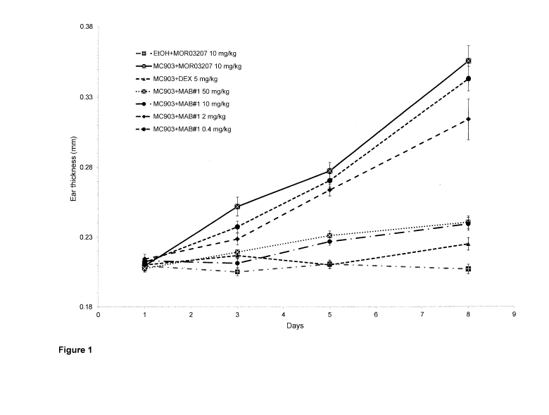
Brief description of the drawings
Figure 1: MAB#1 dose-dependently prevents the ear thickening induced by
topical
application of MC903 on ear skin.
Data are expressed as mean values standard error of the mean (SEM) (n=8 per
group).
Statistical significance versus M0903+M0R03207 was calculated using ANOVA and
Dunnett's multiple comparison test: * p<0.05; ** p<0.01; *** p<0.001. (DEX:
dexamethasone;
EtOR ethanol)
Figure 2: MAB#1 dose-dependently reduces the ear inflammation induced by
topical
application of MC903 on ear skin.
Ear inflammation was assessed at Day 5 using in vivo imaging. Left panel:
quantification of
signal intensity in ears. Individual data points (n=8 per group) represent the
average intensity
3
CA 03014842 2018-08-16
WO 2017/140831 PCT/EP2017/053592
of both ears; data are also shown as mean values (horizontal lines) SEM.
Statistical
significance versus MC903+M0R03207 was calculated using ANOVA and Dunnett's
multiple
comparison test: * p<0.05; ** p<0.01; *** p<0.001. Right panel: representative
images of mouse
ears from animals from different treatment groups were acquired on the Bruker
In-vivo Xtreme
Imager 24h after injection of the Prosense 680 probe. (DEX: dexamethasone;
Et0H: ethanol)
Figure 3: MAB#1 dose-dependently reduces the thickening of epidermal and
dermal
skin layer induced by topical application of MC903 on ear skin.
Data are presented as individual data points (n=8 per group) and mean values
(horizontal
lines) SEM. Statistical significance versus MC903+M0R03207 was calculated
using ANOVA
and Dunnett's multiple comparison test: * p<0.05; ** p<0.01; *** p<0.001. Left
panel: data for
epidermal thickness; Right panel: data for dermal thickness.
Figure 4: MAB#1 dose-dependently inhibits the MC903-mediated increase in TSLP
and
IL-33 expresssion in ear and TARC levels in plasma.
Data are presented as individual data points (n=8 per group) and mean values
(horizontal
lines) SEM. Statistical significance versus MC903+M0R03207 group was
calculated using
ANOVA and Dunnett's multiple comparison test: * p<0.05; ** p<0.01; ***
p<0.001. Top left
panel: data for TSLP protein expresson in ear; Bottom left panel: data for IL-
33 protein
expression in ear; Top right panel: data for TARC protein levels in plasma.
Figure 5: Therapeutic administration of MAB#1 dose-dependently reduces the ear
thickening induced by topical application of MC903 on ear skin.
Data are expressed as mean values SEM (n=10 per group). Statistical
significance versus
MC903+M0R03207 was calculated using ANOVA and Dunnett's multiple comparison
test: *
p<0.05; ** p<0.01; *** p<0.001. DEX: dexamethasone; Et0H: ethanol.
Figure 6: Therapeutic administration of MAB#1 dose-dependently reduces the ear
inflammation induced by topical application of MC903 on ear skin.
Ear inflammation was assessed at Day 12 using in vivo imaging and the signal
intensity in ears
is graphically represented. Individual data points (n=10 per group) represent
the average
intensity of both ears; data are also shown as mean values (horizontal lines)
SEM. Statistical
significance versus MC903+M0R03207 was calculated using ANOVA and Dunnett's
multiple
comparison test: * p<0.05; ** p<0.01; *** p<0.001. DEX: dexamethasone; Et0H:
ethanol.
4
CA 03014842 2018-08-16
WO 2017/140831 PCT/EP2017/053592
Figure 7: Therapeutic administration of MAB#1 dose-dependently reduces the
thickening of epidermal and dermal skin layer induced by topical
administration of MC903 on ear skin.
Data are presented as individual data points (n=10 per group) and mean values
(horizontal
lines) SEM. Statistical significance versus MC903+M0R03207 was calculated
using ANOVA
and Dunnett's multiple comparison test * p<0.05; ** p<0.01; *** p<0.001. Left
panel: data for
epidermal thickness; Right panel: data for dermal thickness. DEX:
dexamethasone; Et0H:
ethanol.
Figure 8: Therapeutic administration of MAB#1 dose-dependently reduces the
dermal
infiltration of eosinophils, T cells and mast cells.
Data are presented as individual data points (n=10 per group) and mean values
(horizontal
lines) SEM. Statistical significance versus MC903+M0R03207 was calculated
using ANOVA
and Dunnett's multiple comparison test: * p<0.05; ** p<0.01; *** p<0.001. Top
left panel: data
for eosinophils; Top right panel: data for mast cells; Bottom left panel: data
for T cells. DEX.
dexamethasone; Et0H: ethanol.
Figure 9: Therapeutic administration of MAB#1 reduces expression of 1L-33, IL-
4 and
S100A9, which were still increased at Day 16 (11 days after stopping MC903
application).
Data are presented as individual data points (n=10 per group) and mean values
(horizontal
lines) SEM. Statistical significance versus MC903+M0R03207 group was
calculated using
ANOVA and Dunnett's multiple comparison test: * p<0.05; ** p<0.01; ***
p<0.001. Top left
panel: data for S100A9 mRNA expression in ear; Bottom left panel: data for IL-
4 mRNA
expression in ear; Top right panel. data for IL-33 protein levels in ear.
Figure 10: MAB#1 reduces macroscopic clinical signs of AD-like inflammation in
the
spontaneous & chronic Flaky Tail model
Clinical scoring of cutaneous inflammation for each mouse was done at start
(week 0) and at
end (week 6) of treatment. Data are the mean SD for each treatment group
(n=8 per group).
Statistical significance versus the isotype antibody treated group was
calculated using ANOVA
and Dunnett's multiple comparison test (* p<0.05; ** p< 0.01; *** p< 0.001)
CA 03014842 2018-08-16
WO 2017/140831 PCT/EP2017/053592
Figure 11: MAB#1 reduces eczematous-like eyelid inflammation in the
spontaneous &
chronic Flaky Tail model.
Skin eyelid inflammation was scored at end of the treatment (week 6). Data are
the mean
SD for each treatment group (n=8 per group). Statistical significance versus
the isotype
antibody treated group was calculated using ANOVA and Dunnett's multiple
comparison test
(* p<0.05; ** p< 0.01 ; *** p< 0.001).
Detailed description of the invention
The disclosure pertains to a number of antibodies or antibody fragments that
recognize
human 1L-17C.
Definitions.
The term "IL-17C" refers to a protein known as interleukin 17C
Human IL-17C has the amino acid sequence of (UniProt Q9P0M4).
MTLLPGLLFLTVVLHTCLAHHDPSLRGHPHSHGTPHCYSAEELPLGQAPPHLLA
RGAKWGQALPVALVSSLEAASHRGRHERPSATTQCPVLRPEEVLEADTHQR
SISPWRYRVDTDEDRYPQKLAFAECLCRGC1DARTGRETAALNSVRLLQSLLV
LRRRPCSRDGSGLPTPGAFAFHTEFIHVPVGCTCVLPRSV (SEQ ID No.: 1)
Mouse IL-17C has the amino acid sequence of (UniProt Q8K4C5).
MSLLLLGWLPTGMTHQDPPSWGKPRSHRTLRCYSAEELSHGQAPPHLLTRS
ARWEQALPVALVASLEATGHRRQHEGPLAGTQCPVLRPEEVLEADTHERSIS
PWRYRIDTDENRYPQKLAVAECLCRGCINAKTGRETAALNSVQLLQSLLVLRR
QPCSRDGTADPTPGSFAFHTEFIRVPVGCTCVLPRSTQ (SEQ ID No.: 2)
Cynomolgus monkey IL-17C has the amino acid sequence of (XP_005592825.1):
MILLPGLLFLTVVLHACLAHQOPFLRGHPHTHGTPRCYSAEELPLGQAPPHLLA
RGAKWGQALPVALVSSLEAAGHRRRHDRPSAATQCPVLRPEEVLEADTHQR
6
CA 03014842 2018-08-16
WO 2017/140831 PCT/EP2017/053592
SISPWRYRVDTDEDRYPQKLAFAECLCRGCIDPRTGRETAALNSVRLLQSLLV
LRRRPCSRDGSGLPTPGAFAFHTEFIRVPVGCTCVLPRSV (SEQ ID No.: 3)
The term "IL17RA" refers to a protein known as interleukin 17 receptor A.
Human
IL17RA has the amino acid sequence of (UniProt Q96F46):
MGAARSPPSAVPGPLLGLLLLLLGVLAPGGASLRLLDHRALVCSQPGLNCTVKNSTC
LDDSWIHPRNLTPSSPKDLQIQLHFAHTQQGDLFPVAHIEVVTLQTDASILYLEGAELS
VLQLNTNERLCVRFEFLSKLRHHHRRWRFTFSHFVVDPDQEYEVTVHHLPKPIPDG
DPNHQSKNFLVPDCEHARMKVTTPCMSSGSLWDPNITVETLEAHQLRVSFTLWNES
THYQILLTSFPHMENHSCFEHMHHIPAPRPEEFHQRSNVILTLRNLKGCCRHQVQ1Q
PFFSSCLNDCLRHSATVSCPEMPDTPEPI PDYMPLWVYWFITGISI LLVGSVILLIVCM
TVVRLAGPGSEKYSDDTKYTDGLPAADLIPPPLKPRKVW1lYSADHPLYVDVVLKFAQ
FLLTACGTEVALDLLEEQAISEAGVMTWVGRQKQEMVESNSKIIVLCSRGTRAKWQ
ALLGRGAPVRLRC DHGKPVGDLFTAAMN MI LPDFKRPACFGTYVVCYFSEVSCDGD
VPDLFGAAPRYPLMDRFEEVYFRIQDLEMFQPGRMHRVGELSGDNYLRSPGGRQL
RAALDRFRDWOVRCPDWFECENLYSADDQDAPSLDEEVFEEPLLPPGTGIVKRAPL
VREPGSQACLAIDPLVGEEGGAAVAKLEPHLQPRGQPAPQPLHTLVLAAEEGALVA
AVEPGPLADGAAVRLALAGEGEACPLLGSPGAGRNSVLFLPVDPEDSPLGSSTPMA
SPDLLPEDVREHLEGLMLSLFEQSLSCQAQGGCSRPAMVLTDPHTPYEEEQRQSV
QSDQGYISRSSPQPPEGLTEMEEEEEEEQDPGKPALPLSPEDLESLRSLQRQLLFR
QLQKNSGWDTMGSESEGPSA (SEQ ID No.: 4)
The term "IL17RE" refers to a protein known as interIeukin 17 receptor E.
Human
IL17RE has the amino acid sequence of (UniProt Q8NFR9):
MGSSRLAALLLPLLLIVIDLSDSAGIGFRHLPHWNTRCPLASHTDDSFTGSSAYIPCRT
VVVVALFSTKPWCVRVVVHCSRCLCQHLLSGGSGLQRGLFHLLVQKSKKSSTFKFYRR
HKMPAPAQRKLLPRRHLSEKSHHISIPSPDISHKGLRSKRTQPSDPETWESLPRLDS
QRHGGPEFSFDLLPEARAI RVTISSGPEVSVRLCHQWALECEELSSPYDVQKIVSGG
HTVELPYEFLLPCLCIEASYLQEDTVRRKKCPFQSWPEAYGSDFWKSVHFTDYSQH
TQMVMALTLRCPLKLEAALCQRHDWHTLCKDLPNATARESDGVVYVLEKVDLHPQL
CFKFSFGNSSHVECPHQTGSLTSWNVSMDTQAQQLILHFSSRMHATFSAAWSLPG
LGQDTLVPPVYTVSQARGSSPVSLDLI I PFLRPGCCVLVVVRSDVQFAWKH LLCPDVS
YRHLGLLILALLALLTLLGVVLALTCRRPQSGPGPARPVLLLHAADSEAQRRLVGALA
ELLRAALGGGRDVIVDLWEGRHVARVGPLPWLWAARTRVAREQGTVLLLWSGADL
RPVSGPDPRAAPLLALLHAAPRPLLLLAYFSRLCAKGDIPPPLRALPRYRLLRDLPRL
7
CA 03014842 2018-08-16
WO 2017/140831 PCT/EP2017/053592
LRALDARPFAEATSWGRLGARQRRQSRLELCSRLEREAARLADLG (SEQ ID No.: 5)
Murine IL17RE has the amino acid sequence of (UniProt Q8BH06):
MGSPRLAALLLSLPLLLIGLAVSARVACPCLRSWTSHCLLAYRVDKRFAGLQWGWF
PLLVRKSKSPPKFEDYWRHRTPASFQRKLLGSPSLSEESHRISIPSSAISHRGQRTK
RAQPSAAEGREHLPEAGSQKCGGPEFSFDLLPEVQAVRVTIPAGPKASVRLCYQW
ALECEDLSSPFDTQKIVSGGHTVDLPYEFLLPCMCIEASYLQEDTVRRKKCPFQSWP
EAYGSDFWQSIRFTDYSQHNQMVMALTLRCPLKLEASLCWRQDPLTPCETLPNATA
QESEGVVYILENVDLHPQLCFKFSFENSSHVECPHQSGSLPSWTVSMDTQAQQLTL
H FSSRTYATFSAAWSDPGLGPDTPMPPVYSI SQTQGSVPVTLDLI I PFLRQENC I LVVV
RSDVHFAWKHVLCPDVSHRHLGLLILALLALTALVG\NLVLLGRRLLPGSGRTRPVLL
LHAADSEAQRRLVGALAELLRTALGGGROVIVDLWEGTHVARIGPLPWLWAARERV
AREQGTVLLLWNCAGPSTACSGDPQAASLRTLLCAAPRPLLLAYFSRLCAKGDI PRP
LRALPRYRLLRDLPRLLRALDAQPATLASSWSHLGAKRCLKNRLEQCHLLELEAAKD
DYQGSTNSPCGFSCL (SEQ ID No.: 6)
The terms "antagonist of IL-17C" and an "IL-17C antagonist", are used
interchangeably
herein and refer to any molecule which inhibits the activity or function of IL-
17C. The term
"IL-170 antagonist" includes, but is not limited to, antibodies or antibody
fragments specifically
binding to IL-17C. Preferably, an IL-17C antagonist in the present disclosure
is an antibody
specific for human IL-17C. Such an antibody may be of any type, such as a
murine, a rat, a
chimeric, a humanized or a human antibody.
The term "antibody" as used herein refers to a protein comprising at least two
heavy
(H) chains and two light (L) chains inter-connected by disulfide bonds which
interacts with an
antigen. Each heavy chain is comprised of a heavy chain variable region
(abbreviated herein
as VH) and a heavy chain constant region. The heavy chain constant region is
comprised of
three domains, CH1, CH2 and CH3. Each light chain is comprised of a light
chain variable
region (abbreviated herein as VL) and a light chain constant region. The light
chain constant
region is comprised of one domain, CL. The VH and VL regions can be further
subdivided into
regions of hypervariability, termed complementarity determining regions (CDR),
interspersed
with regions that are more conserved, termed framework regions (FR). Each VH
and VL is
composed of three CDRs and four FR's arranged from amino-terminus to carboxy-
terminus in
the following order: FR1, CDR1, FR2, CDR2, FR3, CDR3, and FR4. The variable
regions of
the heavy and light chains contain a binding domain that interacts with an
antigen. The
8
CA 03014842 2018-08-16
WO 2017/140831 PCT/EP2017/053592
constant regions of the antibodies may mediate the binding of the
immunoglobulin to host
tissues or factors, including various cells of the immune system (e.g.,
effector cells) and the
first component (Clq) of the classical complement system. The term "antibody"
includes for
example, monoclonal antibodies, human antibodies, humanized antibodies,
camelised
antibodies and chimeric antibodies. The antibodies can be of any isotype
(e.g., IgG, IgE, IgM,
IgD, IgA and IgY), class (e.g., IgG1, IgG2, IgG3, IgG4, lgA1 and IgA2) or
subclass. Both the
light and heavy chains are divided into regions of structural and functional
homology.
The phrase "antibody fragment", as used herein, refers to one or more portions
of an
antibody that retain the ability to specifically interact with (e.g., by
binding, steno hindrance,
stabilizing spatial distribution) an antigen. Examples of binding fragments
include, but are not
limited to, a Fab fragment, a monovalent fragment consisting of the VL, VH, CL
and CH1
domains; a F(ab)2 fragment, a bivalent fragment comprising two Fab fragments
linked by a
disulfide bridge at the hinge region; a Fd fragment consisting of the VH and
CH1 domains; a
Fv fragment consisting of the VL and VH domains of a single arm of an
antibody; a dAb
fragment (Ward et al., (1989) Nature 341:544-546), which consists of a VH
domain; and an
isolated complementarity determining region (CDR). Furthermore, although the
two domains
of the Fv fragment, VL and VH, are coded for by separate genes, they can be
joined, using
recombinant methods, by a synthetic linker that enables them to be made as a
single protein
chain in which the VL and VH regions pair to form monovalent molecules (known
as single
chain Fv (scFv); see e.g., Bird et al., (1988) Science 242:423-426; and Huston
et al., (1988)
Proc. Natl. Acad. Sci. 85:5879-5883). Such single chain antibodies are also
intended to be
encompassed within the term "antibody fragment". These antibody fragments are
obtained
using conventional techniques known to those of skill in the art, and the
fragments are
screened for utility in the same manner as are intact antibodies. Antibody
fragments can also
be incorporated into single domain antibodies, maxibodies, minibodies,
intrabodies, diabodies,
triabodies, tetrabodies, v-NAR and bis-scFv (see, e.g., Hollinger and Hudson,
(2005) Nature
Biotechnology 23:1126-1136). Antibody fragments can be grafted into scaffolds
based on
polypeptides such as Fibronectin type III (Fn3) (see U.S. Pat. No. 6,703,199,
which describes
fibronectin polypeptide monobodies). Antibody fragments can be incorporated
into single chain
molecules comprising a pair of tandem Fv segments (VH-CH1-VH-CH1) which,
together with
complementary light chain polypeptides, form a pair of antigen-binding sites
(Zapata et a/.,
(1995) Protein Eng. 8:1057-1062; and U.S. Pat. No. 5,641,870).
A "human antibody" or "human antibody fragment", as used herein, includes
antibodies
and antibody fragments having variable regions in which both the framework and
CDR regions
are derived from sequences of human origin. Furthermore, if the antibody
contains a constant
9
CA 03014842 2018-08-16
WO 2017/140831 PCT/EP2017/053592
region, the constant region also is derived from such sequences. Human origin
includes, e.g.,
human germline sequences, or mutated versions of human germane sequences or
antibody
containing consensus framework sequences derived from human framework
sequences
analysis, for example, as described in Knappik etal., (2000) J Mol Biol 296:57-
86).
The structures and locations of immunoglobulin variable domains, e.g., CDRs,
may be
defined using well known numbering schemes, e.g., the Kabat numbering scheme,
the Chothia
numbering scheme, or a combination of Kabat and Chothia (see, e.g., Sequences
of Proteins
of Immunological Interest, U.S. Department of Health and Human Services
(1991), eds. Kabat
et al.; Lazikani et a/., (1997) J. Mol. Bio. 273:927-948); Kabat et al.,
(1991) Sequences of
Proteins of Immunological Interest, 5th edit., NIH Publication no. 91-3242
U.S. Department of
Health and Human Services; Chothia et at, (1987) J. Mol. Biol. 196:901-917;
Chothia et a/.,
(1989) Nature 342:877-883; and Al-Lazikani etal., (1997) J. Mol. Biol. 273:927-
948.
A "humanized antibody" or "humanized antibody fragment" is defined herein as
an
antibody molecule which has constant antibody regions derived from sequences
of human
origin and the variable antibody regions or parts thereof or only the CDRs are
derived from
another species. For example a humanized antibody can be CDR-grafted, wherein
the CDRs
of the variable domain are from a non-human origin, while one or more
frameworks of the
variable domain are of human origin and the constant domain (if any) is of
human origin.
The term "chimeric antibody" or "chimeric antibody fragment" is defined herein
as an
antibody molecule which has constant antibody regions derived from, or
corresponding to,
sequences found in one species and variable antibody regions derived from
another species.
Preferably, the constant antibody regions are derived from, or corresponding
to, sequences
found in humans, and the variable antibody regions (e.g. VH , VL , CDR or FR
regions) are
derived from sequences found in a non-human animal, e.g. a mouse, rat, rabbit
or hamster.
The term "isolated" refers to a compound, which can be e.g. an antibody or
antibody
fragment, that is substantially free of other antibodies or antibody fragments
having different
antigenic specificities. Moreover, an isolated antibody or antibody fragment
may be
substantially free of other cellular material and/or chemicals. Thus, in some
aspects, antibodies
provided are isolated antibodies which have been separated from antibodies
with a different
specificity. An isolated antibody may be a monoclonal antibody. An isolated
antibody may be
a recombinant monoclonal antibody. An isolated antibody that specifically
binds to an epitope,
isoform or variant of a target may, however, have cross-reactivity to other
related antigens,
e.g., from other species (e.g., species homologs).
CA 03014842 2018-08-16
WO 2017/140831 PCT/EP2017/053592
The term "recombinant antibody", as used herein, includes all antibodies that
are
prepared, expressed, created or segregated by means not existing in nature.
For example
antibodies isolated from a host cell transformed to express the antibody,
antibodies selected
and isolated from a recombinant, combinatorial human antibody library, and
antibodies
prepared, expressed, created or isolated by any other means that involve
splicing of all or a
portion of a human immunoglobulin gene, sequences to other DNA sequences or
antibodies
isolated from an animal (e.g., a mouse) that is transgenic or transchromosomal
for human
immunoglobulin genes or a hybridoma prepared therefrom. Preferably, such
recombinant
antibodies have variable regions in which the framework and CDR regions are
derived from
human germline immunoglobulin sequences. In certain embodiments, however, such
recombinant human antibodies can be subjected to in vitro mutagenesis (or,
when an animal
transgenic for human 1g sequences is used, in vivo somatic mutagenesis) and
thus the amino
acid sequences of the VH and VL regions of the recombinant antibodies are
sequences that,
while derived from and related to human germline VH and VL sequences, may not
naturally
exist within the human antibody germline repertoire in vivo. A recombinant
antibody may be a
monoclonal antibody. In an embodiment, the antibodies and antibody fragment
disclosed
herein are isolated from the Ylanthia antibody library as disclosed in US
13/321,564 or US
13/299,367, which both herein are incorporated by reference.
The term "monoclonal antibody" as used herein refers to a preparation of
antibody
molecules of single molecular composition. A monoclonal antibody composition
displays a
unique binding site having a unique binding specificity and affinity for
particular epitopes.
As used herein the term "binds specifically to", "specifically binds to", is
"specific to/for"
or "specifically recognizes", or the like, refers to measurable and
reproducible interactions such
as binding between a target and an antibody or antibody fragment, which is
determinative of
the presence of the target in the presence of a heterogeneous population of
molecules
including biological molecules. For example, an antibody or antibody fragment
that specifically
binds to a target (which can be an antigen or an epitope of an antigen) is an
antibody or
antibody fragment that binds this target with greater affinity, avidity, more
readily, and/or with
greater duration than it binds to other targets. In certain embodiments, an
antibody or antibody
fragment specifically binds to an epitope on a protein that is conserved among
the protein from
different species. In another embodiment, specific binding can include, but
does not require
exclusive binding. The antibodies or antibody fragments disclosed herein
specifically bind to
human IL-17C. Preferably, the disclosed antibodies or antibody fragments
specific for human
IL-17C specifically bind to IL-17C of another species, such as IL-170 from
mouse, rat, rhesus
monkey and/or cynomolgus monkey. Even more preferred the antibodies or
antibody
fragments disclosed herein are specific for human IL-17C, cynomolgus monkey IL-
17C and
11
CA 03014842 2018-08-16
WO 2017/140831 PCT/EP2017/053592
mouse IL-17C. Methods for determining whether two molecules specifically bind
are well
known in the art and include, for example, a standard EL1SA assay. The scoring
may be carried
out by standard color development (e.g. secondary antibody with horseradish
peroxide and
tetramethyl benzidine with hydrogen peroxide). The reaction in certain wells
is scored by the
optical density, for example, at 450 nm. Typical background (=negative
reaction) may be 0.1
OD; typical positive reaction may be 1 OD. This means the difference
positive/negative can be
more than 5-fold. Typically, determination of binding specificity is performed
by using not a
single reference antigen, but a set of about three to five unrelated antigens,
such as milk
powder, BSA, transferrin or the like.
The term "avidity" is used to describe the combined strength of multiple bond
interactions between proteins. Avidity is distinct from affinity which
describes the strength of a
single bond. As such, avidity is the combined synergistic strength of bond
affinities (functional
affinity) rather than the sum of bonds. With the antibodies of the present
disclosure, both
antigen-binding sites from the VHNL pairs simultaneously interact with IL-17C
. Whilst each
single binding interaction may be readily broken (depending on the relative
affinity), because
many binding interactions are present at the same time, transient unbinding of
a single site
does not allow the molecule to diffuse away, and binding of that site is
likely to be reinstated.
The overall effect is synergistic, strong binding of antigen to antibody.
As used herein, the term "affinity" refers to the strength of interaction
between the
polypeptide and its target at a single site. Within each site, the binding
region of the polypeptide
interacts through weak non-covalent forces with its target at numerous sites;
the more
interactions, the stronger the affinity.
The term "KD", as used herein, refers to the dissociation constant, which is
obtained
from the ratio of Kd to Ka (i.e. Kd/Ka) and is expressed as a molar
concentration (M). KD values
for antigen binding moieties like e.g. monoclonal antibodies can be determined
using methods
well established in the art. Methods for determining the KD of an antigen
binding moiety like
e.g. a monoclonal antibody are SET (soluble equilibrium titration) or surface
plasmon
resonance using a biosensor system such as a Biacore system. In the present
disclosure an
antibody specific to IL-17C typically has a dissociation rate constant (K0)
(koff/kon) of less than
5x10⁻²M, less than 10-2M, less than 5x10⁻³M, less than 10-3M, less than 5x10-
4M, less than
10-4M, less than 5x10⁻⁵M, less than 10-5M, less than 5x10⁻⁶M, less than 10-6M,
less than
5x10⁻⁷M, less than 10-7M, less than 5x10⁻⁶M, less than 10-8M, less than 5x10-
9M, less than
10-9M, less than 5x10⁻¹⁰m, less than 10-10M, less than 5x10-11M, less than 10-
11M, less than
12
CA 03014842 2018-08-16
WO 2017/140831 PCT/EP2017/053592
5x10-12M, less than 10-12M, less than 5x10-13M, less than 10-13M, less than
5x10-14M, less than
10-14M, less than 5x10-15M, or less than 10-15M or lower.
'Cross competes" means the ability of an antibody, antibody fragment or other
antigen-
binding moieties to interfere with the binding of other antibodies, antibody
fragments or antigen-
binding moieties to a specific antigen in a standard competitive binding
assay. The ability or
extent to which an antibody, antibody fragment or other antigen-binding
moieties is able to
interfere with the binding of another antibody, antibody fragment or antigen-
binding moieties
to a specific antigen, and, therefore whether it can be said to cross-compete
according to the
invention, can be determined using standard competition binding assays. One
suitable assay
involves the use of the Biacore technology (e.g. by using the BlAcore 3000
instrument
(Biacore, Uppsala, Sweden)), which can measure the extent of interactions
using surface
plasmon resonance technology. Another assay for measuring cross-competing uses
an
ELISA-based approach. A high throughput process for "epitope binning"
antibodies based
upon their cross-competition is described in International Patent Application
No.
WO 2003/48731. Cross-competition is present if the antibody or antibody
fragment under
investigation reduces the binding of one of the antibodies described in Table
1 to IL-17C by
60% or more, specifically by 70% or more and more specifically by 80% or more
and if one of
the antibodies described in Table 1 reduces the binding of said antibody or
antibody fragment
to IL-17C by 60% or more, specifically by 70% or more and more specifically by
80% or more.
The term "epitope" includes any proteinacious region which is specifically
recognized by
an antibody or fragment thereof or a T-cell receptor or otherwise interacts
with a molecule.
Generally epitopes are of chemically active surface groupings of molecules
such as amino
acids or carbohydrate or sugar side chains and generally may have specific
three-dimensional
structural characteristics, as well as specific charge characteristics. As
will be appreciated by
one of skill in the art, practically anything to which an antibody can
specifically bind could be
an epitope.
"Binds the same epitope as" means the ability of an antibody, antibody
fragment or other
antigen-binding moiety to bind to a specific antigen and binding to the same
epitope as the
exemplified antibody when using the same epitope mapping technique for
comparing the
antibodies. The epitopes of the exemplified antibody and other antibodies can
be determined
using epitope mapping techniques. Epitope mapping techniques are well known in
the art. For
example, conformational epitopes are readily identified by determining spatial
conformation of
amino acids such as by, e.g., hydrogen/deuterium exchange, x-ray
crystallography and two-
dimensional nuclear magnetic resonance.
13
CA 03014842 2018-08-16
WO 2017/140831 PCT/EP2017/053592
Compositions of the present disclosure may be used for therapeutic or
prophylactic
applications. The present disclosure, therefore, includes a pharmaceutical
composition
containing an antibody (or functional antibody fragment) as disclosed herein
and a
pharmaceutically acceptable carrier or excipient therefor. In a related
aspect, the present
disclosure provides a method for treating an inflammatory disorder. Such
method contains the
steps of administering to a subject in need thereof an effective amount of the
pharmaceutical
composition that contains an antibody (or functional antibody fragment) as
described or
contemplated herein.
The present disclosure provides therapeutic methods comprising the
administration of
a therapeutically effective amount of an IL-170 antibody as disclosed to a
subject in need of
such treatment. A "therapeutically effective amount" or "effective amount", as
used herein,
refers to the amount of an IL-17C antibody necessary to elicit the desired
biological response.
In accordance with the subject invention, the therapeutic effective amount is
the amount of an
IL-17C antibody necessary to treat and/or prevent a disease.
"Subject" or "species", as used in this context refers to any mammal,
including rodents,
such as mouse or rat, and primates, such as cynomolgus monkey (Macaca
fascicularis),
rhesus monkey (Macaca mulatta) or humans (Homo sapiens). Preferably the
subject is a
primate, most preferably a human.
Embodiments:
In one embodiment, the present disclosure refers to an antibody or antibody
fragment specific
for IL-170 wherein said antibody or antibody fragment comprises
(a) a HCDR1 region comprising the amino acid sequence of SEQ ID No.: 7, a
HCDR2
region comprising the amino acid sequence of SEQ ID No.: 8, a HCDR3 region
comprising the amino acid sequence of SEQ ID No.: 9, a LCDR1 region comprising
the amino acid sequence of SEQ ID No.: 13, a LCDR2 region comprising the amino
acid sequence of SEQ ID No.: 14 and a LCDR3 region comprising the amino acid
sequence of SEQ ID No.: 15, or
(b) a HCDR1 region comprising the amino acid sequence of SEQ ID No.: 20, a
HCDR2
region comprising the amino acid sequence of SEQ ID No.: 21, a HCDR3 region
comprising the amino acid sequence of SEQ ID No.: 22, a LCDR1 region
comprising the amino acid sequence of SEQ ID No.: 26, a LCDR2 region
14
CA 03014842 2018-08-16
WO 2017/140831 PCT/EP2017/053592
comprising the amino acid sequence of SEQ ID No.: 27 and a LCDR3 region
comprising the amino acid sequence of SEQ ID No.: 28.
In one embodiment, the present disclosure refers to an antibody or antibody
fragment
specific for 1L-17C, wherein said antibody or antibody fragment comprises
the HCDR1 region of SEQ ID No.: 7, the HCDR2 region of SEQ ID No.: 8, the
HCDR3
region of SEQ ID No.: 9, the LCDR1 region of SEQ ID No.: 13, the LCDR2 region
of SEQ ID
No.: 14 and the LCDR3 region of SEQ ID No.: 15, or
the HCDR1 region of SEQ ID No.: 20, the HCDR2 region of SEQ ID No.: 21, the
HCDR3
region of SEQ ID No.. 22, the LCDR1 region of SEQ ID No.: 26, the LCDR2 region
of SEQ ID
No.: 27 and the LCDR3 region of SEQ ID No.: 28.
In another embodiment, the present disclosure refers to an antibody or
antibody
fragment specific for IL-17C wherein said antibody or antibody fragment
comprises
(a) a HCDR1 region comprising the amino acid sequence of SEQ ID No.: 10, a
HCDR2
region comprising the amino acid sequence of SEQ ID No.: 11, a HCDR3 region
comprising the amino acid sequence of SEQ ID No.: 12, a LCDR1 region
comprising the amino acid sequence of SEQ ID No.: 13, a LCDR2 region
comprising the amino acid sequence of SEQ ID No.: 14 and a LCDR3 region
comprising the amino acid sequence of SEQ ID No.: 15, or
(b) a HCDR1 region comprising the amino acid sequence of SEQ ID No.: 23, a
HCDR2
region comprising the amino acid sequence of SEQ ID No.. 24, a HCDR3 region
comprising the amino acid sequence of SEQ ID No.: 25, a LCDR1 region
comprising the amino acid sequence of SEQ ID No.: 26, a LCDR2 region
comprising the amino acid sequence of SEQ ID No.: 27 and a LCDR3 region
comprising the amino acid sequence of SEQ ID No.: 28.
In a further embodiment, the present disclosure refers to an antibody or
antibody
fragment specific for IL-17C, wherein said antibody or antibody fragment
comprises
the HCDR1 region of SEQ ID No.: 10, the HCDR2 region of SEQ ID No.: 11, the
HCDR3
region of SEQ ID No.: 12, the LCDR1 region of SEQ ID No.: 13, the LCDR2 region
of SEQ ID
No.: 14 and the LCDR3 region of SEQ ID No.: 15, or
the HCDR1 region of SEQ ID No.: 23, the HCDR2 region of SEQ ID No.: 24, the
HCDR3
region of SEQ ID No.: 25, the LCDR1 region of SEQ ID No.: 26, the LCDR2 region
of SEQ ID
No.: 27 and the LCDR3 region of SEQ ID No.: 28.
CA 03014842 2018-08-16
WO 2017/140831 PCT/EP2017/053592
In one embodiment, the present disclosure refers to an antibody or antibody
fragment
specific for 1L-17C wherein said antibody or antibody fragment comprises
(a) a HCDR1 region comprising the amino acid sequence of SEQ ID No.: 7, a
HCDR2
region comprising the amino acid sequence of SEQ ID No.: 8, a HCDR3 region
comprising the amino acid sequence of SEQ ID No,: 9, a LCDR1 region comprising
the amino acid sequence of SEQ ID No.: 13, a LCDR2 region comprising the amino
acid sequence of SEQ ID No.: 14 and a LCDR3 region comprising the amino acid
sequence of SEQ ID No.: 15, or
(b) a HCDR1 region comprising the amino acid sequence of SEQ ID No.: 10, a
HCDR2
region comprising the amino acid sequence of SEQ ID No.: 11, a HCDR3 region
comprising the amino acid sequence of SEQ ID No.: 12, a LCDR1 region
comprising the amino acid sequence of SEQ ID No.: 13, a LCDR2 region
comprising the amino acid sequence of SEQ ID No.: 14 and a LCDR3 region
comprising the amino acid sequence of SEQ ID No.: 15, or
(c) a HCDR1 region comprising the amino acid sequence of SEQ ID No.: 20, a
HCDR2
region comprising the amino acid sequence of SEQ ID No.: 21, a HCDR3 region
comprising the amino acid sequence of SEQ ID No.: 22, a LCDR1 region
comprising the amino acid sequence of SEQ ID No.: 26, a LCDR2 region
comprising the amino acid sequence of SEQ ID No.: 27 and a LCDR3 region
comprising the amino acid sequence of SEQ ID No.: 28.
(d) a HCDR1 region comprising the amino acid sequence of SEQ ID No.: 23, a
HCDR2
region comprising the amino acid sequence of SEQ ID No,: 24, a HCDR3 region
comprising the amino acid sequence of SEQ ID No.: 25, a LCDR1 region
comprising the amino acid sequence of SEQ ID No.. 26, a LCDR2 region
comprising the amino acid sequence of SEQ ID No. 27 and a LCDR3 region
comprising the amino acid sequence of SEQ ID No.: 28.
In one embodiment, the present disclosure refers to an antibody or antibody
fragment
specific for IL-17C, wherein said antibody or antibody fragment comprises
the HCDR1 region of SEQ ID No.: 7, the HCDR2 region of SEQ ID No.: 8, the
HCDR3
region of SEQ ID No.: 9, the LCDR1 region of SEQ ID No.: 13, the LCDR2 region
of SEQ ID
No.. 14 and the LCDR3 region of SEQ ID No.: 15, or
the HCDR1 region of SEQ ID No.: 10, the HCDR2 region of SEQ ID No.: 11, the
HCDR3
region of SEQ ID No.: 12, the LCDR1 region of SEQ ID No.: 13, the LCDR2 region
of SEQ ID
No.. 14 and the LCDR3 region of SEQ ID No.: 15, or
16
CA 03014842 2018-08-16
WO 2017/140831 PCT/EP2017/053592
the HCDR1 region of SEQ ID No.: 20, the HCDR2 region of SEQ ID No.: 21, the
HCDR3
region of SEQ ID No.: 22, the LCDR1 region of SEQ ID No.: 26, the LCDR2 region
of SEQ ID
No.: 27 and the LCDR3 region of SEQ ID No.: 28, or
the HCDR1 region of SEQ ID No.: 23, the HCDR2 region of SEQ ID No.: 24, the
HCDR3
region of SEQ ID No.: 25, the LCDR1 region of SEQ ID No.: 26, the LCDR2 region
of SEQ ID
No.: 27 and the LCDR3 region of SEQ ID No.: 28.
In another embodiment of the present disclosure the antibody or antibody
fragment
specifically binds to human IL-17C.
In another embodiment of the present disclosure the antibody or antibody
fragment is
a monoclonal antibody or antibody fragment.
In another embodiment of the present disclosure the antibody or antibody
fragment is
a human, humanized or chimeric antibody or antibody fragment. In another
embodiment of the
present disclosure the antibody or antibody fragment is of the IgG isotype. In
another
embodiment the antibody or antibody fragment is IgG1.
In one embodiment, the present disclosure refers to an antibody or antibody
fragment
specific for IL-17C, wherein said antibody or antibody fragment comprises
the HCDR1 region of SEQ ID No.: 7, the HCDR2 region of SEQ ID No.: 8, the
HCDR3
region of SEQ ID No.: 9, the LCDR1 region of SEQ ID No.: 13, the LCDR2 region
of SEQ ID
No.: 14 and the LCDR3 region of SEQ ID No.: 15, and further comprises a heavy
chain of SEQ
ID No.: 17 or a light chain of SEQ ID No.: 16, or
the HCDR1 region of SEQ ID No.: 10, the HCDR2 region of SEQ ID No.: 11, the
HCDR3
region of SEQ ID No.: 12, the LCDR1 region of SEQ ID No.: 13, the LCDR2 region
of SEQ ID
No.: 14 and the LCDR3 region of SEQ ID No.: 15 and further comprises a heavy
chain of SEQ
ID No.: 17 or a light chain of SEQ ID No.: 16, or
the HCDR1 region of SEQ ID No.: 20, the HCDR2 region of SEQ ID No.: 21, the
HCDR3
region of SEQ ID No.: 22, the LCDR1 region of SEQ ID No.: 26, the LCDR2 region
of SEQ ID
No.: 27 and the LCDR3 region of SEQ ID No.: 28 and further comprises a heavy
chain of SEQ
ID No.: 30 or a light chain of SEQ ID No.: 29, or
the HCDR1 region of SEQ ID No.: 23, the HCDR2 region of SEQ ID No.: 24, the
HCDR3
region of SEQ ID No.: 25, the LCDR1 region of SEQ ID No 26, the LCDR2 region
of SEQ ID
No.: 27 and the LCDR3 region of SEQ ID No.: 28, and further comprises a heavy
chain of SEQ
ID No.: 30 or a light chain of SEQ ID No.: 29.
17
CA 03014842 2018-08-16
WO 2017/140831 PCT/EP2017/053592
In one embodiment, the present disclosure refers to an antibody or antibody
fragment
specific for IL-17C, wherein said antibody or antibody fragment comprises a
heavy chain of
SEQ ID No.: 17 and a light chain of SEQ ID No.: 16.
In one embodiment, the present disclosure refers to an antibody or antibody
fragment
specific for IL-17C, wherein said antibody or antibody fragment comprises a
heavy chain of
SEQ ID No.: 30 and a light chain of SEQ ID No.: 29.
In a further embodiment, the present disclosure refers to an antibody or
antibody
fragment specific for IL-170, wherein said antibody or antibody fragment
comprises a heavy
chain of SEQ ID No.: 43 and a light chain of SEQ ID No.: 42.
In one embodiment, the present disclosure refers to an antibody or antibody
fragment
specific for IL-17C, wherein said antibody or antibody fragment comprises a
heavy chain of
SEQ ID No.: 56 and a light chain of SEQ ID No.: 55.
In another embodiment of the present disclosure the antibody or antibody
fragment is
an isolated antibody or antibody fragment.
In another embodiment of the present disclosure the antibody or antibody
fragment is
a recombinant antibody or antibody fragment.
In one embodiment, the present disclosure refers to an antibody or antibody
fragment
specific for IL-17C for use in the treatment of a disorder or condition
associated with the
undesired presence of IL-17C.
In one embodiment, the present disclosure refers to a nucleic acid composition
comprising a nucleic acid sequence or a plurality of nucleic acid sequences
encoding an
antibody or antibody fragment specific for IL-17C, wherein said antibody or
antibody fragment
corn prises
the HCDR1 region of SEQ ID No.: 7, the HCDR2 region of SEQ ID No.: 8, the
HCDR3
region of SEQ ID No.: 9, the LCDR1 region of SEQ ID No.: 13, the LCDR2 region
of SEQ ID
No.: 14 and the LCDR3 region of SEQ ID No.: 15, or
the HCDR1 region of SEQ ID No.: 10, the HCDR2 region of SEQ ID No.: 11, the
HCDR3
region of SEQ ID No.: 12, the LCDR1 region of SEQ ID No.: 13, the LCDR2 region
of SEQ ID
No.: 14 and the LCDR3 region of SEQ ID No.: 15, or
18
CA 03014842 2018-08-16
WO 2017/140831 PCT/EP2017/053592
the HCDR1 region of SEQ ID No.: 20, the HCDR2 region of SEQ ID No.: 21, the
HCDR3
region of SEQ ID No.: 22, the LCDR1 region of SEQ ID No.: 26, the LCDR2 region
of SEQ ID
No.: 27 and the LCDR3 region of SEQ ID No.: 28, or
the HCDR1 region of SEQ ID No.: 23, the HCDR2 region of SEQ ID No.: 24, the
HCDR3
region of SEQ ID No.: 25, the LCDR1 region of SEQ ID No.: 26, the LCDR2 region
of SEQ ID
No.: 27 and the LCDR3 region of SEQ ID No.: 28.
In another embodiment, the present disclosure refers to a vector composition
comprising a vector or a plurality of vectors comprising the nucleic acid
sequence or plurality
of nucleic acid sequences encoding an antibody or antibody fragment as
disclosed in Table 1.
In one embodiment, the present disclosure refers to a cell comprising a vector
composition comprising a vector or a plurality of vectors comprising the
nucleic acid sequence
or plurality of nucleic acid sequences encoding an antibody or antibody
fragment as disclosed
in Table 1.
In another embodiment, the present disclosure refers to a pharmaceutical
composition
comprising an antibody or antibody fragment as disclosed in Table 1 and a
pharmaceutically
acceptable carrier or excipient.
In one embodiment, said antibody or antibody fragment specific for IL-17C
blocks the
binding of 1L-17C to the receptor of IL-17C. In a further embodiment, said
antibody or antibody
fragment specific for IL-17C blocks the binding of IL-17C to the receptor of
IL-17C, wherein
said receptor is IL17RE. In another embodiment the present disclosure refers
to an antibody
or antibody fragment specific for 1L-170, wherein said antibody or antibody
fragment blocks
the binding of 1L-17C to IL17RE. In another embodiment said antibody or
antibody fragment
comprises the HCDR1 region of SEQ ID No.: 7, the HCDR2 region of SEQ ID No.:
8, the
HCDR3 region of SEQ ID No.: 9, the LCDR1 region of SEQ ID No.: 13, the LCDR2
region of
SEQ ID No.: 14 and the LCDR3 region of SEQ ID No.: 15, or
the HCDR1 region of SEQ ID No.: 10, the HCDR2 region of SEQ ID No.: 11, the
HCDR3
region of SEQ ID No.: 12, the LCDR1 region of SEQ ID No.: 13, the LCDR2 region
of SEQ ID
No.: 14 and the LCDR3 region of SEQ ID No.: 15, or
the HCDR1 region of SEQ ID No.: 20, the HCDR2 region of SEQ ID No.: 21, the
HCDR3
region of SEQ ID No.: 22, the LCDR1 region of SEQ ID No.: 26, the LCDR2 region
of SEQ ID
No.: 27 and the LCDR3 region of SEQ ID No.: 28, or
19
CA 03014842 2018-08-16
WO 2017/140831 PCT/EP2017/053592
the HCDR1 region of SEQ ID No.: 23, the HCDR2 region of SEQ ID No.: 24, the
HCDR3
region of SEQ ID No.: 25, the LCDR1 region of SEQ ID No.: 26, the LCDR2 region
of SEQ ID
No.: 27 and the LCDR3 region of SEQ ID No.: 28.
In another embodiment said antibody or antibody fragment comprises a heavy
chain of
SEQ ID No.: 17 and a light chain of SEQ ID No.: 16 or a heavy chain of SEQ ID
No.: 30 and a
light chain of SEQ ID No.: 29 or a heavy chain and a light chain that has at
least 60%, at least
70 %, at least 80%, at least 90% or at least 95% identity to the a heavy chain
of SEQ ID No.:
17 or 30 and to the light chain of SEQ ID No.: 16 or 29.
In another embodiment the present disclosure refers to an antibody or antibody
fragment specific for IL-17C wherein said antibody or antibody fragment
bivalently binds to an
IL-170 homodimer and forms a complex consisting of said antibody or antibody
fragment and
one IL-17C homodimer and wherein said antibody or antibody fragment blocks the
binding of
IL-17C to IL17RE.
In certain embodiments, said antibody or antibody fragment specific for IL-17C
blocks
the binding of IL-17C to one or more receptors of IL-17C. In another
embodiment said antibody
or antibody fragment comprises the HCDR1 region of SEQ ID No.: 7, the HCDR2
region of
SEQ ID No.: 8, the HCDR3 region of SEQ ID No.: 9, the LCDR1 region of SEQ ID
No.: 13, the
LCDR2 region of SEQ ID No.: 14 and the LCDR3 region of SEQ ID No.: 15, or
the HCDR1 region of SEQ ID No.: 10, the HCDR2 region of SEQ ID No.: 11, the
HCDR3
region of SEQ ID No.. 12, the LCDR1 region of SEQ ID No.: 13, the LCDR2 region
of SEQ ID
No.: 14 and the LCDR3 region of SEQ ID No.: 15, or
the HCDR1 region of SEQ ID No.: 20, the HCDR2 region of SEQ ID No.: 21 the
HCDR3
region of SEQ ID No.: 22, the LCDR1 region of SEQ ID No.: 26, the LCDR2 region
of SEQ ID
No.: 27 and the LCDR3 region of SEQ ID No.: 28, or
the HCDR1 region of SEQ ID No.: 23, the HCDR2 region of SEQ ID No.: 24, the
HCDR3
region of SEQ ID No.: 25, the LCDR1 region of SEQ ID No.: 26, the LCDR2 region
of SEQ ID
No.: 27 and the LCDR3 region of SEQ ID No.: 28.
In another embodiment said antibody or antibody fragment comprises a heavy
chain of
SEQ ID No.: 17 and a light chain of SEQ ID No.: 16 or a heavy chain of SEQ ID
No.: 30 and a
light chain of SEQ ID No.: 29 or a heavy chain and a light chain that has at
least 60%, at least
70 %, at least 80%, at least 90% or at least 95% identity to the a heavy chain
of SEQ ID No.:
17 or 30 and to the light chain of SEQ ID No.: 16 or 29.
CA 03014842 2018-08-16
WO 2017/140831 PCT/EP2017/053592
In alternative embodiments, said antibody or antibody fragment specific for
the receptor
of IL-17C blocks the binding of IL-17C to receptors of IL-17C, wherein the
receptors of IL-17C
include IL17RE and IL17RA. In alternative embodiments, said antibody or
antibody fragment
specific for the receptor of 1L-170 blocks the binding of IL-17C to IL17RE and
IL17RA. In
certain embodiments, said antibody or antibody fragment specific for IL-17C
blocks the binding
of IL-17C to IL17RE with an IC50 concentration of less than 100nM. 90nM, 80nM,
70nM, 60nM,
50nM, 40nM, 30nM, 20nM, 10nM, 9nM, 8nM, 7nM, 6nM, 5nM, 4nM, 3nM, 2nM, 1nM,
100pM,
90pM, 80pM, 70pM, 60pM, 50pM, 40pM, 30pM, 20pM, 10pM, 9pM, 8pM, 7pM, 6pM, 5pM,
4pM, 3pM, 2pM or 1pM. In certain aspects the IC50 concentration can be
determined by ELISA;
SET, FACS or MSD (Meso Scale Discovery). In another aspect the IC50
concentration can be
determined by the method as described herein in Example 3. In another
embodiment said
antibody or antibody fragment comprises a heavy chain of SEQ ID No.: 17 and a
light chain of
SEQ ID No.: 16 or a heavy chain of SEQ ID No.: 30 and a light chain of SEQ ID
No.: 29 or a
heavy chain and a light chain that has at least 60%, at least 70 %, at least
80%, at least 90%
or at least 95% identity to the a heavy chain of SEQ ID No.: 17 or 30 and to
the light chain of
SEQ ID No.: 16 or 29.
In one embodiment the disclosed antibody or antibody fragment is specific for
human
IL-17C. In a further embodiment the disclosed antibody or antibody fragment
specific for IL-17C
is cross-reactive with IL-17C of another species, such as IL-17C from mouse,
rat, rhesus
monkey and/or cynomolgus monkey. In another embodiment the antibody or
antibody
fragment is specific for human IL-17C, cynomolgus monkey IL-17C and mouse IL-
17C. In a
further embodiment the antibody or antibody fragment is specific for human IL-
17C,
cynomolgus monkey IL-17C and mouse IL-17C. In another embodiment the antibody
or
antibody fragment is specific for human IL-17C, cynomolgus monkey IL-17C and
mouse
IL-17C, wherein said antibody or antibody fragment comprises
the HCDR1 region of SEQ ID No.: 7, the HCDR2 region of SEQ ID No.: 8, the
HCDR3
region of SEQ ID No.: 9, the LCDR1 region of SEQ ID No.: 13, the LCDR2 region
of SEQ ID
No.: 14 and the LCDR3 region of SEQ ID No.: 15, or
the HCDR1 region of SEQ ID No.: 10, the HCDR2 region of SEQ ID No.: 11, the
HCDR3
region of SEQ ID No.: 12, the LCDR1 region of SEQ ID No.: 13, the LCDR2 region
of SEQ ID
No.: 14 and the LCDR3 region of SEQ ID No.: 15, or
the HCDR1 region of SEQ ID No.: 20, the HCDR2 region of SEQ ID No.: 21, the
HCDR3
region of SEQ ID No.: 22, the LCDR1 region of SEQ ID No.: 26, the LCDR2 region
of SEQ ID
No.: 27 and the LCDR3 region of SEQ ID No.: 28, or
the HCDR1 region of SEQ ID No.: 23, the HCDR2 region of SEQ ID No.: 24, the
HCDR3
region of SEQ ID No.: 25, the LCDR1 region of SEQ ID No.: 26, the LCDR2 region
of SEQ ID
21
CA 03014842 2018-08-16
WO 2017/140831 PCT/EP2017/053592
No.: 27 and the LCDR3 region of SEQ ID No.: 28. In another embodiment said
antibody or
antibody fragment comprises a heavy chain of SEQ ID No.: 17 and a light chain
of SEQ ID
No.: 16 or a heavy chain of SEQ ID No.: 30 and a light chain of SEQ ID No.: 29
or a heavy
chain and a light chain that has at least 60%, at least 70 /0, at least 80%,
at least 90% or at
least 95% identity to the a heavy chain of SEQ ID No.: 17 or 30 and to the
light chain of SEQ
ID No.: 16 or 29.
In yet another embodiment the disclosed antibody or antibody fragment
specifically
binds to human IL-17C, cynomolgus monkey IL-17C and mouse IL-17C and blocks
the binding
of human IL-17C, cynomolgus monkey IL-17C and mouse IL-17C to its specific
receptor
IL17RE with an IC50 concentration of less than 100nM, 90nM, 80nM, 70nM, 60nM,
50nM,
40nM, 30nM, 20nM, 10nM, 9nM, 8nM, 7nM, 6nM, 5nM, 4nM, 3nM, 2nM, 1nM, 100pM,
90pM,
80pM, 70pM, 60pM, 50pM, 40pM, 30pM, 20pM, 10pM, 9pM, 8pM, 7pM, 6pM, 5pM, 4pM,
3pM,
2pM or 1pM. In another aspect said antibody is in IgG1 format. In another
embodiment said
IC50 concentration is determined in a Receptor Inhibition Assay as described
herein in
Example 3.
In yet another embodiment the disclosed antibody or antibody fragment
specifically
binds to human IL-17C and blocks the binding of human IL-170 to human IL17RE
with an IC50
concentration of less than 100nM, 90nM, 80nM, 70nM, 60nM, 50nM, 40nM. 30nM,
20nM,
10nM, 9nM, 8nM, 7nM, 6nM, 5nM, 4nM, 3nM, 2nM, 1nM, 100pM, 90pM, 80pM, 70pM,
60pM,
50pM, 40pM, 30pM, 20pM, 10pM, 9pM, 8pM, 7pM, 6pM, 5pM, 4pM, 3pM, 2pM or 1pM.
In
another aspect said antibody is in IgG1 format. In another embodiments said
IC50 concentration
is determined in a Receptor Inhibition Assay as described herein in Example 3.
In yet another embodiment the disclosed antibody or antibody fragment
specifically
binds to cynomolgus monkey IL-17C and blocks the binding of cynomolgus monkey
IL-17C to
cynomolgus monkey IL17RE with an IC50 concentration of less than 100nM, 90nM,
80nM,
70nM, 60nM, 50nM, 40nM, 30nM, 20nM, 10nM, 9nM, 8nM, 7nM, 6nM, 5nM, 4nM, 3nM,
2nM,
1nM, 100pM, 90pM, 80pM, 70pM, 60pM, 50pM, 40pM, 30pM, 20pM, 10pM, 9pM, 8pM,
7pM,
6pM, 5pM, 4pM, 3pM, 2pM or 1pM. In another aspect said antibody is in IgG1
format. In
another embodiments said IC50 concentration is determined in a Receptor
Inhibition Assay as
described herein in Example 3.
In yet another embodiment the disclosed antibody or antibody fragment
specifically
binds to human IL-17C, cynomolgus monkey IL-17C and mouse IL-17C, and blocks
the binding
of human IL-17C, cynomolgus monkey IL-17C and mouse IL-17C, to human IL17RE,
22
CA 03014842 2018-08-16
WO 2017/140831 PCT/EP2017/053592
cynomolgus monkey IL17RE and mouse IL17RE, respectively, each with an IC50
concentration
of less than 100nM, 90nM, 80nM, 70nM, 60nM, 50nM, 40nM, 30nM, 20nM, 10nM, 9nM,
8nM,
7nM, 6nM, 5nM, 4nM, 3nM, 2nM, 1nM, 100pM, 90pM, 80pM, 70pM, 60pM, 50pM, 40pM,
30pM, 20pM, 10pM, 9pM, 8pM, 7pM, 6pM, 5pM, 4pM, 3pM, 2pM or 1pM. In a
preferred
embodiment said antibody or antibody fragment comprises
the HCDR1 region of SEQ ID No.: 7, the HCDR2 region of SEQ ID No.: 8, the
HCDR3
region of SEQ ID No.: 9, the LCDR1 region of SEQ ID No.: 13, the LCDR2 region
of SEQ ID
No.: 14 and the LCDR3 region of SEQ ID No.: 15, or
the HCDR1 region of SEQ ID No.: 10, the HCDR2 region of SEQ ID No.: 11, the
HCDR3
region of SEQ ID No.: 12, the LCDR1 region of SEQ ID No.: 13, the LCDR2 region
of SEQ ID
No.: 14 and the LCDR3 region of SEQ ID No.: 15, or
the HCDR1 region of SEQ ID No.: 20, the HCDR2 region of SEQ ID No.: 21, the
HCDR3
region of SEQ ID No.: 22, the LCDR1 region of SEQ ID No.: 26, the LCDR2 region
of SEQ ID
No.: 27 and the LCDR3 region of SEQ ID No.: 28, or
the HCDR1 region of SEQ ID No.: 23, the HCDR2 region of SEQ ID No.: 24, the
HCDR3
region of SEQ ID No.: 25, the LCDR1 region of SEQ ID No.: 26, the LCDR2 region
of SEQ ID
No.: 27 and the LCDR3 region of SEQ ID No.: 28. In another embodiment said
antibody or
antibody fragment comprises a heavy chain of SEQ ID No.: 17 and a light chain
of SEQ ID
No.: 16 or a heavy chain of SEQ ID No.: 30 and a light chain of SEQ ID No.. 29
or a heavy
chain and a light chain that has at least 60%, at least 70 %, at least 80%, at
least 90% or at
least 95% identity to the heavy chain of SEQ ID No.: 17 or 30 and to the light
chain of SEQ ID
No.: 16 or 29. In another aspect said antibody is in IgG1 format In another
embodiments said
IC50 concentration is determined in a Receptor Inhibition Assay as described
herein in
Example 3.
In yet another embodiment the disclosed antibody or antibody fragment inhibits
human
IL-17C, cynomolgus monkey IL-17C and mouse IL-17C driven activation of a NF-
k13 reporter
gene in NIH3T3 cells with an IC50 concentration of less than 100pM, 90pM,
80pM, 70pM, 60pM,
50pM, 40pM, 30pM, 20pM, 10pM, 9pM, 8pM, 7pM, 6pM, 5pM, 4pM, 3pM, 2pM or 1pM.
In a
preferred embodiment said antibody or antibody fragment comprises
the HCDR1 region of SEQ ID No.: 7, the HCDR2 region of SEQ ID No.: 8, the
HCDR3
region of SEQ ID No.: 9, the LCDR1 region of SEQ ID No.: 13, the LCDR2 region
of SEQ ID
No.: 14 and the LCDR3 region of SEQ ID No.: 15, or
the HCDR1 region of SEQ ID No.: 10, the HCDR2 region of SEQ ID No.: 11, the
HCDR3
region of SEQ ID No.: 12, the LCDR1 region of SEQ ID No.: 13, the LCDR2 region
of SEQ ID
No.: 14 and the LCDR3 region of SEQ ID No.: 15, or
23
CA 03014842 2018-08-16
WO 2017/140831 PCT/EP2017/053592
the HCDR1 region of SEQ ID No.: 20, the HCDR2 region of SEQ ID No.: 21, the
HCDR3
region of SEQ ID No.: 22, the LCDR1 region of SEQ ID No.: 26, the LCDR2 region
of SEQ ID
No.: 27 and the LCDR3 region of SEQ ID No.: 28, or
the HCDR1 region of SEQ ID No.: 23, the HCDR2 region of SEQ ID No.: 24, the
HCDR3
region of SEQ ID No.: 25, the LCDR1 region of SEQ ID No.: 26, the LCDR2 region
of SEQ ID
No.: 27 and the LCDR3 region of SEQ ID No.: 28. In another embodiment said
antibody or
antibody fragment comprises a heavy chain of SEQ ID No.: 17 and a light chain
of SEQ ID
No.: 16 or a heavy chain of SEQ ID No.. 30 and a light chain of SEQ ID No.: 29
or a heavy
chain and a light chain that has at least 60%, at least 70 %, at least 80%, at
least 90% or at
least 95% identity to the heavy chain of SEQ ID No.: 17 or 30 and to the light
chain of SEQ ID
No.: 16 or 29. In another aspect said antibody is in 1gG1 format. In another
embodiments said
IC50 concentration is determined in a IL-17C-driven NE-KB reporter assay as
described herein
in Example 4.
In one embodiment the disclosed antibody or antibody fragment is specific for
human
1L-17C encoded by the amino acid sequence of SEQ ID No.: 1. In one embodiment
the
disclosed antibody or antibody fragment is specific for a polypeptide
comprising the amino acid
sequence of SEQ ID No.: 1. In a further embodiment said monoclonal antibody or
antibody
fragment is a monoclonal antibody specific for a polypeptide consisting of the
amino acid
sequence of SEQ ID No.: 1. In another embodiment the disclosed antibody or
antibody
fragment is specific for human IL-17C encoded by the amino acid sequence of
SEQ ID No.: 1
and is a monoclonal antibody or antibody fragment.
In one embodiment the disclosed antibody or antibody fragment specific for IL-
17C is
a monoclonal antibody or antibody fragment.
In one embodiment the disclosed antibody or antibody fragment specific for IL-
17C is
a human, humanized or chimeric antibody. In certain embodiments, said antibody
or antibody
fragment specific for IL-17C is an isolated antibody or antibody fragment. In
another
embodiment said antibody or antibody fragment is a recombinant antibody or
antibody
fragment. In a further embodiment said antibody or antibody fragment is a
recombinant human
antibody or antibody fragment. In a further embodiment said recombinant human
antibody or
antibody fragment is an isolated recombinant human antibody or antibody
fragment. In a further
embodiment said recombinant human antibody or antibody fragment or isolated
recombinant
human antibody or antibody fragment is monoclonal.
24
CA 03014842 2018-08-16
WO 2017/140831 PCT/EP2017/053592
In another embodiment the disclosed antibody or antibody fragment comprises a
heavy
chain of SEQ ID No.: 17 and a light chain of SEQ ID No.: 16 or a heavy chain
of SEQ ID No.:
30 and a light chain of SEQ ID No.: 29 or a heavy chain and a light chain that
has at least 60%,
at least 70 /0, at least 80%, at least 90% or at least 95% identity to the a
heavy chain of SEQ
ID No.: 17 or 30 and to the light chain of SEQ ID No.: 16 or 29.
In one embodiment the disclosed antibody or antibody fragment comprises a
human
heavy chain constant region and a human light chain constant region. In a
further embodiment
said human heavy chain constant region comprises the amino acid sequences of
SEQ ID No.:
17 and the human light chain constant region comprises the amino acid
sequences of SEQ ID
No.: 16 or said human heavy chain constant region comprises the amino acid
sequences of
SEQ ID No.: 30 and the human light chain constant region comprises the amino
acid
sequences of SEQ ID No.: 29.
In one embodiment the disclosed antibody or antibody fragment is of the IgG
isotype.
In another embodiment said antibody is IgG1.
In one embodiment said antibody fragment is a bivalent antibody fragment.
In another embodiment, the present disclosure refers to an antibody or
antibody
fragment that cross-competes with an antibody described in Table 1. In one
embodiment the
present disclosure refers to an antibody or antibody fragment, wherein said
antibody or
antibody fragment cross-competes with an antibody or antibody fragment
comprising 6 CDRs
defined by Kabat of one of the antibodies in Table 1. In another embodiment
the present
disclosure refers to an antibody or antibody fragment specific for human IL-
17C wherein said
antibody or antibody fragment bivalently binds to an IL-17C homodimer and
forms a complex
consisting of said antibody or antibody fragment and one IL-17C homodimer and
wherein said
antibody or antibody fragment cross-competes with an antibody or antibody
fragment
comprising 6 CDRs defined by Kabat of one of the antibodies in Table 1.
In another embodiment the present disclosure refers to an antibody or antibody
fragment, wherein said antibody or antibody fragment cross-competes with an
antibody or
antibody fragment comprising 6 CDRs, wherein the HCDR1 is the amino acid
sequence of
SEQ ID No.. 7, the HCDR2 is the amino acid sequence of SEQ ID No.: 8, the
HCDR3 is the
amino acid sequence of SEQ ID No.: 9, the LCDR1 is the amino acid sequence of
SEQ ID No.:
13, the LCDR2 is the amino acid sequence of SEQ ID No.: 14 and the LCDR3 is
the amino
acid sequence of SEQ ID No.: 15. In another embodiment the present disclosure
refers to an
CA 03014842 2018-08-16
WO 2017/140831 PCT/EP2017/053592
antibody or antibody fragment, wherein said antibody or antibody fragment
cross-competes
with an antibody or antibody fragment comprising the VH according to SEQ ID
No.: 17 and the
VL according to SEQ ID No.: 16.
In another embodiment the present disclosure refers to an antibody or antibody
fragment, wherein said antibody or antibody fragment cross-competes with an
antibody or
antibody fragment comprising 6 CDRs, wherein the HCDR1 is the amino acid
sequence of
SEQ ID No.: 20, the HCDR2 is the amino acid sequence of SEQ ID No.: 21, the
HCDR3 is the
amino acid sequence of SEQ ID No.: 22, the LCDR1 is the amino acid sequence of
SEQ ID
No.: 26, the LCDR2 is the amino acid sequence of SEQ ID No.: 27 and the LCDR3
is the
amino acid sequence of SEQ ID No.: 28. In another embodiment the present
disclosure refers
to an antibody or antibody fragment, wherein said antibody or antibody
fragment cross-
competes with an antibody or antibody fragment comprising the VH according to
SEQ ID No.:
30 and the VL according to SEQ ID No.: 29.
In another embodiment, the present disclosure refers to an antibody or
antibody
fragment that cross-competes with an antibody described in Table 1. In one
embodiment the
present disclosure refers to an antibody or antibody fragment, wherein said
antibody or
antibody fragment cross-competes with an antibody or antibody fragment
comprising 6 CDRs
defined by Chothia of one of the antibodies in Table 1. In another embodiment
the present
disclosure refers to an antibody or antibody fragment specific for human IL-
170 wherein said
antibody or antibody fragment bivalently binds to an IL-17C homodimer and
forms a complex
consisting of said antibody or antibody fragment and one IL-17C homodimer and
wherein said
antibody or antibody fragment cross-competes with an antibody or antibody
fragment
comprising 6 CDRs defined by Chothia of one of the antibodies in Table 1.
In another embodiment the present disclosure refers to an antibody or antibody
fragment, wherein said antibody or antibody fragment cross-competes with an
antibody or
antibody fragment comprising 6 CDRs, wherein the HCDR1 is the amino acid
sequence of
SEQ ID No.: 10, the HCDR2 is the amino acid sequence of SEQ ID No.: 11, the
HCDR3 is the
amino acid sequence of SEQ ID No.: 12, the LCDR1 is the amino acid sequence of
SEQ ID
No.: 13, the LCDR2 is the amino acid sequence of SEQ ID No.: 14 and the LCDR3
is the
amino acid sequence of SEQ ID No.: 15. In another embodiment the present
disclosure refers
to an antibody or antibody fragment, wherein said antibody or antibody
fragment cross-
competes with an antibody or antibody fragment comprising the VH according to
SEQ ID No.:
17 and the VL according to SEQ ID No.: 18.
26
CA 03014842 2018-08-16
WO 2017/140831 PCT/EP2017/053592
In another embodiment the present disclosure refers to an antibody or antibody
fragment, wherein said antibody or antibody fragment cross-competes with an
antibody or
antibody fragment comprising 6 CDRs, wherein the HCDR1 is the amino acid
sequence of
SEQ ID No.: 23, the HCDR2 is the amino acid sequence of SEQ ID No.: 24, the
HCDR3 is the
amino acid sequence of SEQ ID No.: 25, the LCDR1 is the amino acid sequence of
SEQ ID
No.: 26, the LCDR2 is the amino acid sequence of SEQ ID No.: 27 and the LCDR3
is the
amino acid sequence of SEQ ID No.: 28. In another embodiment the present
disclosure refers
to an antibody or antibody fragment, wherein said antibody or antibody
fragment cross-
competes with an antibody or antibody fragment comprising the VH according to
SEQ ID No.:
30 and the VL according to SEQ ID No.: 29.
In a certain embodiment, the disclosure refers to an antibody or antibody
fragment that
cross-competes with an antibody described in Table 1 and reduces the specific
binding of one
of the antibodies described in Table 1 by at least 70%, 80% or 90% in an ELISA-
based cross-
competition assay. In a certain embodiment, the present disclosure refers to
an monoclonal
antibody or antibody fragment that cross-competes with an antibody described
in Table 1 and
reduces the specific binding of one of the antibodies described in Table Ito
IL-170 by at least
70%, 80% or 90% in an ELISA-based cross-competition assay. A representative
assay set-up
is illustrated in Example 6 in the present disclosure.
In another embodiment, the present disclosure refers to an antibody or
antibody
fragment that binds to (e.g., by binding, stabilizing, spatial distribution)
the same epitope as
one of the antibodies in Table 1. In a further embodiment said antibody or
antibody fragment
binds to (e.g., by binding, stabilizing, spatial distribution) the same
epitope as an antibody or
antibody fragment comprising 6 CDRs defined by Kabat of one of the antibodies
in Table 1. In
yet another embodiment said antibody or antibody fragment binds to (e.g., by
binding,
stabilizing, spatial distribution) the same epitope of IL-17C as an antibody
or antibody fragment
comprising 6 CDRs defined by Kabat of one of the antibodies in Table 1. In yet
another
embodiment said antibody or antibody fragment binds to (e.g., by binding,
stabilizing, spatial
distribution) the same epitope of polypeptide comprising the amino acid
sequence of SEQ ID
No.: 1 as an antibody or antibody fragment comprising 6 CDRs defined by Kabat
of one of the
antibodies in Table 1.
In another embodiment the present disclosure refers to an antibody or antibody
fragment, wherein said antibody or antibody fragment binds to the same epitope
as an antibody
or antibody fragment comprising 6 CDRs, wherein the HCDR1 is the amino acid
sequence of
SEQ ID No.: 10, the HCDR2 is the amino acid sequence of SEQ ID No.: 11, the
HCDR3 is the
27
CA 03014842 2018-08-16
WO 2017/140831 PCT/EP2017/053592
amino acid sequence of SEQ ID No.: 12, the LCDR1 is the amino acid sequence of
SEQ ID
No.: 13, the LCDR2 is the amino acid sequence of SEQ ID No.: 14 and the LCDR3
is the
amino acid sequence of SEQ ID No.: 15. In another embodiment the present
disclosure refers
to an antibody or antibody fragment, wherein said antibody or antibody
fragment binds to the
same epitope as an antibody or antibody fragment comprising the VH according
to SEQ ID
No.: 17 and the VL according to SEQ ID No.: 18.
In another embodiment the present disclosure refers to an antibody or antibody
fragment, wherein said antibody or antibody fragment binds to the same epitope
as an antibody
or antibody fragment comprising 6 CDRs, wherein the HCDR1 is the amino acid
sequence of
SEQ ID No.: 23, the HCDR2 is the amino acid sequence of SEQ ID No.: 24, the
HCDR3 is the
amino acid sequence of SEQ ID No.: 25, the LCDR1 is the amino acid sequence of
SEQ ID
No.: 26, the LCDR2 is the amino acid sequence of SEQ ID No.: 27 and the LCDR3
is the
amino acid sequence of SEQ ID No.: 28. In another embodiment the present
disclosure refers
to an antibody or antibody fragment, wherein said antibody or antibody
fragment binds to the
same epitope as an antibody or antibody fragment comprising the VH according
to SEQ ID
No.: 30 and the VL according to SEQ ID No.: 29.
Regions of a given polypeptide that include an epitope can be identified using
any
number of epitope mapping techniques, well known in the art See, e.g., Epitope
Mapping
Protocols in Methods in Molecular Biology, Vol. 66 (Glenn E.Morris, Ed., 1996)
Humana Press,
Totowa, New Jersey. For example, epitopes may be determined by e.g.,
concurrently
synthesizing large numbers of peptides on solid supports, the peptides
corresponding to
portions of the protein molecule, and reacting the peptides with antibodies
while the peptides
are still attached to the supports. Such techniques are known in the art and
described in, e.g.,
U.S. Patent No. 4,708,871 ; Geysen et al., (1984) Proc. Natl. Acad. Sci. USA
813998-4002;
Geysen et al., (1985) Proc. Natl. Acad. Sci. USA 82:78-182; Geysen et al.,
(1986) Mol.
lmmunol. 23:709-715. Similarly, epitopes are readily identified by determining
spatial
conformation of amino acids such as by, e.g., hydrogen/deuterium exchange, x-
ray
crystallography and two-dimensional nuclear magnetic resonance. See, e.g.,
Epitope Mapping
Protocols, supra. Antigenic regions of proteins can also be identified using
standard
antigenicity and hydropathy plots, such as those calculated using, e.g., the
Omiga version 1.0
software program available from the Oxford Molecular Group. This computer
program employs
the Hopp/Woods method, Hopp et al., (1981) Proc. Natl. Acad. Sci USA 78:3824-
3828; for
determining antigenicity profiles, and the Kyte-Doolittle technique, Kyte et
at, (1982) J.Mol.
Biol. 157:105-132; for hydropathy plots.
28
CA 03014842 2018-08-16
WO 2017/140831 PCT/EP2017/053592
In one embodiment, the present disclosure refers to an antibody or antibody
fragment
comprising 6 CDRs defined by Kabat of any of the antibodies in Table 1. In
another aspect,
the disclosure pertains to an isolated monoclonal antibody or fragment thereof
comprising 6
CDRs defined by Kabat of each of the antibodies in Table 1.
In another embodiment, the present disclosure refers to an antibody or
antibody
fragment comprising 6 CDRs defined by Chotia of any of the antibodies in Table
1. In another
aspect, the disclosure pertains to an isolated monoclonal antibody or fragment
thereof
comprising 6 CDRs defined by Chotia of each of the antibodies in Table 1.
In certain embodiments, the present disclosure refers to the antibodies or
antibody
fragments disclosed in in Table 1, wherein said antibodies or antibody
fragments can bind to
IL-17C with an affinity of about less than 100 nM, more preferably less than
about 60 nM, and
still more preferably less than about 30 nM. Further preferred are antibodies
or antibody
fragments that bind to IL-17C with an affinity of less than about 10 nM, and
more preferably
less than about 3 nM.
In certain embodiments, the present disclosure refers to the antibodies or
antibody
fragments disclosed in Table 1, wherein said antibodies or antibody fragments
can bind to
IL-17C with a monovalent affinity of about less than 100 nM, more preferably
less than about
60 nM, and still more preferably less than about 30 nM. Further preferred are
antibodies or
antibody fragments that bind to 1L-17C with a monovalent affinity of less than
about 10 nM,
and more preferably less than about 3 nM.
In another embodiment, the present disclosure refers to antibodies or antibody
fragments specific for IL-17C, wherein said antibodies or antibody fragments
have a
monovalent affinity to IL-170 with a dissociation rate constant (KO of less
than 5x10⁻²M, less
than 10-2M, less than 5x10⁻³M, less than 10-3M, less than 5x10⁻⁴M, less than
10-4M, less than
5x10⁻⁵M, less than 10-5M, less than 5x10⁻⁶M, less than 10-6M, less than 5x10-
7M, less than
10-7M, less than 5x10⁻⁸M, less than 10-8M, less than 5x10⁻⁹M, less than 10-9M,
less than
5x10⁻¹⁰M, less than 10-10M, less than 5x10-11M, less than 10-11M, less than
5x10-12M, less than
10-12M, less than 5x10-13M, less than 10-13M, less than 5x10⁴M, less than 10-
14M, less than
5x10-15M, or less than 10-15M and wherein said antibodies or antibody
fragments in a bivalent
format have an affinity to IL-17C with a dissociation rate constant (KO which
is at least 2-fold,
5-fold, 10-fold, 100-fold, 1000-fold, 10000-fold, 100000-fold lower than the
dissociation rate
constant (KD) in a monovalent format. In a further embodiment the bivalent
affinity of said
29
CA 03014842 2018-08-16
WO 2017/140831 PCT/EP2017/053592
antibodies or antibody fragments is determined in IgG-format, wherein the
monovalent affinity
of said antibodies or antibody fragments is determined in Fab-format.
The compositions of the present disclosure are preferably pharmaceutical
compositions
comprising an antibody or antibody fragment specific for IL-17C as disclosed
herein and a
pharmaceutically acceptable carrier, diluent or excipient, for the treatment
of an inflammatory
disorder or cancer. Such carriers, diluents and excipients are well known in
the art, and the
skilled artisan will find a formulation and a route of administration best
suited to treat a subject
with the IL-17C antibodies or antibody fragments of the present disclosure.
In another embodiment the present disclosure refers to pharmaceutical
compositions
comprising an antibody or antibody fragment specific for IL-17C as disclosed
herein for the
use in the treatment of a disorder or condition associated with the undesired
presence of
IL-17C. In another embodiment said condition associated with the undesired
presence of
IL-170 is an inflammatory disorder or cancer. In another embodiment said
inflammatory
disorder is rheumatoid arthritis, psoriasis, pulmonary inflammation, COPD
and/or atopic
dermatitis (AD), including moderate-to-severe AD. In a preferred embodiment
said
inflammatory disorder is atopic dermatitis (AD) and/or moderate-to-severe AD.
In another embodiment the present disclosure refers to the use of said
pharmaceutical
compositions comprising an antibody or antibody fragment specific for IL-17C
as disclosed
herein in the preparation of a medicament for the treatment of a disorder or
condition
associated with the undesired presence of IL-17C. In another embodiment said
condition
associated with the undesired presence of IL-17C is an inflammatory disorder
or cancer. In
another embodiment said inflammatory disorder is rheumatoid arthritis,
psoriasis, pulmonary
inflammation, COPD and/or atopic dermatitis (AD), including moderate-to-severe
AD. In a
preferred embodiment said inflammatory disorder is atopic dermatitis (AD)
and/or moderate-
to-severe AD.
In another embodiment the present disclosure refers to the use of said
pharmaceutical
composition for the treatment of a disorder or condition associated with the
undesired presence
of IL-170. In another embodiment said condition associated with the undesired
presence of
IL-170 is an inflammatory disorder or cancer. In another embodiment said
inflammatory
disorder is rheumatoid arthritis, psoriasis, pulmonary inflammation, COPD
and/or atopic
dermatitis (AD), including moderate-to-severe AD. In a preferred embodiment
said
inflammatory disorder is atopic dermatitis (AD) and/or moderate-to-severe AD.
CA 03014842 2018-08-16
WO 2017/140831 PCT/EP2017/053592
In another aspect, provided herein is a method of treating atopic dermatitis
(AD) and/or
moderate-to-severe atopic dermatitis (AD) in a subject, the method comprising
administering
a pharmaceutical composition comprising a therapeutically effective amount of
the IL-17C
antibodies or antibody fragments of the present disclosure. In one embodiment
said subject is
resistant, non-responsive or inadequately responsive to treatment by either a
topical
corticosteroid (TCS) or a calcineurin inhibitor. In another embodiment the
subject is a subject
in need thereof. In a preferred embodiment said subject is a human. In
alternative aspects said
subject is a rodent, such as a rat or a mouse.
In another embodiment, the present disclosure refers to a method for the
prophylaxis
of an inflammatory disorder in a subject, said method comprising administering
an IL-17C
antagonist to said subject. "Prophylaxis" as used in this context refers to
methods which aim
to prevent the onset of a disease or which delay the onset of a disease. In
some embodiments
said subject is a human. In alternative aspects said subject is a rodent, such
as a rat or a
mouse.
In some embodiments, the antibodies or antibody fragments specific for 1L-17C
of the
present disclosure are administered subcutaneously. In other aspects the
antibodies or
antibody fragments specific for IL-17C of the present disclosure are
administered intra-
venously, intra-anicularly or intra-spinally.
In one embodiment, the disclosure pertains to an isolated monoclonal antibody
or
fragment thereof comprising a VH and a VL of any of the antibodies in Table 1.
In another embodiment, the disclosure pertains to an isolated monoclonal
antibody or
fragment thereof comprising a Heavy chain (IgG1) and a Light chain of any of
the antibodies
in Table 1.
In another embodiment, the disclosure refers to an isolated nucleic acid
encoding a
heavy chain sequence and/or light chain sequence of an antibody that binds to
IL-170 the
nucleic acid comprising
a HCDR1 region of SEQ ID No.: 58, the HCDR2 region of SEQ ID No.: 59, the
HCDR3
region of SEQ ID No.: 60, the LCDR1 region of SEQ ID No.: 64, the LCDR2 region
of SEQ ID
No.: 65 and the LCDR3 region of SEQ ID No.: 66, or
31
CA 03014842 2018-08-16
WO 2017/140831 PCT/EP2017/053592
a HCDR1 region of SEQ ID No.: 61, the HCDR2 region of SEQ ID No.: 62, the
HCDR3
region of SEQ ID No.: 63, the LCDR1 region of SEQ ID No.: 64, the LCDR2 region
of SEQ ID
No.: 65 and the LCDR3 region of SEQ ID No.: 66, or
a HCDR1 region of SEQ ID No.: 33, the HCDR2 region of SEQ ID No.: 34, the
HCDR3
region of SEQ ID No.: 35, the LCDR1 region of SEQ ID No.: 39. the LCDR2 region
of SEQ ID
No.: 40 and the LCDR3 region of SEQ ID No.: 41, or
a HCDR1 region of SEQ ID No.: 36, the HCDR2 region of SEQ ID No.: 37, the
HCDR3
region of SEQ ID No.: 38, the LCDR1 region of SEQ ID No.: 39, the LCDR2 region
of SEQ ID
No.: 40 and the LCDR3 region of SEQ ID No.: 41, or
a HCDR1 region of SEQ ID No.: 46, the HCDR2 region of SEQ ID No.: 47, the
HCDR3
region of SEQ ID No.: 48, the LCDR1 region of SEQ ID No.: 52, the LCDR2 region
of SEQ ID
No.: 53 and the LCDR3 region of SEQ ID No.: 54, or
a HCDR1 region of SEQ ID No.: 49, the HCDR2 region of SEQ ID No.: 50, the
HCDR3
region of SEQ ID No.: 51, the LCDR1 region of SEQ ID No.: 52, the LCDR2 region
of SEQ ID
No.: 53 and the LCDR3 region of SEQ ID No.: 54.
In another embodiment, the disclosure refers to a nucleic acid encoding an
isolated
monoclonal antibody or fragment thereof wherein the nucleic acid comprises a
VH region of
SEQ ID No.: 19 and a VL region of SEQ ID No.: 18, or a VH region and a VL
region that has
at least 60%, at least 70 %, at least 80%, at least 90% or at least 95%
identity to the VH region
of SEQ ID No.: 19 and/or the VL region of SEQ ID No.: 18.
In another embodiment, the disclosure refers to a nucleic acid encoding an
isolated
monoclonal antibody or fragment thereof wherein the nucleic acid comprises a
VH region of
SEQ ID No.: 68 and a VL region of SEQ ID No.: 67, or a VH region and a VL
region that has
at least 60%, at least 70 `)/0, at least 80%, at least 90% or at least 95%
identity to the VH region
of SEQ ID No.: 68 and/or the VL region of SEQ ID No.: 67.
In another embodiment, the disclosure refers to a nucleic acid encoding an
isolated
monoclonal antibody or fragment thereof wherein the nucleic acid comprises a
VH region of
SEQ ID No.: 32 and a VL region of SEQ ID No.: 31, or a VH region and a VL
region that has
at least 60%, at least 70 %, at least 80%, at least 90% or at least 95%
identity to the VH region
of SEQ ID No.: 32 and/or the VL region of SEQ ID No.: 31.
In another embodiment, the disclosure refers to a nucleic acid encoding an
isolated
monoclonal antibody or fragment thereof wherein the nucleic acid comprises a
Heavy chain
(IgG1) of SEQ ID No.: 45 and a Light chain of SEQ ID No.: 44.
32
CA 03014842 2018-08-16
WO 2017/140831 PCT/EP2017/053592
In another embodiment, the disclosure refers to a nucleic acid encoding an
isolated
monoclonal antibody or fragment thereof wherein the nucleic acid comprises a
Heavy chain
(IgG1) of SEQ ID No.: 70 and a Light chain of SEQ ID No.: 69.
In another embodiment, the disclosure refers to a nucleic acid encoding an
isolated
monoclonal antibody or fragment thereof wherein the nucleic acid comprises a
Heavy chain
(IgG1) of SEQ ID No.: 71 and a Light chain of SEQ ID No.: 57.
In another embodiment, the disclosure refers to a nucleic acid encoding an
isolated
monoclonal antibody or fragment thereof wherein the nucleic acid comprises a
VH and a VL
of any of the antibodies in Table 1.
In another embodiment, the disclosure refers to a nucleic acid encoding an
isolated
monoclonal antibody or fragment thereof wherein the nucleic acid comprises a
Heavy chain
(IgG1) and a Light chain of any of the antibodies in Table 1.
In another embodiment, the disclosure refers to a method of producing an
isolated
monoclonal antibody or fragment thereof of any of the antibodies in Table 1.
Table 1: Antibody sequences
r Antibody
SEQ ID No.: [aa] / DNA
MAI3#1 HCDR1(Kabat) SEQ ID No.:7 DYAMH
HCDR2(Kabat) SEQ ID No. :8 YIGGVGEGTQYAESVKG
HCDR3(Kabat) SEQ ID No.:9 GFAIRYYGFDY
________ HCDR1 (Chothia) SEQ ID No.:10 GFTVSDY
____________________________________ HCDR2 (Chothia) SEQ ID No.:11 GGVGEG
HCDR3 (Chothia) SEQ ID No.:12 GFAIRYYGFDY
LCDR1(Kabat &
SEQ ID No.:13 SGDKLGDKYAY
Chothia)
LCDR2(Kabat & SEQ ID No.:14 QDSKRPS
Chothia)
LCDR3(Kabat & SEQ ID No.:15 QVFTFPLVTT
_______ LChothia) __
33
CA 03014842 2018-08-16
WO 2017/140831 PCT/EP2017/053592
Antibody
SEQ ID No.: [all] / DNA
SYELTQPPSVSVSPGQTASITCSGDKLGDKYAYW
YQQKPGQSPVLVIYQDSKRPSGIPERFSGSNSGN
VL SEQ ID No.:16
TATLTISGTQAEDEADYYCQVFTFPLVTTVFGGGT
KLTVLGQ
EVQLLESGGGLVQPGGSLRLSCAASGFTVSDYAM
H1NVRQAPGKGLEVVVSYIGGVGEGTQYAESVKGR
VH SEQ ID No.:17
FTISRDNSKNTLYLQMNSLRAEDTAVYYCARGFAI
RYYGF DYWGQGTLVTVSS
agctatgaactgacccagccgccgagcgttagcgttagcccaggcca
gaccgccagcattacctgtagcggcgacaaactgggcgacaaatac
gcctactggtatcagcagaaaccgggccagagcccggtgctggttatc
VL (DNA) SEQ ID No.:18
tatcaggatagcaaacgcccgagcggcattccagaacgctttagcgg
cagcaacagcggcaacaccgccaccctgaccattagcggcaccca
ggccgaagacgaagccgattattactgccaggttttcactttcccgctgg
ttactactgtgtttggcggcggtaccaagctgaccgtgctgggccag
gaagtgcagctgctggaaagcggtggcggtctggtgcagccaggtgg
tagcctgcgcctgagctgtgccgcaagcggcttcacagtgtccgacta
cgcaatgcattgggtgcgccaagcaccaggcaaaggcctggaatgg
VH (DNA) SEQ ID No, =19
gtgagttacataggtggcgtgggtgaggggacacaatatgcagagag
cgtgaaaggtcgctttaccattagtcgcgataacagcaaaaacaccct
gtatctgcaaatgaacagcctgcgggcagaagataccgcagtttattat
tgcgcgcgtggatcgcaatccgttattatggatttgattattggggccagg
ca ccctggttactgtctcgagc
HCDRI (Kabat) SEQ ID No.:33 gactacgcaatgcat
HCDR2 (Kabat) SEQ ID No.:34
tacataggtggcgtgggtgaggggacacaatatgcagagagcgtga
aaggt
HCDR3 (Kabat) [SEQ ID No. L35. ggtttcgcaatccgttattatggatttgattat
HCDR1 (Chothia) SEQ ID No. :36 ggcttcacagtgtccgactac
HCDR2 (Chothia) SEQ ID No.:37 ggtggcgtgggtgagggg
HCDR3 (Chothia) , SEQ ID No. :38 ggtttcgcaatccgttattatggatttgattat
_
LCDR1 (Kabat &
Chothia) SEQ ID No.:39 ' agcggcgacaaactgggcgacaaatacgcctac
LCDR2 (Kabat &
SEQ ID No. :40 caggatagcaaacgcccgagc
Chothia)
LCDR3 (Kabat &
SEQ ID No. :41 caggttttcactttcccgctggttactact
_______ Choth ia)_ __
SYELTQPPSVSVSPGQTASITCSGDKLGDKYAYW
YQQKPGQSPVLVIYQDSKRPSGIPERFSGSNSGN
TATLTISGTQAEDEADYYCQVFTFPLVTTVFGGGT
Light chain SEQ ID No.:42 KLTVLGQPKAAPSVTLFPPSSEELQANKATLVCLIS
DFYPGAVTVAWKADSSPVKAGVETTTPSKQSNNK
YAASSYLSLTPEQWKSHRSYSCQVTHEGSTVEKT
VAPTECS
34
Se
D6e3616e600noo33661600e6ene6616-
pon6e3666e6oeoepeo166e336135eoepole6epeomi6
ee6616e36e60003oe61306e6poep6e36e3o6336oelee
neneeo6e6eo6en6epo333eooe3oeee66;63660066
ee616poo6e96e;e6o066e86613366;63oe6;6306366po
oelopoe6o6e3;e6m646463popeo366eeoeeD36683op
ee66e6o6eobee00000m1610338616o6e1333363o6eeeo
1717:.0N CI 03S (VNICI) u!et-10 101
opbeao666p6;603e6p6enoe1563653664161613epe4
66p6Do3mon41466e036penew6006ee63e6ee5o366
e000e366o6e4epoe6po3e3p6o3e3ee36636e3ee36eo
6636mo6oee6e33pe36636e633363eeeo6ele66eo1el
ole064061663336e6e33666Doeeebeo6eole266pep35
3eleeeoe636663eeeoe636636e25p3epe36eoo600e6
eoo66eoo36e036e036e63o6006eoope6pee6lep6e
NOcIS1S
1SNO1AHNH1V3HLAJAS3SANDOOM2:1S>10A11>1
SA1ddSO0SCI1AddLDIANN3d0ONS3M3AVICIS
NVNSdAdONA1011SAONNIVOalSddlliAbdald09
11>131dVd1V>i NSAN ONA3NON1MCIOH1A1.1
ASAAWaSNA033eld>IDIVNHA3AOCIAAMNdNA3
dCIAHSACINV\01/'OcIMS10111C1>id)1ddd1JASd0 (1.061)
Etr'
91-0dVdOcidaLHINCIOSNd3A?:INCIAN : oN 01 03S
INSd>iHN umtio
ANJ3IA10101SSSdA_U\ASS1SA1OSSO1AVddlH
ADS11VOSNMSA1AdddAGNA1001WI00SI9
NSSdbi1ddASdONISYSSAIA11009MACIJOAA8
1VdM:IVOAAAVICI3VH1SNINO1AILNNSNCNSIld
IONASAVA01030A001ASAAA21ONedVOWV\AH
INVAOSAJAOSW3S1:11S99d0A1909S3110A3
vNa reel roN 01 03S Apocultay
Z6SESO/LIOLIWIDd
IF8O1- I/L I OZ OM
9T-80-8TOZ Z1,81,TOCO V3
9C
6803666p61633e6pbee3oeo66e66366o46163oe
ooeo166poomumeo0166eoo6pepepe63366e6oe66
863366eoopeo6600ple33e6pooeoo6o3eoeeo6600pe
epolo6600pu6636e6l000wo6600l0006636eeoope66e L9: ON 01 03S (VN0)
3oep4eo166p6i63p000i6e33663oo6ee6eo6eole466pe
po63816eele6o666p6eeoe6o6633461opeolepop3630
86e3366pop46160316263op000006e000e6p6e6oelool
ce!yloyo
33eooe3166133333p33e3ll6166e3 gg.-oN at tns
lecieN) Djaoi
(eiLooL10
3ppo36606ee33pe66eo 99* oN at 03S lecle>1) nmoi
(81410140
pepo6oe;6eqe6o66636eeoe6366301 179.*ON 01 03S TecieN) mom
oe;oe6o1p66oeioe166001e0o6oup66 29 oN at 03S (e!LIT01-10) ENOOH
3666e6366616o66366 Z9 *oN at 038 (e!LO0L10) ZHC1OH
oe3e6301616opem4366 oN 01 03S (01-110L10) LH001:1'
pel3e6312366oepe1663ole336341366 09: oN at 03S (eWN) CHOOH
3666-
69:.011 01 03S (lecleN)
ZHOOH
ee6i6p6e6p6oel6eomeo666e6o666;6366o66olele;
3eo6;e1363epe6 Ã19:13N 01 03S (lecle>1) LHOOH
(PazIwildo) 11NO
6eeo6693m6e63o6e6po3i6ee6e3
33e3e3e33e3e3610006Befi3e36m61603136136eop646
3eeo666e36e366;66006e6eeoe661600e64o6eeo6eoe1
6loopolleop66oe636eoe6613616poommeooebeeoelo
eeoee6e6poo6e33663eeo6e6e6661886616336i3eoe63
6emooeplio6668e61634336133e6m346166emeebeeo
3e6le6e6ee666336ep30006peoeoe16166e00006e6o6
opoo6e3366eeeoo66eep6e31emeeee6e6o1em000613
o61o3o66ee3e83316166ee9616eeoe16e6eeeo66oee613
6613e66eooeo61361633e61o61600l6166166600eme36e
peepel6eoee66e6e6e3ao6eeooe6eeoo6oeeoe3616ee
66463660e6646oe1664eep16ee616ee6pooe6686oe000
1616386616616616361ooe616ee600000e66336e3le61e6
pooeoe6fieepo6eeep00000p6pop61600p00066e666 (NINO '1.961)
1o6pee610000613o36poo3oo16poeoemoe6eeoe6o610 9t7* ON 01 039 Limy AneaH
6e6eepooee66166636ee3e66166eeeoemeo6e0006ee
oeooee6Aoeeo6pleoepoe6e3ope3666po6eo6eo6eo
oo64600e61631636e36e6loo6eoe161336636e36e6e33)3
6163o6epowoneD616o6636eooe6loo363661opee661
30161633e51690362630003e43e66ee3166133619666133
06336e3ee6636613p3e36e6ee36e36e33333661343331
16;606e333066eeeope6326o636e632346;oe1166133oe36
' 65e3o6666=e641e661eliep63meep6om66163636361
, mile1116eo600ele6ee6e3666364336eoee6leee36ple16
po3epeeeeeo6eoeme636316enemen363166eee6163
6e6e6e361eleeaeoe6666e61666;6366;66eleoell6eW
66we6613366eee366e33e36eeD363616664e361ee369
epe63016;6eoe34366o6eeo633616p6e6po636loo6e1
66166embeo616613166o6646636eee661o6p6eo616ee6
=
VNO / feel :oN 01 CGS Apoculuy
Z6SESO/LIOLIWIDd
IESOt I/LIOZ OM
9T-80-8TOZ Z1,81,TOCO V3
LE
68836633336863
33161333168868000epepeope838361333668638361864
6331361031301638e3666eo6e366266333168838662633
e 61368833;3816133113483p 6 608 633138 6643616133mm
0833e6B838438838e6863306800660883316e66648e66
1633621808 63313300813110 6 6 6 88316 613164338 643331616
6e338e6883386486868e66603681333336pe383816166
e333369636033068336668833668eoppe33868e8860
le3333361336133366eeoeeo0464668e36168eoei686888
366oee613661086683383613616308613616331616616663
oepoe3313883816838866e686833368e3oe6823363e8
pe062688664636608661638166488311689616ee6pme6
686pe000461638661664664636poe61688633303e65333
134864864333838668e33368e833333op613011616301poe (VNO l. D61)
663666106pee6p033613036133333316poe3eo33e6e8 OL:'0N 01 03S u1et.43 AABOH
3863643315e83338866466636e80e66166e83383emop
336e838338e61638836pleoe100862330806661313136e
o3p336163386163169313346130ope46433660313316836p
616336poopooe3e36163663313326433363661313886613
3161633861633368633334238138668e31664036406661330
61363383663664opoe03168eo6e3313333366pl000p6;6
331333366688338332336368p6463383166133383666e
33666613813e6011066oe1oe26633183363436686e336164
oei3e;61633600838 6 68 633 666o6p3opee 6836433e
16333eoee68830pee0e666033101803e3p6633666ee6
1643;68643638168333e3666e63666160660663;81813016
16661ee66433666e8366133336683860316661080618436
oepe63316163383413663319360363643261086e6poolo6
6366133683616613866e660663318e6613613683646686
m135162600813303664633
e6eeee66163383313666863e3e3834669336136238331
6633833346886616836863333386133316poe1001334336
3060emee0e80e800168068833poo3oe0383388866163
66036688646433134368386336688661306616338646336
6 61333843438 6031348 6133 61316 640308336 68 2088336 6
e36138866e63343318333333461333e6463310301360366
8813368306664364633e643688338066e6636634161600e 69:0N 01 03S (VW) u!eq0
114611
338316 61330331;3083016 6eop 613 epepe 6036 68 638 6 6
e 6336 68330 836 6334oieooe 61300E0360080e eo 6 60olo e
833136 603ion 6 60 68 6pooieo 6 600poo 6 60 6e800pe 6 6e
338pleo166106163333316e336633368868368318266138
loo63816881863666136883863663311613383)833133630
e6e336613313161633;61633130303368333e6406863epol
3681341-6383456133083 = .
036666138108634136638p81669318336311366E6e036464
oepe161633600e38668603666361333138861868061038
161003e38868803138838666333131833804660366620
(
46401626;363846800383666863666163659663181813316 99:0N 01 03S VNO) HA
466648e66130666883664333366eoe63316664380618136
38;08633;61633831436633133633636113;613e6e64393136
6366100683616613266866066331886613613683646686
VNO / feel :=oN a On Apoqguy
Z6SESO/L I OZdaLLD41 I
E8Ot I a I OZ OM
91-80-810Z Z686T0E0 ItO
CA 03014842 2018-08-16
WO 2017/140831 PCT/EP2017/053592
Antibody
SEQ ID No.: [aa] / DNA
MAB#2 HCDR1 (Kabat) SEQ ID No. :20 SDHYIS
HCDR2 (Kabat) SEQ ID No.:21 YISSSGSTTYYAESVKG
HCDR3 (Kabat) SEQ ID No.:22 QSYYFLPYFDV
HCDR1 (Chothia) SEQ ID No.:23 GFTFSDH
HCDR2 (Chothia) SEQ ID No.:24 SSSGST
HCDR3 (Chothia) SEQ ID No. :25 , QSYYFLPYFDV
LCDR1 (Kabat &
SEQ ID No :26 TGTSSDVGSYNLVS
Choth*) ________________
LCDR2 (Kabat &
SEQ ID No.:27 EGSKRPS
Chothia)
LCDR3 (Kabat &
SEQ ID No. :28 ASRGSRRVLYV
Chothia)
QSALTQPASVSGSPGQSITISCTGTSSDVGSYNLV
SVVYQQHPGKAPKLMIYEGSKR PSGVSNRFSGSK
VL SEQ ID No. :29
SGNTASLTISGLQAEDEADYYCASRGSRRVLYVFG
_____________________________________ GGTKLTVLGQ _________________________
QVQLVESGGGLVKPGGSLRLSCAASGFTFSDHYI
SWI RQAPG KG LEVVVSYI SSSGSTTYYAESVKGR F
VH SEQ ID No. :30
TISRDNAKNSLYLQMNSLRAEDTAVYYCARQSYYF
LPYFDVWGQGTLVTVSS
cagagcgccctgacccagccagccagcgttagcggtagcccaggcc
agagcattaccattagctgcaccggcaccagcagcgacgtgggcag
ctataacctggttagctggtatcagcagcatccgggcaaagccccgaa
VL (DNA) SEQ ID No. :31
actgatgatctatgaaggcagcaaacgcccgagcggcgttagcaac
cgctttagtggcagcaaaagcggcaacaccgccagcctgaccattag
cggcctgcaagccgaagacgaagccgattattactgcgcaagtcgg
ggaagccgtcgtgtgctgtatgtffitggcggcggtaccaagctgaccgt
_____________________________________ gctgggccag
caggtgcagctggtggaaagcggcggtggcctggtgaaaccaggcg
gtagcctgcgcctgagctgcgccgccagcggctttacctttagcgatca
ttacattagctggattcgccaggccccaggcaaaggcctggaatgggt
VH (DNA) SEQ ID No.: 32
tagctatattagcagcagtggcagcaccacctattacgccgagagcgt
gaaaggccgctttaccattagccgcgataacgccaaaaacagcctgt
atctgcaaatgaacagcctgcgggccgaagataccgccgtgtattatt
gcgcgcgacaatcctactatttcctgcchatttcgacgtttggggccagg
gcaccctggttactgtctcgagc
HCDR1 (Kabat) SEQ ID No.:46 agcgatcattacattagc
HCDR2 (Kabat) SEQ ID No.:47
tatattagcagcagtggcagcaccacctattacgccgagagcgtgaa
aggc
HCDR3 (Kabat) SEQ ID No. :48 caatcctactatttcctgccttatttcgacgtt
HCDR1 (Chothia) SEQ ID No.:49 ggctttacctttagcgatcat
HCDR2 (Chothia) SEQ ID No.:50 agcagcagtggcagcacc
HCDR3 (Chothia) SEQ ID No.:51 caatcctactatttcctgccttatttcgacgtt
LCDR1 (Kabat &
SEQ ID No. :52
accggcaccagcagcgacgtgggcagctataacctggttagc
Chothia) _______________
38
CA 03014842 2018-08-16
WO 2017/140831 PCT/EP2017/053592
Antibody
SEQ ID No.: [aa] / DNA
LCDR2 (Kabat &
Chothia) SEQ ID No.:53 gaaggcagcaaacgcccgagc
I
LCDR3 (Kabat &
_______ Chothia) SEQ ID No..54 gcaagtcggggaagccgtcgtgtgctgtatgtt
QSALTQPASVSGSPGQSITISCTGTSSDVGSYNLV
SVVYQQHPGKAPKLMIYEGSKRPSGVSNRFSGSK
SGNTASLTISGLQAEDEADYYCASRGSRRVLYVFG
Light chain SEQ ID No.:55 GGTKLTVLGQPKAAPSVTLFPPSSEELQANKATLV
CLISDFYPGAVTVAWKADSSPVKAGVETTTPSKQS
NNKYAASSYLSLTPEQWKSHRSYSCQVTHEGSTV
EKTVAPTECS
QVQLVESGGGLVKPGGSLRLSCAASGFTFSDHYI
SWIRQAPGKGLEVVVSYISSSGSTTYYAESVKGRF
TISRDNAKNSLYLQMNSLRAEDTAVYYCARQSYYF
LPYFDVVVGQGTLVTVSSASTKGPSVFPLAPSSKS
TSGGTAALGCLVKDYFPEPVTVSWNSGALTSGVH
TFPAVLQSSGLYSLSSVVTVPSSSLGTQTYICNVN
Heavy chain No :56 HKPSNTKVDKRVEPKSCDKTHTCPPCPAPELLGG
SEQ ID .
(IgG1) PSVFLFPPKPKDTLMISRTPEVTCVVVDVSHEDPE
VKFNVVYVDGVEVHNAKTKPREEQYNSTYRVVSVL
TVLHQDWLNGKEYKCKVSNKALPAPIEKTISKAKG
QPREPQVYTLPPSREEMTKNQVSLTCLVKGFYPS
DIAVEWESNGQPENNYKTTPPVLDSDGSFFLYSKL
TVDKSRWQQGNVFSCSVMHEALHNHYTQKSLSL
____________________________________ SPGK
cagagcgccctgacccagccagccagcgttagcggtagcccaggcc
agagcattaccattagctgcaccggcaccagcagcgacgtgggcag
ctataacctggttagctggtatcagcagcatccgggcaaagccccgaa
actgatgatctatgaaggcagcaaacgcccgagcggcgttagcaac
cgctttagtggcagcaaaagcggcaacaccgccagcctgaccattag
cggcctgcaagccgaagacgaagccgattattactgcgcaagtcgg
ggaagccgtcgtgtgctgtatgtttttggcggcggtaccaagctgaccgt
Light chain (DNA) SEQ ID No.:57
gctgggccagcccaaagccgcccctagcgtgaccctgttccccccaa
gcagcgaggaactccaggccaacaaggccaccctcgtgtgcctgat
cagcgacttctaccctggcgccgtgaccgtggcctggaaggccgata
gcagccctgtgaaggccggcgtggaaaccaccacccccagcaagc
agagcaacaacaaatacgccgccagcagctacctgagcctgaccc
ccgagcagtggaagtcccacagatcctacagctgccaggtcacaca
cgagggcagcaccgtggaaaagaccgtggcccccaccgagtgcag
39
Ot7
UOl4eAlla0U00 anpa4.1a %O
osoa
S
wn!pan i6e3 pawpon s,000aqina VOLAICI
6upolieos Igeon opeuAG Sic
uo!bei 6u!upialap Alpeluawaidwoo JCIO
lqopm Apoq M9
quinqie wrues auvwq VS9
aouepen 40 s!sAleue VAONV
SNOL1VIAJZ:188V JO 1S11
saidwex3 Buppom
BenHopoo"6-66m6e-BpoofSeeEeo
3pe3epe33eeoe36;33066e6peoble6;63op6136834516
3eo666e36e366166336e6ee3e66163oe61o6eeo6eoel
6loollowolo66oe636eoe661o616p0000meooebeeoeio
eeoee6e60006e3366oeeD6e6e666lee6616336118386o
6e0000elow666ee616oloo6poe6pool6166eooee6eeo
oe6m6e6ee666306epoo336peoeoe16166emoo6e6o6
opo36e3366eeeop66ee36eoleomeee6e63le3000p64o
36133365eeoee3o16166e836;6eeoe1bebeeeo66oee610
6613e66epoeo6p6163oe6p6;633261661666opepoeo6e
oepoel6e3ee66e6ebe000beemebeeop6oene3616ee
66163660e66163q664eem6Be626ee613o3e66e6oe000
161638661661661636poe6i6ee633333e66336eoleNe6
pooeoe66881336eeeo33333116433461600poo366e666
(1.961
io6pee6p0006poo6p00000l6poepe000ebeeoe6obp l.L:soN GI 03S
H
' Anea
Meeomee661666o6eepe66166eeeoemeo6e3336ee VNO) tpetio
peooee616oeeo6loleoepoe6eomeo666pobeobeo6e3
3361633e61601636e36e6p36eoel6m6636eo6e6epop
616006eoommeoe361636636e33e6l0006366ppee664
op1626ope6160006e60000pepe66en166p36;o666133
o60063ee66366ploon6e6eeD6eo6eop3o36613p3o1
16;6o6e00006beeepoe6o16o6o6e631315pep661ameo6
66eop6666m63e6ameuo36;oolnepepoleeoe6o6D636
pe4e161633600eie6ee600666361336epeebleeep6ple
Ibloo6neeeee3363eele636306e4ep3e4p63366eee6
1606e6e6006oe4epoeopeobeD6616eobeo6e4eielobel
1666lee6613365eeeo66eoopo66e33634e66p6eueoeu
eole6o6em3Demp66o6e3o6o36o636e62o6o6m6q6
6o66emeee616613366166o66obeee66166436e36166eo
vNaaeel oi 03S Apoq guy
Z6SESO/LIOLIWIDd
I 80t I a I OZ OM
9T-80-8TOZ Z1,81,TOCO V3
CA 03014842 2018-08-16
WO 2017/140831 PCT/EP2017/053592
ECD extracellular domain
ECL electrochemiluminescence
EL1SA enzyme-linked immunosorbent assay
Et0H Ethanol
Fab antigen-binding fragment
FBS Foetal Bovine Serum
Fc constant fragment
HPLC High Performance Liquid Chromatography
i.p. intraperitoneal(ly)
iv. intravenous(ly)
IC50 50% inhibitory concentration
IFN-y Interferon-gamma
Ig immunoglobulin
IHC immunohistochemistry
IL-17R interleukin 17 receptor
IL-xx interleukin xx
dissociation constant
MC903 calcipotriol
MPEK mouse primary keratinocytes
mRNA messenger ribonucleic acid
NF-KB nuclear factor kappa B
p.o. per os, oral(ly)
PBS phosphate buffered saline
qPCR quantitative polymerase chain reaction
qRT-PCR quantitative real-time polymerase chain reaction
SEM standard error of the mean
Th2 Type 2 helper T cells
Example 1: Generation of Antigen, Fab fragments and antibodies
1.1 Antigen Generation and Quality control
Amino acid sequences of IL-170 from human, cynomolgus monkey and mouse, were
aligned.
41
CA 03014842 2018-08-16
WO 2017/140831 PCT/EP2017/053592
Without leader sequence, homology of 79% is shared among all three species.
Human Cynomolgus Mouse
Human 100% ______ 95% ______ 79% ____
Cynomolgus 100% 79%
Mouse 100%
IL-17C form different species was purchased from different providers or
produced in-
house and solubilised, if necessary. Per 100 pg protein 4 pl biotinylation
reagent of the ECLTM
biotinylation module were added and incubated for 60 min at room temperature
in the dark
with gentle agitation. Subsequently biotinylated protein was purified using
ZebaTM Desalt spin
columns and 00280nm was determined.
Antigens were biotinylated by using the ECLTM biotinylation module (GE
Healthcare;
#1061918). After biotinlyation the product was purified using ZebaTM Desalt
spin columns
(Pierce; #89889).
Biotinylated and non-biotinylated mouse, cynomolgus and human IL-170 were
subjected to a quality control comprising analyses under denaturing, reducing
and denaturing,
non-reducing conditions in SDS-PAGE and in native state by High Pressure-Size
Exclusion
Chromatography (HP-SEC) and Dynamic Light Scattering (DLS).
HP-SEC was performed on a Dionex UltiMate 3000 Titanium HPLC system (Dionex
Corporation, Germering, Germany) in combination with Wyatt miniDAWN Treos and
Wyatt
Optilab rEX (Wyatt Technology Europe, Dernbach, Germany). For separation a
Tosoh TSK-
Gel G3000SWx1 column was used (Tosoh Bioscience, Stuttgart, Germany). For each
sample
15 pg of protein was loaded onto the column, separation was performed at a
flow rate of 0.5
ml/min and recorded analyzing the UV absorption at 280 nm. The running buffer
was
composed of 49 mM NaH2PO4, 51 mM Na2HPO4, 100 mM K2SO4, 0.0005% Tween-80 at
pH 6.8.
All DLS experiments were performed using a DynaPro Titan cuvette system (Wyatt
Technology Europe, Dernbach, Germany) with protein concentrations between 0.2
and 1.0
mg/ml. In case of precipitation or particle formation, the sample was
centrifuged at 10.000 g
for 5 minutes prior to the experiment.
42
CA 03014842 2018-08-16
WO 2017/140831 PCT/EP2017/053592
The extracellular domain (ECD) of mouse IL-17 receptor E (UniProt Q8BH06,
isoform
1) and human IL-17 receptor E (UniProt Q8NFR9) were cloned in the expression
vector
pMAX_vk_Fc2_His using Kpril and EcoRV resulting in C-terminal Fc2_H fusion
constructs.
Beside the natural leader (AG00158) a second construct with a Vk-Leader was
generated
(AG00159).
Both constructs were transiently expressed in HKB11 cells. The cell suspension
was
scaled up three days post transfection and the cell culture supernatant was
harvested 6 days
post transfection. After sterile filtration, the solution was subjected to
protein A affinity
chromatography. Buffer exchange was performed to PBS and samples were sterile
filtered
(0.2 pm pore size). Protein concentrations were determined by UV-
spectrophotometry. Purity
of the products was analysed under denaturing, reducing and denaturing, non-
reducing
conditions in SDS-PAGE and in native state by HP-SEC and DLS.
1.2. Pannings and Fab / Antibody generation
For the antibody generation the MorphoSys Ylanthia library was used to select
Fab
fragments against human IL-17C. The MorphoSys Ylanthia library (Tiller etal.
mAbs 5:3, 1-
26; May/June (2013) and U.S. Patent No. 8,728,981) is a commercially available
phagemid
library and employs the CysDisplay technology for displaying the Fab on the
phage surface
(Lohning et al., W02001/05950).
To identify 1L-17C - specific antibodies different panning strategies were
used. Each
panning strategy comprised at least 3 individual rounds of panning against the
respective
antigens including human IL-170 (SEQ ID NO.: 1) and mouse 1L-17C.
The isolated clones identified were maturated, engineered and/or germlined in
order to
increase affinity and/or functionality. Thereafter several hundred clones were
screened and
functionality was rigorously tested in in vitro assays comprising e.g. the
evaluation of binding
to human, cynomolgus monkey and mouse 1L-17C via SET, inhibition of binding of
human,
cynomolgus monkey and mouse 1L-17C to its respective IL-17RE and functional
inhibition of
IL-17C (IL-17RE-driven NF-KB reporter assay in mouse NIH3T3 cells and IL-17C
mediated
CSF3 expression in mouse primary keratinocytes (MPEK)).
Finally two preferred lead molecules (MAB#1 and MAB#2) were selected and are
further described in the working examples as outlined below.
43
CA 03014842 2018-08-16
WO 2017/140831 PCT/EP2017/053592
Example 2: Affinity determination in monovalent Fab and bivalent IqG format
Monovalent Fab and bivalent IgG affinity was determined by SET. Therefore
purified
Fabs were titrated on human, cynomolgus or mouse IL-170 for determination of
KD.
Respectively purified IgGs were titrated on human, cynomolgus or mouse IL-17C
for EC50
determination.
Solution equilibrium titration (SET) was basically performed as described in
the
literature (Fnquet et al., (1985) J. Immunol. Meth. 77: 305-19). In order to
improve the
sensitivity and accuracy of the SET method, it was transferred from classical
ELISA to ECL
based technology (Haenel etal. (2005) Anal Biochem. 339.1: 182-84).
Respective results for MAB#1 and MAB#2 are shown in Table 2 and Table 3
respectively.
Example 3: Characterization of IL-17C specific Fabs or IqGs for receptor
inhibition
activity
Purified IL-17C specific Fabs or IgGs, respectively were tested for its
capacity to inhibit
the binding of IL-17C to its specific receptor IL-17RE. Therefore MA6000 384-
well plates (Mesa
Scale Discovery, MSD) were coated with 30 pl of mouse IL17RE/Fc chimeric
protein at 75
ng/ml in PBS at 4 C overnight. The next day a serial antibody dilution
(concentrations from
0.001 to 100 nM) were pre-incubated for 30 min at RT with an equal volume of
biotinylated
human/cynomolgus or mouse 1L-17C to determine an IC50 concentration for
receptor binding
inhibition. After blocking of plates for 1h with 2.5% BSA in PBST, previously
formed antibody-
ligand complexes were added for 1h to coated IL17RE/Fc and receptor binding
was detected
via Streptavidin-ECL using MSD Sector Imager.
Both antibodies (MAB#1 and MAB#2) equally inhibited interaction of
human/cynomolgus or mouse IL-170 with IL-17RE in Fab and in IgG format. IC50
concentrations in the double-digit pM-range were observed for both antibodies
in IgG format
throughout all three clinically relevant species. Results are shown in rows 3
and 4 in Table 2
and Table 3 respectively.
44
CA 03014842 2018-08-16
WO 2017/140831 PCT/EP2017/053592
Example 4: Functional testing in IL-17C-driven NF-03 reporter assay
Purified IL-17C specific IgGs were further tested for their ability to inhibit
the biological
activity of human, mouse and cynomolgus IL-17C in a functional cell based
assay that monitors
the IL-17C driven activation of a NF-KB reporter gene in NIH3T3 cells
overexpressing the
murine IL-17RE.
NIH3T3 cells were cultured in DMEM supplemented with 10% FBS and 1% Pen/Strep
at 37 C, 5% CO2. For the assay, N1H3T3 cells were transfected in suspension
with total amount
of 10Ong DNA (20 ng mouse 1L-17RE expression construct, 50ng NF-KB luciferase
reporter
construct and 30ng pBluescript) using the Polyplus jet-PEI transfection agent.
In brief, the DNA
was diluted in 5p1150 mM NaCI (per well) and 0.2p1 jet-PEI in 8p1150 mM NaCl
(per well) was
prepared. After 5 minutes incubation at room temperature, the JetPEI solution
was added to
the DNA solution and further incubated for 20-30 minutes at room temperature.
NIH3T3 cells
were diluted to have -40,000 cells in 87p1 medium. The cells were added to the
DNA-JetPEI
I mix (87p1 cells and 13p1 DNA-JetPEI I mix/well) and the final volume was
transferred into
96 well plates.
After an overnight incubation at 37 C in a humidified 5% CO2 incubator, the
medium
was removed and replaced jetPEI with 90p1 medium containing 5% FBS and 1%
Pen/Strep.
10p1 of a serial antibody dilution made in DPBS that was pre-incubated for 30
minutes at room
temperature with an equal volume of purified recombinant IL-17C (either human
IL-17C (Novus
#NBP1-42910), mouse mIL-17C (R&D Systems #2306-m1-025) or cynomolgus IL-17C
(produced in house)), was added to the cells. The final concentration of IL-
17C was 0.5 ng/ml.
After incubating the plates at 37 C in CO2 incubator, 100 pl SteadyLite Plus
(Perkin
Elmer) is added followed by readout of the luminescence on the Envision
(Perkin Elmer).
Both antibodies effectively reduced NF-KB reporter gene activation mediated by
IL-17RE in the presence of human, mouse and cynomolgus IL-170. Respective
results can be
found in row 5 of Table 2 and Table 3 respectively.
Example 5: IL-17C-driven gene expression in primary human keratinocytes
Keratinocytes endogenously express the IL-17RE and IL-17RA receptors, both of
which are needed for functional signaling of IL-17C. Human primary
keratinocytes derived from
CA 03014842 2018-08-16
WO 2017/140831 PCT/EP2017/053592
healthy individuals and mouse primary keratinocytes derived from C57BL/6 mice
were
therefore used to determine the capacity of MAB#1 IgG1 and MAB#2 IgG1 to
inhibit the
biological activity of respectively human and mouse 1L-17C in a more
physiological context.
NHEK (normal human epidermal keratinocytes) were obtained from Lonza and
cultured
in Keratinocyte Growth Medium with supplements (KGM-GoldTm Bullet kit, Lonza)
following
manufacturer's protocol. NHEKs that were sub-cultured to and cryopreserved at
passage 3
were thawed and immediately seeded in KGM cell culture medium at 30,000
cells/well in a 96
well cell culture plate. After 2 days, the medium was removed and changed to
KGM-Gold w/o
hydrocortisone prior to addition of h1L-17C (Novus #NBP1-42910) and hTNFa
(Peprotech
#300-01A) to final concentrations of 10 ng/ml each.
For testing antibodies, the human IL-17C was first pre-incubated for 30
minutes at room
temperature with an equal volume of a serial dilution of antibody made in
DPBS. Cells were
stimulated with recombinant IL-17C (pre-incubated with or without a dilution
of antibody) in
presence of TNFa. Co stimulation with IL-170 and TNFa is known to result in
synergistic
induction of various genes and was shown to be necessary as IL-17C alone has
limited effect
on the expression of investigated genes.
Cells were cultured for 48 hours and then total RNA was extracted using RNeasy
96
Kit (Qiagen), reverse transcribed using Taqman Reverse Transcription Reagents
(Applied
Biosystems) and the expression of beta-defensin 2 DEFB4A (human) or CSF3
(mouse) genes
was determined by quantitative polymerase chain reaction (qPCR). In brief,
10p1 PCR
reactions were prepared using Taqman universal PCR master mix/No AmpErase
UNG and
predesigned Assay-on-demand Gene expression primer/probe sets for DEF4B or
CSF3
(#Hs00823638_m1, all Applied Biosystems). qPCR was performed on the ViiA7 TM
Real-Time
PCR instrument (Applied Biosystems). Gene expression was normalized to a
housekeeping
gene either 8-actin (Taqman primer set #4310881E) or GAPDH (Taqman primer set
#4310893E).
Both antibodies (MAB#1 and MAB#2) were shown to effectively reduce 2 DEFB4A
(human) or CSF3 (mouse) gene expression respectively and confirmed their
ability to
neutralize the biological activity of human IL-170 and also mouse IL-17C
(Table 2 and Table
3).
46
CA 03014842 2018-08-16
WO 2017/140831
PCT/EP2017/053592
Overview of functional in vitro characterization:
Respective results from in vitro testing are summarized in Table 2 for MAB#1
and in
Table 3 for MAB#2. The amino acid and the nucleic acid sequences of the
variable regions
and the CDRs of those two antibodies are shown in Table 1. Both antibodies
fulfilled respective
criteria and were considered as potential molecules for clinical development.
Table 2: Summary in vitro characterization MAB#1
MAB#1 Human IL-17C Mouse IL-17C
Cynomolgus IL-17C
SET monovalent affinity KD = 3950 50 pM Ko =28000 4000 pM Ko = 22000
100 pM
(Fab) (n=2) (n=2) (n=2)
EC50 = 19 6.9 pM EC50 = 387 63 pM
SET apparent affinity (19G) EC50 = 257 59 pM
(n=3) (n=3)
Receptor inhibition assay ¨ IC50 = 2900 pM IC50 = 6000 pM ICso =
2700 pM
Fab format
Receptor inhibition assay ¨ IC50 = 59 pM IC50 =
55 pM IC50= 44 pM
IgG format
NE-KB reporter assay IC50= 31.3 12.1 pM IC50 =47.6 9.4 pM
IC50 = 12,8 1.9 pM
(n=3) (n=3) (n=3)
IC50 = 319.2 86.3 IC50= 112.1 26.7
Keratinocyte assay n.d.
pM (n=3) pM (n=3)
Table 3: Summary in vitro characterization MAB#2
MAB#2 Human IL-17C Mouse IL-17C Cynomolgus IL-17C
SET monovalent Ko=37 17 pM Kr) = 460 90 pM Kb= 63
20 pM
affinity (Fab) (n=2) (n=2) (n=2)
SET apparent affinity EC50 = 16 4 pM EC50 = 323 12 pM
(IgG) (n=3) (n=3) NT
Receptor inhibition
IC50 = 76 pM IC50 = 150 pM ICso = 50 pM
assay ¨ Fab format
Receptor inhibition
IC50 = 58 pM IC50 = 48 pM IC50 = 57 pM
assay ¨ IgG format
NF-KB reporter assay IC50= 18.5 2.2 pM IC50 =64.8 8.6 pM
IC50= 12.9 4.5 pM
(n=3) (n=3) (n=3)
IC50= 279.9 161.7 IC50 = 51.0 9.0
Keratinocyte assay n.d.
pM (n=3) pM(n=3)
47
CA 03014842 2018-08-16
WO 2017/140831 PCT/EP2017/053592
Example 6: ELISA-based cross-competition assay
Cross-competition of an anti-IL-17C antibody or another IL-170 binding agent
may be
detected by using an ELISA assay according to the following standard
procedure.
The general principle of the ELISA-assay involves coating of an anti-IL-17C
antibody
(such as MAB#1 or MAB#2) onto the wells of an ELISA plate. An excess amount of
a second,
potentially cross-competitive, anti-1L-170 antibody is then added in solution
(i.e. not bound to
the ELISA plate). Subsequently a limited amount of IL-1 7C-Fc is then added to
the wells.
The antibody which is coated onto the wells and the antibody in solution will
compete
for binding of the limited number of IL-17C molecules. The plate is then
washed to remove
IL-17C molecules that has not bound to the coated antibody and to also remove
the second,
solution phase antibody as well as any complexes formed between the second,
solution phase
antibody and IL-17C. The amount of bound IL-17C is then measured using an
appropriate
1L-17C detection reagent. Therefore, IL-17C may be fused with a tag, e.g. Fc,
Flag, etc. which
can be detected via an appropriate tag-specific agent.
An antibody in solution that is cross-competitive to the coated antibody will
be able to
cause a decrease in the number of IL-17C molecules that the coated antibody
can bind relative
to the number of IL-17C molecules that the coated antibody can bind in the
absence of the
second, solution phase antibody.
This assay is described in more detail further below for two antibodies termed
Ab-X and
Ab-Y. In the instance where Ab-X is chosen to be the immobilized antibody, it
is coated onto
the wells of the ELISA plate, after which the plates are blocked with a
suitable blocking solution
to minimize non-specific binding of reagents that are subsequently added. An
excess amount
of Ab-Y is then added to the ELISA plate such that the moles of Ab-Y IL-17C
binding sites per
well are at least 10 fold higher than the moles of Ab-X IL-17C binding sites
that are used, per
well, during the coating of the ELISA plate. IL-17C is added such that the
moles of 1L-170
added per well were at least 25-fold lower than the moles of Ab-X IL-17C
binding sites that are
used for coating each well. Following a suitable incubation period, the ELISA
plate is washed
and a detection reagent specific for the IL-17C antigen is added to measure
the amount of
IL-17C molecules specifically bound by the coated anti-IL-17C antibody (in
this case Ab-X).
The background signal for the assay is defined as the signal obtained in wells
with the coated
antibody (in this case Ab-X), second solution phase antibody (in this case Ab-
Y), buffer only
(i.e. no IL-17C) and detection reagents. The positive control signal for the
assay is defined as
48
CA 03014842 2018-08-16
WO 2017/140831 PCT/EP2017/053592
the signal obtained in wells with the coated antibody (in this case Ab-X),
second solution phase
antibody buffer only (i.e. no second solution phase antibody), IL-17C
detection reagents. The
ELISA assay needs to be run in such a manner so as to have the positive
control signal be at
least 6 times the background signal.
To avoid any artifacts (e.g. significantly different affinities between Ab-X
and Ab-Y for
IL-17C) resulting from the choice of which antibody to use as the coating
antibody and which
to use as the second (competitor) antibody, the cross-blocking assay needs to
be run in two
formats: 1) format 1 is where Ab-X is the antibody that is coated onto the
ELISA plate and Ab-
Y is the competitor antibody that is in solution and 2) format 2 is where Ab-Y
is the antibody
that is coated onto the ELISA plate and Ab-X is the competitor antibody that
is in solution.
Example 7: IL-23 induced psoriasis-model in mice
To examine in vivo efficacy and therapeutic potential, both candidate
antibodies were
further evaluated in an IL-23 induced psoriasis-model in mice.
The IL-23 induced psoriasis-model in mice was essentially carried out as
described by
Rizzo H et a/. J Immunol (2011) Vol. 186(3) pp. 1495-1502.
In brief, skin lesions were induced in Balb/C mice by intradermal injection of
IL-23 into
the ears(1 pg) for 4 consecutive days (day1 to day4) Measurement of gross ear
thickness was
done daily up to day 5 on which the mice were sacrificed. At sacrifice, the
pinna ears were
sampled and cut longitudinally in 2 halves. One half was fixed in formalin for
in depth
histological analysis of epidermal/dermal thickness of ear skin. The second
half was immersed
in RNAlater for quantitative reverse-transcription polymerase chain reaction
(qRT-PCR)
analysis of mRNA expression levels of disease-relevant IL-17A, IL-22 and IL-
1f3 pro-
inflammatory cytokines and LCN2, S100A8 and S100A9 anti-microbial proteins.
Groups
(n=10) of IL-23 injected mice were treated with the MAB#1 or MAB#2 antibodies
which were
administered twice i.p. at a dose of 10 mg/kg (once 3 days before and once
just before the first
IL-23 intradermal injection).
Each antibody was tested in 2 independent experiments divided over 3 different
studies. In each study the following control groups (each n=10) were included:
49
CA 03014842 2018-08-16
WO 2017/140831 PCT/EP2017/053592
A control group of mice did not receive daily injections of IL-23 but instead
received
daily intradermal injection of the same volume of a BSA/PBS solution. This
group was treated
with a negative control isotype antibody M0R03207 (2x 10 mg/kg, i.p.).
For each of the studies the gross ear thickness and epidermal thickness was
followed
over time and individual data points per animal were used to determine the %
prevention of
the IL-23 mediated thickening. qPCR expression data were expressed using the
Rq (relative
quantity) equation (Rq=2-ACt where ACt= Ct (gene of interest) ¨ Ct
(housekeeping gene)) after
normalization to cyclophilin A expression levels used as housekeeping gene.
For all
measurements, mean SEM data of groups were compared with a one-way analysis
of
variance (ANOVA) and Dunnett multiple comparison post hoc test using PRISM
software. A
"p" value of < 0.05 was considered to be statistically significant (*: p<0.05;
**: p<0.01;
***.p<0.001; ns: not significant)
In summary, the administration of both candidates IgGs MAB#1 and MAB#2 (2x10
mg/kg) ameliorated the IL-23 induced skin inflammation, demonstrating the in
vivo efficacy and
therapeutic potential of both antibodies. Both antibodies had similar effects
at level of IL-23
induced epidermal thickening and a similar impact at level of IL-23 induced
gene expression.
(see Table 4).
Table 4: Summary In vivo efficacy in IL-23 model
MAE3#2
% prevention of Gross ear thickness (mean and
Significant Not significant
significance in 2-3 studies)
in 2 out of 2 studies in 2 out of 2 studies
-62 -50
% prevention of epidermal thickness
Significant in 2 out of Significant in 2 out of
(mean and significance in 2-3 studies)
________________________________________ 2 studies 2 studies
Number of disease related genes significantly
nt / 3 / 6 1 / 6 / nt
reduced (in each of the 3 studies performed)
Example 8: MC903 mouse model of atopic dermatitis
The efficacy of the MAB#1 antibody in atopic dermatitis was examined in a non-
infectious cutaneous inflammation mouse model of atopic dermatitis where
topical application
of the low-calcemic vitamin D3 analogue MC903 (calcipotriol) induces atopic
dermatitis like
CA 03014842 2018-08-16
WO 2017/140831 PCT/EP2017/053592
skin lesion characterized by a red and scaly skin, accompanied by an epidermal
hyperplasia
and dermal infiltration of various cell types as well as an increase of Th2
cytokine in skin and
elevated serum IgE (Li M et al. Proc Natl Acad Sci USA ( 2006) Vol. 103(31)
pp. 11736-11741;
Li M etal. J Invest Dermatol. (2009) Vol. 129(2). pp. 498-502).
8.1 Animals
BALB/c mice (female, 8-week old) were obtained from Janvier Labs (France).
Mice
were kept on a 12 hours light/dark cycle (0700-1900). Temperature was
maintained at 22 C,
and food and water were provided ad libitum.
8.2. Experimental procedures
In order to induce an AD-like response, 2 nmol M0903 (Tocris, dissolved in
ethanol)
was topical applied in a volume of 20 pL on both ears of mice for 5
consecutive days. A non-
disease control group received the same quantity of ethanol (Et0H).
The severity of skin inflammation (erythema and thickening) was observed
daily. Ear
thickness was measured with an electronic caliper (Mitutoyo). Inflammation was
further
assessed using an in vivo imaging technique. To that end, Prosense 680 probe
(1.6 nmol,
Perkin Elmer) was administered by intraperitoneal route 24 hours before
imaging. Imaging was
performed with the Bruker In-vivo Xtreme Imaging System. Images were captured
by a deeply
cooled 4MP CCD camera (f-stop 1.1, binning 2x2, 5 sec acquisition time, Ex 630
nm, Em 700
nm). For anatomical co-registration, a reflectance image was taken (f-stop
2.8, 0.175 sec
acquisition time). All images were taken with a 190x190 field of view and
images were
analysed using Molecular Imaging Software version 7.1 (Bruker Biospin,
Billerica, MA, USA).
For each group, the mean values and standard error of mean (sem) was
calculated using for
each mouse the mean value of left and right ear.
At sacrifice, samples form ear skin were collected and fixed in 4%
formaldehyde before
embedding in paraffin. 4 pm-thick slices were stained with hematoxylin and
eosin (H&E stain)
for histomorphometric evaluation of epidermal thickness by image analysis with
Sisn'Com
software (France). Five fields per ear (high power field x20) covering the
whole ear from top to
bottom were measured, and the 5 values were averaged per ear and per mice
(left/right ear).
An additional set of tissue slices were prepared for IHC staining of IL-17C
using the anti-IL-17C
51
CA 03014842 2018-08-16
WO 2017/140831 PCT/EP2017/053592
biotinylated MAB antibody (and biotinylated M0R03207 isotype negative control
antibody).
Processing and staining was essentially done as described above.
Ear skin samples were also taken for analysis of cytokine expression using
qPCR or
ELISA. Ear tissue pieces for qPCR gene analysis were submerged in RNALaterS
stabilisation
solution (Ambion) and stored at -20 C. Ear skin samples for quantification at
protein level were
immediately snap frozen in liquid N2 and stored at -80 C. For gene expression
analysis, tissue
was disrupted and lysed in RNA lysis solution using Precellys homogenisator
(microtubes filled
with 1.4 mm ceramide beads, 3 times 3 cycles of 15 sec at 6000 rpm). Total RNA
was further
extracted using NucleoSpin0 RNA Kit according to manufactures guidelines
(Macherey-
Nagel) and 300 ng was reverse-transcribed using Applied Biosystems T" High-
Capacity cDNA
Reverse Transcription Kit. 5 pL of 10-fold diluted cDNA was used in real-time
quantitative
PCR reactions using SYBR Green technology with gene-specific primers from
Qiagen. qPCR
was performed on the ViiA Tr" 7 Real-Time PCR System (Applied Biosystems).
Gene
expression was normalized to the expression of 3 different house-keeping genes
(cyclophilin,
b-actin and GAPDH) and expressed as relative mRNA level of specific gene
expression as
obtained using the 2.tCt method, with ACt=Ctgene-Geomean Ct-value
(housekeeping genes).
For quantification of expression at protein level, tissues were first
disrupted and lysed in 250
pL lysis buffer (T-PERTm Tissue Protein Extraction Reagent (Pierce)
supplemented with
Protease Inhibitor Cocktail (Roche) and HaltTM Phosphatase Inhibitor Cocktail
(Pierce)) using
Precellys homogenisator (microtubes filled with 2.8 mm metal beads, 10 min
14000 rpm at
4 C). The amount of TSLP in ears was determined using a TSLP mouse DuoSet
ELISA kits
from R&D System. The amount was normalized to total protein content in lysate
which was
determined using Coomassie Protein Assay Reagent (Thermo Fisher) in reference
to BSA
protein standard. Data were expressed as amount of cytokine in ear which was
calculated
using the formula: concentration cytokine in sample / concentration of protein
in sample x total
ear protein content.
The significance of effect of a treatment on each of the readouts was assessed
with
Prism Software using one-way ANOVA followed by a Dunnett's multiple
comparison post-
hoc test versus the MC903 + M0R03207 control group with *: p<0.05; **: p<0.01
; ***:p<0.001.
8.3. Efficacy of MAB#1 in a Mouse Model of Atopic Dermatitis (Prophylactic)
The efficacy of various doses of MAB#1 in the MC903 atopic dermatitis model
was
assessed in a prophylactic setting. In brief, skin lesions were induced in
BALB/c mice by topical
52
CA 03014842 2018-08-16
WO 2017/140831 PCT/EP2017/053592
administration of 2 nmol MC903 (dissolved in ethanol) on both ears for 5
consecutive days;
mice were sacrificed at Day 8 MAB#1 was administered i.p. 3 times, i.e. 3 days
before, at start
of and 4 days after the first MC903 application. Effects on ear swelling, ear
inflammation,
epidermal hyperplasia, dermal thickness and gene expression were evaluated. A
group of mice
which received an isotype negative control antibody (M0R03207) served as
comparator.
Dexamethasone (5 mg/kg in 0.5% methyl cellulose, administered daily by oral
route (p.o.) as
of the first day of MC903 application) was used as active comparator. Mice
were randomly
divided into equal groups (n=10).
The severity of skin inflammation (erythema and thickening) was followed
during the
course of the experiment. The gross ear thickness was measured using a
Mitutoyo thickness
gage during the course of the experiment. Inflammation was further assessed at
Day 5 using
an in vivo imaging technique as described in 8.2).
Ethanol (as vehicle control) had no effect on ears. In contrast, MC903-treated
ears
became red and swollen. Treatment with MAB#1 at doses of 2 mg/kg or higher
significantly
prevented ear thickening. The effect of MAB#1 was maximal at a dose of 10
mg/kg and was
comparable to the effect of dexamethasone (Figure 1). In line with these
observations, ear
inflammation (assessed by in vivo imaging at Day 5) was clearly increased in
MC903-treated
animals and reduced by MAB#1 with significant and near maximal effect observed
at a dose
of 10 mg/kg (Figure 2).
To confirm the reduction of ear thickness by MAB#1, histological sections of
ears
collected at Day 8 were stained with hematoxylin and eosin for
histomorphometric evaluation
of epidermal and dermal thickness. Values of five fields per ear covering the
whole ear from
top to bottom were captured, and averaged per mouse. MAB#1 at doses of 10
mg/kg and
higher significantly prevented the increase in thickness of both epidermal and
dermal layer
(Figure 3).
8.3.1 Effect of MAB#1 on Gene Expression
To further characterize the effect of MAB#1 in the M0903 atopic dermatitis-
like skin
inflammation model, the expression of various atopic dermatitis-relevant genes
was analysed
at mRNA level by qPCR or at protein level by enzyme-linked immunosorbent assay
(ELISA).
TSLP and IL-33 protein expression in ears and TARC level in plasma were
increased by
MC903 application and significantly inhibited upon treatment with MAB#1 at
doses of 10 mg/kg
53
CA 03014842 2018-08-16
WO 2017/140831 PCT/EP2017/053592
or higher (Figure 4). A similar inhibitory effect was observed with MAB#1 on
several other
genes that were upregulated in MC903-treated ears, like IL-31 (a cytokine
linked with itch in
atopic dermatitis), the Th2-cytokine IL-4, and other genes that have been
shown to be
upregulated in human atopic dermatitis, like S100A8/9 and 1FN-y. Vice versa,
MAB#1 was
shown to prevent the downregulation of FLG2 in MC903-treated ears, which might
suggest a
potential role of MAB#1 in restoring barrier function.
8.4 Efficacy of MAB#1 in a Mouse Model of Atopic Dermatitis (Therapeutic)
The efficacy of various doses of MAB#1 in the MC903 atopic dermatitis model
was
assessed in a more disease-relevant therapeutic setting. In brief, skin
lesions were induced in
BALB/c mice before the start of any treatment by topical administration of 2
nmol MC903
(dissolved in ethanol) on both ears for 5 consecutive days. MAB#1 was
administered i.p. 4
times, with the first administration at the last day of MC903 skin application
(Day 5) and
following ones at Day 8, 12 and 15. Mice were sacrificed at Day 16. Effects on
ear swelling,
ear inflammation, epidermal hyperplasia, dermal thickness and gene expression
were
evaluated. In addition, effect on influx of immune cells (eosinophils, T cells
and mast cells) was
also evaluated. A group of mice which received an isotype negative control
antibody
(M0R03207) served as comparator. Dexamethasone (1 mg/kg in 0.5% methyl
cellulose,
administered p.o. starting as of the last day of MC903 application at Day 5
and subsequently
from Day 8 to 12 and from Day 15 to 16) was used as active comparator.
8.4.1 Effect of MAB#1 on Gross Ear Thickness and Inflammation
The gross ear thickness was measured using a Mitutoyo thickness gage during
the
course of the experiment, Topical application of MC903 on ears elicited a
marked increase in
ear thickness as of Day 3 as compared to ears treated with ethanol (as vehicle
control for
MC903). Disease activity was well induced at Day 5 (i.e. day of first
treatment) and continued
to evolve after (even though MC903 application was stopped) as evident from a
continued
increase in ear swelling in the group treated with the negative control
antibody M0R03207
(Figure 5). Treatment with MAB#1 at doses of 0.4 mg/kg or higher significantly
reduced ear
thickening as of Day 12, with the higher doses of MAB#1 having a stronger
effect and
significantly reducing ear thickness even at earlier time points (as of Day 8
for MAB#1 at 50
mg/kg and as of Day 10 for doses of 10 and 2 mg/kg). In line with these
observations, ear
inflammation as assessed by in vivo imaging (as described in 8.2) at Day 12,
was still increased
54
CA 03014842 2018-08-16
WO 2017/140831 PCT/EP2017/053592
in animals treated with negative control antibody and significantly reduced by
MAB#1 at all
doses, with maximal effect observed at dose of 2 mg/kg (Figure 6).
8.4.2 Effect of MAB#1 on Ear Epidermal/dermal Thickness
To confirm the reduction of ear thickness by MAB#1, histological sections of
ears
collected at Day 16 were stained with hematoxylin and eosin for
histomorphometric evaluation
of epidermal and dermal thickness. MAB#1 at doses of 2 mg/kg and higher
strongly reduced
the increase in thickness of both epidermal and dermal layer, in line with the
measurements of
gross ear thickness (Figure 7).
8.4.3 Effect of MAB#1 on dermal cell infiltration
To further characterize the effect of MAB#1on disease processes in the MC903
atopic
dermatitis model, we evaluated the effect on cell infiltrations. More
specifically, we assessed
the effect on eosinophils, T-lymphocytes and mast cells by
immunohistochemistry (INC) and
subsequent quantification of the area that stained with respectively
antibodies detecting
eosinophil peroxidase, T cell marker CD3 and mast cell tryptases (for details
see (Marsais,
2016)). In line with the increased inflammation and ear thickness,
infiltration of eosinophils, T
cells and to lesser extent mast cells was still prominent at Day 16 in ears of
mice in which
disease activity was induced by MC903. Treatment of MAB#1 reduced the dermal
infiltration
of all three cell types examined (Figure 8). A significant reduction in number
of infiltrated
eosinophils and T cells was observed at all MAB#1 doses tested with the higher
dose of 50
mg/kg having the more stronger effect reducing the influx of these cell types
to levels
comparable to the effect of dexamethasone. The effect on mast cell
infiltration was in general
weaker but still significant: influx was significantly reduced by MAB#1at the
higher doses of 50
and 10 mg/kg.
8.4.4 Effect of MAB#1 on Gene Expression
Expression was analysed on ear skin samples or plasma collected at Day 16
either by
qPCR for eight disease-relevant genes or by ELISA for TSLP and IL-33 produced
in ears and
TARC in plasma as described in 8.2.
No significant differential expression could be observed for most of the
analysed genes
between Et0H-treated non-disease control mice and mice in which disease was
induced by
application of MC903 for the first 5 days. Only levels of IL-33 protein and IL-
4, S100A9 and
CA 03014842 2018-08-16
WO 2017/140831 PCT/EP2017/053592
IFN-y mRNA expression levels were still increased. Increased expression levels
for those
genes (except 1FN-y) were reduced by MAB#1 treatment as shown in Figure 9 at
all dose
levels.
8.5. Results
MAB#1 prevented the generation of an atopic dermatitis-like skin inflammation
in the
M0903 model, with a significant impact on epidermal and dermal thickening,
inflammation and
type 2 T helper cell (Th2)-like gene expression when dosed prophylactically at
3x10 mg/kg
intraperitoneally (i.p.). Significant effects on gross ear thickness were
already observed in this
model at doses of 3x2 mg/kg (i.p.). Also in the therapeutic MC903 model MAB#1
improved
skin inflammation. Significant therapeutic effects on gross ear thickness,
epidermal thickening,
inflammation and influx of eosinophils and T cells were observed at doses of
4x0.4 mg/kg (i.p.),
up to doses of 4x2 to 10 mg/kg (i.p.).
Example 9: Flaky tail mouse model of Atopic Dermatitis
The efficacy of the MAB#1 antibody was evaluated in the Flaky Tail model.
Flaky Tail
mice have a mutation in the Flg and Matt (Mattma/maFlg") genes resulting in
skin barrier
dysfunction. These mice spontaneously develop atopy and progressive dermatitis
characterised by a mixed Th2/Th17 inflammatory phenotype and reproduce
cardinal features
of AD in man.
9.1. Animals & experimental procedures
Flaky tail (Mattma/maFIgftift) mice on a congenic C57BL/6J background were
used as
described in Fallon et al (2009) Nat Genet. 41(5): 602-608 and Saunders et al
(2013) J Allergy
Clin Immunol 132(5): 1121-1129. Female Flaky tail mice, 9-10 weeks old, were
randomized
into four groups (8 mice per group) and treated for 6 weeks as follows:
- Group I: lsotype negative control antibody (M0R03207) (30 mg/kg, ip
twice weekly x 6 weeks)
- Group II: MAB#1 (3mg/kg, ip twice weekly x 6 weeks)
- Group III: MAB#1(30mg/kg, ip twice weekly x 6 weeks)
56
CA 03014842 2018-08-16
WO 2017/140831 PCT/EP2017/053592
- Group IV: Dexamethasone 2mg/kg (ip twice per week x 6 weeks) as active
comparator
The severity of AD-like skin inflammation was scored using the macroscopic
diagnostic
criteria adapted from assessment of skin inflammation in the NC/Nga mouse
model as
described in Saunders et al (2013) J Allergy Clin Immunol 132(5): 1121-1129.
In brief, a scoring
system (0: none; 1: mild; 2: moderate; 3: marked) was applied to the signs of
erythema,
excoriation and scaling. The total scores for each mouse (with the maximum of
9) were
calculated from the sum of the individual scores for each of the three
parameters.
The occurrence of eczematous lesions in eyelid skin was monitored at end of
the study
and blepharitis was scored separately for its severity (0: normal, 1: eyelid
erythema and/or
edema, 2: eyelid erythema, edema and scaling, 3: eyelid erythema, edema,
scaling, erosion).
The maximum blepharitis score (mean of both eyes) for each mouse is 3.
9.2. Efficacy of MAB#1 in spontaneous & chronic Flaky tail mouse model of AD
Clinical scoring of skin inflammation shows that Flaky Tail mice had already
signs of
spontaneous eczematous-like dermatitis at start of treatment which further
progressed during
the 6-week follow up period into an overt dermatitis, in non-treated animals.
Treatment with
MAB#1 reduces progression with significant effect comparable to the effect of
dexamethasone
observed at the highest tested dose of 30 mg/kg (Figure 10). A similar effect
of the MAB#1
antibody is observed at the level of blepharitis at the end of the treatment
(Figure 11). .
57