Note: Descriptions are shown in the official language in which they were submitted.
CA 03127800 2021-07-23
WO 2020/160389
PCT/US2020/016071
HIGH DYNAMIC RANGE IMAGING
CROSS REFERENCES TO RELATED APPLICATIONS
[0001] This
application claims the benefit of U.S. Provisional Patent Application
No. 62/800,160 filed on February 1, 2019. The disclosure and entire teachings
of U.S.
Provisional Patent Application 62/800,160 are hereby incorporated by
reference.
FIELD OF THE INVENTION
[0002] The
present invention relates to microscopy, and more specifically to a
system and method for high dynamic range imaging in multi-photon microscopy.
BACKGROUND
[0003]
Molecular excitation by the simultaneous absorption of two photons (or
multiple photons) provides intrinsic three-dimensional resolution in laser
scanning
fluorescence microscopy. Two-photon microscopy and multi-photon microscopy
have
been used extensively to measure dynamic processes, such as calcium dynamics,
in
populations of neurons in the intact brain, even during animal behavior.
[0004]
Photodetectors that detect light down to the single photon level, such as
photomultiplier tubes (PMTs) and silicon photomultipliers (SiPMs), are
commonly used
in two-photon microscopy systems, due to their low cost, high sensitivity and
wide
coverage of wavelengths. In fluorescence microscopy, upon excitation, some
structures
of a sample can fluoresce more photons and other structures. For example, when
imaging
- 1 -
CA 03127800 2021-07-23
WO 2020/160389
PCT/US2020/016071
neuron activities, the tissue body is a lot brighter than the activities in
the neural
networks. The ability of imaging a structure of a sample depends on the number
of
photons detected by a detector, as well as the background signal. In order to
detect a
faint structure in the sample, the gain of the detector needs to be increased,
or the
integration time increased. However, the gain or integration time increase is
limited by
the brightest object in the sample, as the photons emitted by the brightest
object would
saturate the detection system, including detector, amplifier and digitizer,
etc., if the gain
or integration time is adjusted too high. Thus, the photodetectors, amplifiers
and
digitizers used in fluorescence microscopy have a limited detection dynamic
range, as it
is bounded by the maximum and minimum intensities that can be simultaneously
detected
within a field of view. Current imaging techniques can provide up to about 14-
bit of
dynamic range. However, a high dynamic range of 22 to 24-bit is desired in
some
imaging applications, such as optical imaging of neural activities and fine
neural
structures. Therefore, there is a need for a method to overcome the above
limitation, so
that high dynamic range imaging of a sample can be performed.
SUMMARY
[0005] An
embodiment of the present invention provides an imaging system
including: a light source configured to generate successive light pulses of
diminishing
intensity having a pulse interval; and a microscopy system configured to image
a sample
and to process signals detected from the sample based on intensities of the
successive
light pulses.
[0006] Further
with an imaging system, an embodiment of the present invention
provides a light source configured to generate a series of reducing intensity
laser pulses,
including: a pulse laser configured to generate light pulses having a
repetition interval; a
first beam splitter configured to receive a light pulse and to direct a first
percentage of the
- 2 -
CA 03127800 2021-07-23
WO 2020/160389
PCT/US2020/016071
light pulse onto a delay loop and output a second percentage of the light
pulse; wherein
the delay loop is configured to direct the first percentage of light pulse
back to the first
beam splitter with a time delay; and wherein the system is configured to, by
having
continued looping of a light pulse in the delay loop, output successive light
pulses of
diminishing intensity with a pulse interval being equal to the time delay.
[0007] Further
with an imaging system, an embodiment of the present invention
provides a microscopy system including: a sample objective; a detector; and
one or more
optical elements configured to direct the successive light pulses to the
sample objective;
wherein the sample objective is configured to focus the successive light
pulses at a focal
plane within a sample; wherein the detector is configured to detect light
emitted from the
focal plane within the sample in response to the focused successive light
pulses.
[0008] An
embodiment of the present invention provides an imaging method
including: generating, by a light source, successive light pulses of
diminishing intensity
having a pulse interval; and imaging a sample and processing signals detected
from the
sample based on intensities of the successive light pulses.
[0009] Further
with an imaging method, an embodiment of the present invention
provides a method for generating a series of reducing intensity laser pulses,
including:
generating, by a pulse laser, light pulses having a repetition interval;
receiving, by a first
beam splitter, a light pulse and directing a first percentage of the light
pulse onto a delay
loop and outputting a second percentage of the light pulse; directing, by the
delay loop,
the first percentage of light pulse back to the first beam splitter with a
time delay; and by
having continued looping of a light pulse in the delay loop, outputting
successive light
pulses of diminishing intensity with the pulse interval being equal to the
time delay.
[0010] Further
with an imaging method, an embodiment of the present invention
includes: directing, by one or more optical elements, the successive light
pulses to a
sample objective; focusing, by the sample objective, the successive light
pulses at a focal
- 3 -
CA 03127800 2021-07-23
WO 2020/160389
PCT/US2020/016071
plane within a sample; detecting, by a detector, light emitted from the focal
plane within
the sample in response to the focused successive light pulses
BRIEF DESCRIPTION OF THE DRAWINGS
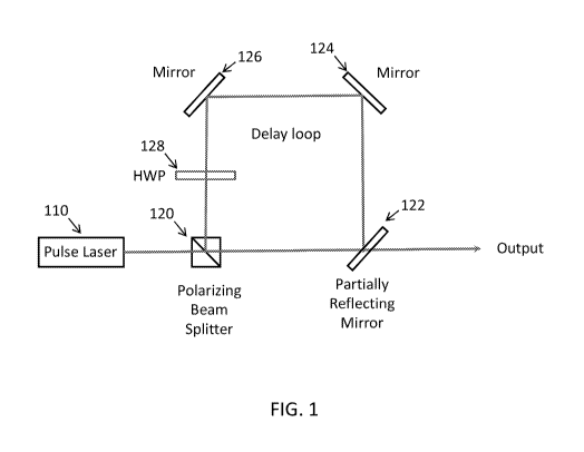
[0011] FIG. 1 shows a diagram of a light source for generating a series
of
reducing intensity laser pulses according to an embodiment.
[0012] FIG. 2 shows a diagram of an imaging system according to an
embodiment.
[0013] FIG. 3 shows a series of modulated light pulses according to an
embodiment.
[0014] FIG. 4 shows a diagram of a light source for generating a series
of
reducing intensity laser pulses according to an embodiment.
[0015] FIG. 5 shows a diagram of a synchronous electro-optic modulator
(SEOM) according to an embodiment.
DETAILED DESCRIPTION OF THE PREFERRED EMBODIMENTS
[0016] The description of illustrative embodiments according to
principles of the
present invention is intended to be read in connection with the accompanying
drawings,
which are to be considered part of the entire written description. In the
description of
embodiments of the invention disclosed herein, any reference to direction or
orientation
is merely intended for convenience of description and is not intended in any
way to limit
the scope of the present invention. Relative terms such as "lower," "upper,"
"horizontal,"
"vertical," "above," "below," "up," "down," "top" and "bottom" as well as
derivative
thereof (e.g., "horizontally," "downwardly," "upwardly," etc.) should be
construed to
- 4 -
CA 03127800 2021-07-23
WO 2020/160389
PCT/US2020/016071
refer to the orientation as then described or as shown in the drawing under
discussion.
These relative terms are for convenience of description only and do not
require that the
apparatus be constructed or operated in a particular orientation unless
explicitly indicated
as such. Terms such as "attached," "affixed," "connected," "coupled,"
"interconnected,"
and similar refer to a relationship wherein structures are secured or attached
to one
another either directly or indirectly through intervening structures, as well
as both
movable or rigid attachments or relationships, unless expressly described
otherwise.
Moreover, the features and benefits of the invention are illustrated by
reference to the
exemplified embodiments. Accordingly, the invention expressly should not be
limited to
such exemplary embodiments illustrating some possible non-limiting combination
of
features that may exist alone or in other combinations of features; the scope
of the
invention being defined by the claims appended hereto.
[0017] This
disclosure describes the best mode or modes of practicing the
invention as presently contemplated. This description is not intended to be
understood in
a limiting sense, but provides an example of the invention presented solely
for illustrative
purposes by reference to the accompanying drawings to advise one of ordinary
skill in the
art of the advantages and construction of the invention. In the various views
of the
drawings, like reference characters designate like or similar parts.
[0018] It is
important to note that the embodiments disclosed are only examples
of the many advantageous uses of the innovative teachings herein. In general,
statements
made in the specification of the present application do not necessarily limit
any of the
various claimed inventions. Moreover, some statements may apply to some
inventive
features but not to others. In general, unless otherwise indicated, singular
elements may
be in plural and vice versa with no loss of generality.
[0019] In one
embodiment, an imaging system includes: a light source configured
to generate successive light pulses of diminishing intensity having a pulse
interval, as
shown in FIG. 1; and a microscopy system configured to image a sample and to
process
- 5 -
CA 03127800 2021-07-23
WO 2020/160389
PCT/US2020/016071
signals detected from the sample based on intensities of the successive light
pulses, as
shown in FIG. 2. FIG. 1 is a diagram of a system for generating a series of
reducing
intensity laser pulses according to an embodiment. The system includes a pulse
laser 110
that generates light pulses having a repetition rate. For example, the pulse
laser may be a
tunable femtosecond Ti:sapphire laser having a repetition rate around 70 to 90
MHz and a
tunable wavelength range of 650 to 1110 nm. It is understood that different
specifications or types of pulse lasers may be used depending on the specific
application.
In one embodiment, the light pulses generated by the pulse laser is directed
to a
polarizing beam splitter (PBS) 120, and the light pulse that is in a first
state of
polarization passes through the PBS to reach a beam splitter 122. In one
embodiment,
the beam splitter 122 is a partially reflecting mirror, for example, a 50/50
mirror. Note
that different reflection/transmission ratios, or percentages, such as 60/40,
80/20, etc., are
also contemplated, depending on specific requirements. The beam splitter 122
splits the
light pulse into two paths: a first percentage to a delay loop and a second
percentage to an
output. In one embodiment, the output goes on to an imaging path for a
microscopy
system. As shown in FIG. 2, in the imaging path, there are one or more optical
elements
for directing the light pulse to a desired location. For example, the one or
more optical
elements may be a lens, mirror, beam splitter, or scanner, etc., or some
combinations of
these elements. In one embodiment, an x-y scanner 130 scans the second
percentage of
the light pulse to cover an area within the plane of a sample 160. By scanning
the
location within the plane, scanning microscopy can be performed. A dichroic
mirror 140
reflects the light pulse into an objective 150. It is noted that a skilled
person would be
able to use a configuration of one or more optical element, or the equivalents
to direct the
light to a location within the plane. The objective 150 then focuses the light
pulse onto
an image plane 170 of a desired depth into the sample 160. It is noted that a
skilled
person would be able to use a configuration of one or more optical element, or
the
equivalents to direct the light to a location within the plane. In one
embodiment, the
wavelength of the laser is selected to cause fluorescence at a focal point in
the sample. In
- 6 -
CA 03127800 2021-07-23
WO 2020/160389
PCT/US2020/016071
one embodiment, the wavelength of the laser is selected to cause a two-photon
excitation
in the sample. Light emitted by the sample 160 is collected by a detector 180.
In one
embodiment, the detector is a photomultiplier tube (PMT). In one embodiment,
the
detector is a silicon photomultiplier (SiPM). In one embodiment, the light
emitted by the
sample 160 passes through the objective 150 and through the dichroic mirror
140 to reach
the detector 160 above. Note that the arrangement of the dichroic mirror and
detector
shown in FIG. 2 is an example setup only. Other optical elements and/or
arrangements
are possible to achieve the desired direction of light to the sample and
detection of the
emitted light from the sample.
[0020] In one
embodiment, the delay loop includes a path traverses the beam
splitter 122 that directs a first percentage of the light pulse to a first
mirror 124, which
reflects the pulse to a second mirror 126, which reflects the pulse through a
half-wave
plate 128 to the PBS 120. The half-wave plate 128 changes the pulse to a
second
polarization state, allowing the PBS 120 to reflect the pulse back to the beam
splitter 122.
Note that the half-wave plate 128 may be placed anywhere within the path. The
arrangement shown in FIG. 1 is an example setup. The delay loop introduces a
desired
delay time to the first percentage of the light pulse relative to the second
percentage of
the light pulse. Note that the delay loop setup shown in FIG. 1 is an
illustrative example.
Other optical elements and/or arrangement may be used to create such a delay
loop, or
equivalents thereof.
[0021] The
beam splitter 122 further splits the delayed light pulse according to its
reflection/transmission ratio, and thus the intensity of the delayed light
pulse transmitted
by the beam splitter 122 is further diminished. Each time the light reflected
by the beam
splitter 122 into the delay loop would be delayed by the desired delay time as
it loops
around the delay loop, and the intensity of the light transmitted by the beam
splitter 122
would be reduced according to the reflection/transmission ratio of the beam
splitter.
Thus, a series of delayed pulses of diminishing intensity is generated from
the initial light
- 7 -
CA 03127800 2021-07-23
WO 2020/160389
PCT/US2020/016071
pulse. FIG. 3 is a plot of intensity over time of the modulated light pulses
according to an
embodiment. As can be seen from FIG. 3, the second light pulse is delayed
relative to the
first light pulse and the intensity of the second light pulse is less than
that of the first light
pulse. The third pulse is delayed by the same amount and with its intensity
further
diminished. The pulses shown in FIG. 3 are for illustration purposes. The
number of
pulses, their relative intensities, delays, etc., may vary depending on
specific setup and
application requirements. Note that FIG. 3 also shows a fourth pulse with the
same
intensity as the first pulse. This fourth pulse is due to a pulse from the
pulse laser
arriving at the next repetition interval.
[0022] Using
knowledge of the pulse intensity, location on the test sample, and
amount of fluorescence measured, an imaging system according to an embodiment
can
create an increased dynamic range of the image relative to what can be
obtained in a
normal two-photon imaging system.
[0023] In one
embodiment, the detector includes a plurality of temporal buffers
which would store signals from the sample. The signal detected by the detector
for the
fluorescence in response to a focused light pulse may be stored in one of the
buffers. If
the delay time introduced by the delay loop is selected such that when the
delayed pulse
arrives at the sample, the fluorescence due to the previous pulse has already
substantially
subsided, then detected signal due to the fluorescence in response to the
focused delayed
light pulse may be stored in another buffer. Thus, at each focal point in the
sample, the
buffers store fluorescence data at different times, each correspond to a light
pulse of
different intensities. Since the intensity of the fluorescence depends on the
intensity
squared of the excitation pulse, for some pulse intensities, the light emitted
by a structure
in the sample may be so high that the buffer is saturated, and for some other
pulse
intensities, the light emitted by a structure in the sample may be so faint
that the signal is
not registered. In one embodiment, the imaging system includes a processor 190
configured to select a buffer among the plurality of temporal buffers, where
the selected
- 8 -
CA 03127800 2021-07-23
WO 2020/160389
PCT/US2020/016071
buffer is not saturated by the brightest object. Thus, for each spot, the
processor has a
choice among different intensity-modulated pulses and can select the
excitation intensity
that corresponds to the most appropriate dynamic range for that spot. In one
embodiment, the processor may select the data in a buffer that corresponds to
the highest
intensity pulse among those pulses that do not cause buffer saturation.
[0024] The
choice of the appropriate buffer may be performed in real time as the
sample is being scanned by the system. In one embodiment, the processor may
include a
field programmable gate array (FPGA), which allows programmable logic to be
incorporated with high speed and flexibility on the processor.
[0025] In
another embodiment, the creation of a series of pulses of reducing
amplitude is realized by a synchronous electro-optic modulator (SEOM) with a
delay
loop as shown in FIG. 4. FIG. 5 shows an example setup of the SEOM according
to an
embodiment. A quarter wave plate 510 and a first PBS or polarizer 520
configured to
receive an input a pulsed laser to provide circularly polarized laser pulses
into the electro-
optic modulator EOM (such as a Pockels cell) 530. If the laser pulses are
reflected the PBS
520, they are directed into a sink 560. The electro-optic modulator 530 is
driven by a
waveform driver 540. The modulated output from the electro-optic modulator is
split by the
second PBS 550 into output 1 and output 2 according to their respective
polarization states.
[0026] The EOM
modulation waveform is generated by the waveform driver 540.
The waveform drive includes a custom circuit to lock to the laser and create a
phase
locked signal, with the ability to step phase shift that signal, to a RF power
amplifier
which drives a transformer, providing the AC voltage to the EOM. In one
embodiment, a
sinusoidal waveform, which is representative of the EOM modulation waveform,
is
expressed as:
= vjain(Iffit + 7),
- 9 -
CA 03127800 2021-07-23
WO 2020/160389
PCT/US2020/016071
where Vp is the amplitude, or "Peak Amplitude" of the waveform, f is frequency
in Hz,
and 9 is the phase in radians.
[0027] A
periodic impulse train, which is representative of pulsed lasers, is
expressed as:
where T, is the period of the pulses going around the delay loop. This means
that the
series of laser pulses arrive at t = 0, t = Ts, t = 271, ..., etc. Now the
phase of the EOM
drive sinusoid is relative to this and is defined by tp. The voltage on the
EOM is relevant
only at the instant in time when the laser pulse is present in the EOM
material (crystal).
That means the voltage on the sinusoidal waveform is relevant only at time t =
0, t = Ts, t
= 2Ts, ..., etc. The drive voltage is thus the sinusoid equation evaluated at
those instants
in time, and now looks like a discrete-time sampled signal:
n:OzTli) = 0, 71 =
[0028] When
the sinusoidal waveform is synchronized with the successive
delayed pulses, we have f = 11T, = repetition rate of the series of pulses of
reducing
amplitude. The amplitude reduction of the series of pulses can be adjusted by
changing
the phase of the EOM drive c3.
[0029] The
present disclosure overcomes the limits in the dynamic range of the
traditional detection/digitization system by creating a series of pulses of
reducing
- 10 -
CA 03127800 2021-07-23
WO 2020/160389
PCT/US2020/016071
amplitude, and then using knowledge of the pulse intensity, location on the
test sample,
and amount of fluorescence measured to assemble a high dynamic range final
image.
Thus, embodiments of the present invention represent significant improvements
over
existing microscopic imaging technology.
[0030] While
the present invention has been described at some length and with
some particularity with respect to the several described embodiments, it is
not intended
that it should be limited to any such particulars or embodiments or any
particular
embodiment, but it is to be construed with references to the appended claims
so as to
provide the broadest possible interpretation of such claims in view of the
prior art and,
therefore, to effectively encompass the intended scope of the invention.
[0031]
Although particular parameters used with particular embodiments of the
microscope are mentioned herein, it is understood that the invention is not
limited to any
particular parameters that have been used with particular embodiments. All
examples
and conditional language recited herein are intended for pedagogical purposes
to aid the
reader in understanding the principles of the invention and the concepts
contributed by
the inventor to furthering the art, and are to be construed as being without
limitation to
such specifically recited examples and conditions. Moreover, all statements
herein
reciting principles, aspects, and embodiments of the invention, as well as
specific
examples thereof, are intended to encompass both structural and functional
equivalents
thereof. Additionally, it is intended that such equivalents include both
currently known
equivalents as well as equivalents developed in the future, i.e., any elements
developed
that perform the same function, regardless of structure.
-11-