Note : Les descriptions sont présentées dans la langue officielle dans laquelle elles ont été soumises.
CA 02339497 2001-02-02
WO 00/07669 PCT/US99/17675
DELIVERY MODIFICATION SYSTEM FOR RADIATION THERAPY
CROSS-REFERENCE TO RELATED APPLICATIONS
STATEMENT REGARDING FEDERALLY
SPONSORED RESEARCH OR DEVELOPMENT
FIELD OF THE INVENTION
This invention relates generally to radiation therapy equipment for the
treatment
of tumors or the like and specifically to a computerized method for rapidly
correcting a
I 0 radiation treatment plan to account for motion or change in shape of
treatment areas.
BACKGROUND OF THE INVENTION
Medical equipment for radiation therapy treats tumorous tissues with high
energy
radiation. Such radiation may be x-ray radiation or accelerated electrons,
protons,
neutrons or heavy ions. The amount of radiation and its placement must be
accurately
15 controlled to ensure both that the tumor receives sufficient radiation to
be destroyed and
that the damage to the surrounding non-tumorous tissue is minimized.
One highly accurate method of controlling the dose to a patient employs a
radiation source that produces many individual rays whose intensity and/or
energy may be
independently controlled. This may be done by a series of shutters each
controlling one
20 ray or by a single modulated ray moved across the patient. The origin of
the rays orbits
the patient within a plane of the rays to illuminate a slice of the patient,
when the orbit is
planar, or several slices of the patient, when the orbit is helical. By
properly selecting the
ray intensities and/or energies at different angles, complex regions within
the slice may be
accurately irradiated. A mapping of the modulation of each beam as a function
of angle
25 forms a "treatment sinogram".
U.S. Patent 5,317,616 issued May 31, 1994 assigned to the same assignee as the
CA 02339497 2003-O1-03
present application describes the constnzction of one
such machine and a method of calculating the necessary beam intensities andlor
energies
as a function of angle.
In order to take advantage of the improved accuracy in dose placement offered
by
such radiation therapy systems, the radiation treatment plan may be based on a
computed
tomography (CT) image of the patient. As is known in the art, a CT image is
produced by
a mathematical reconstruction of many projection images obtained at dii:ferent
angles
about the patient. In a typical fan beam CT acquisition, the origin of the fan
beam orbits
the patient within a plane of the fan to illuminate a slice of the patient,
while the
attenuation of each ray of the fan beam is measured as a function of that
angle to obtain
projections. The geometry of the CT acquisition is thus very similar to the
geometry of
the radiation therapy.
Each CT proj ection forms a one-dimensional line image indicating the
attenuation
of the fan beam by a "slice" of the patient. Together these line images at
each angle form
an "attenuation sinogram" which may be reconstructed using well known
algorithms such
as filtered back projection into two dimensional tomographic images of the
slice. The
sinographic data, which by itself is unintelligible, is normally no longer
used or accessed
by the user.
Using the CT image, the radiologist views the tumorous area andl determines
the
beam angles and intensities and/or energies (identified with respect to the
tumor image)
which will be used to treat the tumor. In an automated system, a computer
program
selects the beam angle and intensities and/or energies after the physician.
creates a dose
map identifying the tumorous region and upper and lower dose limits four
regions of the
treatment.
Preparing a treatment plan based on the dose map is a time consiuning
operation
even on current high speed computers. Accordingly, the CT image of th:e
patient is
acquired before the time of radiation treatment. As a result, the patient will
typically not
be in the same position during the radiation treatment as the patient was
during the CT
imaging: The problem of properly aligning the patient is compounded when the
treatment
occurs in a number of different sessions over time.
U.S. Patent 5,673,300 assigned to the same assignee as the present invention
describes a method of determining patient movement by obtaining a second CT
image
2
CA 02339497 2001-02-02
WO 00/0?669 PCT/US99/17675
immediately prior to radiation therapy and comparing the sinogram of that CT
image to
the sinogram of the original CT image used for radiation treatment planning.
This
comparison yields an indication of patient movement which may be applied
directly to the
treatment sinogram used to control the radiation therapy machine. This
invention, by
recognizing the close analogy between the attenuation sinograms of the CT
image and of
the treatment sinograms of radiation therapy treatment, greatly simplified
detecting and
correcting mis-registrations of the patient to the treatment sinogram.
BRIEF SUMMARY OF THE INVENTION
The present inventors have recognized that the above technique of directly
modifying the treatment sinogram, by bypassing the time-consuming translation
of dose
map to treatment sinogram, makes possible real-time correction for patient
motion. Such
a correction may deduce real-time motion from a concurrent tomographic scan or
from
well known transducers used for measuring physiological motion. An improved
method
for correcting "fan beam" sinograms facilitates this use of the sinogram
directly.
The inventors have also recognized that the ability to manipulate sinograms to
accommodate motion in the underlying structure, allows for a novel method of
generating
a treatment sinogram by combining precalculated partial sinograms representing
treatments of standard elements of the patient. These standard elements may be
moved to
match a particular patient's anatomy and the partial sinograms modified
according to the
techniques described above. The partial sinograms are then combined and used
directly
or as a starting base for iterative treatment planning software.
Specifically, then, the present invention provides a method of operating a
radiation
therapy machine providing a radiation beam of individually intensity and/or
energy
modulated radiation rays separated along a radiation beam axis, the radiation
beam axis
positionable at a range of angles about a patient. A treatment sinogram is
received
providing intensities and/or energies of different rays for a given angle of
the radiation
beam, in a row, and intensities and/or energies of a given ray for different
angles of the
beam axis, in a column, for a patient at a first position. During radiation
treatment, data
indicating patient movement from the first position to a second position is
also received
and for each given beam axis angle of the treatment sinogram, the
corresponding row of
treatment sinogram is altered according to the indicated movement.
3
CA 02339497 2001-02-02
WO 00/07669 PCT/US99/17675
Thus it is one object of the invention to make possible real-time correction
of
patient motion to correct not only patient positioning errors but
physiological motions
such as caused by respiration and cardiac motion. Direct operation on the
treatment
sinogram renders such real-time control possible.
The movement may be detected by comparing a planning tomographic image of
the patient contemporaneous with the preparation of the treatment sinogram to
a
monitoring tomographic image of the patient taken during radiation therapy.
Alternatively, the patient movement may be determined by a model receiving as
an input
a physiological signal such as respiration or heart beat or external fiducial
marks may be
measured.
Thus it is another object of the invention to provide a method of detecting
patient
motion on a real-time basis in a radiation therapy setting.
The modification of the treatment sinogram may shift corresponding rows of
treatment sinogram according to a component of patient motion perpendicular to
the
given beam axis.
Thus it is another object of the invention to provide an extremely simple
operation
on the treatment sinogram such as may be performed in real-time.
The modification of the treatment sinogram may scale corresponding rows of
treatment sinogram according to a component of patient motion parallel to the
given beam
axis.
Thus it is another object of the invention to provide a more sophisticated
modification of the treatment sinogram addressing the geometry of the highly
efficient fan
beam radiation therapy machine.
The present invention also contemplates the preparation of a library of
partial
sinograms, each partial sinogram providing intensities and/or energies of
different rays at
given angles of the radiation beam axis, in sinogram rows, and intensity
andlor energy of
given rays for different angles of the beam axis, in sinogram columns, for a
patient
element in first modes. Sets of representations of patient elements may be
arranged in
combinations at second modes so as to model a given patient requiring
radiation
treatment. Changes in the patient elements between the first and second modes
may be
captured in alteration data. This alteration data may be used to modify the
partial
sinograms of each of the patient elements according to the alteration data and
the partial
4
CA 02339497 2001-02-02
WO 00/07669 PCT/US99/17675
sinograms may be combined to produce a treatment sinogram of the patient.
Thus it is another object of the invention to make use of the ability to
directly
modify treatment sinograms to prepare template sinograms that may be simply
combined
to produce a treatment sinogram without the need for extensive treatment
planning
operations. The alteration data may indicate either change in location or
dimension of the
patient elements, the latter which may be simple geometric shapes or may model
specific
organs.
Thus it is another object of the invention to provide in a finite library of
patient
elements sufficient to permit assembly of an approximate treatment smogram.
The treatment sinogram thus constructed may be further optimized to better
conform with the dose map.
Thus it is another object of the invention to provide an advanced starting
point for
dose optimization such as may reduce the number of iterations and thus the
time required
to prepare the treatment sinogram.
The foregoing and other objects and advantages of the invention will appear
from
the following description. In the description, reference is made to the
accompanying
drawings which form a part hereof and in which there is shown by way of
illustration a
preferred embodiment of the invention. Such embodiment does not necessary
represent
the full scope of the invention, however, and reference must be made to the
claims herein
for interpreting the scope of the invention.
BRIEF DESCRIPTION OF THE DRAWINGS
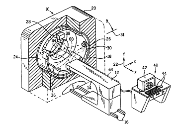
Fig. 1 is a perspective, cut-away view of a radiation therapy system providing
for
the acquisition of radiographic projections and for the generation of high
energy radiation
therapy beams and showing a patient table for supporting a patient thereon;
Fig. 2 is a simplified view of a slice of an object, such as a patient,
showing line
projections of the object taken at two angles 9, with attenuations A along
dimension t
indicated in the vertical axis of each projection;
Fig. 3 is a sinogram formed of multiple line projections such as those
acquired in
Fig. 2, over 360 degrees of angle 8 with the attenuation of the projections
indicated by
shading;
Fig. 4 is a perspective view of a simplified object that may be scanned
showing a
CA 02339497 2001-02-02
WO 00/07669 PCT/US99117675
helical and slice-by-slice scanning path;
Fig. 5 is a set of sinograms of the object of Fig. 4 such as may be obtained
in a
slice-by-slice scanning;
Fig. 6 is a sinogram of the object of Fig. 4 such as may be obtained in a
helical
S scan;
Fig 7 is combination block diagram and flow chart showing the steps of
preparing
a treatment sinogram used for controlling a radiation therapy machine, from a
computed
tomography scan taken on the same or a different machine;
Figs. 8a and 8b are figures similar to Fig. 2 showing the effect of movement
of a
structure in a parallel beam and fan beam system, respectively;
Fig. 9 is a flow chart showing the steps of a first method of correcting for
motion
within a fan beam involving rebinning the fan sinogram to a parallel
configuration;
Fig. 10 is a flow chart similar to that of Fig. 9 showing a second method of
correcting for motion within a fan beam involving direct mathematical
manipulation by
1 S scaling and shifting of the treatment sinogram without rebinning to
parallel beam
configuration;
Fig. 11 is a figure similar to that of Figs. 2, 8a and 8b showing the effect
of in-
place expansion of an object in contrast to translative movement of Figs. 8a
and 8b; and
Fig. 12 is a figure similar to that of Fig. 7 showing elements of the real-
time
motion correction employed by the present invention and the construction of a
treatment
sinogram from precalculated partial sinograms.
DETAILED DESCRIPTION OF THE INVENTION
The Radiotherapy Machine
Referring now to Fig. 1, a radiation therapy machine 10, suitable for use with
the
present invention, includes a radiotranslucent table 12 having a cantilevered
top 14. The
table top I4 is received within a bore 18 of an annular housing 20 of the
radiation therapy
machine 10 with movement of the table 12 along tracks 16 extending along a z-
axis of a
Cartesian coordinate system 22.
Table 12 also includes an internal track assembly and elevator (not shown) to
allow adjustment of the top 14 in a lateral horizontal position (indicated by
the x-axis of
the coordinate system 22) and vertically (indicated by the y-axis of the
coordinate system
6
CA 02339497 2001-02-02
WO 00/07669 PCT/US99/17675
22). Motion in the x and y directions are limited by the diameter of the bore
18.
A rotating gantry 24, coaxial with the bore 18 and positioned within the
housing
20, supports an x-ray source 26 and a high energy radiation source 28 on its
inner surface.
The x-ray source 26 may be a conventional rotating anode x-ray tube, while the
radiation
S source 28 may be any source of treatment radiation including one producing x-
rays,
accelerated electrons, protons or heavy ions such as are understood in the
art. The x-ray
source 26 and a radiation source 28 rotate with the gantry 24 about a center
of rotation 64
near the top of patient table 12 when the table top 14 is positioned within
the bore 18.
The x-ray source 26 is collimated to produce a fan beam 30 lying generally
within
the x-y plane and crossing the bore 18 and thus the table top 14 when table
top 14 is
positioned within the bore 18. The fan beam 30 diverges about a central axis
31 whose
angle is controlled by the position of the gantry 24. The axis 31 will
henceforth be termed
the projection axis.
After exiting the table top 14, the fan beam 30 is received by a linear array
detector 32 positioned diametrically across from the radiation source 28.
Thus, the
rotating gantry 24 permits fan beam radiographic projections of a patient on
the table top
14 to be acquired at a variety of angles 8 about the patient.
The radiation source 28 is mounted so as to project a fan beam of high energy
radiation 34, similar to the fan beam 30, but crossing fan beam 30 at right
angles so as to
be received on the other side of the gantry 24 by radiation detector and stop
36. In an
alternative embodiment, the stop is replaced by a detector to provide an
alternative to the
detector 32 for deducing motion of the patient. The fan beam of high energy
radiation 34
diverges about a radiation axis centered within the beam and perpendicular to
the
projection axis 31.
The radiation source 28 has a collimator 38 mounted in front of it to divide
the
beam of high energy radiation 34 into multiple adjacent rays whose energy
and/or
fluence may be individually controlled. As used herein, control of the energy
and/or
fluence of the rays should be understood to include not only the energy of
individual x-
ray photons (or particles in the case of radiation therapy using electrons,
protons or heavy
ions) but alternatively or in addition the total number of photons or
particles such as is a
function of fluence, fluence rate and exposure time. In the case of
radiotherapy using
particles, the energy of the particles, fluence and fluence rate may be
controlled using
7
CA 02339497 2003-O1-03
sinograms which may be modified by the present invention as will be apparent
from the
following description.
A collimator suitable for flue;nce control type is described in U.S. Patent
5,31 T,6I6 assigned to the assignee of the present case and:
a simple modification of this collimator using wedge filters may be used for
particle energy control. Alternatively, a scanning single beam system rnay be
used, or
other system providing a set of individually modulated rays. The location of
the radiation
source 28 and x-ray source 26 are precisely characterized so that images
obtained from'
the radiation source 28 may be used to aim the radiation source 28.
A computer 40 having,a display screen 42 and user entry mouse; and keyboard 44
well known in the art is connected to the radiation therapy machine 10 to
control motion
of the table I2 and to coordinate operation of the gantry 24 together with the
radiation
source 28 and x-ray source 26 and to, collect data from the linear array
detector 32 during
a scan of the patient according to methods well known in the art.
CT and Radiotherapy Treatment Sinograms
Referring now to Fig. 2, a slice 50 of the patient taken along the x-y plane
includes
two zones 54 within a larger zone 52. Radiation passing along beam axis 31
through the
slice 50 (at a vertical or anteriorlposterior angle ("AP")) produces a
projection 56 which
records the attenuation of x-rays passing through to slice 50 along a single
line
perpendicular to the beam axis 31 (for C'I~ or provides a radiation treatment
projection of
different a energy and/or intensity of beams corresponding to the different
zones 52 and
54 (for radiotherapy). In either case, the distance along this perpendicular
to the
projection axis is designated: t. The zones 54 may be resolved separatelly at
the vertical
angle and heacc two peaks 58 (attenuation or radiation energy andlor
intensity) are
presex~t in the projextion 56.
In contrast at a second projection along a projection axis 31' at an angle 6
from
vertical, the zones 54 are aligned so that the projection 56' shows a single
peak 58 ;
Referring now to Fig. 3, projections at a different angle 8 over 360 degrees,
may
be combined to form an sinogram 60 which is stored temporarily in computer 40
as a
matrix of data. As depicted, this matrix of data is arranged with each row
representing a
different angle 8 and each column a diffexe;nt distance t along the
projection. For a CT
attenuation sinogram, each element of the matrix is a value of attenuation.
For a radiation
8
CA 02339497 2003-O1-03
treatment sinogram, each element of the matrix is an energy and/or fluence of
a ray of the
treatment beam. The values may be stored as numeric variables in the computer
40 and
are shown as shaded curves 62.
The pattern of the sinogram 60 is generally that of superimposed sinusoidal
curves
62 (hence the name) each curve 62 having a fundamental period in 8 of 360
degrees as a
result of the apparent movement of zones 54 in orbit about a center of gantry
rotation 64
as projections are taken at various angles 9. Generally, zones 54 toward the
axis of
rotation 64 of the gaatry trace smaller amplitude sine curves whereas zones 54
farther
from the center of rotation 64 trace greater amplitude sine curves. The ;phase
of the sine
curves depends generally on the initial position of the zones 54 with respect
to the first
proj ection at 8 = 0.
In a conventional CT acquisition, an attenuation sinogram may be reconstructed
into a tomographic image of the slice 50. As is well understood in the art, an
attenuation
sinogram having t values spanning the largest cross-sectional width of a~n
imaged slice SO
and 9 values over 360 degrees is sufficient to reconstruct a tomographic image
of the slice
through, for example, the method of filtered back projection.
In radiation therapy, a treatment sinogram may be used to control the a energy
and/or fluence of adjacent rays of a faun beam of high energy radiation 3~4
transmitted
through the patient. For example, if the zones 54 of Fig. 2 were tumors" a
radiation
treatment plan might well conform generally to curves 62 which would produce
beams of
high intensity radiation that would intersect at the zones S4 at a variety of
different angles
8 to produce a high cumulative dose at the zones S4 but low dose elsewlhere.
Referring again to Fig. I, it follows that the tornographic image produced
from the
sinogram 60 may be employed to establish a radiation treatment plan precisely
related to
that tomographic image. U.S. Patent No. 5,661,773
describes generally an interactive method for
generating a treatment plan in the form of a sinogram 60 based on a
tom.ographic image.
Referring now to Fig. 4, in a "slice-by-slice" tomographic acquisition or in a
slice-
by-slice radiation treatment, the imaged object 51 is divided into a plurality
of slices 70
separated along the z-axis and the acquisition of projections or the radiation
treatment is
obtained with the beam axis 31 constrained to a single plane as it rotates
about the imaged
object 51 indicated generally by arrow 72. At the conclusion of 360 de~ees of
rotation
9
CA 02339497 2001-02-02
WO 00/07669 PCT/US99/17675
the object is moved along the z-axis by movement of the table 12 until the
next slice is
aligned with the beam axis 31.
In an alternative acquisition or treatment method termed "helical scanning",
the
projection axis follows a helical path through the imaged object 51 in which
the table 12
is incremented by a small amount in z with each change in angle 8.
In the former slice-by-slice method, a series of sinograms 60' is used
(attenuation
and treatment), each one identical to that described with respect to Fig. 3
and typically
encompassing 360 degrees of gantry motion. Different slices 70 produce
different ones of
a sequence of sinogram 60' each of which has a different but constant z value.
In contrast, the helical acquisition produces a sinogram 60" in which each row
of
the sinogram 60" represents a different increment in both 8 and in Z.
In the example shown in Fig. 4, a zone 54 extends only through the first two
slices
70. Hence, in Fig. 5, only the first two sinograms 60' show sine curves 62
related to the
zone 54. Likewise, in the helically acquired sinogram 60" of Fig. 6 only the
first 720
degrees of the sinogram 60" show a sign curve 62.
The Treatment Planning Process
Referring now to Fig. 7, the radiation therapy machine 10 or an independent CT
machine (not shown) may be used to acquire tomographic data in the form of an
attenuation sinogram 41 of a patient 43. As described above, the attenuation
sinogram 4I
generally provides rows {here depicted vertically) comprising a set of
attenuation
measurements A(t) received by the detector 32 at different rays at a given
projection angle
8 and columns (here depicted horizontally) representing the same data for
different
projection angles 8.
The attenuation sinogram 41 is received by a tomographic reconstructor 45 such
as one using well known filtered backprojection algorithms to provide a
planning
tomographic image 46 depicting a slice of the patient 43. This and the
following steps
may be performed on computer 40.
The planning tomographic image 46 may be provided to a dose map editor 48
where it provides a background on which a dose map 55 is prepared by a
physician. The
dose map 55 depicts the desired dose in regions within the slice of the
patient 43.
In the preferred embodiment, the dose map 55 is prepared interactively with
editing commands 53 from a keyboard or cursor control device being received by
the dose
CA 02339497 2001-02-02
WO 00/07669 PCT/US99/17675
map editor 48.
The dose map 55 is used to prepare a treatment sinogram 57 describing energy
and/or fluence of plurality of radiation beams from the radiation source 28 at
different
beam angles A that will produce the desired dose of the dose map 55. The
treatment
sinogram 57 is generally arranged with rows (here depicted vertically)
providing values of
a function I(t) indicating beam energy and/or fluence for different rays t
within a beam
and columns (here depicted horizontally) providing values of a function I(8)
indicating
beam energy and/or fluence for different beam angles 8.
Generally the process of converting the dose map 55 to the treatment sinogram
is
performed as an iterative optimization by planning software 59. The planning
software
59 produces a trial sinogram 101 which is provided to a dose calculator 61,
the latter
which determines the dose that would be produced by the trial sinogram and
comparing it
to the desired dose as indicated by comparison node 102. The planning software
59
receiving an indication of the deviation between the dose provided by the
trial sinogram
101 and the dose map 55 then modifies the beam energy and/or fluence of the
trial
sinogram 101 according to that deviation and the process is repeated until a
treatment
sinogram 57 is obtained. The treatment sinogram 57 is then provided to control
the
collimator 38 for treatment of the patient 43.
Because of the time required to perform the actual radiation treatment using
the
treatment sinogram 57, patient movement caused by respiration and other
sources is
inevitable. This movement can be detected, but the time required to change the
dose map
55 to reflect the movement and recalculate the treatment sinogram 57 is too
great to
practically account for short term patient movements. The present invention
has
recognized that with high speed computers, the pretreatment patient position
correction
technique described generally in U.S. Patent 5,673,300 assigned to the same
assignee as
the present invention, can be used to also correct for motion during the
treatment process
itself.
As depicted in Fig. 8a, relative motion of the patient 43 or a portion of the
patient
43 by an amount ~ from a first position 63 to a second position 63' will
require a
shifting of the rows of the sinogram modifying I(t) to be I(t+ DYl ) where DYl
is an amount
proportional to fir= times ~ being the difference between the beam angle 0 for
that row of
the sinogram and the angle of Vii- . This correction alone is sufficient for
parallel ray
11
CA 02339497 2001-02-02
WO 00/07669 PCT/US99/17675
systems in which each of the rays of high energy radiation 34 are parallel
(shown in Fig.
8a) , but is only part of the correction desired for a fan ray system where
each of the rays
of the high energy radiation 34 diverge about the beam axis from a common
origin as
depicted in Fig. 8b.
The present inventors have realized that the divergence of the rays in a fan
beam
system cause a magnification effect which ideally should be compensated.
Refernng then
to Fig. 8b, an arbitrary motion of an object from position 63 to position 63'
by Or
provides not only a shifting of the sinogram rows I(t) but can provide a
magnification of
that function. Thus the object at position 63' moving closer to the origin of
the fan beam
as well as across the origin of the fan beam causes a shifting of the function
I(t+cz) and a
scaling of the function I((3t) reflecting the relative magnification effects
caused by moving
toward and away from the origin of the fan beam of high energy radiation 34.
The
amounts a and ~3 depend on the particular dimensions of the radiation therapy
machine 10
and will generally be functions of the amount of motion and the origin of the
motion and
may be determined by well understood geometric techniques. Generally the
scaling and
shifting need not be linear functions and need not be uniform over the
patient..
Correction of the treatment sinogram, as described above, may be provided in a
number of ways. In a first embodiment shown in Fig. 9, the rows and columns of
the
treatment sinogram are rebinned to reflect a sinogram providing an identical
radiation
pattern but on a machine having parallel rays. This rebinning process is a
geometnc
transformation well understood in the computed tomography art and may be
calculated
on-the-fly using mathematical formulas or by precalculated to produce a table
mapping
elements of the fan beam treatment sinogram 57 to corresponding elements of a
parallel
ray sinogram. Generally, an interpolation step will be required so that the
elements map
to the integer ray and beam angle values of the sinogram. This rebinning is
indicated by
process block 8i.
Next at process block 83 for each beam angle, the component of the motion
perpendicular to the beam angle is determined and used to shift the particular
row of the
sinogram in direct proportion to that component. Motion parallel to the beam
angle may
be disregarded as a result of the parallel ray geometry.
At process block 84, the resultant shifted parallel ray sinogram may be
rebinned to
a divergent ray sinogram using the inverse process described with process
block 81. The
12
CA 02339497 2001-02-02
WO 00/07669 PCT/US99/17675
resultant sinogram will have been corrected both for parallel and
perpendicular motion of
the patient.
Alternatively as depicted by Fig. 10, for each row of the treatment sinogram
related to a particular beam angle, a perpendicular component DYl of motion 0r
may be
determined as indicated by process block 86. Then at process block 88, the
rows may be
shifted proportionally to this component and the magnification caused by the
diverging
rays of the fan beam. Next at process block 90, the parallel component of the
motion Ors
with respect to a beam axis at 8 may be determined and at process block 92,
the rows may
be scaled appropriately. Finally, at process block 94, the scaled and shifted
rows may be
resampled to fit within the integer values allowed in the treatment sinogram
57.
The perpendicular value of the motion Or1 and the parallel component of the
motion 4rr may be deduced for any arbitrary displacement of 4x and Dy within a
fixed x-
y coordinate system lying in the plane of the fan beam according to the
following
formula:
1
~cos~ -sin~~~~~ ~~'1~ ( )
sin ~ cos ~ Dy
For a fan beam system using a multileaved collimator, generally the corrected
sinogram I(~3t+oc) will have discontinuities that do not lie on the separation
between the
leaves of the collimator 38 which define the rays. For this reason, the
sinogram will have
to be resampled to fit within the confines of the treatment sinogram 57.
Standard
methods of interpolation can be used for this resampling. The inventors
recognize that
there are additional errors in this approach including the failure to account
for scatter but
it is believed that these errors are small or can be corrected for.
Referring now to Fig. 12, the above described treatment sinogram modification
technique or the more simple treatment sinogram modification described in
predecessor
parent 5,673,300 may be used to provide for real-time motion correction.
During the
treatment of the patient 43 with radiation therapy machine 10', a regular
tomographic
projection signal may be obtained using detector 32 or a megavoltage
tomographic
projection signal may be obtained by using a megavoltage detector 65. The
images thus
obtained may be used to provide a real-time imaging sinogram 67 which may be
compared to the planning attenuation sinogram 41 as indicated by comparison
block 83
13
CA 02339497 2001-02-02
WO 00/07669 PCT/US99/17675
according to the techniques described in the above mentioned patent to provide
a motion
signal 69.
Generally the comparison between the two attenuation sinograms 41 and 67
correlates rows of the sinograms at particular beam angles to determine
patient motion in
one or more orthogonal axes. More generally, this technique may be expanded to
completely define the motion of the patient 43 in six parameters of x, y and z
and roll,
yaw and pitch.
Alternatively, a patient motion sensor 66 may be used to provide a
physiological
signal from the patient indicating motion. The sensor 66 in its simplest case
may be a
pressure cuff to detect chest wall expansion commensurate with breathing or
may simply
detect an electronic signal such as an ECG signal. The thus detected signal 71
may be
provided to a mathematical model 73 relating the cycle of the signal to
internal changes in
the patient either through the use of a simple mathematical modeling of the
patient, for
example, breathing as an expansion of a generally oval chest wall, or by
keying different
phases of the cycle of the signal to measured tomographic images of the
patient or of a
standard patient that would indicate changes in location or aspect of internal
structures of
the patient 43. The patient motion sensor may alternatively be optical, using
external
fiducial marks optically detected by lasers or the like, or may employ signals
from other
known imaging systems such as magnetic resonance imaging (MRI) or may be
provided
by positioning fixtures attached to the patient employing well know
location/orientation
determining technologies such as those using radio or light transmitters and
receivers as
are known for virtual reality headsets and the like.
These signals 69 from either source may be provided to a sinogram manipulation
program 74 implemented as software in a high speed computer processor
performing the
transformations described above, the inputs describing the ~r Or- value and
absolute
coordinate information. The sinogram manipulation program 74 thus receives a
sinogram
as prepared above as described in Fig. 7 to produce a modified treatment
sinogram 76
which may be provided to the collimator 38 on a real-time basis so as to
modify the actual
radiation treatment on a real-time basis.
The ability to modify the treatment sinogram 57 through simple mathematical
operations such as shifting and scaling provides the ability to make the rapid
corrections
required of real-time. Further in the case where a model 73 is used,
modifications may be
14
CA 02339497 2001-02-02
WO 00/07669 PCT/US99/17675
anticipated and calculated in advance, and/or multiple modified treatment
sinograms 76
may be precalculated and simply switched into communication with the
collimator 38 as
required.
Refernng now again to Fig. 11, the correction process applied to the sinogram
need not only deal with displacement of objects, either within the patient or
including the
entire patient, but may accommodate general dimensional changes of objects
within the
patient to a limited degree. For example, object in mode 63 may inflate to
become object
in mode 63" to the predictable effect of expanding by scaling the sinogram
function I(t)
necessary to continue to treat that structure. Unlike the expansion of Fig. 4,
however, a
similar expansion in this case is found in all beam angles. Thus complex organ
dimensional changes may be accommodated through this technique without the
need for
recalculation of the dose map S5.
This ability to modify the treatment sinogram 57 to change the location and
dimensions of the structures they treat, allows a novel method of rapidly
constructing
treatment sinograms which avoids the need for conventional planning software
or limits
the need for such software's iterations.
Referring again to Fig. 12, such a system prepares a series of partial
sinograms
80(a) through 80(c) each corresponding to a predetermined patient element
82(a) through
82(c) and providing a standard treatment for those elements. These partial
sinograms
80(a) through 80(c) may be prepared using conventional planning software and
optimized
over the course of an arbitrarily long period of time and then stored for
later use in an
electronically accessible library together with representations of patient
elements 82(a)
through 82(c) indicating an area, possibly an expected material, and a desired
treatment
dose.
During the planning process, the dose map editor 48 receives representations
corresponding to the treatment zones and doses of patient elements 82(a)
through 82(c)
and allows them to be manipulated with respect to the planning tomographic
image 46
both by translation with respect to the planning tomographic image 46 and by
expansion
and contraction. These manipulation commands are received as editing commands
53
and are also provided via line 85 to the sinogram manipulation program 74.
As the patient elements 82(a) through 82(c) are manipulated, the sinogram
manipulation program 74, modifies the partial sinograms 80(a) through 80(c) as
has been
CA 02339497 2001-02-02
WO 00/07669 PCT/US99117675
described above to conform to the new spatial locations and dimensions of
their
associated patient elements 82. The sinograms 80(a) through 80(c) for the
selected
patient elements 82(a) through 82(c) used in the creation of a dose map 55 may
then be
summed or otherwise combined to produce the treatment sinogram 76. The
combination
operates on element pairs of corresponding rows and columns.
Thus a physician wishing to treat a tumorous organ might select a patient
element
82(a) representing a preplanned treatment for that organ based on some average
person
and combine it with a second patient element 82(a) representing a nearby
sensitive
structure where radiation is to be avoided. These two portions may be placed
upon a
representation of the patient's torso also modeled by a portion as aligned
against a
tomographic image of an actual patient.
The resulting treatment sinogram 76 may be provided directly to the collimator
38
or may be used as a starting point for further optimizations using the
iteration of the dose
calculator 61 and the planning software 59 as has been described before.
The patient elements may thus represent either standard organs or standard
geometric shapes of predetermined density and a desired dose. To the extent
that many
patient's treatments will be similar, except for minor anatomical dimensional
variations,
such a system allows the physician to use proven radiation therapy techniques
across
patients.
The technique of preparing a treatment sinogram from partial sinograms 80 may
be further augmented by the motion detection provided by signals 69 to the
extent that
motion identified to a particular organ may be identified to a single, partial
sinogram 80
and thus distinguishable from the other components of the treatment sinogram
57
allowing adjustment of that organ in isolation.
The above description has been that of a preferred embodiment of the present
invention, and it will occur to those that practice the art that many
modifications may be
made without departing from the spirit and scope of the invention. In order to
apprise the
public of the various embodiments that may fall within the scope of the
invention, the
following claims are made.
16