Note : Les descriptions sont présentées dans la langue officielle dans laquelle elles ont été soumises.
CA 02441086 2003-09-11
WO 02/074911 PCT/US02/07756
IDENTIFICATION OF GENE EXPRESSION ALTERATIONS UNDERLYING
THE AGING PROCESS IN MAMMALS
CROSS-REFERENCE TO RELATED APPLICATION
[0001] This application claims priority to 60/277,382, filed March 19, 2001
and
incorporated by reference herein.
STATEMENT REGARDING FEDERALLY SPONSORED
RESEARCH OR DEVELOPMENT
[0002] This invention was made with United States government support
awarded by the following agencies: NIH CA79740. The United States has
certain rights in this invention.
BACKGROUND OF THE INVENTION
[0003] A common feature of most multicellular organisms is the progressive
and irreversible physiological decline that characterizes senescence.
Although genetic and environmental factors can influence the aging process,
the molecular basis of senescence remains unknown. Postulated
mechanisms include cumulative damage to DNA leading to genomic
instability, epigenetic alterations that lead to altered gene expression
patterns,
telomere shortening in replicative cells, oxidative damage to critical
macromolecules and nonenzymatic glycation of long-lived proteins (S.M.
Jazwinslei, Science 273:54, 1996; G.M. Martin, et al., Nature Gen. 13:25,
1996; F.B. Johnson, et al., Cell 96:291, 1996; K.B. Becleman and B.N. Ames,
Phyrsiol. Revs. 78:547, 1998). Factors which contribute to the difficulty of
elucidating mechanisms and testing interventions include the complexity of
CA 02441086 2003-09-11
WO 02/074911 PCT/US02/07756
organismal senescence and the lack of molecular markers of biological age
(biomarkers). Aging is complex in that underlying mechanisms in tissues with
limited regenerative capacities (e.g., skeletal and cardiac muscle, brain),
which are composed mainly of postmitotic (non-dividing) cells, may differ
markedly from those operative in proliferative tissues. Accordingly,
approaches which provide a global assessment of senescence in specific
tissues would greatly increase understanding of the aging process and the
possibility of pharmaceutical, genetic or nutritional intervention.
(0004] Genetic manipulation of the aging process in multicellular organisms
has been achieved in Drosophila, through the over-expression of catalase
and Cu/Zn superoxide dismutase (W.C. Orr and R.S. Sohal, Science
263:1128, 1994; T.L. Parkes, et al., Nat. Genet. 19:171, 1998), in the
nematode C. elegans, through alterations in the insulin receptor signaling
pathway (S. Ogg, et al., Nature 389:994, 1997; S. Paradis and G. Ruvkun,
Genes Dev. 12:2488-2498, 1998; H.A. Tissenbaum and G. Ruvkun, Genetics
148:703, 1998), and through the selection of stress-resistant mutants in
either
organism (T.E. Johnson, Science 249:908, 1990; S. Murakami and T.E.
Johnson, Genetics 143:1207, 1996; Y.J. Lin, et al., Science 282:943, 1998).
In mammals, there has been limited success in the identification of genes that
control aging rates. Mutations in the Werner Syndrome locus (WRN)
accelerate the onset of a subset of aging-related pathology in humans, but
the role of the WRN gene product in the modulation of normal aging is
unknown (C.E. Yu, et al., Science 272:258, 1996; D.B. Lombard and L.
Guanrente, Trends Genet. 12:283, 1996).
-2-
CA 02441086 2003-09-11
WO 02/074911 PCT/US02/07756
[0005] In contrast to the current lack of genetic interventions to retard the
aging process in mammals, caloric restriction (CR) appears to slow the
intrinsic rate of aging (R. Weindruch and R.L. Walford, The Retardation of
Aaina and Disease by Dietary Restriction (CC. Thomas, Springfield, IL, 1988;
L. Fishbein, Ed., Biological Effects of Dietary Restriction (Springer-Verlag,
New York, 1991; B.P. Yu, Ed., Modulation of Aaing Processes by Dietary
Restriction (CRC Press, Boca Raton, FL 1994). Most studies have involved
laboratory rodents which, when subjected to a long-term, 25-50% reduction in
calorie intake without essential nutrient deficiency, display delayed onset of
age-associated pathological and physiological changes and extension of
maximum lifespan.
BRIEF DESCRIPTION OF THE SEVERAL VIEWS OF THE DRAWINGS
[0006] Figs. 1 - 23 are individual bar graphs disclosing the fold change of
messages and lines showing signal intensities corresponding to individual
sequences in young and old tissue.
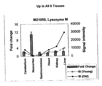
[0007] Fig. 1 discloses changes in M21050.
[0008] Fig. 2 discloses changes in 249204.
[0009] Fig. 3 discloses changes in U49430.
[0010] Fig. 4 discloses changes in K02782.
[0011] Fig. 5 discloses changes in X58861.
[0012] Fig. 6 discloses changes in X66295.
[0013] Fig. 7 discloses changes in M22531.
[0014] Fig. 8 discloses changes in X67809.
_3_
CA 02441086 2003-09-11
WO 02/074911 PCT/US02/07756
[0015] Fig. 9 discloses changes in 019118.
[0016] Fig. 10 discloses changes in M64086.
[0017] Fig. 11 discloses changes in M63695.
[0018] Fig. 12 discloses changes in 039066.
[0019] Fig. 13 discloses changes in X92590.
[0020] Fig. 14 discloses changes in X56518.
[0021] Fig. 15 discloses changes in AA182189.
[0022] Fig. 16 discloses changes in X16493.
[0023] Fig. 17 discloses changes in X60452.
[0024] Fig. 18 discloses changes in 020344.
[0025] Fig. 19 discloses changes in X16834.
[0026] Fig. 20 discloses changes in X82648.
[0027] Fig. 21 discloses changes in D00754.
[0028] Fig. 22 discloses changes in D16313.
[0029] Fig. 23 discloses changes in 15789.
DESCRIPTION OF THE INVENTION
[0030] In order to generate rational interventions to retard aging and
associated diseases, identification of molecular targets is required. To
achieve this goal, we used the new 074 and 11 K Affymetrix (Santa Clara,
CA) murine genome DNA chips to measure the gene expression profile
associated with the aging process for 11,000 genes in six tissues from mice:
cerebral cortex, cerebellum, skeletal muscle (gastrocnemius), heart, liver and
kidney. Six animals were used per experiment (3 young and 3 old), resulting
-4-
CA 02441086 2003-09-11
WO 02/074911 PCT/US02/07756
in a total of 396,000 independent gene expression measurements. To our
knowledge, this study represents the largest search ever performed for gene
expression alterations as a function of age.
[0031] We reasoned that alterations in gene expression that are shared
among 5 to 6 tissues, or among the four post-mitotic tissues studied (i.e.,
cerebellum, neocortex, gastrocnemius and heart) must represent
fundamental changes associated with aging as opposed to tissue-specific
effects that are secondary to the aging process.
[0032] An additional requirement for the evaluation of therapies that retard
the
aging process is the development of aging biomarkers. A suitable biomarker
of the aging process should reflect biological age (physiological condition)
as
opposed to chronological age. Additionally, the biomarker should be
amenable to quantitation and reflect aging-related alterations at the
molecular
level in the tissue under study.
[0033] By "biological age" we mean the physiological state of an animal or
tissue relative to the physiological changes that occur throughout the
animal's
lifespan. By "chronological age" we mean the age of an animal as measured
by a time scale, such as month or years.
[0034] There exists a large and growing segment of the population in
developed countries that is suffering from age-associated disorders, such as
sarcopenia (loss of muscle mass), neurodegenerative conditions, and cardiac
disease. Therefore, the market for compounds that prevent aging-associated
disorders and improve quality of life for the elderly is likely to drive
research
and development of novel drugs by the pharmaceutical industry. As an
-5-
CA 02441086 2003-09-11
WO 02/074911 PCT/US02/07756
example, many drugs, nutraceuticals and vitamins are thought to influence
aging favorably, but their use remains limited due to the lack of FDA
approval.
The inability to assess biological aging in tissues at the molecular level
precludes proper animal and human testing of such compounds.
[0035] In one embodiment, the invention is a method for measuring the
relative biological aging process of a multicellular organism, such as a
mammal, at the organ, tissue or cellular level through the characterization of
the organism's gene expression patterns. This method preferably comprises
obtaining a cDNA copy of the organism's RNA and determining the
expression pattern of at least one of the genes listed in Table 2 (genes which
change in expression with aging in multiple tissues), preferably at leasfi 5
biomarker sequences, most preferably at least 10 biomarker sequences and
more preferably at least 20, 30, 40, or 50 biomarker sequences, within the
cDNA. By "gene expression pattern" we mean to include the change in
pattern of the encoded RNA or protein.
[0036] One may characterize the biological age of the organism by
determining how many and at what level these genes disclosed are altered in
expression. Because the genes listed in Table 2 are age-related alterations
in multiple tissues, one could use the same genes to determine biological
aging in multiple tissues, such as, but not limited to, neocortex, heart,
cerebellum, kidney, liver and skeletal muscle.
[0037] In some embodiments, gene expression is measured by identifying the
presence or amount of one or more proteins encoded by one of the genes
listed in Table 2.
_g_
CA 02441086 2003-09-11
WO 02/074911 PCT/US02/07756
[0038] The present invention also provides systems for detecting two or more
markers of interest (e.g., two or more markers from Table 2). For example,
where it is determined that a finite set of particular markers provides
relevant
information, a detection system is provided that detects the finite set of
markers. For example, as opposed to detecting al! genes expressed in a
tissue with a generic microarray, a defined microarray or other detection
technology is employed to detect the plurality (e.g., 2, 5, 10, 25) of markers
that define a biological condition (e.g., a biological age, a response to a
pharmaceutical or diet that increases or decreases rate of aging, etc.).
[0039] The present invention is not limited by the method in which biomarkers
are detected or measured. In some embodiments, mRNA, cDNA, or protein
is detected in tissue samples (e.g., biopsy samples). In other embodiments,
mRNA, cDNA, or protein is detected in bodily fluids (e.g., serum, plasma,
urine, or saliva). The present invention further provides kits for the
detection
of biomarkers.
[0040] In some preferred embodiments, protein is detected. Protein
expression may be detected by any suitable method. In some embodiments,
profieins are detected by binding of an antibody specific for the protein. For
example, in some embodiments, antibody binding is detected using a suitable
technique, including but not limited to, radioimmunoassay, ELISA
(enzyme-linked immunosorbant assay), "sandwich" immunoassays,
immunoradiometric assays, gel diffusion precipitation reactions,
immunodiffusion assays, in situ immunoassays (e.g., using colloidal gold,
enzyme or radioisotope labels, for example), Western blots, precipitation
_7_
CA 02441086 2003-09-11
WO 02/074911 PCT/US02/07756
reactions, agglutination assays (e.g., gel agglutination assays,
hemagglutination assays, etc.), complement fixation assays,
immunofluorescence assays, protein A assays, immunoelectrophoresis
assays, and proteomic assays, such as the use of gel electrophoresis
coupled to mass spectroscopy to identify multiple proteins in a sample.
[0041] In one embodimenfi, antibody binding is detected by detecting a label
on the primary antibody. In another embodiment, the primary antibody is
detected by detecting binding of a secondary antibody or reagent to the
primary antibody. In a further embodiment, the secondary antibody is
labeled. Many methods are known in the art for detecting binding in an
immunoassay and are within the scope of the present invention.
[0042] In some embodiments, an automated detection assay is utilized.
Methods for the automation of immunoassays include, but are not limited to,
those described in U.S. Patents 5,885,530; 4,981,785; 6,159,750; and
5,358,691, each of which is herein incorporated by reference. In some
embodiments, the analysis and presentation of results is also automated. For
example, in some embodiments, software that generates a diagnosis and/or
prognosis based on the presence or absence of a series of proteins
corresponding to markers is utilized.
[0043] In other embodiments, the immunoassay described in U.S. Patents
5,599,677 and 5,672,480, each of which is herein incorporated by reference,
is utilized. In other embodiments, proteins are detected by
immunohistochemistry.
_g_
CA 02441086 2003-09-11
WO 02/074911 PCT/US02/07756
[0044] In other embodiments, markers are detected at fihe level of cDNA or
RNA. In some embodiments of the present invention, markers are detected
using a direct sequencing technique. In these assays, nucleic acid samples
are first isolated from a subject using any suitable method. In some
embodiments, the region of interest is cloned into a suitable vector and
amplified by growth in a host cell (e.g., bacteria). In other embodiments, DNA
in the region of interest is amplified using PCR. Following amplification, DNA
in the region of interest is sequenced using any suitable method, including
but
not limited to manual sequencing using radioactive marker nucleotides, or
automated sequencing. The results of the sequencing are displayed using
any suitable method.
[0045] In some embodiments of the present invention, markers are detected
using a PCR-based assay. In yet other embodiments, reverse-transcriptase
PCR (RT-PCR) is used to detect the expression of RNA. In RT-PCR, RNA is
enzymatically converted to complementary DNA or "cDNA" using a reverse
transcriptase enzyme. The cDNA is then used as a template for a PCR
reaction. PCR products can be detected by any suitable method, including
but not limited fio, gel electrophoresis and staining with a DNA specific
stain or
hybridization to a labeled probe. In some embodiments, the quantitative
reverse transcriptase PCR with standardized mixtures of competitive
templates method described in U.S. Patents 5,639,606, 5,643,765, and
5,876,978 (each of which is herein incorporated by reference) is utilized.
[0046] In preferred embodiments of the present invention, markers are
detected using a hybridization assay. In a hybridization assay, the presence
_g_
CA 02441086 2003-09-11
WO 02/074911 PCT/US02/07756
of absence of a marker is determined based on the ability of the nucleic acid
from the sample to hybridize to a complementary nucleic acid molecule (e.g.,
an oligonucleotide probe). A variety of hybridization assays using a variety
of
technologies for hybridization and detection are available.
[0047] In some embodiments, hybridization of a probe to the sequence of
interest is detected directly by visualizing a bound probe (e.g., a Northern
or
Southern assay; See e.g., Ausabel, et al. (eds.), Current Protocols in
Molecular Bioloay, John Wiley & Sons, NY [1991]). In these assays, DNA
(Southern) or RNA (Northern) is isolated. The DNA or RNA is then cleaved
with a series of restriction enzymes that cleave infrequently in the genome
and not near any of the markers being assayed. The DNA or RNA is then
separated (e.g., on an agarose gel) and transferred to a membrane. A
labeled (e.g., by incorporating a radionucleotide) probe or probes is allowed
to contact the membrane under low, medium, or high stringency conditions.
Unbound probe is removed and the presence of binding is detected by
visualizing the labeled probe.
[0048] In some embodiments, the DNA chip assay is a GeneChip (Affymetrix,
Santa Clara, CA; See e.g., U.S. Patent Nos. 6,045,996; 5,925,525; and
5,858,659; each of which is herein incorporated by reference) assay. The
GeneChip technology uses miniaturized, high-density arrays of
oligonucleotide probes affixed to a "chip." Probe arrays are manufactured by
Affymetrix's light-directed chemical synthesis process, which combines
solid-phase chemical synthesis with photolithographic fabrication techniques
employed in the semiconductor industry. Using a series of photolithographic
-10-
CA 02441086 2003-09-11
WO 02/074911 PCT/US02/07756
masks to define chip exposure sites, followed by specific chemical synthesis
steps, the process constructs high-density arrays of oligonucleotides, with
each probe in a predefined position in the array. Multiple probe arrays are
synthesized simultaneously on a large glass wafer. The wafers are then
diced, and individual probe arrays are packaged in injection-molded plastic
cartridges, which protect them from the environment and serve as chambers
for hybridization.
[0049] The nucleic acid to be analyzed is isolated, amplified by PCR, and
labeled with a fluorescent reporter group. The labeled DNA is then incubated
with the array using a fluidics station. The array is then inserted into the
scanner, where patterns of hybridization are detected. The hybridization dafia
are collected as light emitted from the fluorescent reporter groups already
incorporated into the target, which is bound to the probe array. Probes that
perfectly match the target generally produce stronger signals than those that
have mismatches. Since the sequence and position of each probe on the
array are known, by complementarity, the identity of the target nucleic acid
applied to the probe array can be determined.
[0050] In other embodiments, a DNA microchip containing electronically
captured probes (Nanogen, San Diego, CA) is utilized (See e.g., U.S. Patent
Nos. 6,017,696; 6,068,818; and 6,051,380; each of which are herein
incorporated by reference). Through the use of microelecfironics, Nanogen's
technology enables the active movemenfi and concentration of charged
molecules to and from designated test sites on its semiconductor microchip.
DNA capture probes unique to a given marker are electronically placed at, or
-11-
CA 02441086 2003-09-11
WO 02/074911 PCT/US02/07756
"addressed" to, specific sites on the microchip. Since nucleic acid molecules
have a strong negative charge, they can be electronically moved to an area of
positive charge.
[0051] In still further embodiments, an array technology based upon the
segregation of fluids on a flat surface (chip) by differences in surface
tension
(ProtoGene, Palo Alto, CA) is utilized (See e.g., U.S. Patent Nos. 6,001,311;
5,985,551; and 5,474,796; each of which is herein incorporated by reference).
Protogene's technology is based on the fact that fluids can be segregated on
a flat surface by differences in surface tension that have been imparted by
chemical coatings. Once so segregated, oligonucleotide probes are
synthesized directly on the chip by ink-jet printing of reagents.
[0052] In yet other embodiments, a "bead array" is used for the detection of
markers (Illumina, San Diego, CA; See e.g., PCT Publications WO 99/67641
and WO 00/39587, each of which is herein incorporated by reference).
Illumina uses a BEAD ARRAY technology that combines fiber optic bundles
and beads that self-assemble into an array. Each fiber optic bundle contains
thousands to millions of individual fibers depending on the diameter of the
bundle. The beads are coated with an oligonucleotide specific for the
detection of a given marker. Batches of beads are combined to form a pool
specific to the array. To perform an assay, the BEAD ARRAY is contacted
with a prepared sample. Hybridization is detected using any suitable method.
[0053] In some embodiments of the present invention, hybridization is
detected by enzymatic cleavage of specific structures (e.g., INVADER assay,
Third Wave Technologies; See e.g., U.S. Patent Nos. 5,846,717, 6,090,543;
-12-
CA 02441086 2003-09-11
WO 02/074911 PCT/US02/07756
6,001,567; 5,985,557; and 5,994,069; each of which is herein incorporated by
reference). In some embodiments, hybridization of a bound probe is detected
using a TaqMan assay (PE Biosystems, Foster City, CA; See e.g., U.S.
Patent Nos. 5,962,233 and 5,538,848, each of which is herein incorporated
by reference). The assay is performed during a PCR reaction. The TaqMan
assay exploits the 5'-3' exonuclease activity of DNA polymerases such as
AMPLITAQ DNA polymerase. A probe, specific for a given marker, is
included in the PCR reaction. The probe consists of an oligonucleotide with a
5'-reporter dye (e.g., a fluorescent dye) and a 3'-quencher dye. During PCR,
if the probe is bound to its target, the 5'-3' nucleolytic activity of the
AMPLITAQ polymerase cleaves the probe between the reporter and the
quencher dye. The separation of the reporter dye from the quencher dye
results in an increase of fluorescence. The signal accumulates with each
cycle of PCR and can be monitored with a fluorimeter.
[0054] Additional detection assays that are produced and utilized using the
systems and methods of the present invention include, but are not limited to,
enzyme mismatch cleavage methods (e.g., Variagenics, U.S. Pat. Nos.
6,110,684; 5,958,692; 5,851,770, herein incorporated by reference in their
entireties); branched hybridization methods (e.g., Chiron, U.S. Pat. Nos.
5,849,481; 5,710,264; 5,124,246; and 5,624,802, herein incorporated by
reference in their entireties); rolling circle replication (e.g., U.S. Pat.
Nos.
6,210,884 and 6,183,960, herein incorporated by reference in their
entireties);
NASBA (e.g., U.S. Pat. No. 5,409,818, herein incorporated by reference in its
entirety); molecular beacon technology (e.g., U.S. Pat. No. 6,150,097, herein
-13-
CA 02441086 2003-09-11
WO 02/074911 PCT/US02/07756
incorporated by reference in its entirety); E-sensor technology (Motorola,
U.S.
Pat. Nos. 6,248,229; 6,221,583; 6,013,170; and 6,063,573, herein
incorporated by reference in their entireties); cycling probe technology
(e.g.,
U.S. Pat. Nos. 5,403,711; 5,011,769; and 5,660,988, herein incorporated by
reference in their entireties); ligase chain reaction (Barnay, Proc. Natl.
Acad.
Sci. USA 88:189-93, 1991); and sandwich hybridization methods (e.g., U.S.
Pat. No. 5,288,609, herein incorporated by reference in its entirety).
[0055] In some embodiments, mass spectroscopy is used to detect markers.
For example, in some embodiments, a MassARRAY system (Sequenom, San
Diego, CA.) is used to detect markers (See e.g., U.S. Patent Nos. 6,043,031;
5,777,324; and 5,605,798; each of which is herein incorporated by reference).
[0056] In some embodiments, the present invention provides kits for the
identification, characterization, and quantitation of markers. In some
embodiments, the kits contain antibodies specific for markers, in addition to
detection reagents and buffers. In other embodiments, the kits contain
reagents specific for the detection of nucleic acid (e.g., oligonucleotide
probes
or primers). In preferred embodiments, the kits contain all of the components
necessary to perform a detection assay, including all controls, directions for
performing assays, and any necessary software for analysis and presentation
of results. In some embodiments, the kits contain instructions including a
statement of intended use as required by the Environmental Protection
Agency or U.S. Food and Drug Administration for the labeling of in vitro
diagnostic assays and/or of pharmaceutical or food products.
-14-
CA 02441086 2003-09-11
WO 02/074911 PCT/US02/07756
[0057] Comparison of the organism's gene expression pattern with the result
expressed in Table 2 would indicate whether the organism has an aberrant
gene expression profile which may indicate that the organism is either
biologically younger or older than the chronological age would indicate.
[0058] In another embodiment, the present invention is a method of screening
a test compound for the ability to inhibit, retard or reverse the aging
process
in mammalian tissue. In a typical example of this embodiment, one would
first treat a test mammal with a test compound and then analyze a
representative tissue of the mammal for the level of expression of the genes
which change in expression in multiple tissues (Table 2). Preferably, the
tissue is selected from the group consisting of brain tissue, heart tissue,
muscle tissue, skeletal muscle, kidney, heart and liver tissue. One then
compares the analysis of the tissue with a control, untreated mammal and
identifies test compounds that are capable of modifying the expression of the
biomarker sequences in the mammalian samples such that the expression is
indicative of tissue that has an inhibited or retarded biological age. This
expression pattern would be more similar to an expression pattern found in
biologically younger subjects.
[0059] As an example, a group of young rodents (e.g., mice) would be divided
into a control and a test group. The test group would receive a test
compound such as a dietary supplement added to food from age 5 months to
30 months, whereas the control group would receive a standard diet during
this time period. At age 30 months, several tissues would be collected from
animals from each group and a gene expression profile of at least one of the
-15-
CA 02441086 2003-09-11
WO 02/074911 PCT/US02/07756
genes listed in Table 2 (preferably at least five genes) would be obtained and
would be compared to the profile of young animals (5 month old). One would
then determine whether, for any of the organs investigated, the gene
expression pattern of the animals receiving the test compound was more
similar to that of young animals, indicating that aging has been retarded.
[0060] In another embodiment of the present invention, one would use the
sequences of the genes disclosed in Table 2 for a therapy for anti-aging or
preventing, retarding or reversing age-associated disorders. In general, one
would try to amplify gene expression for the genes identified herein as
decreasing during aging process and decrease the expression of genes
identified herein as increased during the aging process. For example, one
might try to decrease the expression of lysosyme M (ORFM21050), which is
shown herein to be induced by at least 1.5-fold in all examined tissues. One
would attempt to increase the expression of NADP transhydrogenase
(Z49204).which has been shown to decrease in expression in the tissues.
Common methods of increasing and decreasing expression would be known
to one of skill in the art. Examples for supplementation of expression would
include supplying the organism with additional copies of the gene. A
preferred example for decreasing expression would include RNA antisense
technologies or pharmaceutical intervention.
[0061] The genes disclosed in Table 2 would be appropriate drug
development targets. One would use the information presented in the
present application for drug development by using currently existing, or by
-16-
CA 02441086 2003-09-11
WO 02/074911 PCT/US02/07756
developing, pharmaceutical compounds that either mimic or inhibit the activity
of the genes listed in Table 2, or the proteins encoded by these genes.
[0062] Therefore, the biomarker genes disclosed herein represent targets for
pharmaceutical development and gene therapy or RNA antisense therapy
with the goal of preventing, retarding or reversing the aging process at the
molecular level. These gene expression alterations may also play a role in
age-related diseases of the organs under study. Additionally, these genes
represent biomarkers of the aging process that can be used for diagnostic
purposes.
[0063] In a particularly preferred form of the present invention, the targeted
genes or proteins would be encoded by ORFs M21050, 249204, 049430,
K02782, X58861, X66295, M22531, M64086, 039066, X56518, X16834,
X82648 and L38971.
[0064] The present invention further provides methods for selecting subjects
(e.g., humans and animals) that are appropriate targets for a particular
therapy. In some such embodiments, a sample from the subject is tested for
one or more markers (e.g., markers in Table 2). The expression profile of the
subject is then used to select a therapy appropriate for that individual's
specific condition.
[0065] The present invention also provides expression profiles. In some such
embodiments, a test sample is assayed for the presence of one or more
biomarkers and compared to the expression profile, for example, to determine
the biological age of the sample and/or to determine the effect of a diet or
other therapy on the sample. The present invention is not limited by the form
-17-
CA 02441086 2003-09-11
WO 02/074911 PCT/US02/07756
of the expression profile. In some embodiments, the expression profile is
maintained in computer software. In some embodiments, the expression
profile is written material. The present invention is not limited by the
number
of markers provided or displayed in an expression profile. For example, the
expression profile may comprise two or more markers found in Table 2,
indicating a biological status of a sample.
(0066] The present invention further provides databases comprising
expression information (e.g., expression profiles comprising one or more
markers from Table 2 from one or more samples). In some embodiments,
fihe databases find use in data analysis, including, but not limited to,
comparison of markers to one or more public or private information databases
(e.g., OMIM, GenBank, BLAST, Molecular Modeling Databases, Medline,
genome databases, etc.). In some such embodiments, an automated
process is carried out to automatically associate information obtained from
data obtained using the methods of the present invention to information in
one or more of public or private databases. Associations find use, for
example, in making expression correlations to phenotypes (e.g., disease
states).
[0067] The present invention also provides methods for selecting ingredients
in food or dietary products (e.g., nutraceuticals) and food and dietary
products
thus generated. For example, a food or dietary product is altered (e.g.,
supplemented or depleted) with a factor that increases or decreases, directly
or indirectly, the expression of one or more age-related markers (e.g.,
markers in Table 2). In some embodiments, the food or dietary product is
-18-
CA 02441086 2003-09-11
WO 02/074911 PCT/US02/07756
altered with a factor that might increase or decrease, directly or indirectly,
the
expression of one or more age-related markers (e.g., markers in Table 2).
[0068] For example, it has been shown that apolipoprotein D expression is
induced by retinoic acid (e.g., Lopez-Boado, et al., J. Biol. Chem. 271:32105,
1996). As shown in Table 2, apolipoprotein D expression is altered in an age-
related manner. Thus, in some embodiments of the present invention, food
or dietary products are altered to increase or decrease retinoic acid
concentrations (or compounds with similar biologic activity), directly or
indirectly, and are prescribed, marketed, and/or labeled as having an effect
on biological age. In some preferred embodiments of the present invention
the food or dietary product is altered to affect a plurality of markers (e.g.,
two
or more markers in Table 2).
[0069] We also understand the present invention to be extended to
mammalian homologs of the mouse genes listed in Table 2. One of skill in
the art could easily investigate homologs in other mammalian species by
identifying particular genes with sufficiently high homology to the genes
listed
in Table 2. By "high homology" we mean that the homology is at least 50%
overall (within the entire gene or protein) either at the nucleotide or amino
acid level.
EXAMPLES
Methods
(0070] A. Animal ages, husbandry and dietary manipulations. All aspects
of animal care were approved by the appropriate committees and conformed
-19-
CA 02441086 2003-09-11
WO 02/074911 PCT/US02/07756
with insfiitutional guidelines. Details on the methods employed to house and
feed male C57 BL6 ("B6") mice, a commonly used model in aging research
with an average lifespan of ~30 months, were recently described (Pugh, et al.,
1999). Briefly, mice were purchased from Charles River Laborafiories
(Wilmington, MA) at 1.5 months of age. After receipt in Madison, the mice
were housed singly in the specific pathogen-free Shared Aging Rodenfi
Facility at the Madison VA Geriatric Research, Education and Clinical Center,
and provided a nonpurified diet (PLI 5001 [Purina Labs, St. Louis, MO]) and
acidified water ad libitum for one week. Each mouse in the control group was
fed 84 kcaUweek of the diet (TD91349 [Teklad, Madison, WI]).
[0071] B. Gene Expression Analysis, All experiments use three mice per
experimental group (i.e., young and old). RNA from each animal is
independently hybridized to DNA chips, so that intragroup variability is
known.
Our own data indicate that variability between animals in the same age/diet
group is minimal, since we have never observed correlation coefficients
between two animals to be <0.98. Mice were euthanized by rapid cervical
dislocation and autopsied to exclude animals showing overt disease. The
brain was dissected and sectioned along the midline. One-half of the brain
was used for microarray analysis. The samples were placed in a
microcentrifuge tube, immediately flash-frozen in liquid nitrogen, and stored
at
-80°C.
[0072] Total RNA was extracted from frozen tissues using TRIZOL reagent
(Life Technologies) and a power homogenizer (Fisher Scientific) with the
addition of chloroform for the phase separation before isopropyl alcohol
-20-
CA 02441086 2003-09-11
WO 02/074911 PCT/US02/07756
precipitation of total RNA. Poly (A)+ RNA is purified from the total RNA with
oligo-dT linked Oligotex resin (Qiagen). Two micrograms of poly (A)'" RNA
are converted into double-stranded cDNA (ds-cDNA) using Superscript
Choice System (Life Technologies) with an oligo dT primer containing a T7
RNA polymerase promoter region (Genset). After second strand synthesis,
the reaction mixture is extracted with phenol/chloroform/isoamyl alcohol.
Phase Lock Gel (5 Prime -~ 3 Prime, Inc.) is used to increase ds-cDNA
recovery. The ds-cDNA is collected by ethanol precipitation. The pellet is
resuspended in 3 p1 of DEPC-treated wafer. In vitro transcription is
performed using a T7 Megascript Kit (Ambion) with 1.5 p1 of ds-cDNA
template in the presence of a mixture of unlabeled ATP, CTP, GTP, and UTP
and biotin-labeled CTP and UTP (bio-11-CTP and bio-16-UTP [Enzo]).
Biotin-labeled cRNA is purified using a Rneasy affinity column (Qiagen). The
amount of biotin-labeled cRNA is determined by measuring absorbency at
260 nm. Biotin-labeled cRNA is fragmented randomly to sizes ranging from
35 to 200 bases by incubating at 94°C for 35 minutes in 40 mM
Trisacetate
pH 8.1, 100 mM potassium acetate, and 30 mM magnesium acetate. The
hybridization solutions contain 100 mM MES, 1 M [Na+], 20 mM EDTA, and
0.01 % Tween 20. The hybridization solutions also contained 50 pM
oligonucleotide B2 (a biotin-labeled control oligonucleotide used for making
grid alignments), 0.1 mg/mL herring sperm DNA, and 0.5 mg/mL acetylated
BSA. The final concentration of fragmented cRNA is 0.05 pg/pl in the
hybridization solutions. Hybridization solutions are heated to 99°C for
5
minutes followed by 45°C for 5 minutes before being placed in the gene
chip.
-21-
CA 02441086 2003-09-11
WO 02/074911 PCT/US02/07756
pg of cRNA is placed in the gene chip. Hybridizations were carried out at
45°C for 16 hours with mixing on a rotisserie at 60 rpm. Following
hybridization, the hybridization solutions are removed, and the gene chips
installed in a fluidics system for wash and stain. The fluidics system
(Affymetrix GeneChip Fluidics Station 400) performs two post hybridization
washes (a non-stringent wash and a stringent wash), staining with
streptavidin-phycoerythrin, and one post-stain wash. The gene chips were
read at a resolution of 6 pm using a Hewlett Packard GeneArray Scanner.
Data collected from two scanned images are used for the analysis.
[0073] C. Data analysis performed by Affymetrix~ software. Detailed
protocols for data analysis of Affymetrix microarrays and extensive
documentation of the sensitivity and quantitative aspects of the method have
been described (Lockhart, et al., 1996). The U74 and the 11 K series are
derived from UniGene (http://www.ncbi.nlm.nih.gov/UniGene/). Briefly, each
gene is represented by the use of ~20 perfectly matched (PM) and an equal
number of mismatched (MM) control probes. The MM probes act as
specificity controls that allow the direct subtraction of both background and
cross-hybridization signals. The number of instances in which the PM
hybridization signal is larger than the MM signal is computed along with the
average of the logarithm of the PM:MM ratio (after background subtraction)
for each probe set. These values are used to make an arbitrary matrix-based
decision concerning the presence or absence of an RNA molecule which
serves as an indicator of data quality. All calculations are performed by
Affymetrix software. To determine the quantitative RNA abundance, the
-22-
CA 02441086 2003-09-11
WO 02/074911 PCT/US02/07756
average of the differences representing PM minus MM for each gene-specific
probe family is calculated, after discarding the maximum, the minimum, and
any outliers beyond three standard deviations. This value, termed the
Average Intensi~ Difference (S1) is a function of mRNA abundance. In order
to make comparisons between data-sets, the Average Intensity Differences
for each gene are normalized to the total fluorescence intensity of the array.
This is similar to the concept of normalizing signal to a reference mRNA, such
as (3-actin in a typical Northern blot.
[0074] In order to calculate fold changes (FC) between data sets (after
normalization) obtained from young (y) vs. old (o) mice, the following formula
is used by the software:
FC = Slo - Sly + 1 if Slo >_ Slo or -1 if Slo < Sly
the smallest of either SIY or 5,0
Where Slo is the average signal intensity from a gene-specific probe family
from an old mouse and SIv is that from a young mouse. Alternatively, if the
Qfacton a measure of the non-specific fluorescence intensity background, is
larger the smallest of either SIY or Slo, the FC is calculated as:
FC = Slo - Sly
factor
[0075] The Qfactor is automatically calculated for different regions of the
microarray and, therefore, minimizes the calculation of spurious fold changes.
Average of pairwise comparisons are made between study groups, each
composed of three animals, using Excel software. For example, each tissue
from 5-month-old mice (n=5) is compared to 30-month-old mice (n=3),
generating a total of 9 pairwise comparisons. No correlation coefficient
-23-
CA 02441086 2003-09-11
WO 02/074911 PCT/US02/07756
between two animals in the same age/diet group was less than 0.98,
suggesting that variations between individuals are small within the same
age/diet group.
(0076] D. Numbers of Genes Selected as Biomarkers. The numbers of
genes identified showing shared changes in expression with aging in 5-6 of
the tissues examined are summarized in Table 1. We have also included the
genes that showed either up-regulation or down-regulation in all four tissues
studied that are composed mainly of post-mitotic cells (non-dividing),
gastrocnemius, heart, cerebellum and neocortex. The procedure involved a
computer search of our database to identify those genes which showed 1.3-
fold or greater increases or decreases in expression with aging in either five
or all six of the tissues examined. The data supporting the change was then
critically evaluated for data quality based on information provided by
Affymetrix software as well as signal intensity (which also provides
information on tissue-specific expression levels), and variation (standard
error).
Table 1: Overview of Numbers of Genes Displaying
~narea ~n an m~umpie
es i issues
in
expression
wizn
Hgm
m
Direction NumberofTissuesShowingAgingChange
of Age
Chan a
Six Five Four
(G,
H,N,C
onl
All Selected*All SelectedAll Selected
Increase 1 1 9 8 3 3
Decrease 2 1 12 6 11 4
*Only genes
that displayed
SEM + 1.3
< observed
fold change
in at least
3 tissues
were
selected
for inclusion
in this
table.
Synopsis of Shared Chan~cies in Gene Expression with Aging.
(0077] A. Genes altered in expression in all six tissues. Only one gene,
Lysozme M (ORF M21050), was induced by 1.5-fold (50%) or higher in all
-24-
CA 02441086 2003-09-11
WO 02/074911 PCT/US02/07756
tissues, whereas only one gene, NADP transhydrogenase, was decreased in
expression by 50% or more in all tissues studied.
[0078] Lysozyme M is a proinflammatory mediator associated with the
monocyte-macrophage system (Cross, et al., 1988). Lysozymes have
primarily bacteriolytic function; those in tissues and body fluids are
associated
with the monocyte-macrophage system and enhance the activity of
immunoagents. The enzyme catalyzes the hydrolysis of the 1,4-beta-linkages
between N-acetyl-d-glucosamine and n-acetylmuramic acid in peptidoglycan
heteropolymers of prokaryotic cell walls.
[0079] NADP transhydrogenase Z49204~ catalyzes transhydrogenation
between NADH and NADP and is coupled to respiration and ATP hydrolysis.
The enzyme functions as a proton pump across the outside mitochondrial
membrane. Depending on metabolic conditions, the enzyme may be involved
in NADPH generation for detoxification of peroxides and free radicals and
protection from ischemic damage. Hence, given current views on the
importance of oxidative stress/damage in aging, this decline in gene
expression may be highly important.
[0080] B. Genes upregulated in five of the six tissues. Several genes
were either upregulated or downregulated in five of the six tissues studied.
Specifically, 8 genes were upregulated in 5 of 6 tissues. These included four
members of the complement pathway and Ceruloplasmin (which encodes a
copper-binding protein that may act as a physiological antioxidant).
[0081] Complement C3 (K027821, Complement C1Qa (X5886, Complement
C1Qc (X66295) Complement C1Qb (M22531~: Genes encoding four
-25-
CA 02441086 2003-09-11
WO 02/074911 PCT/US02/07756
components of the complement cascade: The clustering of upregulated
complement genes is striking and highly significant. The classical
complement pathway plays a central role in antibody-mediated cell toxicity.
New studies suggest that the role of the pathway is not limited to antibody-
mediated reactions. Complement-mediated tissue damage contributes to the
myocardial injury associated with ischemia-reperfusion, and in brain injury
subsequent to stroke. Augmented membrane attack complex formation
through complement activation and assembly has been observed in
irreversibly injured myocytes during reperfusion. There is evidence that
inhibitors of complement activation attenuate myocardial reperfusion injury
(Murohara, et al., 1995; Kirschfink, 1997) and stroke (Huang, et al., 1999) in
vivo. Although it was assumed that complement components are deposited
from the plasma, resulting in membrane attack complex formation and,
ultimately, cell lysis, it is now established that several tissues, including
the
heart and brain, can synthesize complement components locally. To our
knowledge, and based on literature searches, our results provide the first
direct evidence that activation of genes encoding several components of the
complement pathway is a shared event in the aging process among multiple
tissues. Given the ability of complement components to induce cell death,
complement induction may be an underlying factor in age-related diseases
such as Alzheimer's disease, Parkinson's disease and heart failure.
[0082] Ceruloplasmin~U49430~: Ceruloplasmin is a blue, copper-binding (6-7
atoms per molecule) glycoprotein found in plasma. Four possible functions
are ferroxidase activity, amine oxidase activity, copper transport and
-26-
CA 02441086 2003-09-11
WO 02/074911 PCT/US02/07756
homeostasis, and superoxide dismutase activity. These represent an
impressive range of functions with the potential to exert a strong influence
on
pathophysiological changes associated with the aging process in multiple
tissues.
[0083] Mama (X67809: This molecule is also known as peptidylprolyl
isomerase C- associated protein (AF065438), pancreas cancer-associated
protein and galectin 6 binding protein. This gene encodes an mRNA that is
increased very strongly by adherence and moderately by exposure to tumor
necrosis factor and interferon-gamma. The nucleotide sequence extends for
2168 bases and encodes a protein of 559 amino acids with six potential
glycosylation sites. The first 100 NH2-terminal amino acids represent a single
scavenger receptor cysteine-rich domain. Mama is a normally produced in a
variety of tissues and down-modulates endotoxin and proinflammatory
responses in vivo (Trahey and Weissman, 1999).
[0084] LRG-21 (U19118): This gene encodes a transcription factor known to
be upregulated in stress responses.
[0085] Serine protease inhibitor 2-2 (M64086) (also known as contrapsin-like
protease inhibitor 6). This gene encodes a protein that inhibits trypsin, but
not chymotrypsin or elastase. It is induced by acute inflammation and
belongs to the serpin family.
[0086] C, Genes downregulated in five of the six tissues.
[0087] CD1d1 antigen M63695): This gene encodes the mouse homolog to
human CD1. it is a nonpolymorphic nonciassical main histocompatibility
complex (MHC) class I-like molecule encoded outside the MHC.
-27-
CA 02441086 2003-09-11
WO 02/074911 PCT/US02/07756
[0088] MAP kinase kinase 6 (U39066): This gene is also known as (mapkk 6)
(mapklerk kinase 6) (sapkk3) and appears to function in mediating stress
responses.
[0089] HIRA protein X92590): This gene is a HIRA, a DiGeorge syndrome
candidate gene (Farrell, et al., 1999). DiGeorge syndrome is a congenital
disease characterized by defects in organs and tissues that depend on
contributions by cell populations derived from neural crest for proper
development. HIRA could play a part in mechanisms of transcriptional
regulation similar to that played by yeast hirl and hir2 together.
[0090] Ace~lcholinesterase precursor (X56518): This gene encodes a
protein that rapidly hydrolyzes choline released into the synapse. The
catalytic activity is acetylcholine + H20 ~ choline + acetate. Thus, changes
in
the expression of this gene have the potential to markedly influence neural
transmission.
[0091] ZFP-1 (X16493): Belongs to the Krueppel family of c2h2-type zinc-
finger proteins which are highly conserved in evolution. The protein encoded
by this gene may be involved in transcriptional regulation.
[0092] Unknown (AA182189): No significant homology to any gene exists on
the public database.
[0093] D. Genes upregulated in the four post-mitotic tissues examined
(gastrocnemius, heart, cerebellum and neocortex). Three such genes
were discovered.
[0094] Gut-enriched Kruppel-like factor~U20344~: May act as a
transcriptional activator. Binds the CACC core sequence. May be involved in
_28_
CA 02441086 2003-09-11
WO 02/074911 PCT/US02/07756
the differentiation of epithelial cells and may also function in the
development
of the skeleton and kidney. Belongs fio the Kruppel family of C2H2-type zinc-
finger proteins.
[0095] Galectin-3 (90% homology)~X16834): Galactose-specific lectin which
binds IgE. The c-terminal domain belongs to the galaptin (S-lectin) family.
Galectin-3 appears to play a role in the endocytosis of both advanced
glycation end products (which are widely thought to be involved in the aging
process) and modified low density lipoproteins (involved in atherosclerosis)
(Zhu, et al., 2001 ).
[0096] Apolipoprotein D X82648): Apolipoprotein D (apoD) is a 29-kDa
glycoprotein that is primarily associated with high density lipoproteins in
human plasma (reviewed in Rassart, et al., 2000). It is an atypical
apolipoprotein and, based on its primary structure, apoD is predicted to be a
member of the lipocalin family. The physiological ligand for apoD is unclear.
ApoD is present at high concentrations at sites of regenerating peripheral
nerves and in the cerebrospinal fluid of patients with neurodegenerative
conditions, such as Alzheimer's disease. While its role in metabolism has yet
to be defined, apoD is likely to be a multi-ligand, multi-functional
transporter.
[0097] E. Genes downregulated in the four post-mitotic tissues
examined (gastrocnemius, heart, cerebellum and neocortex). Four such
genes were discovered.
[0098] Acrosin X80% identical~D00754~: Acrosin is the major protease of
mammalian spermatozoa. It is a serine protease of trypsin-like cleavage
specificity which is synthesized in a zymogen form, proacrosin and stored in
-29-
CA 02441086 2003-09-11
WO 02/074911 PCT/US02/07756
the acrosome. Little is known about its functions in cells other than
spermatozoa.
[0099] C~itokeratin 15 homolog (56% identity~(D16313~ Little is known about
this molecule. Cytokeratin 15 may be preferentially expressed in epithelial
stem cells (Lyle, et al., 1999).
[0100] Integral Membrane Protein 2A (Itm2A, 96% identit~r~(L38971~ This
gene encodes a type II membrane protein. It is expressed in mandibular
condyles, in bone and in hair follicles. Strong expression is seen in
osteogenic tissues, such as neonatal calvaria, paws, tail and skin.
[0101] Retinoic Acid-Binding Protein 1 (CRABP-1)X15789): Cytosolic
CRABPs may regulate the access of retinoic acid to the nuclear retinoic acid
receptors. It belongs to the fabplp2/crbp/crabp family of transporter. It has
recently been discovered that this protein is associated with mitochondria
(Ruff and Ong, 2000).
[0102] Conclusion. To our knowledge, the genes described in this
application provide the first genetic evidence for common gene expression
alterations involved in aging. An upregulation of genes involved in
inflammatory processes is obvious, providing novel targets for genetic and
pharmacological interventions. Genes that decrease in expression with aging
may underlie age-associated defects that could also be corrected by specific
interventions, such as gene therapy. Importantly, by identifying these genes,
we have identified specific targets for intervention in aging and associated
diseases.
-30-
CA 02441086 2003-09-11
WO 02/074911 PCT/US02/07756
References
Ausabel, et al., (eds.), "Current Protocols in Molecular Biology," John Wiley
&
Sons, NY, 1991.
Barnay, Proc. Natl. Acad. Sci. USA 88:189-93, 1991
Cross, M., Mangelsdorf, I., Wedel, A., Renkawitz, R., "Mouse lysozyme M
gene: isolation, characterization and expression studies," Proc. Natl. Acad.
Sci. USA 85(17):6232-6, 1988.
Drysdale, B.E., Howard, D.L. and Johnson, R.J., "Identification of a
lipopolysaccharide inducible transcription factor in murine macrophages," Mot.
Immunol. 33:939-998, 1996.
Farrell, M.J., Stadt, H., Wallis, K.T., Scambler, P., Hixon, R.L., Wolfe, R.,
Leatherbury, L., Kirby, M.L., "HIRA, a DiGeorge syndrome candidate gene, is
required for cardiac outflow tract septation.
Huang, J., Kim, L.J., Mealey, R., Marsh, H.C., Jr., Zhang, Y., Tenner, A.J.,
Connolly, E.S., Jr, Pinsky, D.J., "Neuronal protection in stroke by an sLex-
glycosylated complement inhibitory protein," Science 285(5427):595-9, 1999.
Kirschfink, M., "Controlling the complement system in inflammation.
Immuno~~harmacoloay 38:51-62, 1997.
Lopez-Boado, et al., J. Biol. Chem. 271:32105, 1996
Lyle, S., Christofidou-Solomidou, M., Liu, Y., Elder, D.E., Albelda, S.,
Cotsarelis, G.J., "Human hair follicle bulge cells are biochemically distinct
and
possess an epithelial stem cell phenotype," Investia. Dermatol. S~rm~ Proc.
4(3):296-301, 1999.
Murohara, T., J.P. Guo, J.A. De(yani, and A.M. Lefer, "Cardioprotective
effects of selective inhibition of the two complement activation pathways in
myocardial ischemia and reperfusion injury," Meth. Find. EXp. Clin.
Pharmacol. 17:449-507, 1995.
Pugh, T.D., Klopp, R.G.,and Weindruch, R., "Controlling caloric consuption:
Protocols for rodents and rhesus monkeys," Neurobiol. Ac~ina 20:157-165,
1999.
Rassart, E., Bedirian, A., Do, Carmo, S., Guinard, O., Sirois, J., Terrisse,
L.,
Mllne, R., "Apolipoprotein D," Biochim. Bi~hys. Acta 1482(1-2):185-98, 2000.
Ruff, S.J. and Ong, D.E., "Cellular retinoic acid binding protein is
associated
with mitochondria," FEBS Lett. 487(2):282-286, 2000.
-31-
CA 02441086 2003-09-11
WO 02/074911 PCT/US02/07756
Trahey and Weissman, "Cyclophilin C-associated protein: a normal secreted
glycoprotein that down-modulates endotoxin and proinflammatory responses
in vivo," Circ. Res. 84(2):127-35, 1999.
Zhu, W., Sano, H., Nagai, R., Fukuhara, K., Miyazaki, A., Horiuchi, S., "The
Role of Galectin-3 in Endocytosis of Advanced Glycation End Products and
Modified Low Density Lipoproteins," Biochem. Biophys. Res. Commun.
280(4):1183-1188, 2000.
-32-
CA 02441086 2003-09-11
WO 02/074911 PCT/US02/07756
Table 2
Shared Agie-Associated Changies in Gene Expression Among Four-to-
Six Tissues
Six tissues were studied: cerebellum, neocortex, gastrocnemium,
heart, kidney and liver. The changes listed for four tissues were confined to
those shared among the four post-mitotic tissue analyzed (heart, cerebellum,
neocortex and gastrocnemius). The data for each tissue represent the fold
change with aging and the standard error for the nine pair-wise comparisons
(see "Methods").
ORF Gene CerebellumNeocortexGasteroc.Heart KidneyLiver
Up
in
6
of
6
tissues
M21050Lysozyme 2.2 (0.3)10.9 1.8 1.5 2.4 2.3R
M (1.3) (0.3) (0.2) (0.4) (1.3)
Down
in
6
of
6
tissues
249204NADP -1.4 -1.4 -2.9 -1.4 -2.5 -4.3
Transhydrogenase(0.1) (0.1) (0.4) (0.5) (0.2) (0.5)
Up
in
of
6
tissues
049430Ceruioplasmin 1.7 2.1 3.0 2.1 1.9
(0.8) (0.3) (0.4) (0.3) (1.1)
K02782Complement 3.5 (0.9)2.3 2.0 11.1 1.7
C3 (0.7) (0.1 (1.5) (0.7)
)
X58861Complement 4.8 (0.5)1.7 1.5 7.3 4.4
C1Qa (0.1) (0.1) (1.5) (4.0)
X66295Complement 3.1 (0.4)1.4 1.5 1.7 2.6
CIQc (0.1) (0.1) (0.2) (2.0)
M22531Complement 2.4 (0.1)1.9 1.4 1.2 1.6 1.7
C1Qb (0.1) (2.1) (0.5) (0.2) (1.6)
X67809mama 24.3 4.1 1.5 14.0 1.8
(8.1) (2.9) (0.6) (3.5) (3.2)
019118LRG-21 1.5 (1.1)2.6 3.0 1.3 1.5
(0.3) (1.0) (0.1) (0.1)
M64086Spit proteinase6.1 (2.7)5.6 4.3 9.6 1.7
(1.7) (1.7) (2.5) (4.6)
Down
in
5
of
6
tissues
M63695CD1.1 -1.8 -1.4 -3.7 -1.7 -1.3
(0.3) (1.0) (2.0) (0.2) (0.7)
039066MAP kinase -2.1 -1.8 -4.5 -1.3 -1.6
kinase (0.4) (0.2) (1.5) (0.6) (1.4)
X92590HIRA -1.6 -4.8 -7.1 -2.9 -4.3
(0.1) (1.2) (3.7) (3.4) (1.3)
X56518Acetylcholinesterase-2.1 -10.6 -10.1 -8.3 -4.8
(0.5) (4.7) (3.3) (6.5) (0.5)
AA182189Unknown -1.5 -1.4 -1.7 -2.3 -1.3
(1.4) (0.6) (0.3) (0.6) (0.7)
X16493ZFP-1 -1.7 -1.2 1.7 -1.3 -2.8
(1.9) (0.1) (0.7) (0.1) (0.5)
Up
in
4
Postmitotic
Tissues
020344Kruppel-like1.5 (0.2)1.8 2.5 1.6
Factor 4 (0.3) (0.5) (0.2)
X16834Galectin-3 2.2 (0.8)1.6 6.0 5.2
(MAC-2) (0.8) (1.5) (2.7)
X82648Apolipoprotein1.5 (0.12.2 2.6 8.3
D ) (0.1 (0.4) (2.1
) )
Down
in
4
Postmitotic
Tissues
-33-
CA 02441086 2003-09-11
WO 02/074911 PCT/US02/07756
ORF Gene CerebellumNeocortexGasteroc.Heart KidneyLiver
D00754Acrosin -3,0 -5.7 -4.2 -5.1
(2.0) (3,3) (2.5) (2.0)
D16313Cytokeratin -1.6 -4.9 -3.9 -4.2
15 (1,1) (1,5) (1.2) (2,2)
L38971Integral -1.7 -1.4 -1.7 -2,9
Membrane (0.1) (0.1) (0,2) (1.1)
Protein 2A
X15789CRABP-1 -1.7 -2.7 -1.7 -1,8
(1,3j (0.4) (0.3) (0.8)
-34-