Note : Les descriptions sont présentées dans la langue officielle dans laquelle elles ont été soumises.
CA 02460192 2004-03-10
WO 03/022435 PCT/US02/28951
SAMPLE VESSELS
Background
[01] Sample preparation and handling generally includes sample collection and
any
preprocessing required for subsequent biological and chemical assays. Sample
collection and
handling is an important part of in vitro diagnostic (IVD) testing, and is an
important factor in
determining the feasibility of test automation. With the advancement of
medicine, the number of
possible assays available to perform is continually increasing. In parallel,
sample collection
methods have evolved over the last several decades. In the case of blood
sample collection, for
example, disposable plastic syringes first replaced glass syringes to improve
safety. Later
developments had vacuum tubes replacing the traditional syringes to simplify
the blood
collection process. However, a vacuum tube is generally not suitable for use
as an IVD test
reaction chamber. Thus, a re-sampling process is necessary for delivery of the
sample to distinct
assay containers for each of a variety of IVD tests. Automation of these
processes is a daunting
task. Indeed, in large clinical testing centers giant automation testing
systems costing several
million dollars are currently used. The major automated task in these machines
is liquid
handling, which entails the pipetting of the sample from sample tubes to 96-
well plates, the
addition of the reagent(s) to the wells, as well as moving reaction mixtures
from well to well.
[02] Recently, nanotechnology has emerged to revolutionize automation and
testing formats.
In this direction, by using silicone micro-fabrication and etching technology,
the lab-on-a-chip
platform was developed in an attempt to integrate and miniaturize certain
parts of the automation
process into a chip with dimensions less than 2 mm by 2 min. Liquid processing
rates for certain
lab-on-a-chip platforms can be on the scale of nanoliters per second. However,
it is often
difficult for users to interface with this type of platform to, for example,
deliver the sample to the
chip.
[03] Another concern of current sample handling devices is the large sample
volume routinely
drawn from a patient for IVD testing. In the case of blood sample collection,
for example, a
small vacuum tube may take close to 5 ml whole blood. When multiple samples
are required in
the testing of various assays, several tubes of blood are frequently ordered.
However, only a
small amount is needed for each assay. The drawing of a large volume of blood
for multiple
CA 02460192 2009-12-01
tests is a concern for pediatric patients as it can lead to iron deficiency
anemia. It is even
more critical for patients with pre-existing anemia or a bleeding disorder.
Summary
[04] The present disclosure is directed to sample vessels that can permit the
collection and
the processing of biological and chemical samples, such as, for example,
blood, saliva, tissue,
or urine, in a closed system. Sample devices disclosed herein may provide a
uniform sample
handling system that simplifies the sample collection process and reduces
exposure to
biohazards. One or more of the sample vessels disclosed herein can accommodate
multiple
fluid samples and a plurality of assays of different types, while
concomitantly reducing the
volume of sample necessary for testing.
[4A] Thus, in accordance with one exemplary embodiment, the invention provides
a
sample vessel comprising: a segmented tubule having an opening for receiving a
sample
material and at least one compressible section, the at least one compressible
section having a
wall constructed at least partially from a material having sufficient
flexibility to permit
compression of opposed sections of the wall into contact, and at least two
segments of the
tubule being fluidically isolated from one another by a bonding of a fluid-
tight seal between
opposed sections of the tubule wall, wherein said fluid-tight seal opens upon
application of
fluid pressure greater than a threshold value to permit selective fluid
communication between
the opposed sections; and an interface in fluid communication with the opening
in the tubule,
the interface facilitating delivery of a sample material to the tubule through
the opening.
[05] In accordance with another exemplary embodiment, a sample vessel may
comprise a
tubule having an opening for receiving a sample material and at least one
compressible
section, a generally rigid container receiving at least a portion of the
tubule, and an interface
in fluid communication with the opening in the tubule. The at least one
compressible section
may have a wall constructed at least partially from a material having
sufficient flexibility to
permit compression of opposed sections of the wall into contact. The interface
may facilitate
delivery of a sample material to the tubule through the opening.
[06] In accordance with another exemplary embodiment, a sample vessel may
comprise a
tubule having a plurality of lumens and a wall constructed at least partially
from a material
having sufficient flexibility to permit compression of opposed sections of the
wall into
contact with one another, and a pressure gate connecting at least two lumens
of the plurality
- 2-
CA 02460192 2009-12-01
of lumens. The pressure gate may permit selective fluid flow between the at
least two
lumens.
[07] In accordance with another exemplary embodiment, a sample vessel may
comprise a
tubule having a wall that forms a lumen when the tubule is in an open
configuration. The
wall may have a plurality of sections including at least a first section of
the wall having
sufficient flexibility to permit compression of a portion of the tubule and at
least a second
section of the wall having sufficient rigidity to support a flow channel
within the tubule
during compression of the tubule.
-2a-
CA 02460192 2004-03-10
WO 03/022435 PCT/US02/28951
[08] In accordance with another exemplary embodiment, an apparatus for drawing
a sample
into a sample vessel may comprise a cylindrical housing having an opening for
receiving the
sample vessel, first means for compressing a first portion of the sample
vessel, and second means
for compressing a second portion of the sample vessel. The first compression
means may be
positioned at a proximal end of the housing and the second compression means
may be
positioned at a distal end of the housing.
Brief Description of the Drawings
[09] These and other features and advantages of the sample vessels disclosed
herein will be
more fully understood by reference to the following detailed description in
conjunction with the
attached drawings in which like reference numerals refer to like elements
through the different
views. The drawings illustrate principles of the sample vessels and methods
disclosed herein
and, although not to scale, show relative dimensions.
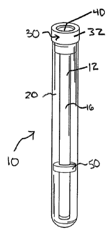
[10] FIGURE IA is a perspective view of an exemplary embodiment of a sample
vessel;
[11] FIGURE 1B is a side-elevational view in cross-section of the sample
vessel of FIGURE
IA;
[12] FIGURE 1C is an exploded view of the sample vessel of FIGURE 1A,
illustrating the
tubule and collar removed from the container;
[13] FIGURE 2A is a perspective view of an exemplary embodiment of a sample
vessel;
[14] FIGURE 2B is a side-elevational view in cross-section of the sample
vessel of FIGURE
2A;
[15] FIGURE 2C is an exploded view of the sample vessel of FIGURE 2A,
illustrating the
tubule and collar removed from the container;
[16] FIGURES 3A-3B are side-elevational views in cross-section of an exemplary
embodiment of a sample vessel having a pair of lumens separated by a pressure
gate;
[17] FIGURE 4 is a side-elevational view in cross-section of an exemplary
embodiment of a
sample vessel having three lumens separated by a pair of pressure gates;
[18] FIGURE 5 is a side-elevational view in cross-section of another exemplary
embodiment
of a sample vessel having three lumens separated by a pair of pressure gates,
illustrating a self-
sealing, reinforced wall section for facilitating injection by a needle;
-3-
CA 02460192 2004-03-10
WO 03/022435 PCT/US02/28951
[19] FIGURE 6A is a perspective view of an exemplary embodiment of a sample
vessel
having a pair of lumens connected by a micro-fluidic channel;
[20] FIGURES 6B-6C are digital photographs of the sample vessel of FIGURE 6A
illustrating
fluid flow through the lumens of the sample vessel;
[21] FIGURE 7A is a perspective view of an exemplary embodiment of a segmented
sample
vessel having a plurality of lumens;
[22] FIGURES 7B and 7C are cross-sectional views of the sample vessel of
FIGURE 7A;
[23] FIGURE 8 a perspective view of another exemplary embodiment of a
segmented sample
vessel having a plurality of lumens, illustrating a hinged cover for the
sample vessel;
[24] FIGURE 9 a perspective view of an exemplary embodiment of a segmented
sample
vessel having a plurality of lumens, illustrating alternative interfaces for
the sample vessel;
[25] FIGURE 10A is a side elevational view in partial cross-section of an
exemplary
embodiment of a sample vessel, illustrating the compression of the sample
vessel;
[26] FIGURE I OB is a cross-sectional view of the sample vessel of FIGURE I OA
taken along
a line transverse to the longitudinal axis of the tubule 12;
[27] FIGURES 11A-11C are side elevational views in cross-section of an
exemplary
embodiment of a sample vessel, illustrating compression of the sample vessel
into a plurality of
configurations;
[28] FIGURE 12 is a side elevational view in cross-section of an exemplary
embodiment of a
sample vessel having a composite cross-section and a micro-fluidic flow
channel;
[29] FIGURES 13A-13B are side elevational views in cross-section of another
exemplary
embodiment of a sample vessel having a composite cross-section and a micro-
fluidic flow
channel, illustrating the sample vessel in a open configuration (FIGURE 13A)
and a compressed
configuration (FIGURE 13B);
[30] FIGURE 14A is a side elevational view in cross-section of an exemplary
embodiment of
a sample vessel having a plurality micro-fluidic flow channels interconnecting
a plurality of
depressions formed on an interior wall surface of the sample vessel;
[31] FIGURE 14B is a top view of an interior wall surface of the sample vessel
of FIGURE
14A;
-4-
CA 02460192 2004-03-10
WO 03/022435 PCT/US02/28951
[32] FIGURES 15A and 15B are side elevational views in cross-section of an
exemplary
embodiment of a sample vessel having a composite cross-section including
opposed planar wall
sections, illustrating the sample vessel in an open configuration (FIGURE 15A)
and a
compressed configuration (FIGURE 15B);
[33] FIGURE 16A is a perspective view of an exemplary embodiment of a sample
vessel
having an adapter for facilitating handling of the sample vessel and/or
connecting of the sample
vessel to an external device;
[34] FIGURES 16B-16E are side elevational views in cross-section of a
plurality of
exemplary embodiments of an adapter connected to the sample vessel illustrated
in FIGURE
16A;
[35] FIGURES 17A-17E are perspective views of an apparatus for drawing a
sample into a
sample vessel, illustrating the operation of the apparatus;
[36] FIGURE 18 is a perspective view of another exemplary embodiment of a
sample vessel,
illustrating the sample vessel with a portion of the wall removed to show a
microarray on an
interior surface of the wall of the sample vessel.
Detailed Description
[37] To provide an overall understanding, certain exemplary embodiments will
now be
described; however, it will be understood by one of ordinary skill in the art
that the sample
vessels and methods described herein can be adapted and modified to provide
devices and
methods for other suitable applications and that other additions and
modifications can be made
without departing from the scope of the present disclosure.
[38] Unless otherwise specified, the exemplary embodiments described below can
be
understood as providing exemplary features of varying detail of certain
embodiments, and
therefore, unless otherwise specified, features, components, modules, and/or
aspects of the
exemplary embodiments can be otherwise combined, separated, interchanged,
and/or rearranged
without departing from the scope of the present disclosure. Additionally, the
shapes and sizes of
components are also exemplary and unless otherwise specified, can be altered
without affecting
the disclosed devices or methods.
-5-
CA 02460192 2004-03-10
WO 03/022435 PCT/US02/28951
[39] The present disclosure is directed to sample vessels that may be utilized
to collect and
process one or more samples in a closed system. Exemplary samples that may be
collected,
processed, or otherwise contained by the sample vessels disclosed herein
include biological
samples, such as blood, urine, saliva, tissue, cell suspensions, microbial
organisms, viruses,
nucleic acids, and oligonucleotides samples; soil; water; and any other sample
materials that may
be assayed using known assays. The term "collection" as used herein generally
refers to the
extraction or gathering of the sample from a sample source, the subsequent
transfer of the sample
into the sample vessel, or the combination of extraction and subsequent
transferring of the
sample into the sample vessel. Exemplary sample gathering may include
pipetting, biopsying,
swabbing, drawing a fluid sample or other methods for extracting a sample from
a sample
source. Exemplary sample sources may include humans or other animals, plants,
water sources,
cell cultures, food, other sample vessels, and chemical and biological assays.
Sample sources
may also include interim storage media, for example, test tubes, syringes,
absorbent applicators
and other interim storage media for containing a sample of interest. The term
"processing" as
used herein generally refers to the preparation, treatment, analysis, and/or
the performance of
other testing protocols or assays on a content of the sample vessel in one or
more steps.
Exemplary processing steps include, for example: displacing a content, e.g.,
the sample or a
reagent, of the sample vessel within the sample vessel to, for example, adjust
the volume of the
content, separate content components, mix contents within the sample vessel;
effecting a
chemical or biological reaction within a segment of the sample vessel by, for
example,
introducing a reagent to the sample, agitating the sample, transferring
thermal energy to or from
the sample, incubating the sample at a specified temperature, amplifying
components of the
sample, extracting, separating and/or isolating components of the sample; or
analyzing the
sample to determine a characteristic of the sample, such as, for example, the
quantity, count,
volume, mass, concentration, or expression level of a molecule, a target, a
content, a marker or
an analyte, binding activity, nucleic acid sequence, or nucleic acid size or
other analyte size, of
the sample. One skilled in the art will appreciate that the forgoing exemplary
processing steps
are described herein for illustrative purposes only. Other processing steps
may be employed
without departing from the scope of the present disclosure.
-6-
CA 02460192 2004-03-10
WO 03/022435 PCT/US02/28951
[40] Figures 1A-1C illustrate an exemplary embodiment of a sample vessel 10
for collecting
and processing one or more samples. The illustrated sample vessel 10 comprises
a tubule 12 that
provides a disposable, single use container and collection and processing
vessel for the sample.
The tubule 12 may be constructed from any biocompatible material and may be
manufactured by
injection molding, insert molding, dip molding, blow-molding, extrusion, co-
extrusion,
lamination, assembling from a sheet material, or other processes generally
used to manufacture
medical devices and implants. The tubule 12 may receive sample in solid or
liquid form and, in
certain embodiments, may be sized to collect and/or process sample volumes in
the range of 2
microliters to 2000 microliters.
[41] The tubule 12 may be used with any known sample testing or processing
system,
including, for example, the systems described in U.S. Patent Number 6,318,191,
U.S. Patent
Application Serial No. 09/339,055, and U.S. Patent Application Serial No.
09/782,732. Each of
the aforementioned patents and patents applications is incorporated herein by
reference.
[42] In the exemplary embodiment illustrated in FIGURES 1 and 2, the tubule 12
may include
an opening 14 for receiving a volume of sample material. The tubule 12 may
include a
compressible segment 16 having a wall 18 constructed at least partially from a
material having
sufficient flexibility to permit compression of the opposed segments of the
wall 18 into contact.
For example, the wall 18 may be constructed to converge when the compressible
segment 16 of
the tubule 12 is compressed in a direction perpendicular to the longitudinal
axis of the tubule
such that the volume of the compressed segment 16 of the tubule 12 decreases,
without
fracturing of the sample vessel. The walls 18 of the compressible segment 16
may be
constructed of a resiliently compressible, flexible, and ultra-high strength
material, such as
polyethylene, polyurethane, polyvinyl chloride, polypropylene, or any other
plastic material
suitable for biomedical or chemical assaying applications. In one illustrative
embodiment, the
walls 18 of the compressible segment 16 have a wall thickness of approximately
0.01mm to
0.5mm. Experimental results indicate that constructing a compressible segment
of a tubule
having a wall thickness within this range significantly increases the
efficiency of sample
processing, such as heat transfer to the sample and sample transfer between
the segments, and
detection. In the illustrated embodiment, the compressible segment 16 of
tubule 12 extends the
entire length of the tubule 12. Alternatively, as discussed below, the tubule
12 may include one
-7-
CA 02460192 2004-03-10
WO 03/022435 PCT/US02/28951
or more discrete compressible segment 16 spaced apart from one or more
segments having
different (e.g., non-flexible) properties.
[43] In other exemplary embodiments, the tubule 12 may comprise a multi-layer
wall
structure. For example, the tubule 12 may include an inner layer providing bio-
compatibility,
using material such as polyethylene or polyurethane, and an outer layer
providing lower
permeability, using material such as high density polyethylene or a metal
foil, such as aluminum
foil or a metal deposition. One skilled in the art will appreciate that one or
more additional
layers may also be employed, depending on, for example, the sample type, the
reagent(s)
employed, and the assay(s) being performed.
[44] The material selected to construct portions of the wall of the tubule 12,
for example an
optional detection segment of the tubule 12, can be optically transmissive
over a selected
wavelength range to facilitate optical analysis of the sample within the
tubule 12.
[45] The sample vessel 10 of the exemplary embodiment illustrated in FIGURES
lA-1C may
comprise a general rigid container 20 for receiving all or at least a portion
of the tubule 12. In
the illustrated embodiment, the container 20 is sized to receive the complete
length of the tubule
12. The container 20 may be constructed of a material having increased
rigidity compared to the
material of the tubule 12 to facilitate handling of the tubule 12. In certain
embodiments, the
container 20 may be constructed of a material having a lower permeability than
the material of
the tubule 12. In the illustrated embodiment, the container 20 is a glass
vacuum tube. Suitable
glass vacuum tubes are available under the trademark VACUTAINER from Becton-
Dickenson. The sample vessel 10 can be used in a manner similar to a glass
vacuum tube to
collect a sample, such as a blood sample. A container 20 may be optionally
used with any of the
tubule embodiments disclosed herein.
[46] The sample vessel 10 may comprise an interface 30 that is in fluid
communication with
the opening 14 in the tubule 12. The interface 30 may permit collection of the
sample within the
tubule 12 by facilitating delivery of the sample material to the tubule 12
through the opening 14.
In certain exemplary embodiments, the interface 30 may include an instrument
for collecting the
sample form a sample source. In the exemplary embodiment illustrated in
FIGURES 1A and 1B,
the interface 30 is a stopper 32 that may be coupled to the tubule 12 and may
selectively seal the
opening 14 in the tubule 12 to facilitate collection of the sample from a
separate instrument. In
-8-
CA 02460192 2004-03-10
WO 03/022435 PCT/US02/28951
the exemplary embodiment, the stopper 32 is removably and replaceably
connected to the rigid
container 20 and seals an opening 22 in the container 20. The stopper 32 may
include a first
annular portion 34 having an opening 36 sized and shaped to receive the tubule
12 in a fluid tight
relationship. The first annular portion 34 is further sized and shaped to
engage the walls of the
container in a fluid tight relationship. The stopper 32 may include a second
annular portion 38
that has a diameter greater than the diameter of both the first annular
portion 34 and the container
20. The opening 36 extends through the second annular portion 38 to form an
interface channel
37. A penetrable, self-sealing portion 40, such as a self-sealing membrane,
may be provided to
selectively seal the opening 36 and, thus, permit selective transfer of the
sample (from, for
example, the sample collection instrument) through the interface channel 37
into the tubule 12.
The self-sealing portion 40 may be constructed of any biocompatible,
resilient, self-sealing
material that can be penetrated by a needle or other sample collection
instrument. Suitable
materials may include rubber and silicon. In certain embodiments, the stopper
32 may be
constructed completely from a biocompatible, resilient, self-sealing material
such as rubber or an
elastomeric polymer. The interface channel 37 may taper or otherwise narrow
through the cross-
section of the stopper 32 to provide a guide for a needle or other instrument
transferring the
sample to the tubule 12.
[47] Alternatively, the interface 30 may include other mechanisms for
selectively sealing the
opening 14 in the tubule 12. For example, the interface may include a self-
sealing elastomeric
duckbill closure. Alternatively, the interface 30 may include a valve for
selectively closing and
opening the interface channel 37.
[48] The sample vessel 10 may include a clamp 50 for compressing the
compressible segment
18 of the tubule to adjust the volume of the tubule 12. The clamp 50 may be
configured to
compress opposing wall portions of the compressible section 16 into contact
thereby dividing the
tubule 12 into two segments, 16A and 16B, as best illustrated in FIGURE 1 B.
When the clamp
50 is employed, the segment 16A remains in fluid communication with the
interface channel 37
and segment 16B is sealed from segment 16A by the clamp 50. Once the sample is
delivered to
the segment 16A of the tubule 12, the clamp 50 may be removed, providing
additional volume in
the tubule 12 that may permit future segmentation of the tubule and
displacement of the sample
within the tubule 12 by compression of the tubule 12.
-9-
CA 02460192 2009-12-01
[49] The clamp 50 may be positioned at any location along the longitudinal
axis of the tubule
12. Additional clamps may also be employed to divide the tubule into
additional segments. In
illustrated exemplary embodiment, the clamp 50 is disk-shaped and includes a
radial slot 52 that
is sized to receive the tubule 18 in a compressed state. One skilled in the
art will appreciate that
other devices may used to compress and, thereby, divide the tubule 12.
[501 In certain exemplary embodiments, the tubule 12 may be wholly or
partially evacuated to
place the lumen 42 of the tubule 12 under negative pressure, e.g., at a
pressure less than
atmospheric pressure, to facilitate fluid flow into the tubule 12. Negative
pressure can be
generated by, for example, compressing the tubule 12 to collapse the lumen 42.
An apparatus
suitable for compressing the tubule is illustrated in FIGURES 17A-17C,
described below.
Alternatively the tubule 12 may be compressed by hand. The tubule 12 may also
be
manufactured to include a negative pressure.
[511 In certain embodiments, the container 20 may be wholly or partially
evacuated to a
negative pressure. For example, the container 20 may be evacuated to inhibit
loss of negative
pressure within the tubule 12 and to hold the shape of the tubule 12 during
storage.
[52] A reagent may be pre-packaged in the tubule 12 or can be introduced to
the tubule 12
after the sample is introduced to the tubule 12. For example, a reagent can be
introduced using a
reagent injector cartridge associated with the sample processing system, by a
needle, or by
another device capable of fluid communication with the tubule 12. The reagent
can be, for
example, an anticoagulant, a cell lyses reagent, a nucleotide, an enzyme, a
DNA polymerase, a
template DNA, an oligonucleotide, a primer, an antigen, an antibody, a dye, a
marker, a
molecular probe, a buffer, or a detection material. The reagent can be in
liquid or solid form. In
the case of a solid reagent, the reagent may be coated onto the walls of the
tubule 12.
1531 In certain exemplary embodiments, the interface 30 may include one or
more chambers
44 that are in fluid communication with the tubule 12 to selectively receive a
volume of fluid,
such as the sample material or a reagent, from the tubule 12. In certain
exemplary embodiments,
the chamber 44 may be evacuated or constructed to have a substantially small
initial volume and
may be expendable when receiving fluid. The chamber 44 can be used as a waste
container to
receive and store overflow sample, wash buffer, or reaction mixture during the
sample
- 10-
CA 02460192 2004-03-10
WO 03/022435 PCT/US02/28951
processing. For example, compressing a segment of the tubule 12 may move a
portion of the
sample to the chamber 44.
[54] In the exemplary embodiment illustrated in FIGURES 2A-2C, for example,
the stopper
32 includes an annular chamber 44 that is in fluid communication with the
interface channel 37
in the stopper 32, and, thus, the tubule 12, through a pressure gate 48. In
certain embodiments
described herein, one or more pressure gates may be employed to selectively
control the flow of
fluid between segments, lumens, and other portions of the tubule, as well as
between the tubule
and external devices. For example, the illustrated pressure gate 48 provides a
fluid tight seal
between the chamber 44 and the interface channel 37 under normal operating
conditions. The
pressure gate 48 may open upon the application of a fluid pressure greater
than a certain
threshold pressure, for example, approximately 3 atmospheres. When a fluid
pressure equal to or
greater than the threshold pressure is applied to the pressure gate 48, the
pressure gate 48 can
open, allowing the sample or a reagent to flow from the high-pressure
compartment, e.g., from
the tubule 12 or from the chamber 44, to the low-pressure compartment. In
certain
embodiments, the pressure gate may be reversible, i.e., the pressure gate may
be configured to
re-close if the fluid pressure is reduced to value less than the threshold
pressure. In other
embodiments, the pressure gate may be irreversible, i.e., the pressure gate
may be initially closed
and may remain open once opened. For example, once a threshold pressure is
exceeded the
irreversible pressure gate remains open, even if the pressure applied to the
pressure gate is
reduced to below the threshold pressure. One example of an irreversible
pressure gate is the
pressure gate described below in connection with FIGURES 3A-3B.
[55] In the illustrated embodiment of FIGURES 2A and 2B, the pressure gate 48
is a slit
formed in the stopper 32 between the interface channel 37 and the chamber 44.
The material
forming the stopper 32 may be selected to be sufficiently flexible and
resilient to allow the slit to
open at the threshold pressure and to close at pressures lower than the
threshold pressure.
[56] A label 60 identifying the sample within the sample vessel 12 may be
attached to the
interface 30, the container 20, or the tubule 12. The label 60 can be a bar
code or other indicia
for identifying the sample.
[57] FIGURES 3A and 3B illustrate another exemplary embodiment of a sample
vessel 100.
The sample vessel 100 comprises a tubule 112, which can be analogous in
construction to the
- 11 -
CA 02460192 2004-03-10
WO 03/022435 PCT/US02/28951
tubule 12, having a plurality of lumens 142A and 142B. The plurality of lumens
142A and 142B
can be separated by a pressure gate 148 that permits selective fluid flow
between the lumens
142A and 142B. FIGURE 3A illustrates the pressure gate 148 in a closed
position and FIGURE
3B illustrates the pressure gate in an open position that permits fluid flow
between the lumens.
[58] In the exemplary embodiment, the lumens 142A and 142B are parallel to
each other and
extend in a direction generally parallel to the longitudinal axis of the
tubule 12. One skilled in
the art will appreciate that other lumen orientations are possible. The lumens
142A and 142B
may be uniform in size (e.g., diameter, width, length) and shape or,
alternatively, the lumens
142A and 142B may be different in size and shape, as in the illustrated
embodiment. For
example, in the illustrated embodiment, the lumen 142B has a smaller diameter
than the lumen
142A. Although two lumens are illustrated in the exemplary embodiment, one
skilled in the art
will appreciate that the tubule 12 maybe constructed of any number of lumens.
[59] The pressure gate 148 in the present embodiment is coextensive with the
lumens 142A
and 142B, i.e. the pressure gate 148 extends along the entire length of the
lumens. Alternatively,
the pressure gate 148 may extend along only a portion or portions of the
lumens, particularly in
embodiments in which the tubule 12 is segmented into discrete longitudinally
extending
segments, as in the case of the embodiment illustrated in FIGURES 7A-7C. In
such
embodiments, one or more pressure gates may be provided between adjacent
lumens.
[60] In the exemplary embodiment, the opposed portions of the wall 118 of the
tubule 112 are
compressed into contact to form a longitudinally extending seam 170 that
divides the tubule 112
into two lumens, lumens 142A and 142B. In addition to dividing the tubule 112
into multiple
lumens, the seam 170 may further provide an irreversible pressure gate,
pressure gate 148,
between the lumens 142A and 142B. The seam 170 may be formed by mechanically
clamping
or otherwise compressing a cylindrical tubule or by applying vacuum pressure
to the interior of a
cylindrical tubule. Alternatively, the seam 170 may be formed during
manufacturing of the
tubule by, for example, extrusion, molding, or lamination processes. The
opposed wall portions
that are compressed into contact to form the seam 170, and the pressure gate
148, may be bonded
together by mechanical or chemical bonding, by heating sealing, for example,
by bringing hot
surfaces into contact with the tubule wall immediately after extrusion, by
ultrasonic welding, by
-12-
CA 02460192 2004-03-10
WO 03/022435 PCT/US02/28951
mechanical interlocking, or both other connection mechanisms, to create the
irreversible pressure
gate 148.
[61] The pressure gate 148 is initially in a closed configuration that
inhibits fluid flow
between the lumens 142A and 142B. The pressure gate 148 may open by separating
the
compressed opposed walls forming the pressure gate 148. Applying a threshold
pressure to the
pressure gate 148, as described above, may open the pressure gate 148.
Alternatively, energy
may be applied to the pressure gate 148 to weaken the bond between the
compressed opposed
walls. For example, thermal energy or light, e.g., ultra-violet light, may be
applied to the
pressure gate 148 or to selected portions or all of the tubule 112. The
threshold pressure and/or
the amount energy to open the pressure gate 148 may vary depending on the type
and strength of
the bond. Alternatively, the bond between the compressed opposed wall portions
may be
weakened or broken by chemical reaction with reagent or the sample.
[62] In certain exemplary embodiments, one or more of the lumens may include
one or more
reagents. Reagents may be provided to one or more lumens prior to sample
collection, e.g., one
or more reagents pre-packaged with the tubule, or after sample collection. In
the exemplary
embodiment illustrated in FIGURES 3A and 3B, for example, a reagent may be
provided in
lumen 142B. Lumen 142A may be utilized for sample collection and processing.
Sample
collection may occur with the pressure gate 148 in a closed configuration, as
illustrated in
FIGURE 3A. Upon transfer of the sample to lumen 142A, the pressure gate 148
may be opened
automatically due to release of pressure within the lumen 142A, or selectively
by applying
energy to the pressure gate and/or a threshold fluid pressure. In other
embodiments, the lumen
142A or 142B may be compress to provide the threshold pressure. Upon opening
the pressure
gate 148, the reagent(s) can mix with and interact with the sample in the
lumen 142A, as
illustrated in FIGURE 3B. Automatic release of the pressure gate 148 and
mixing of the reagent
with the sample may be beneficial in certain applications, such as the mixing
of an anticoagulant
with a blood sample.
[63] FIGURE 4 illustrates another embodiment of a multi-lumen tubule 112 that
includes
three lumens, namely a first lumen 142A, a second lumen 142B, and a third
lumen 142C. Each
lumen may be separated a pressure gate 148, for example, an irreversible
pressure gate, as
described above. Each of the lumens 142A, 142B, and 142C may be provided with
one or more
-13-
CA 02460192 2004-03-10
WO 03/022435 PCT/US02/28951
reagents and/or may be used for sample collection and processing. For example,
second lumen
142B may be provided with one or more prepackaged reagents and first lumen
142A may be
used for sample collection and processing. Upon sample collection in first
lumen 142A, pressure
gate 148A may be opened allowing fluid communication between the second lumen
142B and
the first lumen 142B. FIGURE 4 illustrates the pressure gate 148A in an open
configuration.
Lumen 142C may be utilized as an injection channel for receiving one or more
reagents,
typically, but not necessarily, after sample collection in first lumen 142A.
The lumen 142C may
be free of sample material until pressure gate 148A is transitioned to an open
configuration.
FIGURE 4 illustrates pressure gate 148B in a closed configuration that
inhibits fluid
communication between third lumen 142C and first lumen 142A. Reagent may be
delivered to
the third lumen 142C by a needle 190, such as a needle from a reagent
injection cartridge, or by
other instruments that can penetrate the lumen or otherwise provide fluid
communication
between a reagent source and the lumen 142C. The lumen 142C may be free of
sample and
reagent material until reagent is injected to avoid cross contamination of the
injection needle
190. The portion of the wall 118C proximal the third lumen 142C may be
constructed of a
resilient, self-sealing material to facilitate re-sealing of the wall 118
after penetration to deliver
reagent.
[64] One or more lumens of the tubule 112 may include a reinforced wall
portion 171, as
illustrated in FIGURE 5. The reinforced wall portion 171 may have an increased
wall thickness
compared with the remainder of the tubule wall 118 to facilitate needle
penetration and re-
sealing. For example, the reinforced portion may have a wall thickness of
approximately 1 mm
to 5 mm grater than other portions of the wall. The reinforced portion 171 may
be constructed
from a different material, having increased strength and/or resiliency, for
example, than the
remainder of the tubule wall 118. Needle guides 172 may be provided to direct
needle
penetration and inhibit tearing of the tubule wall 118.
[65] FIGURES 6A-6E illustrate another exemplary embodiment of a multi-lumen
tubule 112
that includes a pair of parallel lumens, namely first lumen 142A and 142B. In
the illustrated
embodiment, the lumens 142A and 142B are connected parallely by a thin layer
fluid channel
176 in the form of a slit opening that extends the length of the tubule 112.
Although one fluid
channel is illustrated, additional fluid channels may be provided. The fluid
channel 176 permits
-14-
CA 02460192 2009-12-01
the sample to be moved between the first lumen 142A and the second lumenl42B
and to occupy
both lumens simultaneously. For example, during sample collection, portions of
the sample, or
the entire sample, can be transferred from the opening 114 along the length of
the first lumen
142A, through the fluid channel 176, and along the length of the second lumen
142B. FIGURES
6B-6E illustrate the flow of a sample, in fluid form, through the first lumen
to the end of the first
lumen due to relatively low flow resistance of the lumen relative to the fluid
channel 176
(FIGURE 6B), through a portion 174 of the fluid channel 176 distal to the
opening in the first
lumen 142A (FIGURE 6C), along the fluid channel 176 and through the second
lumen 142B
(FIGURE 6D) to fill both lumens (FIGURE 6D). In embodiments in which a solid
reagent is
packed into the lumens 142A and/or 142B of the tubule 112, flow of the sample
through the
lumens via the fluid channel 176 can facilitate mixing of the solid reagent
with the sample. For
example, in the case of blood samples, the inventors have determined that by
allowing the blood
sample to flow through the first lumen 142A and the second lumen 142B via the
fluid channel
176 can improve mixing of the sample with an anticoagulant coated on the inner
walls of the two
lumens.
[661 FIGURES 7A-7C illustrate another exemplary embodiment of a multi-lumen
tubule 112
having three parallel lumens, namely a first lumen 142A, a second lumen 142B,
and a third
lumen 142C. In the exemplary embodiment, each lumen of the tubule 112 is
divided into a
plurality of longitudinally extending segments 180. For example, the third
lumen 142C,
illustrated in cross-section in FIGURE 7B, includes five segments 180A-E. Each
of the
segments 180 can be used for one or more sample collection and/or sample
processing steps,
including the processing steps described above. In PCR (polymerase chain
reaction) testing, for
example, one segment may used for sample collection, one segment may be used
for sample
pretreatment, e.g., nucleic acid extraction, one or more segments may used for
sample
processing, e.g., thermocycling, and one or more segments may be used for
sample analysis.
Any number of segments may be provided. In addition, one or more segments may
be used to
store reagent or as an injection channel for the delivery of reagent. The
number of segments may
be varied depending of the sample being processed and the processing steps
selected.
[671 Each of the segments 180 may be separated by a seal 182 that provides a
temporary or
permanent fluid seal between adjacent segments 180. A seal 182 may be a
pressure gate, such as
- 15-
CA 02460192 2004-03-10
WO 03/022435 PCT/US02/28951
the reversible and irreversible pressure gates described above. Alternatively,
a seal 182 may be
formed by bonding or fusing of compressed opposed wall sections of the tubule.
The seal 182
may be formed by applying energy, such as thermal energy or RF energy, by
ultrasonic welding,
or by using a bonding agent. A clamp may also be applied to the exterior of
the tubule to
compress the wall of the tubule and form a seal separating the segments in the
tubule. For
example, the clamp may be an electro-mechanical clamping mechanism as
described below in
connection with FIGURE 10. Any other mechanism for provided an external
compressive force
on the tubule may be employed as the clamping mechanism. One or more clamps
may be
provided as part of the sample processing system used to process the sample
within the tubule
112. The segments may be connected by one or more micro-fluidic flow channels
that provide
selective fluid connection between the segments, as described below. A seal
182 may be a filter
disposed within the tubule to separate selected components of a fluid within
the tubule from
other segment or components of the fluid within the tubule.
[68] In the illustrated exemplary embodiment, the interface 30 for
facilitating delivery of the
sample to the tubule 112 includes a needle 184 for direct collection of the
sample to be processed
with the sample vessel 100. The needle 184 is positioned a proximal end of the
tubule and is
fluid communication with an opening in the tubule 112. In the illustrated
exemplary
embodiment, the needle 184 is in fluid communication with an opening in the
first lumen 142A,
however, the needle 184 may be connected to any one or all of the lumens 142
of the tubule 112.
A removable and replaceable needle cover 186 may be provided to secure the
needle 184 prior to
and after use. Alternatively, the needle cover 186 may be connected by a
hinge, as shown in
FIGURE 8, or by another mechanism that allows to the cover 186 to be moved
into and out of
position about the needle 184. A needle safety mechanism may be coupled to the
needle and the
sample vessel.
[69] FIGURE 9 illustrates another embodiment of the cover 186 in which the
sample
collection instrument, e.g., the needle 184, is connected to the cover 186. In
the illustrated
exemplary embodiment, the proximal end 184A of the needle 184 may be used for
sample
collection from a sample source and the distal end 184B of the needle 184 may
be used to
provide a fluid connection with an opening in the tubule 112 through interface
30. For example,
the distal end 184B may be used to penetrate a self-sealing membrane 40
provided in the
-16-
CA 02460192 2004-03-10
WO 03/022435 PCT/US02/28951
interface 30. In another embodiment, a cover 190 may include a sample
instrument in the form
of a needle 184 and may have a compressible portion in fluid communication
with the needle to
facilitate drawing a fluid sample into the needle 184 and transferring the
sample to the sample
vessel 110. Cover 190 may be particularly useful as a finger prick for
collection a blood sample.
[70] FIGURE 10 illustrates a processing station 300 of an exemplary sample
processing
device, such as a sample processing device described in U.S. Patent
Application No. 6,318,191
and U.S. Patent Application Serial Number 09/782,732, filed February 13, 2001.
The exemplary
processing station 300 includes multiple compression members, namely first
compression
member 302A, second compression member 302B, and third compression member
302C. Each
compression member 302 is adapted to compress a sample vessel, for example,
the tubule 12 of
sample vessel 10 described above, and thereby displace the contents of the
sample vessel, e.g.
reagent or sample, within the sample vessel. Although the exemplary processing
station 300 is
illustrated in connection with sample vessel 10, one skilled in the art will
appreciate that any of
the sample vessels disclosed herein may be used in with the exemplary
processing station 300.
A plurality of compression members 302 may be oriented parallel to the
longitudinal axis of the
tubule 12, as illustrated in FIGURE 10A. Alternatively, a plurality of
compression members 302
may be oriented transverse to the longitudinal axis of the tubule, i.e.,
oriented latitudinally, as
illustrated in FIGURE 15B described below, or in other orientations depending
on the
compression configuration desired. A driver may be coupled to one or more of
the compression
members 302 to selectively move the compression member into contact with the
sample vessel.
The driver can be, for example, an electromagnetic actuating mechanism, a
motor, a solenoid, or
any other device for imparting motion on the compression members 302. A
stationary member
304 or another compression member may be provided opposite compression member
302.
[71] A compression member 302 may be employed to compress a portion of the
wall 18 of the
tubule 12 into contact with another portion of the wall 118 of the tubule 12
to form a seal in the
tubule 12 and thereby divide the tubule 12 into multiple segments. In
alternative embodiments, a
compression member 302 may compress a portion of the wall 18 of the tubule 12
into proximity
with another portion of the wall 18 of the tubule 12 to form a micro-fluidic
channel 306 between
segments of the tubule 12. For example, in the embodiment illustrated in
FIGURE 10,
compression member 302B compresses a portion of wall 18 into proximity with
another portion
-17-
CA 02460192 2004-03-10
WO 03/022435 PCT/US02/28951
of the wall to create a micro-fluidic channel 306 that connects a first
segment 180A and a second
segment 180B of the tubule 12. The width of the micro-fluid channel 306 may be
adjusted by
displacing the compression member 302B towards or away from the tubule 12.
Micro-fluid
channels may be formed having a gap less than 200 microns, preferably 10 to 30
microns.
[72] The compression members 302 may be arranged in a variety of orientations
to compress
the tubulel2 into a variety of configurations. For example, in FIGURE I OB,
the width of the
micro-fluidic channel 306 extends across the entire width of the tubule 12.
Such a compressed
configuration may be formed by a compression member 302B having a planar
compression
surface 308 for engaging the tubule 12 that is sized to engage the entire
compressed wall surface
of the tubule. In other embodiments, the size or shape of the compression
surface 308 may be
varied and the number and orientation of compression members 302B may be
varied. For
example, FIGURE 11 A illustrates a compressed tubule 12 having a centrally
located flow
channel 306 that may be formed by a compression member 302 having a groove
formed on the
bottom surface thereof or by three compression members 302 aligned transverse
to the
longitudinal axis of the tubule 12. A centrally positioned compression member
may compress
wall portion 18A into proximity with an opposed wall portion, while a pair of
compression
members, one on either side of the central compression member, may compress
side wall
portions 18B and 18C, respectively. FIGURE 11B illustrates a compressed tubule
12 having a
centrally located lumen 306 formed by compressing the tubule 12 into a non-
planar
configuration. In this illustrated embodiment, a triangular profile flow
channel is formed, which
inherently forces a cell or particle to flow through the central line of the
channel, thus reducing
the need to regulate the tolerance in forming the flow channel. FIGURE 11 C
illustrates a
compressed tubule 12 having a flow channel 306 formed off-set from the center
of the tubule 12.
In the illustrated embodiment, the flow channel is formed on a lateral edge of
the tubule 12.
[73] At least a portion of the wall of the tubule 12 may be optically
transparent to allow
monitoring or detection of the sample or reaction. The transparent portion of
the wall may be
located in the flow channel section, thus allowing the monitoring of sample or
reaction under
flow or through a thin layer of liquid, for processes such as counting cells,
reaction hybridization,
or detection, for example, microarray spots.
-18-
CA 02460192 2004-03-10
WO 03/022435 PCT/US02/28951
[74] One skilled in the art will appreciate that while it may be desirable in
certain applications
for the wall of the tubules disclosed herein to be uniform along the
circumference and the
longitudinal axis of the tubule, only a portion of the wall along the
circumference and/or
longitudinal axis of the tubule need be resilient and compressible. Thus, the
tubule need not
have a uniform cross-section, either along the longitudinal axis or transverse
to the longitudinal
axis. In certain exemplary embodiments, for example, a section of the wall of
the tubule may be
formed of a material selected to provide a property distinct from a property
of another section of
the wall. Exemplary properties that may be varied include permeability,
flexibility, hardness,
resiliency, optical transparency, biocompatibility, surface smoothness of the
inner wall, and
surface chemistry of the inner wall, for example the hydrophobic or
hydrophilic properties of the
inner wall surface. Surface properties may be rendered by coating with a layer
of material, such
as a thermoset urethane aired by UV energy or other cross linking methods.
[75] FIGURES 15A and 15B illustrate an exemplary embodiment of a sample vessel
10 in the
form of a tubule 12 having wall sections 18A that are formed of a material
selected to provide a
property distinct from a property of a plurality of other wall sections 18B of
the tubule 12. Wall
sections 18A may be opposed to one another, as illustrated, or positioned at
other positions in the
cross section of the tubule 12. Wall sections 18A may similar in size, shape
and material
properties, as illustrated, or may vary in size, shape, and material
properties from one another. In
the illustrated embodiment, wall sections 18A are selected from a material
having sufficient
flexibility to permit compression of the tubule 12, as illustrated in FIGURE
15B. Wall sections
18B are formed of a material having increased rigidity compared to the
material of wall sections
18A. In the illustrated embodiment, wall sections 18B preferably have
sufficient rigidity to resist
flexing during compression and thereby maintain a planar configuration. Wall
sections 18B may
be opposed to one another, as illustrated, or positioned at other positions in
the cross section of
the tubule 12. Wall sections 18B may be similar in size, shape and material
properties, as
illustrated, or may vary in size, shape, and material properties from one
another. In the
illustrated embodiment, the wall sections 18A and 18B are spaced
latitudinally, i.e., about the
circumference of the tubule 12 and transverse to the longitudinal axis. Wall
sections 18A and
18B are interposed between one another in an alternating arrangement about the
circumference
of the tubule 12. Wall sections 18A and 18B may be formed from the same
material or a
-19-
CA 02460192 2009-12-01
different material. For example, wall sections 18A may be formed of a
relatively low durometer
polyurethane, for example, in the range of from 40A to 90A depending on
thickness, and wall
sections 18B may be formed of a polyurethane having a relatively higher
durometer, for
example, in the range of from 40D to 90D depending on thickness. A tubule
having wall
sections of varying properties may be manufactured by conventional extrusion,
co-extrusion,
injection molding, insert molding, dip molding, blow molding, transfer
molding, or lamination
processes.
[76J During compression of the tubule 12 illustrated in FIGURE ISA and 15B,
the wall
sections 18A flex allowing a first wall section 18B to be moved into proximity
or contact with
second wall section 18B'. Wall sections 18B may provide improved sealing
surfaces due to the
increased rigidity compared with wall sections 18A. In addition, walls
sections 18B permit the
formation of a precisely defined micro-fluid flow channel 306, as illustrated
in FIGURE 15B.
The increased rigidity of the wall sections 18B allows for the formation of a
smaller and more
uniform flow channel than more flexible wall sections. FIGURE 15B illustrates
the formation of
a micro-fluidic flow channel 306 between segments 180A and 180B of the tubule
12. In the
illustrated embodiment, the compression members 302A-C are oriented transverse
to the
longitudinal axis of the tubule to form a flow channel 306 that extends
latitudinally, i.e.,
transverse to the longitudinal axis, between first segment 180A and second
segment 180B.
[771 In other exemplary embodiments, the number of wall sections of differing
properties may
be varied. For example, a single wall section 18B having increased rigidity
may be provided or
three or more wall sections having increased rigidity may be provided.
[781 In certain exemplary embodiments, a flow channel 306 may be pre-formed in
a section of
the wall 18 of the tubule as illustrated in FIGURES 12 and 13A-B. The pre-
formed flow channel
306 may be a groove 316 formed in a wall section of the tubule 12. The groove
316 may be
formed by scoring or etching the wall 18 of the tubule 12 or may be formed
during the extrusion
or molding of the tubule 12. The groove 316 in the illustrated embodiments
extends
longitudinally, however, the groove 316 may be formed in any direction,
including latitudinally.
More than one groove 316 may be provided. The groove 316 may have a variety of
cross-
section shapes and sizes. In the embodiment illustrated in FIGURE 12, the
groove 316 has a
triangular cross-section. In the embodiment illustrated in FIGURES 13A-13B,
the groove 316
- 20-
CA 02460192 2004-03-10
WO 03/022435 PCT/US02/28951
has a rectangular cross-section. The cross-sectional size of the groove 316
can be selected based
on desired shear rate profile of the flow channel 306.
[79] The groove 316 maybe formed in any section of the wall 18 of the tubule
12. For
example, the groove 316 may be formed in a wall section 1 8B having increased
rigidity
compared to other wall sections of the tubule 12, as is the case for the
illustrated embodiments of
FIGURES 12 and 13A-B. During compression of the tubule 12, as illustrated in
FIGURE 13B,
the wall section 18B contacts wall section 18B' to provide a fluid tight seal.
Groove 316
provides a flow channel 306 that extends longitudinally through the fluid
tight seal.
[80] FIGURES 14A and 14B illustrate an exemplary embodiment of a sample vessel
400
comprising a tubule 412 having a plurality of flow channels 306 and one or a
plurality of
depressions 408 formed on an interior wall surface 410 of the wall 418 of the
tubule 412. Each
depression 408 can form a micro-cup during compression of the tubule 412 that
can hold a fixed
volume of sample or reagent. The volume of a depression forming a micro-cup
can be from 0.1
microliter to 10 microliter, preferably, from 0.5 microliter to 4 microliter.
A pattern of one or
more grooves 316 and depressions 408 may be formed on the interior wall
surface 410 of the
tubule 412 and may interconnect to provide a network of micro-cups
interconnected by micro-
fluidic flow channels 306, as best illustrated in FIGURE 14B. Such a network
may be used to
perform a variety of processing steps within one or more micro-cups and may
permit the
transport of small, precise volumes of sample and reagent between the micro-
cups via micro-
fluid flow channels by selectively compressing the tubule 412. The network of
grooves and
depressions may be formed using semi-conductor processing techniques. For
example, a mask
pattern may be applied to an interior wall surface of the tubule 12 using
conventional
photolithographic techniques. The grooves and depressions may then be formed
by etching or
otherwise removing portions of the interior wall surface based on the pattern
imaged onto the
interior wall surface. It may be desirable to form the network of grooves and
depressions on a
planar substrate 418A constructed of a material suitable for use in the tubule
412, as illustrated in
FIGURES 14A and 14B. A second layer 418B of material can be attached to the
planar substrate
418A to form the wall 418 of the tubule 412.
[81] Referring to FIGURES 14A and 14B, one or more sample or reagent
processing devices
414 may be provided on the interior wall surface 410 of the tubule 412. For
example, a
-21-
CA 02460192 2004-03-10
WO 03/022435 PCT/US02/28951
microarray device may be embedded on the interior wall surface 410 of the
tubule 412. An
exemplary microarray device 414 may comprise a plurality of reagent coated
zones for
simultaneous analysis of a plurality of analytes within a sample. The
processing device 414 may
also be a micro-fluid device or a lab-on-a-chip device, or any other device
for processing a
sample. The processing device 414 may be interconnected with one or more
depressions 408 or
other processing devices via flow channels 306. Any number of processing
devices of any type
may be provided in the tubule 412.
[82] Referring to FIGURE 18, a sample vessel 700 comprising a tubule 712
divided into
multiple segments 780A-C. Segment 780B may be constructed of a rigid,
generally non-flexible
material and may have a processing device, such as a microarray 714, embedded
on the interior
wall thereof. The segment 780B may provide a pre-formed flow channel between
two
compressible segments 780A and 780C. By alternately compressing the two
flexible segments
708A and 780C, the sample may flow through the flow channel 706 to facilitate
high efficient
hybridization or binding of analytes to the reagent spots of the microarray
714. A flow channel
706 having a small gap may also increase wash efficiency as a laminar flow is
formed.
[83] FIGURES 16A-16E illustrates an exemplary embodiment of a sample vessel 10
comprising a tubule 12 and an adapter 500 that is connected to the tubule 12
of the sample vessel
10. The adapter 500 maybe provided to facilitate handling of the sample vessel
10 and/or to
facilitate connection of the tubule 12 to an external device, such as a micro-
fluid device, a lab-
on-a-chip device, a microarray device, a reagent source, another sample
vessel, or any other
device suitable for containing or processing a sample. In the illustrated
embodiments, the
adapter 500 is a generally planar tab that is coextensive with the tubule 12.
One skilled in the art
will appreciate that the adapter 500 need not be coextensive with the tubule
and may be
constructed of varying sizes and shapes depending upon the application.
Moreover, more than
one adapter may be provided.
[84] The adapter 500 may be constructed of any material suitable for use in
construction the
tubule 12. For example, the adapter may be constructed of polyurethane. The
adapter 500 may
be constructed of the same or a different material than the tubule 12. To
facilitate handling, the
adapter 500 may be constructed of a material having increased rigidity
compared to the material
of the tubule 12, for example a high durometer polyurethane. In certain
embodiments, the
-22-
CA 02460192 2009-12-01
adapter 500 may be manufactured with the tubule 12 in, for example, a co-
extrusion process or
an injection molding process. Alternatively, the adapter 500 may be
manufactured
independently and attached to the tubule 12 in a post-forming process by, for
example, bonding.
[851 The exemplary embodiment of FIGURES 16A-16E also includes a container 20
and an
interface 30, as described above. The container 20 removably and replaceably
encloses the
tubule 12 to protect the sample tubule 12 and when removed, may allow direct
manipulation of
tubule 12. A portion of adapter 500 may not be enclosed by container 20. The
exposed portion
of the adapter 500 can be directly accessed by a user for labeling, handling
and other processing.
The interface 30 includes an interface channel 37 that communicates with an
opening in the
tubule 12 to facilitate delivery of a sample to the tubule 12. In the
illustrated embodiment, a
removable and replaceable cover 586 is provided to selectively open and close
the interface
channel 37. The exemplary cover 586 includes a sample collection instrument in
the form of a
tissue swab 584 for collecting tissue samples from a sample source.
[861 FIGURES 16A-B illustrate an embodiment of the adapter 500 that is
constructed to
facilitate handling of the sample vessel 10.
[871 FIGURE 16C illustrates an embodiment of the adapter 500C that is designed
to facilitate
delivery of a reagent or a sample from an external device, such as a needle 90
from a reagent
injector cartridge. The adapter 500C includes a reversible or irreversible
pressure gate 48 that
provides a fluid channel to permit selective displacement of a fluid, e.g., a
sample or reagent,
between the tubule 12 and the external device, in the present embodiment,
needle 90. The
adapter 500C may include a self-sealing membrane 540, valve, or other sealing
mechanism to
facilitate selective communication with the external device. A reservoir 502
may be provide to
contain a fluid delivered from the external device or fluid from the tubule
12. In use, the needle
90 may penetrate the self-sealing membrane 540 to deliver fluid to the
reservoir 502 or to
withdraw fluid from the reservoir 502. Pressure gate 48 may be opened in the
manner described
above, e.g. by compressing the tubule 12 or the reservoir 502, to withdraw
fluid from the
reservoir 502 or to deliver fluid to the reservoir 502 from the tubule 12. The
needle 90 may be
coupled with a sensor, such as an electrode, a fiber optical sensor, for
penetrating the self sealing
membrane 540 and measuring a sample property.
-23-
CA 02460192 2004-03-10
WO 03/022435 PCT/US02/28951
[881 FIGURE 16D illustrates an embodiment of the adapter 500D that comprises a
compressible reservoir 506 and a reversible or irreversible pressure gate 48
that provides a fluid
channel to permit selective displacement of a fluid, e.g., a sample or
reagent, to the tubule 12
from the compressible reservoir 506. The compressible reservoir 506 may
contain a pre-packed
reagent. In certain embodiments, the compressible reservoir 506 may be a
blister pack. Upon
compression of the compressible reservoir 506, pressure gate 48 may open and
fluid with the
compressible reservoir 506 can be displaced in to the tubule 12.
[89] FIGURE 16E illustrates an embodiment of the adapter 500E that comprises a
reservoir
502, a first reversible or irreversible pressure gate 48A that provides a
fluid channel to permit
selective displacement of a fluid, e.g., a sample or reagent, between the
tubule 12 and the
reservoir 502, and a second reversible or irreversible pressure gate 48B that
provides a fluid
channel to permit selective displacement of a fluid, e.g., a sample or
reagent, between an external
device 508 and the reservoir 502. A connector 509 may be provided to interface
with the
external device 508. Such device may be an AccessTM card for Micronics Inc., a
LabChip
product from Caliper, Inc. or a GeneChip from Affymetrix, Inc.
[90] FIGURES 17A-E illustrate another exemplary embodiment of a sample vessel
600 that
comprises a tubule 10 and an apparatus 602 for drawing a sample into the
tubule 12 of the
sample vessel 10. The apparatus 602 includes a cylindrical housing 604 having
an opening 606
for receiving the tubule 12. The opening 602 extends from a proximal end 608
to a distal end
610 of the housing 604. Both the housing 604 and the opening 606 can be sized
and shaped to
accommodate the size and shape of the tubule 12 or other sample vessels. For
example, the
housing 604 and opening 606 are cylindrical in shape and have a circular cross-
section analogous
to that of the tubule 12. The adapter 600 comprises first means 612 for
compressing a first
portion of the tubule 12 and second means 614 for compressing a second portion
of the tubule
12. The first compression means 612 may be spaced apart from the second
compression means
614. For example, the first compression means 612 may be positioned at the
proximal end 608
of the housing 602 and the second compression means 614 may be positioned at
the distal end
610 of the housing 602. The spacing between the first and second compression
means may be
selected based on the desired sample collection volume in the tubule 12.
-24-
CA 02460192 2004-03-10
WO 03/022435 PCT/US02/28951
[91] The first compression means 612 may comprise a first pair of spaced apart
rollers, 616A
and 616B. At least one of the rollers 616A-B may be selectively movable into
contact with the
other roller to compress the tubule 12 between the rollers 616A-B. A first
activator 620 may be
coupled to the rollers 616A, 616B to effect separation or compression of the
rollers. The second
compression means 612 may comprise a second pair of spaced apart rollers, 618A
and 618B. At
least one of the rollers 618A-B may be selectively movable into contact with
the other roller to
compress the tubule 12 between the rollers 618A-B. A second activator 622 may
be coupled to
the rollers 618A, 618B to effect separation or compression of the rollers. In
addition to rollers,
or other compression mechanisms may be employed for the first and second
compression means,
including the compression members described above. Any structure suitable for
selective
compression of the tubule 12 may be employed. The first and second compression
means need
not be the same structure.
[92] In use, the tubule 12 is inserted into the opening 606 at the proximal
end 608 of the
housing 604 and drawn completely through the opening 606 to the distal end 610
of the housing
604. As the tubule 12 is drawn through the housing 604, the tubule 12 is
flatten and compressed,
as illustrated in FIGURE 17B, to evacuate the tubule 12. At the time of sample
collection, a
cover 686 may be removed to expose a sample collection instrument, such as a
needle 684, that
is in fluid connection with the tubule 12. The needle 684 can be inserted into
the sample source
and the first compression means 612 may be separated to draw the sample into
the tubule 12, as
illustrated in FIGURE 17C. The sample vessel 10 may then be inserted into a
device 630 for
removing the needle 684, or other sample collection instrument, as illustrated
in FIGURE 17D.
The device 630 may also include a mechanism for sealing the proximal end of
the tubule 12 after
the needle 684 is removed, by, for example, compressing and heating the wall
of the tubule 12 at
the proximal end to bond or fuse the walls together. The second compression
means 614 may
separate and the adapter 600 may be removed from the tubule 12, as illustrated
in FIGURE 17E.
[93] While the sample vessels disclosed herein have been particularly shown
and described
with references to exemplary embodiments thereof, it will be understood by
those skilled in the
art that various changes in form and details may be made therein without
departing from the
spirit and scope of the disclosure. Those skilled in the art will recognize or
be able to ascertain
using no more than routine experimentation, many equivalents to the exemplary
embodiments
-25-
CA 02460192 2004-03-10
WO 03/022435 PCT/US02/28951
described specifically herein. Such equivalents are intended to be encompassed
in the scope of
the present disclosure.
-26-