Note : Les descriptions sont présentées dans la langue officielle dans laquelle elles ont été soumises.
CA2727498
SYSTEMS AND METHODS FOR AUTOMATED MUSCLE STIMULATION
BACKGROUND
[0001] Neuromuscular electrical stimulation (NMES) (also referred to as
powered muscle stimulation, functional
muscle stimulation, electrical muscle stimulation, and other terms) is an
established technology capable of
activating a person's muscles involuntarily and non-invasively. NMES is
typically delivered as an intermittent and
repeating series of short electrical pulses. A complicating factor is that
each person responds differently to NMES.
Thus, it is often required to adjust stimulation parameters on a case-by-case
basis to ensure that a person receives
effective therapy that is both safe and well-tolerated. During adjustment for
traditional NMES, a trained operator
must be present to guarantee that stimulation parameters remain within a safe
range of values. Even with a trained
operator, parameter adjustment to achieve optimal results is typically an
iterative and time-consuming process.
100021 Zanotti and colleagues (Chest 124:292-296, 2003) have demonstrated
improved functional outcomes and
accelerated patient rehabilitation by applying NMES to the leg muscles of bed-
bound COPD patients. Despite this
and other clinical evidence showing improved patient outcomes, NMES technology
has not been transferred for
use in the intensive care unit (ICU) setting (where critically ill patients
are cared for), although it has been
hypothesized that doing so could improve patient care (Morris et al., Critical
Care Clinics, 23:1-20, 2007). In its
current state, NMES is inadequate for use in the ICU setting.
[0003] Two major factors provide a barrier to the use of NMES in the ICU: 1)
the need for user training for safe
and effective delivery of therapy and 2) the labor-intensive nature of current
NMES devices. Most FDA-approved
electrical muscle stimulators are designed for use in more than one
application (ex. pain management, sports
rehabilitation, muscle atrophy), and therefore leave many stimulation settings
described above under the control of
the operator (ex. a nurse, physical therapist). Virtually all nurses, as well
as most physical and occupational
therapists, are not trained to deliver NMES therapy and therefore do not have
the knowledge base required to
adjust stimulation parameters safely and effectively or to tailor energy
levels on a patient-by-patient basis. A
second muscle stimulation task requiring training involves placement of
stimulation electrodes over the motor
points of muscles. With traditional NMES, precise electrode placement is
required if muscles are to be activated
1
CA 2727498 2019-02-25
CA 02727498 2016-06-07
=
CA 2727498
effectively in a manner such that the person receiving therapy experiences
minimal discomfort. Current methods
to determine electrode placement involve initial estimations based upon
anatomical markers, followed by iterative
trial-and-error based adjustments based upon an observed muscle response.
Again, most nurses and physical
therapists are not trained to perform these adjustments.
100041 The second barrier to the use of NMES in the ICU is the lack of
available personnel to deliver therapy.
Even if current electrical stimulation devices were straightforward to use,
stimulation electrode re-positioning and
stimulation parameter adjustment is a labor-intensive activity. A recent study
(Lacey et al., North Carolina Center
for Nursing, 2002 ¨ http://vvwvv.nursenc.org/research/chgs_time_alloctn.pdt),
found that time for direct patient
care by nurses declined by 6% during the period of 1999 ¨2001. Given
skyrocketing health-care costs, many
institutions cannot afford or cannot justify hiring additional help,
especially well-compensated advanced operators
trained in delivering NMES therapy. In particular, critical care nurses have
their time fully committed, and cannot
take on a new patient care activity without discarding another. Because NMES
is not vital to a critically ill
person's immediate survival, it's delivery would need to be very time-
efficient in order for it to be implemented in
the ICU setting. Existing electrical muscle stimulation devices found in the
prior art do not meet this standard.
[0005] Within the ICU, the patient cohort comprised of sedated, comatose, or
otherwise non-interactive patients
poses unique challenges that further render existing electrical stimulation
devices and treatment paradigms
ineffective. Because patients are non-interactive, direct patient assessment
of muscle contraction strength (often
used to aid judgments of stimulation effectiveness) is unavailable. This
leaves the onus of judgment to a device
operator who is most often left with only visual evidence of contraction
(i.e., looking for muscle and/or body part
movement in treated regions). A striking example of the effect of this
limitation arises if the target muscle group
for stimulation is the quadriceps. As the overwhelming majority of sedated or
comatose ICU patients lie in bed
with legs extended, little to no physical movement is activated by stimulating
quadriceps muscles, even though the
process of stimulation is effectively preserving muscle mass and strength.
Particularly in older patients with low
baseline muscle mass, induced muscle contractions may be very difficult to
distinguish visually. These difficulties
exacerbate problems related to a modality that is already riddled with
shortcomings.
2
CA 02727498 2016-06-07
= CA 2727498
[0006] Furthermore, although generally considered safe, NMES therapy is
occasionally associated with skin
and/or tissue burns. There are multiple potential causes of burns. One common
cause is an excessive amount of
current flowing through a small area of tissue (i.e., large current density).
While the risk of this type of burn can be
minimized through the use of large dispersive electrodes and mechanisms to
ensure good electrode contact with a
person's skin, burns of this type continue to occur. Another type of bum is
associated with abnormally large
temperature increases in the electrode itself, oftentimes due to an electrode
malfunction. In this scenario, increases
in electrode temperature may result in superficial skin bums, or more serious
burns if the situation is not
addressed. Over time, temperatures at the skin surface may also increase when
using normally functioning
electrodes simply due to resistive heating in skin, although in this scenario
temperatures rarely reach dangerous
levels. Stecker and colleagues (Am J END Tech., 43:315-342, 2006), provide an
extensive review of the potential
mechanisms of injury when using electrical skin electrodes.
100071 Temperature control requirements during NMES seek to constrain
temperatures within a range that is
safe to avoid tissue burns. As noted by Prausnitz (Advanced Drug Delivery
Reviews 18:395-425,2006), the
required temperature rise for tissue damage is a function of the duration
which the temperature rise is applied to
tissue. For surface electrodes, temperature rises are generally desired to be
less than 6 C during NMES therapy.
Given that average baseline skin surface temperatures generally do not exceed
33 C, it is desirable that
temperatures above 39 C should be avoided.
[0008] For most users, the risk of serious skin or tissue bum due to an
abnormally hot electrode or a severe
temperature rise at the skin interface is minimal. This is because the
electrode can be removed or the NMES
system disabled by the user before temperature rises become significant. For
example, most persons receiving
NMES would detect a painful or unpleasantly hot temperature shortly after an
electrode malfunction (as the
electrode begins to warm) and would be able to terminate therapy (or inform a
trained operator that something is
wrong) before temperatures continued to rise to more serious levels. In this
scenario, minor skin irritation or skin
burns could occur, but more serious skin or deep tissue burns are avoided.
[0009] Immobilized persons, however, are at increased risk for serious skin
and deep tissue burns. A large
proportion of immobilized persons are medical patients who are suffering from
conditions such as coma or who
3
CA 02727498 2016-06-07
CA 2727498
are receiving interventions (such as mechanical ventilation) that generally
require sedation and/or analgesia. These
patients are likely to have abnormal skin sensation and/or a reduced sensory
threshold. As a result, these patients
have a reduced capacity to acknowledge that an electrode or region of skin is
increasing in temperature. Thus, the
risk mitigation mechanisms described above that exist for most users are not
available to these persons.
Accordingly, the U.S. FDA places a labeling requirement on marketing
literature for powered muscle stimulators,
indicating that caution must be utilized when electrodes are placed over skin
areas lacking normal sensation.
[0010] The potential for severe burns is one of the major reasons that NMES
therapy is not typically delivered to
comatose, sedated, or analgesed patients in the intensive care units (ICUs) of
most hospitals. Recent peer-reviewed
medical literature has confirmed the potential benefits of NMES for
immobilized ICU patients. However, in these
very ill patients, the consequences of a serious bum (and subsequent risk of
further infection, etc) are potentially
devastating. Given that the focus of ICU care is to maintain life and
stabilize a patient's vital signs, the risk of
harmful bums outweighs any downstream benefits related to the maintenance of
muscle strength. In the high
demand environment of the ICU, a nurse or other care provider does not have
the time or resources to constantly
check stimulation electrodes to ensure proper functioning and a safe range of
operating temperatures. Existing
electrical stimulation devices do not provide adequate protection against
burns when used with this vulnerable
group of persons.
[0011] Therefore, a need exists for improved NMES systems and methods, which
may be delivered to comatose,
sedated, or analgesed subjects.
SUMMARY
[0012] The disclosure provides systems and methods for neuromuscular
electrical stimulation to muscle and/or
nervous tissue. Various aspects of the disclosure may be applied to any of the
particular applications set forth
below or for any other types of electrical stimulation and sensing systems or
methods. The disclosure may be
applied as a standalone system or method, or as part of an integrated medical
treatment system. It shall be
understood that different aspects of the disclosure can be appreciated
individually, collectively, or in combination
with each other.
4
CA 02727498 2016-06-07
CA 2727498
[0013] Detailed within are systems and methods for delivering NMES to a
critically ill person or other person
who is sedated, comatose, or has abnormal skin sensation globally or locally.
A logical extension of the systems
and methods would also prove beneficial for applying NMES to healthy persons
or persons with non critical care
medical conditions. Also disclosed is an example system that would empower the
use of an NMES method. The
method allows for an operator not trained in NMES to deliver safe and
effective electrical stimulation therapy to a
person. The method also enables NMES therapy to be delivered in a time-
efficient manner in environments, such
as a hospital ICU, that are incompatible with most labor-intensive procedures.
[0014] An NMES method may include several steps intended to provide
performance, ease of use, and safety
improvements over technology known in the prior art. One step involves placing
an array of stimulation electrodes
in contact with a patient's skin in the vicinity of muscles it is desired to
stimulate. A later step involves using a
device that communicates with the array to automatically optimize the
electrical stimulation parameters, the
location of stimulus application, or both. Following optimization, safe and
effective NMES may be initiated
automatically without requiring additional involvement of the operator.
[0015] In an example scenario, the operator begins by using imprecise
estimates to place adhesive pads
containing a number of stimulation electrodes and sensing element(s) on a
person's skin in the region of the target
muscle(s) and the mechanically connected tendon. This array of electrodes may
be connected to a control unit
comprising components such as a signal generator, memory, processor, and power
supply. When activated, the
system may generate stimulating signals described by variable parameters that
may be optimized based upon the
feedback from the sensing components. Parameters that may be optimized include
anatomical location of the
applied stimulus, amplitude of stimulus, shape of stimulus waveform, duration
of stimulus signal, and stimulus
signal frequency. The operator may only be required to place the electrode
array and activate the control unit in
order to initiate safe and effective therapy. After a specified amount of
time, the control unit shuts down
automatically so that the operator does not have to be present to terminate
therapy at the desired time. Given this
method, NMES therapy may be delivered effectively by an untrained operator in
a manner that requires very little
effort and/or time.
CA 02727498 2016-06-07
CA 2727498
[0016] In one embodiment of the system, the stimulating parameters can be
optimized as follows: An electrical
stimulus signal, described by a default set of stimulation parameters, may be
delivered to the stimulating
electrodes. The stimulating electrodes may be utilized to couple the generated
stimulus to underlying muscles
and/or nerves, causing the muscle(s) to contract. In such a manner, the
stimulating electrodes may be in electrical
contact with underlying muscle and/or nervous tissue. Simultaneously, the
sensing components may detect signals
that are representative of the electrical properties of the tendon, the
geometric path of current flow, the electrical
stimulus coupled to the body by the stimulation electrodes, and other factors.
The sensing components may deliver
these signals to the control unit, which stores them for later comparison. The
stimulus parameters can be cycled
through a series of predetermined settings, and this process may be repeated.
The control unit may then compare
the signals recorded by the sensing components under different stimulation
conditions in order to determine
optimal settings for the stimulation parameters. Several optimization
algorithms may be utilized depending on the
outcome that is desired to achieve.
[0017] The method described may be useful because it eliminates the need for
an operator to be trained in
NMES in order to deliver safe and effective electrical stimulation therapy. As
described previously, surface
electrode placement and electrical stimulation parameter selection can be non-
intuitive and time consuming for
even trained operators. No method, system, or device described in the prior
art allows for an inexperienced
operator to deliver optimal NMES therapy to non-interactive persons. The
method described in this document may
expand the use of NMES therapy into facilities where trained NMES operators
are not available. The current
method may also increase the efficiency of trained operators by simplifying
the NMES procedure and decreasing
the optimization and setup time required to deliver safe and effective
therapy.
[0018] The system described in this document may be useful because it is an
example of a system that would
empower the described method to be utilized. The system may also allow for non-
invasive determination of
muscle contraction strength, tendon tension, and other parameters, making it
useful as a stand-alone system
independent of the described method. Key features of the system may include
the use of a selectively-activated
array of surface electrodes, muscle sensor(s) and feedback mechanism(s), and
automated optimization algorithms
in the control unit. The array of surface electrodes allows for imprecise
placement of the stimulation pad,
6
CA 02727498 2016-06-07
= CA 2727498
eliminating the need for iterative stimulation electrode adjustment in order
to optimize the efficacy of the delivered
energy. The control unit could use measurements from both the stimulation
electrodes and sensing element(s) to
automatically select which stimulating electrodes in the array should be
active for a given person and stimulation
pad placement. Similarly, information from the sensing element(s) is used to
automatically optimize the electrical
stimulation parameters used to produce muscle contraction. This rapid
optimization may ensure that safe and
effective therapy is delivered to the person without requiring a trained NMES
operator to adjust parameters
manually. The optimization algorithm also introduces quantitative criteria
into the process of selecting stimulation
parameters, eliminating qualitative and subjective measures of performance and
removing the significant intra-
operator variability associated with the devices described in the prior art.
[0019] The presently described method and system has several benefits: 1) It
provides a novel approach to
feedback-based NMES optimization that will be more robust for use with obese,
edematous, and other persons, 2)
It takes advantage of stress-induced electrical property changes in tendon
tissue to improve performance relative to
other methods of NMES optimization found in the prior art, 3) It has been
designed such that it may be used for
therapy in persons that may be located in a hospital setting and may be very
ill, 4) It has been designed so that it is
safe to use in critically ill individuals, 5) It has been designed such that
physician access to vital anatomy (such as
major vessels for catheter placement) is not compromised, and 6) Any
components of the presently described
system that come in direct contact with the person receiving therapy are
designed such that they are disposable
and/or sterilizable.
[0020] Also detailed within are a device, system, and method for automatically
preventing skin or deep tissue
bums in persons receiving NMES. Use of the device, system, and method will
decrease the risk profile of NMES
use in critically ill, sedated, or comatose patients and allow for more
patients to benefit from this therapy. The
device and system will enable NMES therapy to be routinely delivered in
persons with abnormal skin sensation
without fear of inflicting bums or other side effects associated with
abnormally high operating temperatures.
Specifically, the device and system will enable NMES therapy to be
automatically terminated in the event of
potentially dangerous increases in temperature local to the stimulation site.
In this scenario, a person receiving
NMES therapy and/or the medical care provider (if applicable) would not need
to actively disable the stimulation
7
CA 02727498 2016-06-07
CA 2727498
device if unsafe operating conditions are encountered. Implementation of the
method will allow for safer
application of NMES to persons who are vulnerable to tissue burns.
[0021] The device and system may include two main components: an electrical
stimulation pad and a 'smart'
control unit that contains microprocessor or other control elements that can
both generate a waveform/signal for
NMES therapy and also execute safety measures in response to signals received
from electronics and/or sensors in
the pad. The pad may contain two or more stimulation electrodes as well as
temperature sensitive elements that are
capable of measuring either absolute or relative temperatures. Connecting the
pad and the control unit may be a
means for transmitting and receiving electrical signals, such as NMES
waveforms and data signals produced by
temperature sensitive elements. The connection means could be a standard cable
connection, a wireless connection
such as Blue-tooth, WiFi, infrared, or other similar connections.
[0022] As an example of the usefulness of the device, system, and method,
consider the following example
medical scenario: The stimulation pad is placed on a desired skin region of a
comatose patient by a care provider.
Once the pad is secured, NMES therapy is initiated by the provider by
performing the appropriate actions on the
control unit. At this point, the care provider returns his or her attention to
their other patient care activities.
Sometime during the NMES therapy session, patient or equipment conditions
change in a way that affects system
operation (ex. an electrode malfunctions, patient incontinence makes
stimulation region wet/moist, excessive
sweating changes skin conditions). Skin regions near the stimulation
electrodes (or the stimulation electrodes
themselves) begin to rise in temperature and very quickly approach unsafe
levels. Temperature sensors in the pad,
in the electrodes, or both record this rise in temperature and send signals to
the control unit, which automatically
terminates NMES therapy. In this case, serious burns and pain to the patient
are avoided. Contrast this scenario to
the situation where a conventional NMES device is used: without a warning or
safety system in place, a care
provider would likely not notice an unsafe operating condition until
significant tissue damage had occurred.
[0023] The device and system are useful because when implemented they will
allow for NMES therapy to
reach a new subset of persons. For example, it is well described in the
medical literature that comatose
patients or patients who are sedated for extended periods of time suffer
tremendously from the effects of
muscle atrophy. NMES therapy is known to prevent or retard muscle atrophy.
Despite this, NMES has found
8
CA 02727498 2016-06-07
CA 2727498
extremely limited use in this patient population. A major reason for this is
the belief that burns, although rare,
would prove devastating to these very ill patients. Additionally, these
patients may have abnormal skin
sensation and may be non-interactive/communicative, placing them at increased
risk for serious burns. A
device or system that could provide an extra layer of protection against burns
would tilt the cost-benefit ratio
in the favor of delivering NMES therapy. Thus, the device and system described
herein will allow for a
greater number of persons to receive the benefits of NMES therapy. The device
and system are also useful
for use in non-sedated or comatose persons. For these users, the device and
system could be used to terminate
NMES therapy earlier than a person may do so on their own, helping to avoid
even minor burns. Thus, use of
the device and system will reduce the incidence of burns in the general use
population.
10024] The presently described device and system have a number of benefits,
including: 1) They allow for
improvements in the safety and efficacy of patient care, 2) They allow an
existing therapy to be applied safely to a
new patient group to treat a terrible problem with limited existing
interventions, and 3) Effective use does not
require extensive operator interaction or decision making, and will not
increase care provider workload.
10025] The claimed invention relates to a muscle stimulation system,
including: 1) at least one stimulating
electrode adapted to apply a stimulating electrical signal to a muscle at a
stimulation region; 2) at least one sensor
adapted to sense the stimulating electrical signal or a change in the
stimulating electrical signal at a sensing region;
and 3) a control unit adapted to analyze the sensed stimulating electrical
signal or the change in the stimulating
electrical signal that occurs between the stimulation region and the sensing
region.
[0025a] The claimed invention relates to a system for electrical muscle
stimulation, comprising: 1) a first sensing
pad comprising at least one electrical sensor and an anatomical marker adapted
to align with a readily identifiable
anatomical feature, wherein the anatomical marker allows the stimulation 2)
pad to be positioned on a patient's
body in a particular location; and a stimulation pad comprising at least one
electrical stimulation electrode, 3)
wherein the first sensing pad further comprises a first alignment marker that
corresponds with a
second alignment marker on the stimulation pad, wherein the corresponding
first and second alignment markers
allow a desired positioning of the stimulation pad on the user's body by
aligning the first and second markers.
9
CA 02727498 2016-06-07
CA 2727498
=
100261 The claimed invention relates to a muscle stimulation system
comprising: 1) a stimulation electrode
adapted to be placed in electrical contact with a muscle of a patient; 2) a
sensing electrode adapted to be placed
over or near a tendon associated with the muscle; 3) an electrical stimulation
energy source selectively
communicable with the stimulation electrode; and 4) a controller configured to
adapt stimulation energy applied to
the stimulation electrode by the energy source in response to a signal sensed
by the sensing electrode.
[0026a] The claimed invention relates to a muscle stimulation system
comprising: I) a stimulation electrode
adapted to be placed in electrical contact with a muscle of a patient; 2) a
sensing electrode adapted to be placed
over or near a tendon associated with the muscle; 3) an electrical stimulation
energy source selectively
communicable with the stimulation electrode; and 4) a controller configured to
adapt stimulation energy applied to
the stimulation electrode by the energy source in response to a signal sensed
by the sensing electrode, 5) wherein
the sensing electrode is adapted to sense an electrical signal representative
of the effectiveness of stimulation
energy directed towards the muscle by the stimulation electrode.
[0027] Other goals and advantages will be further appreciated and understood
when considered in conjunction
with the following description and accompanying drawings. While the following
description may contain specific
details describing particular embodiments, this should not be construed as
limitations to the scope of the disclosure
but rather as an exemplification of preferable embodiments. For each aspect of
the disclosure, many variations
are possible as suggested herein that are known to those of ordinary skill in
the art. A variety of changes and
modifications can be made within the scope of the disclosure.
BRIEF DESCRIPTION OF THE DRAWINGS
[0028] The novel features of the invention are set forth with particularity in
the appended claims. A better
understanding of the features and advantages of the present invention will be
obtained by reference to the
following detailed description that sets forth illustrative embodiments, in
which the principles of the
invention are utilized, and the accompanying drawings of which:
100291 Fig. 1 provides an overview of a neuromuscular electrical stimulation
(NMES) system in accordance
with an embodiment of the invention.
9a
CA 02727498 2016-06-07
= CA 2727498
[0030] Fig. 2 provides an example of how an NMES method and system could be
used.
[0031] Fig. 3 shows an example of how a control unit may be integrated with an
electrode array.
[0032] Fig. 4 shows a flow chart illustrating a possible series of steps that
may occur during an NMES
method.
[0033] Fig. 5 illustrates an overview of a preferable embodiment of an NMES
system with main
components.
[0034] Fig. 6 shows a preferable embodiment of a stimulation pad that may
enable efficient use of the
method.
[0035] Fig. 7 illustrates an alternative embodiment of an NMES system with a
control unit integrated into a
stimulation pad.
[0036] Fig. 8 is flow chart illustrating a number of the major steps in a
preferable embodiment of an NMES
method.
CA 02727498 2010-12-09
WO 2010/003106 PCT/US2009/049601
[0037] Fig. 9 shows examples of electrical activity response waveform
shapes, with coffesponding
electrical stimulation locations to produce said waveform shapes.
[0038] Fig. 10 shows an example functionality of a variation of a
preferable embodiment with
example electrical activity data demonstrating usefulness and functionality.
[0039] Fig. 11 provides an overview of an NMES device and system with the
main components
[0040] Fig. 12, several variations of a preferable embodiment of an NMES
device and system.
[0041] Fig. 13, an embodiment of an NMES device and system with a control
unit integrated with an
electrode array.
[0042] Fig. 14, an embodiment of an NMES device and system that utilizes an
actively cooled
stimulation pad.
[0043] Fig. 15 provides an example of how an NMES device and system could
be used.
DETAILED DESCRIPTION OF THE INVENTION
[0044] While preferable embodiments of the invention have been shown and
described herein, it will
be obvious to those skilled in the art that such embodiments are provided by
way of example only.
Numerous variations, changes, and substitutions will now occur to those
skilled in the art without
departing from the invention. It should be understood that various
alternatives to the embodiments of the
invention described herein may be employed in practicing the invention.
[0045] Stimulation and sensing electrodes within same pad
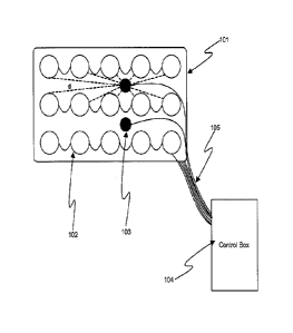
[0046] Figure 1 shows, in accordance with an embodiment of the invention,
an array of electrodes
placed within a thin, flexible housing 101. The thin flexible housing may be
connected to one or more
stimulating electrode 102 and/or one or more sensing electrode 103. A control
box 104 may be
electrically connected to the one or more stimulating electrode and/or the one
or more sensing electrode.
The control box may communicate with the electrodes through a series of wire
connections 105. The
system may be used for neuromuscular electrical stimulation (NMES) of muscle
tissue.
[0047] In a preferable embodiment of the invention, the thin, flexible
housing 101 may form a
substrate or support for an electrode pad. The thin flexible housing may be
formed of a material that may
enable the pad to conform to an anatomical placement on a subject. For
example, the housing may
include a deformable or elastic component. The placement of the pad may
determine which muscle tissue
11
CA 02727498 2010-12-09
WO 2010/003106 PCT/US2009/049601
of the subject may be stimulated by the NMES device. For instance, the muscle
tissue proximate to the
pad may be stimulated.
[0048] The one or more stimulating electrode 102 may be mechanically
attached or integrated into
the pad 101. In a preferable embodiment, an array of stimulating electrodes
may be provided on the pad.
Any number of electrodes may be provided on the array. For example, an array
may be formed of n rows
and m columns, where n and in are any integer with a value of one, two, three,
four, five, six, seven, eight,
nine, ten, or greater. In other embodiments, the array of stimulating
electrodes need not be arranged into
rows and columns and may have any placement on a pad.
[0049] Sensing electrodes 103 may be located near the center of the
stimulating electrode array 102.
For example, one, two, three, four, or more sensing electrodes may be placed
near the center of the pad
101. However, sensing electrodes may be placed anywhere on the pad, and need
not be at the center. For
example, the sensing electrodes may be distributed substantially even over the
surface of the pad, between
any of the stimulation electrodes, along the border of the electrodes, or
beyond the area defined by the
stimulation electrodes. Optionally, the sensing electrodes may be distributed
so that they fall within the
array of electrodes, and not outside an area defined by the array of
electrodes. Similarly, any number of
sensing electrodes may be provided.
[0050] In some embodiments, a sensing electrode may function as a reference
electrode. The
reference electrode may be positioned such that the reference electrode is at
a predetermined distance or
range of distances from the stimulation electrodes. Also, the reference
electrode may be positioned to be
at a predetermined distance or range of distances from other sensing
electrodes. In some embodiments,
the reference electrodes may be at a greater distance from the stimulation
electrodes than other sensing
electrodes are from the stimulation electrodes. Thus, a reference electrode
may be further from
stimulation electrodes than other sensing electrodes. In some embodiments, the
reference electrode may
be at a distance or relative position from the stimulation electrodes such
that the effects of the stimulation
electrodes on the signals picked up by the reference electrodes are reduced,
minimized, or non-existent.
[0051] The control box 104 may contain pulse generation electronics as well
as digital and/or analog
signal processing components. The control box may provide electrical
stimulation signals to a
stimulation electrode 102 and/or receive signals from a sensing electrode 103.
In preferable
12
CA 02727498 2016-06-07
CA 2727498
embodiments, the signals provided to the stimulation electrode may depend on
signals received from the
sensing electrode. Thus, the system may provide a feedback, to control the
electrical stimulation provided.
[0052] In some embodiments, each of the stimulation electrodes may be
individually controllable by the
control box. For example, the stimulation electrodes may be connected to the
control box in such a way
that for each stimulation electrode, whether any stimulation is provided, the
level of stimulation provided,
or pulse width, duration, frequency, amplitude, waveform, or any other
characteristic of the stimulation
provided to the stimulation electrode may be individually controlled by the
control box. Thus,
customization and localization of stimulation may be closely controlled. See,
e.g., PCT Publication No.
WO 2007/017778 and PCT Publication No. WO 2005/075018.
[0053] There may be a number of parameters that describe the stimulation
electrical pulses. As previously
mentioned these include voltage amplitude, current amplitude, waveform shape
(e.g., square, sinusoidal,
exponential, monophasic/biphasic, symmetric/asymmetric), pulse length, pulse
repetition frequency, and
the relative on/off times between repeating series of pulses. Depending on the
mode of operation (e.g.,
constant current vs. constant voltage stimulation), some of these parameters
may be independently user-
controlled, while others are dependent on external factors such as the
electrical impedance between
electrodes.
[0054] There may also be several stimulation parameters that are typically
operator-controlled. The values
of these parameters directly impact the safety, efficacy, and relative comfort
of an electrical stimulation
therapy session. For example, stimulation pulse length and waveform shape have
been shown to
significantly impact comfort and tolerability. Pulse repetition frequency and
voltage/current amplitude are
correlated with strength of muscle contraction and thus treatment efficacy.
Current density, a function of
injected charge, must be carefully controlled to avoid burns, nerve injury,
and other potential complications
(as detailed by Prausnitz Advanced Drug Delivery Reviews 18:395-425,2006 and
Stecker et al Am J END
Tech., 43:315-342, 2006). Additional parameters may impact other features of
NMES therapy, including
the required duration of therapy and the suitability for use in certain
patient populations.
13
CA 02727498 2010-12-09
WO 2010/003106 PCT/US2009/049601
[0055] In some other embodiments, subsets of the stimulation electrodes
provided may be
controlled. For example, if three subsets of stimulation electrodes are
provided, each of the electrodes
within the same subset may receive the same stimulation (or lack thereof).
Thus, for example, the first
subset may lie dormant and not receive any stimulation, the second subset may
receive a stimulation of a
high amplitude and great frequency, and the third subset may receive a
stimulation with a lower
amplitude and lesser frequency.
[0056] In an alternate embodiment, all of the stimulation electrodes on a
pad may receive the same
stimulation.
[0057] Similarly, signals from the sensing electrodes may be individually
analyzed, or partially
aggregated and then analyzed, or completely aggregated and analyzed. The
control box may perform
signal processing steps to the signals from the sensing electrodes, examples
of which are to be discussed
in greater detail below.
[0058] Wire connections 105 may enable the control box 104 to communicate
with the electrodes.
In some alternate embodiments, the stimulation and/or sensing electrodes may
be able to communicate
with the control box wirelessly.
[0059] In accordance with an embodiment of the invention, an NMES system
may be provided for
electrically stimulating a selected muscle-tendon region. In some instances, a
muscle-tendon region may
include a muscle group. A muscle-tendon region may also refer to a general
region encompassing a
muscle group and associated tendons. The NMES system may be provided for
electrically stimulating a
targeted muscle and/or nervous tissue. Any description of an NMES targeted
region or tissue may also
refer to any other type of NMES targeted region or tissue.
[0060] In some embodiments, an integral electrical stimulation unit, which
may include at least one
stimulating electrode and at least one sensing electrode may be provided. The
integral electrical
stimulation unit may be unitary and provided as one piece. The stimulating
electrode and the sensing
electrode may be integrated within a substrate or pad. The integral electrical
stimulation unit may be
provided so that a plurality of sensors are positioned within an array of
stimulating electrodes. The
plurality of sensors may be positioned so that they fall between stimulating
electrodes and/or are within
an area defined by the array of stimulating electrodes.
14
CA 02727498 2010-12-09
WO 2010/003106 PCT/US2009/049601
[0061] The position of the stimulating electrodes may depend on target
muscle and/or nervous tissue.
The stimulating electrodes may have contact portions that are positioned on
the skin over the target
muscle and/or nervous tissue. The stimulating electrodes may be in electrical
contact with the underlying
target tissue, even if they are not in direct physical contact with the
tissue. Thus, the stimulating
electrodes may be able to electrically communicate with target tissue
transdennally.
[0062] Each stimulating electrode may be a predetermined distance from a
sensing electrode. For
example, in some embodiments, the position of a stimulating electrode may be
selected to be a
predetermined distance from the sensing electrode. For example, in forming the
integral electrical
stimulation unit, a desired distance d or relative position of the stimulating
electrodes relative to one or
more sensing electrodes may be calculated, and the electrodes may be
positioned accordingly. in some
embodiments, the placement of each stimulating electrode may be predetermined
to fall within a spaced
apart distance from a plurality of sensors. In some embodiments, a desired
range of distances may be
provided, where electrodes may fall within that distance range from the
plurality of sensors.
[0063] In some embodiments, the predetermined distance d or desired
distance range for the
electrodes from the sensors may depend on the target muscle-tendon region.
Based on the underlying
target anatomy, desired placements of the stimulating and sensing electrodes
may be determined and/or
calculated, and the integral electrical stimulation unit may be formed
accordingly.
100641 In some embodiments, one or more sensor may be a reference sensor. A
reference sensor
may also be a predetermined distance or desired distance range from the
electrodes. In some instances,
the distance of a reference sensor from the electrodes may be greater than the
distance between the other
sensors and the electrodes. The position of a reference sensor may be
predetermined or predefined
depending on the expected anatomical features.
100651 The NMES system may also include a controller which may be in
electrical communication
with the stimulating electrodes and sensing electrodes of an integral
electrical stimulation unit. The
controller may provide electrical signals to the array of electrodes based on
electrical signals received
from the plurality of sensors. In some instances, the controller may provide
electrical signals to a subset
of the stimulating electrodes in the array of stimulating electrodes based on
instructions from the
controller.
CA 02727498 2010-12-09
WO 2010/003106 PCT/US2009/049601
[0066] Figure 2, provides an example of one potential anatomic placement of
the embodiment
described in Figure 1. For instance, a pad may be placed on the front of the
thigh of a subject, which may
result in electrical stimulation of quadriceps. The pad may be placed above
the knee of the subject. The
pad, which may include stimulation electrodes and/or sensing electrodes may
contact the skin of the
patient. Electrical stimulation signals may reach the underlying muscle and/or
nervous tissue
transdermally. Thus, since electrical conduction may be provided from the
stimulation electrode to the
underlying tissue, the stimulation electrode may be in electrical contact with
the underlying tissue.
[0067] In other implementations, the pad may be placed at another location
on a patient. For
example, the pad may be used to stimulate other leg muscles, or muscle and/or
nervous tissue provided in
a subject's arms or torso. For example, the pad may be placed at the rear of
the thigh of a subject, around
an entire thigh of the subject, in the front of back of the power leg of the
subject, at the upper arm of a
subject, at the lower arm of a subject, at the waist of a subject, at the
upper torso of a subject, or below the
waste of a subject.
[0068] Figure 3 shows an embodiment where the electrode array and the
control box are comprised
within a single unit. For example, a thin, flexible housing may be provided,
which may act as a substrate
or support for the unit. The thin, flexible housing may form a pad for the
unit. One or more stimulating
electrodes and/or one or more sensing electrodes may be provided on the
flexible housing. A control box
may also be provided on the housing, to form the single unit. Preferably,
electrical connections between
the stimulating electrodes and/or sensing electrodes with the control box may
be provided by wires that
may be integrated into the single unit.
[0069] The control box may be affixed to the pad of the unit. In some
embodiments, the control box
may be integrally connected to the pad of the unit so that it may not be
removed. In other embodiments,
the control box may be removably attached to the pad of the unit. For example,
the control box may snap
into and out of a connection provided on the unit, may be velcroed to the
unit, may be clipped or clamped
to the unit, or may be removably attached to the unit in any other manner.
Removable or fixed control
units may electrically communicate with the rest of the unit via wire or
wireless connections.
[0070] As shown in Figure 4, a preferable embodiment of the described
method could be described
by a simple flow chart. For example, a method of performing NMES may be
provided where the method
16
CA 02727498 2010-12-09
WO 2010/003106 PCT/US2009/049601
may include an operating pushing a button device 401, beginning a calibration
sequence, where sensors in
a stimulation pad may automatically determine a person's muscle activity 402,
optimizing a location of
active electrodes and electrical stimulation parameters to deliver safe and
efficient therapy 403, delivering
an electrical stimulation to a person's body via one or more stimulation pad
404, and terminating therapy
when complete 405.
[0071] Only the first step 401 may require effort on the part of the
operator. The subsequent steps
402-405 may be automatically implemented though algorithms executed by the
control box.
[0072] During a method of NMES, an operator may place a stimulation pad on
a desired anatomical
region of a subject. The subject may be a patient, such as a comatose,
sedated, analgesed patient, or a
patient at the ICU, or may be a clinical test subject, or any other human,
mammal, or any other animal
that may receive NMES.
[0073] In a preferable embodiment, the operator may place the stimulation
pad at an estimated
location to stimulate the target muscle tissue, without having to place it at
a precise location. The
operator may place the stimulation pad to contact the skin approximately over
the target muscle tissue.
The stimulation pad may be attached to the skin using an adhesive or straps,
or any other mechanism that
may enable the stimulation pad to remain in contact with the skin. Once one or
more stimulation pad is
secured at the desired locations, an operator may activate the stimulation
pad. The operator may initiate
the activation by interfacing with a control box, such as by pushing a button
device 401, or flipping a
switch, touching a touchscreen, or performing any other such action that may
enable the operator to
initiate action through the control box.
[0074] Once it receives instructions to begin, the control box may begin a
calibration sequence 402.
The calibration sequence may include providing stimulation signals to one or
more stimulating electrode
and/or receiving signals from sensors of the stimulation pad. In some
embodiments, the calibration
sequence may include providing signals to stimulating electrodes in accordance
with a predetermined
sequence and at predetermined waveform characteristics. Alternatively, the
calibration sequence may
include providing signals to stimulating electrodes based on signals received
from sensors. The signals
received from the sensors may be analyzed by the control box to determine the
subject's muscle activity.
17
CA 02727498 2016-06-07
CA 2727498
100751 The control box may optimize a location of active electrodes and
electrical stimulation parameters to
deliver safe and efficient therapy 403. For example, based on analysis
performed during the calibration
sequence, electrodes may be selected to provide electrical stimulation to
target muscle tissue. Such electrodes
may be a subset of the stimulating electrodes provided on the stimulation pad.
In some embodiments, the
selected electrodes may remain the same throughout the therapy. In other
embodiments, the selected electrodes
may vary over the course of therapy. Such variation may depend on signals
provided by sensors in the
stimulation pad, or may be predetermined based on the calibration sequence. In
addition to variation of selected
stimulating electrodes, variation may be provided to parameters of the applied
stimulus. Thus, parameters that
may be optimized include anatomical location of the applied stimulus,
amplitude of stimulus, shape of stimulus
waveform, duration of stimulus signal, and stimulus signal frequency. See,
e.g., U.S. Patent No. 4,838,272, U.S.
Patent No. 6,324,432 and U.S. Patent No. 7,499,746.
[0076] The control box may deliver an electrical signal which may cause
delivery of electrical stimulation to
the subject's body via one or more stimulation pads 404. The electrical
stimulation provided to the subject body
may depend on algorithms performed by the control box to determine optimized
parameters. For example, if the
control box determines that an increased stimulation frequency is desirable,
the stimulation pads may deliver
electrical stimulation to the subject's body at an increased stimulation
frequency.
[0077] The control box may automatically terminate therapy when it is complete
405. The control box may
determine that therapy is complete based on predetermined instructions. For
example, the therapy may be
complete after a predetermined amount of time has elapsed, or after a
predetermined amount of electrical
stimulation has been provided. Alternatively, the control box may determine
that therapy is complete based on
signals received from the sensors. For example, the therapy may be complete
when the sensors reach a
particular threshold for an electrical signal.
[00781 In accordance with another embodiment of the invention, a method of
electrically stimulating a selected
muscle-tendon region of a body may be provided. The method may include placing
a stimulation assembly in
electrical contact with muscle tissue, wherein the stimulation assembly may be
formed with a plurality of
electrodes and at least one sensor. The sensor may be positioned at a
18
CA 02727498 2010-12-09
WO 2010/003106 PCT/US2009/049601
predefined range away from each electrode to monitor the remote effects of
electrical stimulation from
the electrodes on the selected muscle-tendon region of the body.
[0079] In some implementations, the predefined range of distance for each
electrode may be
determined prior to forming the stimulation assembly. For example, prior to
manufacturing or building
the stimulation assembly, it may be desirable to calculate the predefined
range of distance, and place each
electrode and sensor within the stimulation assembly accordingly. In some
embodiments, the predefined
range of distance may depend on the anticipated anatomical placement of the
stimulation assembly, while
in other embodiments, the predefined range may be placement agnostic.
100801 The method may also include receiving a feedback signal from at
least one sensor of the
stimulation assembly, and providing an electrical stimulation signal to at
least one of the plurality of
stimulation electrodes based on the received feedback signal.
Stimulation and sensing electrodes in separate pads
100811 In a preferable embodiment, an NMES system may be comprised of two
main functional
components: a stimulation unit comprising an array of stimulation electrodes
and sensor element(s) and a
control unit. In a preferable embodiment, the stimulation unit may be
comprised of two separate pads that
can contact a person's body: a stimulation pad containing an array of
stimulation electrodes and
associated electronics, and a sensing pad containing muscle sensors and
associated electronics.
Alternatively, the stimulation pad and sensing pad may be composed of a single
unit, or the two pads may
be connected mechanically through straps, buttons, hooks, or other suitable
means. The control unit may
communicate with the stimulation unit through a wired connection,
radiofrequency transmission, optical,
acoustic, or electromagnetic signals, or another suitable mechanism. The
control unit may be a separate
unit that may be located some distance from the person receiving therapy. In
an alternate embodiment, the
control unit may be integrated into a housing unit containing the stimulating
electrode and sensing
component(s).
[0082] In some alternate embodiments of the invention, the stimulation pad
and the sensing pad may
be integrated into one piece. For example, the stimulation pad comprising
stimulation electrodes and the
sensing pad comprising sensors may share a common substrate. The stimulation
pad and the sensing pad
may be distinct regions on the common substrate or may be placed on a common
substrate.
19
CA 02727498 2010-12-09
WO 2010/003106 PCT/US2009/049601
[0083] Any of the components, steps, features, or advantages provided in an
embodiment where the
stimulation and sensing electrodes are within the same pad may also be applied
or combined to an
embodiment with separate stimulation and sensing pads.
[0084] Figure 5 shows the major components of a preferable embodiment of
the system. An NMES
system may include a control unit 501, a stimulation pad 502, and a sensing
pad 503. Thus, in some
embodiments, a separate stimulation pad and sensing pad may be provided. Any
discussion of the
system, device, or method relating to a system with a single integrated
stimulation pad comprising both
stimulation electrodes and sensors may also apply to a system with separate
stimulation and sensing pads,
and vice versa. Furthermore, in alternate embodiments of the invention, any
number of stimulation and
sensing pads may be provided within an NMES system, and any discussion herein
may also apply to such
embodiments.
[0085] The NMES system may include a control unit 501, which may include a
display and limited
operator controls. Some examples of operator controls may include an on/off
button or switch, a stop
button, or a time knob. Any other device interface mechanisms, including
buttons, switches, knobs,
touchscreens, light sensors, microphones, speakers, voice-recognition devices,
or any other mechanisms
known in the art may be utilized to enable an operator to interact with the
control unit.
[0086] The control unit may also include components such as a signal
generator, memory, processor,
and power supply. The primary operation of the control unit may be provided by
a microprocessor, field
programmable gate array (FPGA), application specific integrated circuit, or
other suitable mechanism.
When activated, the control unit may generate electrical stimulation signals
that may be transmitted to the
stimulation pad, which couple the energy into the body to activate muscles.
Some electrical stimulation
parameters, including the duration of therapy, are adjustable by the operator
through buttons, knobs, dials,
or switches on the control unit. Other electrical stimulation parameters may
be optimized through
automatic algorithms implemented by the control unit, as outlined below.
[0087] An operator may interact with the control unit to initiate the
performance of an NMES
therapy. For example, in some embodiments, an operator may simply turn the
control unit on, and not
need to interfere any further throughout the course of the therapy.
CA 02727498 2010-12-09
WO 2010/003106 PCT/US2009/049601
[0088] In some embodiments, controls may be provided which may enable an
operator to intervene
during an NMES therapy. For example, a control unit may alert an operator to
an alarm condition, which
may cause an operator to adjust a parameter, or terminate the therapy. In
other examples, the control unit
may automatically adjust the parameter or terminate the therapy. In some
instances, an operator may
adjust a parameter (e.g., the duration of therapy).
[0089] The control unit may also enable an operator to enter subject-
specific information. For
example, an operator may enter personal information about the subject, which
may be linked to data
generated and/or stored during NMES therapy. In some instances, the
information about the subject may
affect a therapy parameter.
[0090] The NMES system may also include at least one stimulation pad 502.
The stimulation pad
may include one, two, three, four, or more stimulation electrodes. The
stimulation pad may include a
plurality of stimulation electrodes and no sensors. Alternatively, the
stimulation pad may include both
stimulation electrodes and at least one sensor. In some instances, a
stimulation electrode may act as a
sensor when inactive for delivering electrical stimulation.
[0091] The NMES system may include at least one sensing pad 503. The
sensing pad may include
one, two, or more sensors. The sensors may include sensing electrodes. The
sensing pad may include a
plurality of sensors and no stimulation electrodes. Alternatively, the sensing
pad may include both
sensors and at least one stimulation electrode.
[0092] ln a preferable embodiment, the stimulation and sensing pads may
each comprised of a thin
and flexible housing with an adhesive backing to facilitate maintenance of
skin contact with a person
receiving NMES. The backing may also contain hydrogel or other coupling agents
to enhance the
coupling of electrical energy and signals between sensing or stimulating
electrodes and the person's body.
The adhesive and/or coupling gels may be located along the entirety of the
skin contact side of the pads,
or may be located solely in discrete locations (such as underneath
electrodes).
[0093] The stimulation pad 502 and the sensing pad 503 may be arranged on
the subject in any
desired manner. For example, the stimulation and sensing pads may be placed on
a subject such that the
stimulation and sensing pads are not in contact with one another. The
stimulation and sensing pads could
also be placed on a subject so that there is some overlap of the pads.
Preferably, the stimulation and
21
CA 02727498 2016-06-07
CA 2727498
sensing pads will not be mechanically connected to one another, although in
some embodiments, they may
somehow be interconnected. See, e.g.. U.S. Patent No. 5,549,656.
[0094] Furthermore, the stimulation and sensing pads may be separately
electrically connected to the control
unit, or may share electrical connections to the control unit. In some
embodiments, the stimulation and sensing
pads may be electrically connected to the control unit via wires.
Alternatively, they may be connected
wirelessly, or may comprise one or more integrated control unit.
[0095] Figure 6 shows a geometry of a preferable embodiment of the sensing pad
601, stimulation pad 604,
and the layout of the array of stimulation electrodes in the stimulation pad.
The sensing pad 601 may include
sensing electrodes used for recording electrical activity 602, and a sensing
electrode serving as a ground
electrode 603. The stimulation pad 604 may include upper stimulation
electrodes 605 in a stimulation electrode
array, and lower stimulation electrodes 606 in the stimulation electrode
array.
[0096] The stimulation pad may contains an array of strategically-placed
stimulation electrodes that may be
used to deliver electrical energy to muscles and/or nerves in order to produce
muscle contraction. The array may
be configurable such that, at any given time, only a subset of the electrodes
in the array are actively delivering
energy to a person receiving NMES. However, electrodes inactive for energy
delivery may still be configured to
deliver relevant information (such as the electrical impedance between it and
a second electrode in the array) to
the control unit. The sensing pad may contain one or more sensing element(s)
that can detect and record
biological parameters that describe muscle contraction directly or indirectly.
These sensing parameters may
include measurement of direct or indirect electrophysiological features of the
muscle contraction or
measurements of the mechanical features of the muscle contraction (such as
contraction distance, velocity, and
acceleration) measured with an accelerometer, a pressure sensitive element, or
similar equipment In a preferable
embodiment, electrical signals that are indicative of the state of tension in
a tendon are measured by sensing
electrodes in the sensing pad.
[0097] In a preferable embodiment, sensing electrodes, integrated into a
sensing pad, may be placed on the
surface of the skin in the region of a tendon that is mechanically coupled to
muscles that are stimulated. In some
embodiments, the underlying muscle group may be in electrical contact with the
stimulation electrodes. The
22
CA 02727498 2016-06-07
CA 2727498
underlying muscle group may be encompassed within a muscle-tendon region. For
example, if quadriceps
muscles are stimulated to contract, one suitable location for the sensing
electrodes would be on the surface of the
skin in the region directly over the quadriceps tendon. Tendons are bands of
tough fibrous tissue that are
composed mostly of collagen fibers, and do not produce electrical activity
during muscle contraction in the same
way that muscle tissues do. As a result, the majority of electrical activity
detected by sensing electrodes located
over the tendons arises originally from nearby muscles or from the energy
source used to stimulate muscle
contraction. Accordingly, these electrical signals travel along and/or around
the tendon before reaching the skin
surface, where they are detected by sensing electrodes.
[0098] In some implementations, one or more sensors may be placed over the
tendons to form a linear
arrangement along a tendon. For example, if a tendon has a vertical alignment,
sensors may be placed over the
tendon in a corresponding vertical alignment. The sensors may be placed over
or near a tendon such that they
are arranged to align in the same direction as the propagation of a signal
from a stimulation electrode along the
tendon. A sensing pad may include sensors that are configured to be positioned
over a tendon along a direction
that matches the direction a signal would travel along the tendon.
[0099] In some embodiments, the sensors placed on a sensing pad may be outside
an area defmed by the
stimulation electrodes. For instance, sensors may be placed so that they are
not between stimulation electrodes.
[00100] Without wishing to be bound by any theory, it is believed that the
geometry, mechanical properties,
and/or other characteristics of the tendon influence its electrical properties
(for example, transmission speed and
impedance ¨ see Suganuma et al, J Ortho Science 9: 302-309, 2004). Thus,
changes in the state of the tendon
may affect electrical signals transmitted to the sensing electrodes. Tendons
connect muscles to bones, and
transmit forces that arise due to muscle contraction. Accordingly, tendons are
capable of withstanding tension
during muscle contraction. Without wishing to be bound by any theory, it is
believed that a stronger muscle
contraction produces more tension and a greater geometry change in an
associated tendon and adjacent
anatomical regions than a weaker muscle contraction. It would follow that a
stronger muscle contraction would
alter both the electrical properties of the tendon and the available
electrical transmission pathways, and thus the
23
CA 02727498 2010-12-09
WO 2010/003106 PCT/US2009/049601
electrical activity detected by the sensing electrodes located over or beyond
the tendon, more than a
weaker muscle contraction. Specifically, it is believed (as proposed by
Suganuma et al) that increased
tendon tension leads to increases in tendon electrical impedance. Thus,
electrical activity detected over or
near tendons is suitable for optimization of techniques, including electrical
stimulation, that produce
muscle contraction and induce changes in tendon geometry and tension.
[00101] Sensing electrodes placed over tendons and novel signal processing
algorithms developed to
expose the effects of tendon properties and geometry on recorded signals offer
many advantages over
traditional NMES optimization methods. Standard electromyography (EMG),
defined here as measuring
the electrical activity produced by muscle contraction using surface or needle
electrodes placed in the
region of contracting muscles, has limited usefulness during electrical
stimulation due to interference
between electrical signals injected into the body by the stimulator (on the
order of 10 ¨ 50 V) and
electrical signals produced by muscles (on the order of 5 ¨ 50 mV). Current
clinical and engineering
research has focused upon the development of complex, often adaptive, signal
filters to extract useful
information from EMG data collected during electrical stimulation. However,
problems with extracting
useful EMG data during electrical stimulation are exacerbated when an array of
stimulation electrodes are
used, because the interference pattern between the stimulation energy artifact
and the muscle activity data
will not be constant among all data acquisitions, leaving previously developed
EMG filters generally not
applicable or of limited utility. As described in detail below, signal
processing algorithms that extract
information concerning tendon tension and geometry from electrical signals
recorded over tendons do not
suffer from performance degradations or interpretation uncertainty due to
electrical stimulation/EMG
signal interference.
[00102] When muscle stimulation is applied to critically ill patients, further
advantages of optimization
methods based upon tendon tension and/or geometry are evident. In the ICU and
many other
environments, the use of needle electrodes to measure EMG data is generally
not suitable, and surface
electrodes must be used. Critically ill patients often suffer from tissue
edema (swelling) as a side effect of
treatment. It is believed that the presence of significant edema will
generally result in a greater distance
between muscle tissues and the skin, attenuating and distorting EMG data that
are collected with
electrodes on the skin surface. In many cases, no useful EMG data can be
acquired. Similar problems
24
CA 02727498 2010-12-09
WO 2010/003106 PCT/US2009/049601
exist when using surface electrodes to measure EMG data from obese persons or
persons with low
baseline muscle mass. Deposits of fatty tissue and tissue edema are typically
at local minima around
bony prominences, such as the knee, where tendons insert. Thus, electrical
activity measured over the
tendon (or, as shown below, even over the bony prominence itself) may produce
data more reliable than
EMG data in these persons. Signal processing algorithms developed to interpret
these data may thus
enable indirect measurement of muscle contraction when useful EMG data are
unobtainable.
Additionally, critically ill patients are most often treated while lying in
bed with legs extended. As legs
are already extended, the stimulation of the quadriceps produces little
physical movement, limiting the
utility of sensors such as accelerometers that seek to optimize the electrical
stimulation location or
parameters based upon measurements of muscle dynamics.
[00103] It should be noted that in order to implement a preferable embodiment
of the method, it is not
required that sensing electrodes be placed directly over the tendon with a
high degree of precision. It is
only required that the sensing electrodes be close enough to the target tendon
such that the varying
electrical properties, geometry, and other properties of the tendon during
muscle contraction significantly
impact the electrical activity waveform detected by the sensing electrodes. As
described below, by using
sensor pad geometries tailored to the local anatomy in the region of
stimulation, it is possible to ensure
that sensing electrodes are placed close enough to the target tendon in order
to empower the successful
implementation of the method.
[00104] As shown in Figure 6, a preferable embodiment will utilize stimulation
and sensing pads with a
specific overall geometry, or footprint. The stimulation and sensing pads
shown in Figure 6 may be
tailored for the stimulation of quadriceps muscles, although those skilled in
the art will recognize that
similar principles can be applied to design pad geometries tailored to the
stimulation of other muscle
groups. The geometry of the sensing pad may be designed such that the sensing
electrodes will make
contact with a person's skin superior to the knee, in the region of the
quadriceps tendon.
[00105] The unique shape of the sensing pad 601 may use a readily identifiable
anatomical marker (e.g.,
the knee cap) both to ensure proper placement of the sensing electrodes over
the quadriceps tendon and to
ensure proper alignment with the stimulation pad (as elaborated upon below).
The sensing pad may
include a protruding feature to match the protrusion provided by the knee cap
to create a desirable
CA 02727498 2010-12-09
WO 2010/003106 PCT/US2009/049601
alignment of the sensing pad. In some instances, the anatomical feature
provided on the sensing pad may
be a hole that may enable the knee cap to protrude through the hole. Other
examples of anatomical
features may include a different material over the knee cap region, that may
enable the knee cap to more
easily stretch the sensing pad at the knee cap, or some sort of visual
indicator, such as a color change or
line that may indicate the placement of the knee cap. Thus, the sensing pad
may include an anatomical
placement guide that may assist with placing the sensors at a desired
location.
[00106] The sensing pad 601 may utilize three sensing electrodes: two
electrodes 602 intended to collect
electrical signals to be used as inputs to a differential amplifier and/or
other signal conditioning circuitry,
and a reference electrode 603 placed over a bony prominence some distance from
the other electrodes (for
example, the shin near tibia). Alternatively, as few as one sensing electrode
may be utilized to extract
sufficient information required to optimize electrical stimulation parameters.
Similarly, at least one signal
collecting electrode and at least one ground electrode may be provided.
Variations of a preferable
embodiment of the system can utilize more than three sensing electrodes to
extract information
concerning tendon tension and/or geometry. In these variant embodiments,
information from individual
sensing electrodes may be analyzed individually, and may or may not be
compared with the use of a
differential amplifier or similar hardware or software.
[00107] In some embodiments, the signal collecting electrodes 602 may be
placed on the sensing pad so
that they are at the lower thigh above the knee cap. In some instances, they
may be vertically directly
above the knee cap, while in other embodiments, they may be horizontally
spaced. Preferably, the signal
collecting electrodes may be arranged some distance from the reference
electrode 603 (aka ground
electrode).
[00108] The reference electrode may be spaced apart from the other signal
collecting electrodes. In some
embodiments, a reference electrode may receive very little or no electrical
signals that are provided from
the stimulation electrodes. A reference electrode may pick up inherent
background electrical signals from
the subject body. In some instances, the signals collected by the other signal
collecting electrodes may be
compared to the signals provided by the reference electrode to compare which
signals are provided by the
stimulation electrodes and muscle properties as compared to background
signals. In some instances, the
26
CA 02727498 2010-12-09
WO 2010/003106 PCT/US2009/049601
background signals provided by the reference electrode may be subtracted from
the signals collected by
the other signal collecting electrodes.
[00109] The top portion of the sensing pad may be shaped to have a protrusion
that fits into a notch-
shaped opening in the stimulation pad 604. Any shape that may allow the
sensing pad to align with the
stimulation pad may be provided. For example, the sensing pad and the
stimulation pad may have
complementary shapes, so that an extension from one pad may have a
corresponding indentation in the
other pad. This geometry may allow for the usefulness of the knee cap as an
anatomical marker to be
extended to aid in accurate gross positioning of the stimulation electrode
array. This geometry may
increase both intra- and inter-operator consistency of stimulation pad
placement by creating a virtual
anatomical reference point (the protrusion of the sensing pad) in a region of
the body (the thigh) that lacks
readily identifiable anatomical markers. Additionally, this geometry may be
designed specifically to fix
the position of the stimulation electrodes with regard to the sensing
electrodes, and to minimize
deviations from the ideal spatial relationship between the two sets of
electrodes. In some embodiments,
additional visual indicators or markers, such as arrows on the sensing and
stimulation pads, may provide
additional aids in aligning the sensing and stimulation pads. Thus, visual
markers may aid in fixing the
spatial relationship of the sensing and stimulation pads.
[00110] The stimulation pad 604 may contain an array of stimulation electrode
contacts, each of which
can be individually enabled or disabled automatically by the control unit to
provide energy to the person
receiving NMES. The use of an array of electrode contacts enables the gross
placement of the stimulation
pad on a person in the desired region of stimulation, without requiring
precise alignment of individual
electrode contacts over the motor points of the muscle. A preferable
embodiment of the stimulation pad
may comprise an array of eight square or rectangular stimulation electrode
contacts arranged in a
particular pattern. It will be clear to those skilled in the art that other
arrangements of the stimulation
electrode array and other electrode contact shapes and sizes could be employed
without loss of generality.
Similarly, a different number of individual stimulation electrode contacts in
the array could be employed
to empower the use of the disclosed method, such as one, two, three, four,
six, nine, ten, twelve, fifteen,
twenty, or more stimulation electrode contacts. The stimulation electrode
contacts may be placed at any
27
CA 02727498 2010-12-09
WO 2010/003106 PCT/US2009/049601
location of the stimulation pad. Stimulation pads designed to be used with
muscle groups other than the
quadriceps will likely contain stimulation electrode arrays with markedly
different arrangements.
[00111] The particular arrangement of the stimulation electrode array
portrayed in Figure 6 can be used to
enhance the performance of an automatically optimizing electrical stimulation
system. Stimulation
electrodes comprising the array can be classified as being part of one of two
groups: upper electrode
contacts located near the bulky part of the thigh 605, or lower electrode
contacts located closer to the
quadriceps tendon 606. During NMES therapy, most commonly the energy delivered
by the system will
travel between one or more upper electrode contacts and one or more lower
electrode contacts. Upper
electrode contacts may be positioned to make contact with the middle-outside
of a person's thigh
(expected location of a motor point), and span a length (for example, six
inches) expected to be larger
than the span of possible motor point locations in an average adult. Lower
electrodes may also positioned
close to the location of an expected muscle motor point, and are also
positioned in a manner so as to
dictate the direction of current flow through the leg. In some embodiments,
the larger lower electrode
need not be centered along the midline of the thigh. Instead, it may be
arranged so that its center point is
slightly toward the inside of the thigh, causing energy traveling between
upper and lower electrode
contacts to cross the midline of the thigh. An additional, smaller lower
electrode may be positioned at a
similar distance superior to the knee cap, but more to the inside of the leg.
The electrode contact
placement may depend on the target muscular stimulation.
[00112] In accordance with an embodiment of the invention, an NMES system may
be provided, wherein
the NMES system comprises a stimulation assembly. The stimulation assembly may
be formed with a
plurality of electrodes and at least one sensor, where the sensor may be
positioned within a predefined
range away from each electrode to monitor the remote effects of electrical
stimulation from the electrodes
on a selected muscle-tendon region of the body. The predefined range may
depend on the target muscle-
tendon region of the body. For example, one or more sensing electrode may be
placed near a bony
prominence near an underlying tendon. The stimulation electrodes may be at a
range of distances from
the sensing electrodes. In one example, if the target muscle-tendon region is
the quadriceps, the sensing
electrodes may be place near a knee, such as above the knee, and the
stimulation electrodes may be
placed over the quadriceps, since range of distance d from the sensing
electrodes.
28
CA 02727498 2010-12-09
WO 2010/003106 PCT/US2009/049601
[00113] The NMES system may also include a control unit connected to the
plurality of electrodes, and
the sensor. The control unit may provide stimulation signals to the electrodes
and receive a signal from
the sensor. In some instances, the stimulation signals provided to the
electrodes may depend on the
signals received by the sensor.
[00114] Figure 7 illustrates a side view of an NMES system where a control
unit 701 may be integrated
into a stimulation pad 702. The system may also include a sensing pad 703,
which may or may not be
integrated together with the stimulation pad. The stimulation and sensing pads
may be placed on a leg
704 of a subject.
[00115] The control unit 701 may include a display and any operator interface
devices 701a, 701b, 701c,
701d that may enable an operator to interact with the control unit.
[00116] The stimulation pad may include an array of stimulating electrodes,
which may be arranged in
any matter on the stimulation pad. For example, the stimulating electrodes may
be grouped into upper
electrodes 702a and lower electrodes 702b. The control unit may determine
electrical stimulation
parameters for signals provided to the stimulation electrodes.
[00117] The sensing pad may include one or more signal collector electrodes
703a, and one or more
ground electrode 703b. The ground electrode may be located over a bony
prominence, such as over a
shin. In some embodiments, the ground electrode may be located below a knee,
while the signal collector
electrode may be located above the knee.
[00118] The sensing pad and/or stimulation pad may be able to accommodate
underlying anatomical
features. For example, when the pads are placed on the front of a leg 704, the
sensing pad may be shaped
with a protrusion to accommodate the protrusion provided by a knee cap 705.
[00119] The NMES system may be applicable to other anatomical regions as well.
Such systems may
make use of other anatomical features, such as protrusions, anatomical shaped
regions, bony
prominences, and so forth. For example, the NMES system may target muscle
tissue provided in the
calves. The stimulation and/or sensing pad may utilize a bony prominence, such
as a knee or ankles as an
anatomical marker, and may have an anatomical placement guide that conforms to
the shapes of the
anatomical markers.
29
CA 02727498 2010-12-09
WO 2010/003106 PCT/US2009/049601
[00120] In another example, the NMES system may target muscle tissue in the
upper or lower arms, and
may utilize anatomical features to place the stimulation and/or sensing pads.
For example, the pads may
be shaped to fit over bony prominences such as elbows or wrists, or may use
other anatomical features as
guides, such as armpits.
[00121] The NMES system may also target muscle tissue in the torso of a
subject. For example, the
system may provide stimulation to a subject's waist, and may use the subject's
hip as an anatomical
guide, or may provide stimulation to the subject's upper torso, and may use
anatomical features such as
armpits as a guide. The NMES system may target any other muscle tissue in a
subject's body and may
include a stimulation and/or sensing pad with an anatomical placement guide
that may guide the pad to a
desired location.
[00122] Figure 8 is a flow chart outlining the major steps in a preferable
embodiment of an NMES
method. For example, the major steps may include operator interaction,
automatic optimization, energy
delivery, and termination. Each of the major steps may include minor steps.
Any of the steps described
may be optional, interchangeable with another step, or may occur in a
different order than described.
[00123] For example, operator interaction may include (1) an operator placing
a sensing pad on a subject,
(2) an operator placing a stimulation pad on a subject, and (3) an operator
initiating a control unit.
Automatic optimization may include (1) running a series of default stimulation
events, (2) having sensing
electrodes record data from the default stimulation events, and (3) a control
unit interpreting the sensing
electrode data for an optimal stimulation location. In some instances, this
may be provided as a
calibration sequence. Automatic optimization may also include (4) providing a
series of default
stimulation events, (5) having sensing electrodes record data from the
stimulation events, and (6)
interpreting data for optimal stimulation for energy and waveform. This may
occur repeatedly and/or
continuously throughout the NMES method. An energy delivery step may include
delivering an optimal
NMES therapy. The signal to be provided by the stimulation electrodes may be
determined by a control
unit during automatic optimization. Termination may occur when a control unit
automatically terminates
energy delivery, when an operator terminates therapy through an emergency
stop, or when the control
unit terminates therapy early. The steps may be discussed in greater detail
below.
CA 02727498 2010-12-09
WO 2010/003106 PCT/US2009/049601
[00124] A preferable embodiment of the method of delivering NMES may begin
with an operator placing
the stimulation and sensing pads on the person receiving therapy. Once the
pads are in place, the operator
may initiate therapy by pressing a button, flipping a switch, connecting two
components, or another
suitable action. Following this action, no other operator actions arc required
to optimize and deliver
NMES to the person. After the control unit is initiated, a preset algorithm
may sequentially deliver
stimulation energy waveforms of known shape, duration, and amplitude to pairs
or groups of stimulation
electrodes in the array. Although a great number of stimulation waveforms
could be used for this preset
calibration step, example waveforms could be characterized by a 5 second train
of asymmetric, biphasic
square wave pulses of 300 [is duration and 50 mA average peak electrical
current repeating at a rate of 40
H7, with the train having amplitude ramp-up and ramp-down periods of l second
(i.e., 3 seconds of full
amplitude energy delivery). Sensing electrodes in the sensing pad may
simultaneously record the
electrical activity measured over the tendon during each stimulation event.
This electrical activity may
represent some combination of the stimulation energy directed into the body by
the device and the
underlying muscle electrical activity (i.e. M-waves) resulting from
contraction. Information detected by
the sensing electrodes may be transmitted to the control unit, where it may be
filtered and stored into
memory. Alternatively, electronics for filtering of electrical activity
waveforms may be located on the
sensing pad itself. Filtering steps may include high and/or low pass filters
as well as adaptive and/or non-
linear signal processing to remove electrical activity produced by the
stimulation electrodes and other
electrical noise generated by the surrounding environment.
[00125] Following the execution of a default series of stimulation events and
the storing of the electrical
activity associated with each event in memory, software and/or hardware
mechanisms located in the
control unit or on either the sensing or stimulation pad may be used to
compare the various electrical
activity waveforms recorded by the sensing electrodes. For example, signals
from the sub-threshold or
low amplitude stimulation may be compared to those of super-threshold or high
level stimulation. The
goal of the comparison may be to search for the pair or group of stimulation
electrodes in the stimulation
array that produced the strongest and/or most efficient muscle contraction.
Electrode placement may be
optimized or improved using such techniques.
31
CA 02727498 2010-12-09
WO 2010/003106 PCT/US2009/049601
[00126] In a preferable embodiment, electronics such as a microprocessor,
FPGA, or other suitable means
located in the control unit will execute algorithms intended to measure the
total energy in the sensed
electrical waveforms, the relative energy drop detected between pairs of
sensing electrodes, the energy
located in certain portions of the electrical waveforms, or the slope of
waveform energy change vs. the
amplitude of energy delivered to the body by the stimulation electrodes. Those
skilled in the art will
recognize that alternative embodiments of the described method could use other
types of waveform
evaluations and signal processing steps to achieve similar endpoints.
[00127] Experience has shown that the shape or type of the electrical activity
waveforms measured over
the quadriceps tendon will vary based upon the placement of the active (i.e.,
used to deliver and/or
receive energy) lower electrode(s) relative to the tendon. It is believed that
the shape or type of the
electrical activity waveform is a function of the muscles stimulated by a
given pair or group of electrodes,
the state of the tendon induced by the muscle contraction, the electrical path
between stimulation
electrodes, sensing electrodes, and other local anatomy, and potentially other
factors. It is thus believed
that similar phenomena may alter the shape of electrical activity waveforms
sensed around tendons in
other anatomical regions, as well. Knowledge of the shape or type of the
waveform may in some
instances be vital for successful implementation of a preferable embodiment of
the disclosed method.
Specifically, it may be important and preferable that each potentially used
pair or group of stimulation
electrodes produces the same shape or type of electrical activity waveform, as
measured by sensing
electrodes placed on or nearby the tendon. While waveforms of similar shape or
type can be compared
accurately with signal processing algorithms to search for subtle differences
resulting from differing
electrical properties of the tendon, comparison of waveforms with markedly
dissimilar shapes will
generally miss these subtle differences.
[00128] Figures 9(a) and 9(b) illustrate example electrical activity waveforms
measured by sensing
electrodes placed over a quadriceps tendon. Although each waveform may be a
single electrical trace,
portions of interest (what may be defined as the response pulses) in the
waveforms have been highlighted
in bold for illustrative purposes. The electrical activity waveforms may
include examples of the large
stimulation pulses 901, examples of monophasic response pulses 902, and
examples of biphasic response
pulses 903. Lower electrodes may be positioned to produce the electrical
activity waveforms. A major
32
CA 02727498 2010-12-09
WO 2010/003106 PCT/US2009/049601
factor in the determination of waveform shape may be the location of the lower
stimulation electrode(s)
with respect to the quadriceps tendon.
[00129] Figures 9(a) and 9(b) show examples of two different electrical
activity waveform shapes
resulting from stimulation of the quadriceps muscle. Waveforms were recorded
with sensing electrodes
arranged and placed as shown in Figure 6 and with the two superior located
electrodes 602 serving as
inputs to a differential amplifier circuit. These examples are provided for
illustrative purposes, and those
skilled in the art will recognize that other waveform shapes are probable in
different anatomical locations
and that different stimulation electrode array configurations and sensing
electrode locations will produce
variations of the depicted waveform shapes as detected over the quadriceps
tendon. Depicted in Figure
9(a) is a subset (zoom-in) of the electrical activity sensed when the smaller
lower electrode is disabled
and the center of the larger lower electrode may be located slightly to the
outside side of the quadriceps
tendon. Depicted in Figure 9(b) is a subset (zoom-in) of the electrical
activity sensed when the smaller
lower electrode is disabled and the center of the larger lower electrode may
be located slightly to the
inside side of the quadriceps tendon.
[00130] Both of the electrical activity waveform shapes shown in Figure 9 are
comprised of two sets of
pulses that repeat at the rate of stimulation (in this case, 40 Hz). The
larger amplitude pulses (for
example, 901) may be related to the NMES energy supplied to the person by the
control unit/stimulation
electrodes. The smaller pulses (for example, 902 and 903) may result from the
electrical activity
produced by muscle contraction (i.e. EMG M-waves), any residual effects of the
stimulation energy
supplied to the body, and potentially other sources. These smaller pulses may
also be referred to as
response pulses. The shape of these response pulses may be important to enable
effective comparison
using a preferable embodiment of the method. In this disclosure, the shape of
the response indicated by
902 may be monophasic and the shape of the response indicated by 903 may be
biphasic.
[00131] In a preferable embodiment of the method, it is desirable to use
sensed electrical activity
waveforms with biphasic response pulse shapes. The biphasic waveform response
may optimize the
tradeoff between the predictive power (with regard to ideal muscle stimulation
location and strength) of
the electrical activity detected by the sensing electrodes and the quality of
the muscle contraction induced
in the quadriceps muscle. Without wishing to be bound by any theory, it is
further believed that biphasic
33
CA 02727498 2010-12-09
WO 2010/003106 PCT/US2009/049601
nature of the response pulse arises when conditions allow for a portion of the
original electrical activity
waveform to interfere with other electrical activity measured by the sensing
electrodes. This interfering
electrical activity could originate from M-wave or stimulus pulse reflections
from the tendon or
surrounding anatomy, H-reflex interference, or from other sources.
[00132] Given the stimulation pad and sensing pad geometries shown in Figure
6, most NMES operators
will place the stimulation pad in a way such that the muscle stimulation
induced by sending energy
between any upper electrode in the array and only the larger lower electrode
will produce an electrical
activity waveform detected by the sensing electrodes that has a biphasic
shape. In a preferable
embodiment, software algorithms may verify proper placement of the stimulation
pad by ensuring that the
electrical activity waveform recorded by sensing electrodes contains biphasic
response pulses. For
example, this could be done by comparing the maximum positive and negative
amplitudes of the response
pulses (not of the larger stimulation pulses indicated by 901). If the
response pulses were determined to
not be suitably biphasic, the smaller lower stimulation electrode may be
activated such that stimulation
energy travels between one or more upper stimulation electrodes and both lower
stimulation electrodes in
tandem. This effectively shifts the lower stimulation energy location more to
the inside of the leg,
making the response pulses detected by the sensing electrodes more biphasic,
and thus more useful.
[00133] Following the initial series of preset stimulation events, the storage
of electrical activity
waveforms in memory, and confirmation that the electrical activity waveforms
produced by each pair or
group of stimulation electrodes all contain similar shaped response pulses
(ex. biphasic, monophasic, or
another shape not explicitly illustrated in this disclosure), signal
processing algorithms will compare the
electrical activity waveforms to determine which pair or group of stimulation
electrodes may be ideal for
use (closest to muscle motor points) during NMES therapy. These comparisons
may be performed by
assessing the electrical waveforms for indications of tendon tension. Stronger
muscle contraction may
lead to more tendon tension, leading to both increases in the electrical
impedance of the tendon and
geometry changes in the tendon and surrounding anatomy. It is believed that
these tendon changes may
further lead to an increased amplitude of electrical activity being measured
at the tendon. This increase in
energy may be due to increased reflection of energy at or near the tendon.
Similar to physics governing
transmission line theory, as electrical waveforms encounter resistive loads, a
portion of the waveform is
34
CA 02727498 2010-12-09
WO 2010/003106 PCT/1JS2009/049601
absorbed by the load (in this case, tendon) and a portion is reflected.
Waveform reflections may also
arise due to changes in geometry of the tendon or surrounding anatomical
structures that may change
geometry in response to increased tension in the tendon. It is also possible
that energy increases may
arise from other sources, such as H-reflex interference or other factors.
Signal processing mechanisms
may be utilized to extract the energy contained in response pulses as an
indirect but accurate measure of
muscle contraction strength. This method may offer significant advantages over
EMG and other
measures of muscle contraction strength in critically ill and other groups of
persons, and can thus enable
more accurate and reliable optimization of NMES.
1001341 Thus, in some embodiments, a sensor located over or near a tendon may
measure large amplitude
pulses (e.g., comparable to 901) from a stimulation electrode. In some
embodiments, a tendon sensor
may measure relatively little or no EMG signals. In some instances, no further
amplification of the signal
received from the sensor may be necessary. Thus, preferably, the signal
received by a tendon sensor
(located over or near a tendon) may be unamplified. For instance, no
differential amplifier may be used
to increase smaller pulses (e.g., pulses comparable to 902 and 903). In
alternate embodiments, some
amplification may occur. Thus, as previously mentioned, the use of a tendon
sensor may offer an
advantage over traditional EMG by not requiring additional amplification
components.
1001351 In a preferable embodiment of the system shown in Figure 6, the (non-
ground) sensing electrodes
602 may be configured to each serve as an input to a two-channel differential
amplifier. In this
configuration, the sensing electrodes may be used to collect data that are
indicative of reflection and other
tendon-induced changes in the response pulse shape, amplitude, and other
characteristics. In this
configuration, the stimulation electrode pair or group that produces the
strongest muscle contraction may
produce response pulses that contain the most energy. The major differentiator
in the energy contained in
the response pulses may be related to the amount of tension in the tendon. In
a preferable embodiment,
signal processing methods may extract the total energy contained in the
response pulses by: i) filtering out
or removing larger stimulation pulses (e.g., those of type indicated by 901)
from the recorded electrical
activity waveform, ii) taking the absolute value or the envelope of the
remaining waveform, iii)
integrating or tallying a cumulative sum of remaining waveform data to
estimate the total energy of the
response pulses contained in the waveform. Step iii) allows for small
amplitude differences that occur
CA 02727498 2010-12-09
WO 2010/003106 PCT/1JS2009/049601
repeatedly over many stimulation events to produce more robust (i.e. higher
contrast) energy estimates.
Alternative signal processing methods could only apply steps ii) and iii)
without filtering out or removing
the larger stimulation pulses from the recorded electrical activity waveform.
An alternative embodiment
of the method could involve a more extensive series of preset stimulation
events, with the default
stimulation train applied to each potential pair or group of stimulation
electrodes being repeated with two
or more average electrical current amplitude levels. In this scenario, signal
processing algorithms could
implement steps i) ¨ iii) as outlined above, but may also add a fourth step
that may determine how the
energy contained in the electrical activity waveform changes with changes in
the applied stimulation
energy. Without wishing to be bound by any theory, it is believed that the
pair or group of stimulation
electrodes that produces the largest change in sensed electrical activity
energy with increasing applied
stimulation energy will be the most suitable for providing effective NMES
therapy. In further
embodiments of the method, analysis could involve the use of amplitude
threshold detectors, integrator
circuits or algorithms, comparator circuits or algorithms, or other similar
techniques.
1001361 Given the belief that changes in tendon tension and/or geometry may
cause changes in the degree
of reflection (if any) of the response pulses or other changes (ex. from H-
reflex or other sources) in the
interference pattern measured by the sensing electrodes, a variation of an
embodiment of the system and
signal processing described above is possible.
1001371 As shown in Figure 10(a), a different configuration of the sensing
electrodes is used in this
variation. In this configuration, the three sensing electrodes near the knee
1001, referred to individually as
the superior, middle, and inferior electrodes, may be utilized to collect
electrical signals for analysis. A
fourth sensing electrode 1002 that acts as a reference (or 'ground') may be
located over a bony
prominence further from the region of stimulation, for example over the shin.
Each non-ground electrode
may collect waveform voltage data (with reference to the reference electrode)
individually. In some
configurations, the voltage signals from the superior or middle electrodes may
be used as one input to a
differential amplifier, with the signal from the inferior electrode serving as
the second input to the
differential amplifier, with the ground electrode signal serving as circuit
ground. In some other
configurations, no differential amplifier is used in conjunction with the
superior, middle, and inferior
electrodes. As previously discussed, any arrangement or number of sensing
electrodes may be utilized.
36
CA 02727498 2010-12-09
WO 2010/003106 PCT/US2009/049601
[00138] In one implementation, the superior and middle electrodes may be
placed at the lower thigh right
above the knee. The inferior electrode may be directly on the knee cap. In
some instances, tendon may
be stretch over the knee cap and the inferior electrode over the knee cap may
be in electrical
communication with the underlying tendon. The reference electrode may be at
the shin directly below the
knee. Stimulation electrodes may be positioned on the thigh above the sensing
electrodes. The superior
and middle electrodes, and the inferior electrodes may be able to measure
electrical signals that may be
affected by the stimulation electrodes. In some embodiments, the superior,
middle, and inferior
electrodes may be aligned in a linear fashion. In some embodiments, they may
be aligned to correspond
to the direction of the underlying tendon. For example, one or more sensors
may be aligned over a tendon
to correspond to the direction of a signal traveling along the tendon. In some
instances, this may provide
an alignment that is substantially parallel to a longitudinal axis defined by
the length of a straightened leg.
[00139] At least one stimulation electrode may be placed at some distance from
a sensor. For example, a
stimulation electrode may be placed at least 0.5 inches from a sensor. A
stimulation electrode may be
positioned over muscle and/or nervous tissue, while a sensor electrode may be
positioned over a tendon
(as opposed to the muscle and/or nervous tissue). Preferably, a sensor may be
placed over a tendon
corresponding to the muscle tissue stimulated by the stimulation electrode,
such that the sensor may
receive signals from the corresponding muscle and/or nervous tissue, the
electrical pulses used to excite
the muscle and/or nervous tissue, or both.
100140] A stimulation electrode may apply stimulation to the underlying muscle
and/or nervous tissue,
which may cause the muscles to contract. The signal used to excite tissue or
cause muscle contraction
may travel along the body away from the site of stimulation. This may include
measurable signals
traveling through or near the tendon. The signals may be measurable by the
sensors at the tendon.
Changes in tendon tension, geometry, or other properties caused by the
contraction of a mechanically
connected muscle may alter signals traveling through or near the tendon, and
the amount of change may
be indicative of the degree of muscle contraction. Such signals may be
measurable and useful (e.g. for
optimization) at the tendon, even in a raw state (e.g., without
amplification). This may provide
advantages over traditional EMG systems which were measuring signals that
would usually be too weak
37
CA 02727498 2010-12-09
WO 2010/003106 PCT/US2009/049601
to be measured at that distance in a raw form (i.e. without amplification or
use of a differential amplifier).
This may also be advantageous for subjects with weak muscle interactions.
[00141] The sensors may be directly adjacent to one another with little or no
spacing between them. For
example, the sensors may be approximately 0.5 inches in diameter, so that when
three sensors arc placed
next to one another, they take up about 1.5 inches. Alternatively, the sensors
may be provided to have
some spacing between one another. The sensors may have any dimensions (e.g.,
diameters of about 0.1
inches, 0.2 inches, 0.3 inches, 0.4 inches, 0.5 inches, 0.6 inches, 0.7
inches, 0.8 inches, 0.9 inches, 1.0
inches), and any number of them may be provided spaced at any distance apart.
[00142] The signals measured by the sensors along the length of the tendon may
vary in amplitude and/or
magnitude in accordance with the distance of the sensor from a stimulation
electrode. For example, if a
stimulation electrode is placed over a muscle on a thigh, and delivers an
electrical stimulation signal, the
signal may travel to the corresponding tendon and propagate along the length
of the tendon. The sensor
over the tendon that is closer to the stimulation electrode may measure the
stimulation signal with a
greater amplitude than a sensor that is further from the stimulation electrode
since the stimulation signal
may degrade along the length of the tendon.
[00143] In some embodiments, a reference electrode placed below the knee may
be able to measure
background electrical signals from the subject without measuring (or only
minimally measuring) signals
from the stimulation electrodes. The reference electrode may be at a
sufficient distance from a
stimulation electrode so as to substantially not measure a signal from the
stimulation electrode. The
reference electrode may be placed such that underlying anatomical features or
intervening anatomical
features may substantially prevent a signal from a stimulation electrode from
being measured by the
sensor. In some embodiments, the signals from the reference electrodes may be
subtracted from the
signals measured by the superior, middle, and inferior electrodes, to measure
the activity in the sensing
electrodes minus the natural electrical background.
[00144] In this embodiment, electrical signals measured at each sensing
electrode may each first be
processed individually using steps i) ¨ iii) described above. Following this,
the energy levels in the
processed electrical waveform measured at each electrode may be normalized by
the strongest energy
level (detected from the superior electrode) and compared. The normalization
step is a useful because it
38
CA 02727498 2010-12-09
WO 2010/003106 PCT/US2009/049601
removes any dependency on overall waveform amplitude, which can be noisy or
unreliable, for
optimization of NMES. The strongest muscle contraction may induce the most
tension and greatest
geometry change on the tendon and surrounding anatomy, and thus produce the
greatest amount of energy
change (due to reflection, interference, and/or other factors) between the
middle and inferior electrodes.
Therefore, it is hypothesized that stronger contractions will produce response
pulses with a relatively
larger percentage of the original energy (as detected by the superior
electrode) detected at the middle
electrode and a relatively lower percentage of the original energy detected at
the inferior electrode. Thus,
one suitable algorithm to determine the pair or group of stimulation
electrodes that produces the strongest
muscle contraction would look for the largest difference in energy calculated
at the middle and inferior
electrodes, respectively. Because only relative energies are compared, for
this variation of the system
embodiment it may not be vital to ensure that the response pulse shapes (ex.
monophasic vs. biphasic) are
similar prior to comparison. It is noteworthy that in this variation of the
embodiment, the pair or group of
stimulation electrodes selected as optimally located need not necessarily be
the pair or group that
produces the strongest overall energy amplitude as measured at any individual
sensing electrode. Those
skilled in the art will recognize that many variations of the described signal
processing algorithms for this
embodiment and others can potentially be used to extract information related
to changes in tendon tension
and geometry from available data.
1001451 Figure 10(a), shows an embodiment that utilizes a different
configuration of the sensing
electrodes. Three sensing electrodes around the knee 1001, referred to
individually as the superior,
middle, and inferior electrodes, may be used to measure electrical activity
relative to a ground electrode
1002. As shown in Figures 10(b) and 10(c), example plots of the cumulative
energy contained in
electrical activity response pulses for muscle stimulation that leads to 10(b)
a strong contraction and 10(c)
a weaker contraction.
1001461 Figure 10(b) and 10(c) show example functionality and usefulness of an
embodiment. The
normalized cumulative energy plots (see steps i) ¨
described above) for a series of response pulses are
shown for each of the three sensing electrodes for stimulation electrode pairs
that may produce strong
Figure 10(b) and weaker Figure 10(c) muscle contraction (and thus tendon
tension). Shown are actual
waveforms acquired by applying electrical stimulation to human quadriceps
muscle and recording
39
CA 02727498 2010-12-09
WO 2010/003106
PCT/1JS2009/049601
electrical activity data using the sensing electrode configuration shown in
Figure 10(a). In Figure 10(b),
the middle electrode records ¨60% of the energy recorded by the superior
electrode, while in Figure 10(c)
the middle electrode records ¨55% of the energy recorded by the superior
electrode. Similarly, in Figure
10(b) the inferior electrode records ¨25% of the original energy, while the
inferior electrode in Figure
10(c) records ¨35% of the original energy. Comparing the two stimulation
locations, Figure 10(b) shows
a 35% difference between middle and inferior electrodes, while Figure 10(c)
shows a 20% difference.
This suggests a greater reflection, or a more constructive local energy
interference, and thus a greater
change in tendon tension and/or geometry, and thus a stronger muscle
contraction in location 10(b).
1001471 After the
suitable signal processing algorithms, such as those described above, have
been
used to select the optimally-located pair or group of stimulation electrodes
in the array for energy
delivery, a preferable embodiment of the method may commence a second
optimization process to adjust
the energy level and/or waveform shape that may be used to induce muscle
contraction during the course
of NMES therapy. This could involve applying different signal processing
algorithms to data collected
during the initial series of default stimulation events, or the collection of
new electrical activity data
during a second series of preset stimulation events using only the ideal pair
or group of stimulation
electrodes. Although numerous strategies for energy adjustment are possible,
it is believed that as the
stimulation energy is moved from an inefficient amplitude to a sufficiently
strong amplitude, there may
be a large increase in the amplitude of the response pulses contained in the
electrical activity waveform
measured by the sensing electrodes. In a preferable embodiment, the average
electrical current carried by
the train of stimulation pulses may be increased until the large change in
response pulse amplitude is
detected. Further, the waveform shape could be adjusted based upon feedback
from the electrical activity
waveform. For example, if no large change in response pulse amplitude is
detected, it could indicate that
insufficient electrical energy may be reaching target muscles. In this
scenario, it could be advantageous to
employ the use of a sinusoidal (as opposed to biphasic square wave or other
shaped) stimulation
waveform, a waveform shape that has been shown to more effectively penetrate
fatty tissue and other
intermediate tissue layers that may lie between skin and muscle. As with
previous stages of optimization,
the NMES optimizations described in this paragraph may ideally be controlled
by electronics, hardware,
CA 02727498 2010-12-09
WO 2010/003106 PCT/US2009/049601
and/or firmware/software contained in the control unit. In alternate
embodiments, portions or all of the
controlling technology may be contained on the stimulation or sensing pads.
[00148] In another embodiment, sensing electrodes configured similarly to
the setup shown in Figure
10(a) may be used. Alternatively, additional configurations (such as those
that use three or more
electrodes in the tendon region) of sensing electrodes could be used without
loss of generality. In this
variation, each sensing electrode may record signals relative to a ground
electrode (located, for example,
over lower knee, shin, or other bony prominence) without the use of a
differential amplifier. Accordingly,
signals recorded during stimulation may generally be reflective of the
stimulation pulses applied to the
person, as circuitry may not be sufficiently sensitive to accurately record
the response pulses that arise
from electrical activity components produced by the muscle contraction.
Benefits to this variation may
include i) signal processing advantages associated with larger signal
amplitude (stimulation pulses
relative to EMG response - i.e., no need for amplification that may add noise,
ii) improved performance in
persons with low EMG strength or poor conduction of EMG signals to surface
electrodes, and iii)
consistent shape of recorded signals from the tendon region (ex. no need to
detect whether recorded
response pulses are monophasic, biphasic, etc.).
[00149] In this embodiment, the control unit may execute a default series
of events that may include
the stimulation of the target muscle with each potential pair or group of
electrodes in an array.
Additionally, multiple amplitudes of stimulation current, voltage, or energy
may be used for each
electrode pair or group. Electrical signals in slightly different regions of
the mechanically connected
tendon may be simultaneously recorded by sensing electrodes. These signals may
be imported into signal
processing algorithms, which may automatically choose the ideal anatomical
location for stimulation
and/or the stimulation parameters that are optimal for use.
[00150] For this embodiment, alternative signal processing techniques may
be implemented to
determine optimum electrode location, stimulation amplitude, and other
parameters. Although differing
from the algorithms associated with other variations of a preferable
embodiment described above, these
new algorithms may also use information related to tendon tension, geometry,
and other properties to
optimize stimulation parameters.
41
CA 02727498 2010-12-09
WO 2010/003106 PCT/US2009/049601
[00151] Algorithms used in conjunction with this variation of a preferable
embodiment may analyze
signals collected at individual sensing electrodes, may compare electrical
signals collected at two or more
sensing electrodes, or both. For example, some calculations may involve
analyzing the average voltage
amplitude at a single sensing electrode for different combinations of
stimulation electrodes. Other
calculations may compare waveform amplitudes, shapes, or other characteristics
as stimulation signals
may travel away from the stimulation zone and through the sensing region.
These calculations may
determine the difference, the ratio, or other relationships between voltage
recordings made at multiple
sensing electrodes.
[00152] A preferable approach to signal processing data collected in this
embodiment may involve
applying multiple levels of stimulation voltage, current, or energy to the
person receiving NMES. For
clear explanation, the following example uses the case of a constant-current
stimulator device, although
those skilled in the art will recognize that other type of stimulation devices
(ex. constant-voltage,
constant-power) could also be used without loss of generality. In one signal
processing scheme, a sub-
threshold current (ex. 10 - 20 mA) may first be applied to a person in the
region of the target muscle once
or more for every potential pair or group of stimulation electrodes, with the
cunent level chosen to be
sufficiently low that little to no muscle contraction is induced. Sensing
electrode data may be recorded
during each of these energy delivery periods. Following this, the current
level may be increased to an
amplitude (ex. 50 - 80 mA) capable of producing muscle contraction in most
adults. The process may be
repeated, and sensing electrode data may be recorded for each stimulation
electrode pair or group. In one
implementation, this may be done for two or more super-threshold current
levels.
[00153] Following data collection described in the above paragraph, signal
processing algorithms may
be used to partially isolate electrical properties induced by muscle
contraction from those that are constant
or arising from other sources. This method may help account for factors, such
as differing electrical
impedance between potential pairs or groups of stimulation electrodes, that
could bias results. This may
be done by characterizing voltage data collected with reasonable assumptions
and then mathematically
manipulating the corresponding expressions using known parameters. For
example, it can be reasonably
assumed that the amplitude characteristics of voltage traces recorded by
sensing electrodes during sub-
threshold and super-threshold stimulation, respectively, can be described by:
42
CA 02727498 2010-12-09
WO 2010/003106 PCT/US2009/049601
[00154] V, cc /õZ (1)
[00155] ve cc /eZ +M (2),
[00156] where Vs and Vc are the peak voltage amplitudes resulting from each
stimulation pulse
during sub-threshold and super-threshold contraction, respectively, Is and Ic
are the peak current
amplitudes delivered by the stimulation electrodes during sub-threshold and
super-threshold contraction,
respectively, Z is the electrical impedance between the electrode pair or
group used for stimulation, and
M is a term describing the effect of tendon tension and/or geometry changes
and/or other factors related
to muscle contraction on the recorded signals. In Equation (2), the parameter
M is modeled as an additive
term; those skilled in the art will recognize that minor variations of
Equation (2) and subsequent
processing steps could incorporate the parameter M as a multiplicative term,
exponential term, or several
other mathematical representations.
[00157] Examination of Equation (1) indicates that, during sub-threshold
stimulation, the peak
amplitude of the voltage trace recorded at a sensing electrode is proportional
to the sub-threshold current
amplitude and the electrical impedance between the stimulation electrodes
used. Equation (2) illustrates a
similar case, but with super-threshold stimulation, where now a parameter (M)
can be used to represent
contributions to the recorded voltage trace that are associated with muscle
contraction. Without wishing
to be bound by any theory, it is believed that this parameter may be useful
for determining the ideal pair
or group of stimulation electrodes to be used during NMES and also optimal
muscle stimulation
parameters.
[00158] Using known characteristics of the muscle stimulation pulses and
the sub-threshold recorded
waveforms, it is possible to make mathematical adjustments to Ve so that the
output of these adjustments
is representative of the parameter M. Although many adjustment paradigms are
possible, Equation (3)
provides a representative example of one adjustment:
[00159] V: /Vs )lx ) (3).
[00160] Substitution of relationships that are evident in Equations (1) and
(2) indicates that V,* M.
The process of obtaining V,* from V. will be referred to as normalization.
43
CA 02727498 2010-12-09
WO 2010/003106 PCT/US2009/049601
[00161] A number of calculations or combinations of calculations may be
used with normalized or
non-normalized sensing electrode data to optimize stimulation electrode
location and/or NMES
parameters. For illustrative purposes, several of these calculations are
described below, although those
skilled in the art will recognize that other similar calculations arc also
suitable.
[00162] 1) The relative voltage or energy amplitude recorded at a single
sensing electrode (or, for a
biphasic stimulation pulse, the relative maximum and/or minimum voltages).
Without wishing to be
bound by any theory, it is believed that larger amplitudes will represent more
suitable stimulation
locations.
[00163] 2) The change in voltage or energy amplitude between sensing
electrodes. It is believed that a
larger change in energy recorded by electrodes located a fixed distance apart
will represent more suitable
stimulation locations. Here, 'change' could be analyzed with difference,
ratio, or other operations.
[00164] 3) If two or more super-threshold amplitudes are used, data from
these sets qf stimulation
events (with or without normalization) may be compared. Comparisons are made
with differences, ratios,
or other means, and can be made using electrical data recorded by a single
sensing electrode or using
electrical data recorded at multiple electrodes. For example, if 60 and 80 mA
currents were used to
produce muscle contraction, a promising calculation is:
[00165] (V80 (1) ¨ J/0(3)) ¨ Woo (1) V6o (3)) (4),
[00166] where V(x) represents normalized or non-normalized voltage or
energy for a given
stimulation current value at the xth sensing electrode. Specifically, Equation
(4) describes how the change
in voltage or energy current between sensing electrodes 1 and 3 (for example,
the superior and inferior
electrodes in Figure 10) differs for the 60 and 80 mA current levels of muscle
stimulation. Without
wishing to be bound by any theory, it is believed that larger values of
Equation 4 and similar expressions
will represent more suitable stimulation locations.
[00167] In another embodiment of the method, the control unit need not
perform any sophisticated
optimization of electrical stimulation parameters, such as waveform energy or
shape. Instead, parameters
may be pre-determined and set to default values. In this case, the only
optimization step may involve the
selection of the ideally-located pairs or groups of stimulation electrodes in
the array to use during NMES
therapy.
44
CA 02727498 2010-12-09
WO 2010/003106 PCT/US2009/049601
[00168] In an additional embodiment of the method and system, the control
unit may not optimize
what electrodes in the array are active to provide optimal NMES therapy.
Instead, the control unit may
cycle through a number of predetermined pairs or groups of electrodes, with
each pair or group being
active for a predetermined length of time. Each stimulation region (where the
region is described by the
location of the stimulation electrodes comprising the said pair or group) may
have different
characteristics, such as electrical impedance, that may require stimulation
parameters to vary for each
location in order to deliver equivalent therapy. Optimization algorithms in
the control unit may adjust
electrical stimulation parameters such that a safe and effective stimulation
energy is provided for each
stimulation location.
[00169] Following automated optimization and self-calibration, the control
unit may automatically
initiate energy delivery to the person receiving NMES. NMES may continue for a
predetermined amount
of time that is either specified by the operator or internally set by the
control unit. Under normal modes of
operation, energy delivery may terminate automatically following this
predetermined period of time and
provide an alert in the form of a sound, light, text message, other visual
indicator, or other suitable
mechanism to the operator. Alternatively, energy delivery can be terminated
early under normal operating
conditions by an operator or another person pressing an emergency shutoff
button, knob, dial, switch, or
other control on the control unit.
[00170] In yet another embodiment, the stimulation pads may contain only
two stimulating electrodes,
with one electrode serving as the 'reference' electrode. In this mode of
operation, the operator may
initiate therapy by pressing a button on either the stimulation pad or the
control box or by performing
another action. The control box may automatically optimize electrical
stimulation parameters given
feedback from the sensor element(s) in the pad, then initiate therapy
automatically. In a variation of this
embodiment, the control box may simply initiate NMES therapy automatically
using a default set of
parameters using no optimization.
[00171] Another embodiment of a NMES system could involve a stimulation pad
without any sensor
element(s). In this mode of operation, the operator would apply the
stimulation pad to the target muscle
and press a button on the control box or stimulation pad or perform another
suitable action. The control
box could automatically optimize features of the NMES therapy based upon
information available from
CA 02727498 2010-12-09
WO 2010/003106 PCT/US2009/049601
the stimulation electrodes. One example of this information is the electrical
impedance sensed between
active or non-active stimulation electrodes.
Safety features and burn prevention
[00172] In a preferable embodiment of the method and system, automated
safety features may be
incorporated into a control unit and/or a stimulation pad. For example,
temperature sensitive elements
such as thermistors, thermocouples, infrared detectors, or other common
electronic components known to
those skilled in the art may be contained within or attached to the
stimulation pad. The control unit may
have one or more electrical channels to receive signals originating from these
temperature sensitive
elements. Upon receiving these signals, the control unit may have a means to
process these data and
evaluate whether the data indicate unsafe operating temperatures in the pad.
This evaluation may be
performed by an embedded microprocessor with associated software and/or
firmware, an application
specific integrated circuit, a field programmable gate array, a comparative
means (ex. comparator with or
without hysteresis), or other means that will be apparent to those skilled in
the art.
[00173] If skin or electrode temperatures rise to 4 C above baseline
temperatures, the control unit
may decrease the energy level delivered to the person by lowering the average
electrical current carried
by the train of stimulation pulses. In an alternate embodiment, the control
unit may shift stimulation
energy delivery temporarily or permanently to a different pair or group of
stimulation electrodes in the
array in response to this temperature rise. In a third embodiment, the mode of
action may involve
increasing the off-time (i.e., adjusting the duty cycle) between repeating
series of stimulation events. If
skin or electrode temperatures rise to 6 C above baseline temperatures, the
control unit may
automatically terminate delivery of NMES therapy, and produce an alert that
signals to the operator than
unsafe operating conditions have been detected. Although 4 C and 6 C above
baseline temperature are
mentioned, any threshold temperature may be used to determine whether an
action needs to be taken.
Further discussion of safety features, such as burn prevention features, as
discussed in greater detail
below.
[00174] In one example of an NMES method with a safety feature, a health
care provider untrained in
NMES can begin by identifying a knee cap as a prominent anatomical marker and
place a sensing pad
appropriately as shown in Figure 6. Once the sensing pad is placed, the
notch/protrusion geometries of the
46
CA 02727498 2010-12-09
WO 2010/003106 PCT/US2009/049601
stimulation and sensing pads may facilitate the proper placement of the
stimulation pad roughly over the
center of the quadriceps, the muscle group targeted for NMES. To accomplish
sufficiently accurate
placement, knowledge of muscle motor points is not required. Once the pads are
placed, the operator may
apply NMES therapy to the person by pressing a button located on either the
control unit or stimulation
pad. When pressed, this button may cause a system to automatically calibrate
and subsequently begin
delivering NMES. After a predetermined length of time, the control unit may
automatically halt the
delivery of NMES. The operator can return at their convenience to remove the
stimulation and sensing
pads from the person. In the event that an unsafe operating condition or other
unexpected event is
detected by the control unit, NMES therapy will automatically be terminated by
the control unit, and an
alert such as a periodic beeping noise and accompanying text message on an LCD
screen will inform the
operator of the problem.
[00175] The system may self-calibrate, which may ensure that the electrical
stimulation parameters
used during NMES are specified for maximum effectiveness while ensuring
patient safety. Following
calibration, NMES therapy may be delivered using automatically determined
parameters, which may
eliminate the need for a trained operator to manually select the parameters
for use and markedly reducing
the time required for an operator to implement therapy.
[00176] In another example of the method, a health care provider untrained
in NMES can begin by
identifying the target anatomy and place the stimulation pad roughly over the
center of the muscle
targeted. To do this, knowledge of muscle motor points is not required. Once
the pad is placed, the
operator may apply NMES therapy to the subject by pressing a button located on
either the control box or
stimulation pad. When pressed, this button may cause a system to automatically
calibrate and
subsequently begin delivering NMES. After a predetermined length of time, the
control box may
automatically halt the delivery of NMES. The operator can return at their
convenience to remove the
stimulation pad from the person.
[00177] There are several potential variations of the system embodiments
described above, in addition
to those variations already described in this disclosure. One variation
utilizes an array of stimulating
electrodes and sensing components contained in a soft, sleeve-like housing
that is worn around a limb or
strapped around a target region of interest. It should be apparent to those
skilled in the art that the device
47
CA 02727498 2010-12-09
WO 2010/003106 PCT/US2009/049601
and/or system described in both this paragraph and in the preceding paragraphs
may be affixed to the
person's skin in a variety of ways that may include the use of adhesives,
sleeves, straps, ties, and Velcro
strips. Modes of operation would be similar to those described above.
[00178] It is worth noting that a variation of the system disclosed herein
may be useful as a stand-
alone device independent of NMES therapy. Non-invasive assessment of tendon
tension is seen as a
worthwhile endeavor in the medical community and may have applications in the
fields of orthopedics,
nursing care, and others. For example, tendon tension has been suggested as a
proxy for adequacy of
repair or replacement of the anterior cruciate ligament. Variations of
preferable embodiments of the
system described herein may thus be utilized to measure tendon tension for a
number of purposes other
than the optimization of NMES.
[00179] Figure 11 illustrates an embodiment of the device and system with a
stimulation pad 1101
comprising an array of stimulating electrodes 1102 placed within a thin,
flexible housing 1103. The
stimulation pad may include temperature sensitive elements 1104, which may be
located near the center
of the stimulating electrode array. A control unit 1105 may comprise pulse
generation electronics, a
safety circuit designed to respond to temperature data from the pad, as well
as both digital and analog
signal processing components. In one embodiment, the control unit may
communicate with the electrode
array through a series of wire connections 1106.
1001801 The system may comprise two main functional components: a
stimulation pad containing two
or more stimulation electrodes and sensor element(s), and a control unit. The
control unit may
communicate with the stimulation pad through a wired connection,
radiofrequency transmission, optical,
acoustic, or electromagnetic signals, or another suitable mechanism. The
control unit may be a separate
unit that may be located some distance from the person receiving NMES therapy.
In an alternate
embodiment, the control unit may be integrated into a housing unit containing
the stimulating electrode
and sensing component(s).
[00181] In the described embodiment, the stimulation pad may comprise a
thin and flexible housing
with an adhesive backing that allows it to retain contact with the person
receiving NMES. Alternatively,
straps, hooks, Velcro, or other mechanisms may be used instead of or in
addition to an adhesive backing
to retain contact. Stimulation electrodes may be built into the pad in such a
way that they make good
48
CA 02727498 2010-12-09
WO 2010/003106 PCT/US2009/049601
electric contact with the skin and that they are electrically isolated from
each other. Also contained within
the housing may be one or more temperature sensitive elements and associated
circuitry (if applicable)
that can produce an electronic signal output that is reflective of absolute or
relative local temperatures.
These sensing elements may include thcrmistors, thermocouples, infrared
detectors, or other common
electronic components that will be apparent to those skilled in the art. In
alternate embodiments,
temperature sensitive elements may be provided in the proximity of the
stimulation electrodes, but may or
may not be in physical or thermal contact with the stimulation electrodes
and/or the housing.
[00182] In another embodiment of the invention, the system may comprise a
stimulation pad
comprising two or more stimulation electrodes, a sensing pad comprising at
least one sensor element, and
a control unit. The stimulation pad may also comprise one or more temperature
sensitive elements.
Similarly, the sensing pad may also comprise one or more temperature sensitive
element. In some
embodiments, a separate pad or connection may be provided for temperature
sensitive elements. The
temperature sensitive elements may include thermistors, thermocouples,
infrared detectors, or other
common electronic components that will be apparent to those skilled in the
art.
[00183] In some implementations, the temperature sensitive elements may
have an oblong or
elongated configuration. For example, the temperature sensitive element may
have a lengthwise
dimension that is greater than a widthwise dimension. The temperature
sensitive elements may have any
shape, including but not limited to ovals, rectangles, squares, circles,
triangles, hexagons, or any other
shape. In some instances, the temperature sensitive elements may have a
different shape than a
stimulating electrode, while in other embodiments, their shapes may be the
same. In some instances, the
temperature sensitive elements may be positioned such that they are oriented
parallel to one another
lengthwise. In other embodiments, some of the temperature sensitive elements
may be parallel to one
another while the other temperature sensitive elements may be perpendicular to
one another. The
temperature sensitive elements may have any orientation with respect to one
another at any angle with
respect to one another.
[00184] The temperature sensitive elements may be spaced apart from one
another. In some
embodiments, they may be positioned between stimulating electrodes. In some
instances, the temperature
sensitive elements may be positioned within an array of stimulating
electrodes, or outside an array of
49
CA 02727498 2010-12-09
WO 2010/003106 PCT/US2009/049601
stimulating electrodes, or both. The temperature sensitive elements may be
positioned between
stimulating electrodes and/or stimulating electrodes may be placed between the
temperature sensitive
elements. For example, an elongated temperature sensitive element may extend
between rows of
electrodes.
[00185] Figure 12 provides four example embodiments of the device. The
device may be placed over
the quadriceps, over the front part of a thigh, above the knee. There are
multiple potential variations of a
preferable embodiment.
[00186] Figure 12(a) illustrates an embodiment of the invention wherein
temperature sensitive
elements may embedded into or sit in very close proximity to a stimulation
electrode. The device may
include a thin, flexible housing 1201a, one or more electrode 1202a, and one
or more temperature
sensitive element 1203a. In this variation, temperature sensitive element may
measure electrode or pad
temperature, not the temperature of the skin directly. In some instances, the
temperature sensitive
element may be positioned on the electrode over the center of the electrode.
For example, an elongated
temperature sensitive element may substantially bisect an electrode.
[00187] Figure 12(b) illustrates an embodiment of the invention where
temperature sensitive elements
may be located on the bottom side of a stimulation pad so that they are in
contact or in the vicinity of a
person's skin. The stimulation pad may include a thin, flexible housing 1201b,
one or more electrode
1202b, and one or more temperature sensitive element 1203b. In some instances,
the temperature
sensitive element may measure the skin temperature, or the electrode and/or
pad temperature. The
temperature sensitive elements may be arranged so that they are not directly
contacting the electrodes, but
are disposed between the electrodes. The temperature sensitive elements may
have any orientation
between the electrodes. In some instances, when between electrodes, the
shorter dimension (width) of the
temperature sensitive element may be parallel to the direction between to the
electrodes. Thus, the
orientation of the temperature sensitive element (lengthwise) may extend
perpendicular to an axis defined
between the electrodes. However, any orientation or positioning of temperature
sensitive elements may
be provided.
[00188] Figure 12(c) shows another embodiment of the invention, wherein the
temperature sensitive
elements may be located to detect temperatures at the skin-electrode
interface. A stimulation pad may
CA 02727498 2010-12-09
WO 2010/003106 PCT/US2009/049601
include a thin, flexible housing 1201c, one or more electrode 1202c, and one
or more temperature
sensitive element 1203c. The temperature sensitive element(s) may be built
into bottom side of some or
all of the electrodes so that temperatures are detected at the electrode/skin
interface. In some instances,
the temperature sensitive element may be positioned on the electrode over the
center of the electrode. For
example, an elongated temperature sensitive element may substantially bisect
an electrode.
[00189] In Figure 12(d), temperature sensitive elements may be located to
detect temperature
increases at the skin-electrode interface near the edges of the electrodes. A
stimulation pad may include a
thin, flexible housing 1201d, one or more electrode 1202d, and one or more
temperature sensitive
element 1203d. The temperature sensitive element(s) may be strategically
placed at the skin/electrode
interface near the edges of the electrode, where current density (and
theoretically the risk of burns) is the
highest. In some instances, the temperature sensitive elements may be parallel
to one another.
Alternatively, the temperature sensitive elements may be perpendicular to one
another. The temperature
sensitive elements may have any orientation along the edges of the electrode.
It will be apparent to one
skilled in the art that many other possible variations of these embodiments
exist.
[00190] In a preferable embodiment, the control unit may contain components
such as a signal
generator, memory, processor, and power supply. When activated, the control
unit may generate
electrical stimulation signals that may be transmitted to the stimulation pad,
which may couple the energy
into the body to activate muscles. The electrical stimulation signal may be
determined based on an input
from a sensor, temperature sensitive element or both. In some variations of
this embodiment, parameters
that describe the electrical stimulation signals transmitted to the pad, such
as the amplitude of stimulus,
the shape of stimulus waveform, the duration of stimulus signal, and the
stimulus signal frequency, may
be adjusted by the user or by another mechanism (such as automatic
adjustment/optimization). In this
embodiment, the control unit may have one or more electrical channels to
receive signals originating from
the temperature sensitive elements in the stimulation pad. Upon receiving
these signals, the control unit
may have a means to process these data and evaluate whether the data indicate
unsafe operating
temperatures in the pad. This evaluation may be performed by an embedded
microprocessor with
associated software and/or firmware, an application specific integrated
circuit, a field programmable gate
array, a comparative means (ex. comparator with or without hysteresis), or
other means that will be
51
CA 02727498 2010-12-09
WO 2010/003106 PCT/US2009/049601
apparent to those skilled in the art. If unsafe operating conditions (i.e. a
temperature higher than the
maximum allowable temperature) are discovered upon evaluation, the control
unit may take an action that
immediately terminates or modifies delivery of NMES therapy.
[00191] Multiple variations arc possible with regard to how the control
unit terminates NMES
therapy. In one variation, the control unit halts the output of energy, while
informing a care provider
through an audible alarm, flashing light/LED/LCD, or other alert mechanism
that an unsafe operating
condition has been detected. Another variation could involve halting energy
output while disabling the
stimulation pad and producing an alert signal to the care provider. The
stimulation pad could be disabled
by burning a fuse, destroying a control chip, or another similar means. In a
third variation, the control unit
could switch its energy output to a different electrode in an array of
electrodes housed in the stimulation
pad. In this scenario, if the cause of overheating was due to a malfunctioning
electrode, NMES therapy
could continue in a safe fashion with the use of a different electrode in the
array. Following normal
therapy termination, the control unit could disable the stimulation pad so
that the faulty pad could not be
utilized for an additional therapy session.
[00192] Figure 13 shows an embodiment where the electrode array and the
control unit may be
integrated within a single unit. The single unit may include a control unit
1301, one or more electrodes
1302, and one or more temperature sensitive element 1303. In some embodiments,
a thin, flexible
housing may be provided 1304.
[00193] An NMES device and system could be comprised of a single,
mechanically connected unit. In
this case, the control unit 1301 may be built directly into the stimulation
pad. Functional operation may be
similar to the other embodiments described above. In this embodiment, it is
conceivable that a
temperature sensitive element could also be housed in the control unit itself.
1001941 Figure 14 shows an embodiment of the invention that may utilize an
actively cooled
stimulation pad. In addition to the temperature sensor and software/firmware
safeguards described above,
this embodiment provides an additional layer of burn protection by maintaining
stimulation pad
temperature below body temperature. An active cooling assembly may be provided
for the stimulation
pad. An NMES device may include a control unit 1401, a stimulation pad 1402,
and a fluid receiver
1403.
52
CA 02727498 2010-12-09
WO 2010/003106 PCT/US2009/049601
[00195] The control unit 1401 may be connected to a stimulation pad 1402
via a fluid transfer
assembly 1404. In some embodiments, the fluid transfer assembly may be a tube
or pipe, or any other
structure that may enable fluid flow from the control unit to the stimulation
pad. In some other
embodiments, the fluid source need not be from the control unit, but may be
from a separate fluid source
(e.g., separate pump source) which may be connected to the stimulation pad via
the fluid transfer
assembly.
[00196] The stimulation pad 1402 may be connected to the fluid receiver
1403 via a second fluid
transfer assembly 1405. In some embodiments, the second fluid transfer
assembly may be a tube or pipe,
or any other structure that may enable fluid flow from the stimulation pad to
the fluid receiver. In some
other embodiments, the fluid receiver may be different the fluid source. For
example, if the fluid source
is a control box, the fluid exiting the stimulation pad may flow to a fluid
receiver that is not the control
box. In another embodiment, the fluid receiver may be the same as the fluid
source, or may somehow be
coupled to the fluid source. For example, if the fluid source is a fluid
reservoir, the fluid exiting may flow
back into the fluid reservoir via the second fluid transfer assembly. In such
situations, the fluid may
cycle. In some instances, the fluid may cycle through a heat exchanger, or
other mechanism that may
reduce the temperature of the fluid exiting the stimulation pad.
[00197] The stimulation pad 1402 may comprise one or more electrode 1406,
one or more
temperature sensitive element 1407, and one or more irrigation channels 1408.
The irrigation channels
may be internal to the pad and may enable fluid flow through the pad. In some
embodiments, the
irrigation channels may surround the electrodes and/or temperature sensitive
elements or flow between
them.
[00198] Saline, chilled water, or other cool liquid, or cool air, or any
other suitable gas or fluid may
be pumped from the control unit or a separate pump source through channels
embedded in the stimulation
pad. Temperature sensitive elements located on the stimulation pad may
continue to monitor temperature.
The control unit will terminate NMES therapy if unsafe operating temperatures
are detected.
[00199] Active cooling could also be accomplished through air circulation,
materials, the use of a fan,
heat sinks, and other methods apparent to those skilled in the art. In some
embodiments, parameters
related to active cooling may be variable. For example, the incoming fluid
temperature and/or the fluid
53
CA 02727498 2010-12-09
WO 2010/003106 PCT/US2009/049601
flow rate may be controllable. In some embodiments, which irrigation channels
are accessed by the fluid
may also be controllable. This embodiment reduces the likelihood of burns by
cooling both electrodes
and the skin surface during NMES therapy.
[00200] Figure 15 illustrates one possible scenario where an NMES device
and system could prove
very useful. (1) A medical care provider may place a stimulation pad with
temperature sensors on the leg
of a comatose or sedated patient. (2) The care provider may return to other
patient care activities such as
charting. (3) An electrode may malfunction or skin conditions may change,
leading to an increase in local
temperatures. The care provider may be otherwise occupied and does not notice
promptly; the
sedated/comatose patient has a reduced sensory threshold and does not react
strongly to the local increase
in temperature. (4) The control unit may receive information regarding the
temperature increase from the
sensors in the pad, and terminates NMES therapy automatically. Severe burns in
the patient may be
avoided.
[00201] In one embodiment of an NMES therapy method, an operator may place
an electrode pad,
which may contain stimulation electrodes and temperature sensitive element(s)
on the body of a person
receiving NMES treatment. The operator may initiate NMES, and may be alerted
if temperature rises in
the region of therapy approach unsafe levels. The operator may terminate NMES
once the alert is
provided. Alternatively, the operator may implement a system that may
automatically disable NMES
therapy if local temperatures are sensed to be approaching unsafe levels
(posing a risk for burns).
[00202] In some embodiments, multiple temperature threshold levels may be
provided that may cause
different actions to be taken. For example, a first temperature threshold may
be provided, that when
exceeded, may cause the electrical stimulation parameters to be modified
(e.g., providing lesser frequency
stimulation, lesser amplitude of stimulation, or varying the electrodes that
receive stimulation signals) or
may cause parameters relating to a cooling assembly to be modified (e.g.,
decreasing source fluid
temperature, or increasing fluid flow rate). A second temperature threshold
may be provided, that when
exceeded, may cause an alert to be sent to an operator. A third temperature
threshold may be provided,
that when exceeded, may cause termination of the NMES therapy. Any number of
temperature thresholds
at any desired temperatures may be provided, with corresponding actions when
the temperature sensed by
the temperature sensitive elements exceed the thresholds. In some embodiments,
pre-existing protocols
54
CA 02727498 2010-12-09
WO 2010/003106 PCT/US2009/049601
may be provided for such actions. Alternatively, such protocols may be
operator-defined and
programmed into a control unit.
[00203] In alternate embodiments of the invention, the cooling assembly may
be utilized to vary
and/or maintain the temperature in any manner. For example, in some instances,
it may be desirable to
warm an NMES device. In some embodiments, a target temperature or range of
temperatures may be
provided, and a temperature control assembly may control the device
temperature to fall within the range.
For example, cool fluid may be circulated when a temperature drop is desired,
and warm fluid may be
circulated when a temperature increase is desired.
[00204] It should be understood from the foregoing that, while particular
implementations have been
illustrated and described, various modifications can be made thereto and are
contemplated herein. It is
also not intended that the invention be limited by the specific examples
provided within the specification.
While the invention has been described with reference to the aforementioned
specification, the
descriptions and illustrations of the preferable embodiments herein are not
meant to be construed in a
limiting sense. Furthermore, it shall be understood that all aspects of the
invention arc not limited to the
specific depictions, configurations or relative proportions set forth herein
which depend upon a variety of
conditions and variables. Various modifications in form and detail of the
embodiments of the invention
will be apparent to a person skilled in the art. It is therefore contemplated
that the invention shall also
cover any such modifications, variations and equivalents.