Note : Les descriptions sont présentées dans la langue officielle dans laquelle elles ont été soumises.
CA 02910019 2015-10-21
WO 2014/190322 PCT/US2014/039444
NANOPORE-BASED NUCLEIC ACID ANALYSIS WITH MIXED FRET DETECTION
CROSS-REFERENCE TO RELATED APPLICATIONS
[0001] This application claims benefit of priority to U.S. Provisional Pat.
Appl. No. 61/827,519
filed May 24, 2013, the content of which is incorporated herewith in its
entirety.
BACKGROUND
[0002] DNA sequencing technologies developed over the last decade have
revolutionized the
biological sciences, e.g. Lerner et al, The Auk, 127: 4-15 (2010); Metzker,
Nature Review Genetics,
11: 31-46 (2010); Holt et al, Genome Research, 18: 839-846 (2008); and have
the potential to
revolutionize many aspects of medical practice, e.g. Voelkerding et al,
Clinical Chemistry, 55: 641-
658 (2009); Anderson et al, Genes, 1: 38-69 (2010); Freeman et al, Genome
Research, 19: 1817-1824
(2009); Tucker et al, Am. J. Human Genet., 85: 142-154 (2009). However, to
realize such potential
there are still a host of challenges that must be addressed, including
reduction of per-run sequencing
cost, simplification of sample preparation, reduction of run time, increasing
read lengths, improving
data analysis, and the like, e.g. Baker, Nature Methods, 7: 495-498 (2010);
Kircher et al, Bioessays,
32: 524-536 (2010); Turner et al, Annual Review of Genomics and Human
Genetics, 10: 263-284
(2009). Single molecule sequencing using nanopores may address some of these
challenges, e.g.,
Maitra et al, Electrophoresis, 33: 3418-3428 (2012); Venkatesan et al, Nature
Nanotechnology, 6: 615-
624 (2011); however, this approach has its own set of technical difficulties,
such as, reliable nanopore
fabrication, control of DNA translocation rates, nucleotide discrimination,
detection of electrical
signals from large arrays of nanopore sensors, and the like, e.g. Branton et
al, Nature Biotechnology,
26(10): 1146-1153 (2008); Venkatesan et al (cited above).
[0003] Optical detection of nucleotides has been proposed as a potential
solution to some of the
technical difficulties in the field of nanopore sequencing, e.g. Huber,
International patent publication
WO 2011/040996; Russell, U.S. patent 6,528,258; Pittaro, U.S. patent
publication 2005/0095599;
Joyce, U.S. patent publication 2006/0019259; Chan, US. patent 6,355,420;
McNally et al, Nano Lett.,
10(6): 2237-2244 (2010); and the like. However, optically-based nanopore
sequencing has not been
realized for a variety of reasons, including the lack of suitable fabrication
techniques and
understanding of how elements of such systems interact.
[0004] In view of the above, it would be advantageous to nanopore sensor
technology in general
and its particular applications, such as optically based nanopore sequencing,
if there were available
1
CA 02910019 2015-10-21
WO 2014/190322 PCT/US2014/039444
materials and configurations of optical elements that permitted successful
optical sensing and analysis
of analytes, such as sequences of nucleic acids.
SUMMARY
[0005] Various methods for optical detection and analysis of polymers, such
as polynucleotides, in
microfluidic and/or nanofluidic devices, such as those using nanopores for
determining sequences of
nucleic acids are provided herein.
[0006] In certain variations, a method of determining a nucleotide sequence
of a polynucleotide
comprises the following steps: (a) translocating a polynucleotide, e.g., a
single or double stranded
polynucleotide, through a nanopore so that nucleotides of the polynucleotide
pass in sequence by a
first member of a FRET pair positioned adjacent to the nanopore, a plurality
of the nucleotides being
within a FRET distance of the first member of the FRET pair as the nucleotides
exit the nanopore and
at least a portion of the nucleotides being labeled with a second member of
the FRET pair; (b)
exposing the FRET pairs adjacent to the nanopore to a light beam so that FRET
occurs between the
first and a plurality of second members of the FRET pair within the FRET
distance to generate a
mixed FRET signal; (c) measuring mixed FRET signals as the polynucleotide
translocates through the
nanopore; and (d) determining a nucleotide sequence of the polynucleotide from
the mixed FRET
signals. In some embodiments, the nanopore is disposed in a solid phase
membrane and the first
member of a FRET pair is attached to the solid phase membrane adjacent to said
nanopore. In other
embodiments, the nanopore is a protein nanopore and the first member of a FRET
pair is attached to
the protein nanopore.
[0007] In another variation, a method of determining a nucleotide sequence
of a polynucleotide
comprises the following steps: (a) translocating a polynucleotide, e.g., a
single or double stranded
polynucleotide, through a nanopore having an exit so that nucleotides of the
polynucleotide pass in
sequence through a FRET zone upon exiting the nanopore, the FRET zone
encompassing a plurality of
the nucleotides during such passage and at least a portion of the nucleotides
being labeled with at least
one second member of a FRET pair and at least one first member of the FRET
pair being in the FRET
zone; (b) exposing the first and second members of the FRET pair in the FRET
zone to a light beam so
that FRET occurs between first and second members of the FRET pair to generate
a mixed FRET
signal; (c) measuring mixed FRET signals as the polynucleotide moves through
the FRET zone; and
(d) determining a nucleotide sequence of the polynucleotide from the mixed
FRET signals.
[0008] In another variation, a method of determining a nucleotide sequence
of a polynucleotide
comprises the following steps: (a) translocating a polynucleotide, e.g., a
single or double stranded
polynucleotide, with labeled nucleotides through a nanopore dimensioned so
that labels on the
2
CA 02910019 2015-10-21
WO 2014/190322 PCT/US2014/039444
nucleotides are constrained to suppress FRET reactions, the labels on the
nucleotides being second
members of a FRET pair, and so that nucleotides of the polynucleotide pass in
sequence through a
FRET zone upon exiting the nanopore, the FRET zone encompassing a plurality of
the nucleotides
during such passage and at least one first member of the FRET pair being in
the FRET zone; (b)
exposing the first and second members of the FRET pair in the FRET zone to a
light beam so that
FRET occurs between the first and second members to generate a mixed FRET
signal; (c) measuring
mixed FRET signals as the polynucleotide moves through the FRET zone; and (d)
determining a
nucleotide sequence of the polynucleotide from the mixed FRET signals.
[0009] Various methods, systems and devices are exemplified in a number of
implementations and
applications, some of which are summarized below and throughout the
specification.
BRIEF DESCRIPTION OF THE DRAWINGS
[0010] Fig. lA illustrates schematically one embodiment with mixed FRET
signal collection with
a polynucleotide analyte labeled with a single kind of acceptor molecule.
[0011] Fig. IB shows data of mixed FRET signals of a test polynucleotide
labeled with a single
kind acceptor molecule.
[0012] Fig. IC illustrates schematically another embodiment with mixed FRET
signal collection
with a polynucleotide analyte labeled with two kinds of acceptor molecules.
[0013] Fig. ID shows data of mixed FRET signals of a test polynucleotide
labeled with two kinds
of acceptor molecules.
[0014] Figs. 2A-2C illustrate one embodiment of a hybrid biosensor.
[0015] Fig. 2D illustrates an embodiment of a device with positioning of a
member of a FRET pair
using oligonucleotide hybridization.
[0016] Fig. 2E. illustrates one embodiment of a hybrid nanopore where the
surface of the solid
state membrane (201) is coated with a hydrophobic layer (202) to which a lipid
layer is adhered (203).
The lipids form a gigaolith seal with the inserted pore protein.
DETAILED DESCRIPTION
[0017] While the various methods, systems and devices described herein are
amenable to various
modifications and alternative forms, specifics thereof have been shown by way
of example in the
drawings and will be described in detail. It should be understood, however,
that the intention is not to
be limited to the particular embodiments described. On the contrary, the
intention is to cover all
modifications, equivalents, and alternatives falling within the spirit and
scope of the invention. For
3
CA 02910019 2015-10-21
WO 2014/190322 PCT/US2014/039444
example, particular nanopore types and numbers, particular labels, FRET pairs,
detection schemes, and
fabrication approaches are shown for purposes of illustration. It should be
appreciated, however, that
the disclosure is not intended to be limiting in this respect, as other types
of nanopores, arrays of
nanopores, and other fabrication technologies may be utilized to implement
various aspects of the
systems discussed herein. Guidance for certain aspects is found in many
available references and
treatises well known to those with ordinary skill in the art, including, for
example, Cao,
Nanostnictures & Nanomaterials (Imperial College Press, 2004); Levinson,
Principles of Lithography,
Second Edition (SPIE Press, 2005); Doering and Nishi, Editors, Handbook of
Semiconductor
Manufacturing Technology, Second Edition (CRC Press, 2007); Sawyer et al,
Electrochemistry for
Chemists, 2nd edition (Wiley Interscience, 1995); Bard and Faulkner,
Electrochemical Methods:
Fundamentals and Applications, 2nd edition (Wiley, 2000); Lakowicz, Principles
of Fluorescence
Spectroscopy, 3rd edition (Springer, 2006); Hermanson, Bioconjugate
Techniques, Second Edition
(Academic Press, 2008); and the like, which relevant parts are hereby
incorporated by reference.
[00181 Various methods and systems described herein relate to the use of
nanopores and FRET
pairs to measure properties of analytes, such as polymer analytes. A FRET pair
generally is one or
more FRET donors and one or more FRET acceptors where each donor is capable of
a FRET reaction
with each acceptor. In one aspect, this means that the donors of the FRET pair
have an emission
spectrum that substantially overlaps the absorption spectrum of the acceptors.
In another aspect, the
transition dipole of the donor and the acceptor have to be aligned in a way
that allows efficient energy
transfer. Certain variations in part are based on the recognition and
appreciation of the use of FRET
pairs under conditions where a plurality of FRET acceptors generate FRET
signals during a detection
event so that mixed FRET signals are collected. In some aspects, certain
variations in part are also
based on the discovery and appreciation of a FRET suppressing property of
nanopores and the
application of this property to enable detection of labeled analytes
translocating through a nanopore. It
is believed, although the variations described herein are not intended to be
limited thereby, that a
nanopore may be selected with a bore dimensioned so that a FRET pair label
cannot orient to engage
in a FRET interaction while translocating through the nanopore. The dipoles of
the labels of the
polynucleotide in the bore of the nanopore are constrained in their rotational
freedom based on the
limited diameter of the nanopore. This reduction in dipole alignment with the
alignment of the
corresponding FRET pair attached to the nanopore limits the FRET efficiency
dramatically. Labeled
polynucleotides can engage in a FRET interaction after exiting the nanopore at
which point the FRET
acceptor or donor on the analyte (e.g. polynucleotide) regains rotational
freedom which allows for
mixed FRET events.
4
CA 02910019 2015-10-21
WO 2014/190322 PCT/US2014/039444
100191 A wide range of embodiments are contemplated depending on the type
of analytes being
detected, the types of donors and acceptors employed, the physical arrangement
of the nanopore,
donor and acceptors, whether analytes are labeled with donors or with
acceptors, and the like. In one
embodiment, analytes measured are acceptor-labeled polymers, especially
acceptor-labeled
polynucleotides. In one species of the latter embodiment, different
nucleotides of a polynucleotide
analyte are labeled with one or more different kinds of acceptors, so that a
nucleotide sequence of the
polynucleotide may be determined from measuring mixed FRET signals generated
as it translocates
through a nanopore. In another embodiment, analytes measured are donor-labeled
polymers, especially
donor-labeled polynucleotides. The sequence of the polynucleotide may be
determined from
measuring mixed FRET signals as it translocates through a nanopore. In yet
another embodiment, at
least one of the four nucleotides of a polynucleotide analyte is labeled with
a member of a FRET pair.
The positions of the labeled nucleotides in the polynucleotide are determined
by translocating the
labeled polynucleotide through a labeled nanopore and measuring FRET events.
By labeling the
remaining nucleotides of the same polynucleotide sample and subsequently
translocating said samples
through a labeled nanopore, sub-sequences of the polynucleotide are generated.
Such sub-sequences
can be re-aligned resulting in a full sequence of the polynucleotide.
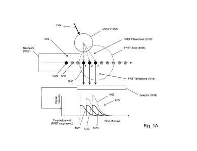
Some of the above aspects and embodiments are illustrated diagrammatically in
Fig. 1A. Polymer
analyte (1000), such as a polynucleotide, is driven, e.g. electrophoretically,
through nanopore (1002),
which constrains the confonnation of polymer (1000) so that its monomeric
units translocate through
the nanopore in the same order as their primary sequence in the polymer.
Moreover, as mentioned
above, whenever an acceptor-labeled monomeric unit is within the bore of
nanopore (1002), FRET
interactions between such acceptors and the donors of its FRET pair (e.g.
1012) are suppressed. Such
suppression typically means that no detectable FRET signal is produced even if
such acceptors are
within a FRET distance of a donor due to unfavorable orientation of the
acceptor and donor dipoles.
On the other hand, as soon as an acceptor-labeled monomeric unit emerges from
the bore of the
nanopore into FRET zone (1008), a strong FRET signal is immediately produced
(due to the proximity
of donor (1012)), after which the signal decreases rapidly as the distance
between the acceptor and
donor increases, because translocation of polymer (1000) carries acceptors out
of FRET zone (1008).
FRET zone (1008), which is a spatial region immediately adjacent to exit
(1015) of nanopore (1002),
is defined by the FRET distances between donor (1012) and the acceptor labels
attached to polymer
(1000) as it translocates through and away from nanopore (1002). In Fig. 1A,
only one type of
monomeric unit, illustrated as solid circles (1004) is labeled; the rest of
the monomeric units,
illustrated as speckled circles (1006), are unlabeled. As illustrated, three
labeled monomeric units
(denoted "1", "2- and "3") are in FRET zone (1008). When donor (1012) is
excited by excitation
CA 02910019 2015-10-21
WO 2014/190322 PCT/US2014/039444
beam (1014), FRET interactions (1010) are generated and the three acceptors on
the monomeric units
produce FRET emissions (1016) that are collected by detector (1018) and
recorded as mixed FRET
signal intensity (1029). Signal intensity contributions from acceptors on
monomeric units 1, 2 and 3
are illustrated by curves (1031, 1032 and 1033, respectively), which are
combined by detector (1018)
to give a mixed FRET signal shown by dashed curve (1022). In the example
described below, an
embodiment corresponding to that of Fig. IA produced data shown in Fig. 1B for
sequence (1082) 3'-
AACGGCCCTICGATCTCATTGAGGATGAGAGGAGAGTCAAAGGAAGA-
ACGAGGATGAGAGGAGAGTGAGAGCAAAGGAAGAACGAGGATGAGAGG-
AGAGTGAGAGCAAAGGAAGAA-5'(SEQ ID NO: 1), in which only cytosines are labeled.
In Fig.
1B, only the relative positions of the labeled C's are shown so that the
correspondence between such
positions and peaks in the data can be appreciated. Intensity peaks are
indicated by asterisks, such as
that of (1080). The data is a plot of relative mixed FRET signal intensity
versus time for the
translocation in a 3'-first orientation of sequence (1082).
[00201 Embodiments are provided where different acceptor labels are
attached to different kinds
of monomeric units, so that signals having different characteristics, e.g.
frequency, intensity,
wavelength, etc., are generated for different kinds of monomeric units,
thereby permitting the different
kinds of monomeric units to be distinguished. In one such embodiment, at least
two different acceptor
labels are used to label different nucleotides of a target polynucleotide. An
apparatus for such an
embodiment is illustrated in Fig. 1C. Polynucleotide (1070) comprises
cytosines (or cytidines or
deoxycytidines) labeled with a first acceptor (solid circles, 1064),
Thymidines or thymines labeled
with a second acceptor (cross-hatched circles, 1066), and Guanines and
Adenines unlabeled (speckled
circles, 1068). As above, as polynucleotide (1070) translocates through
nanopore (1002), nucleotides
exit into FRET zone (1008) where acceptors (if present) become capable of
engaging in a FRET
reaction and generating FRET emissions (1062). Such emissions are collected by
detector (1060)
which has conventional optical components for separating FRET emissions (1062)
in accordance with
the different signal characteristics of the different acceptor labels being
employed, such as wavelength
which can be separated, for example, by a dichroic mirror and/or filters. As a
result, an initially
collected mixed FRET signal is split into two or more signals representing
mixed FRET signals from
different acceptors, which may be further processed by conventional components
(1072). Also
described more fully in the example below, an embodiment corresponding to that
of Fig. IC produced
data shown in Fig. ID for sequence (1092) 5'
GCTAIGTGGCGCGGTATTATTAAGAAGGAGACTGAGAGGAGAGAAGGAGCAAGAAGGA
AATGAGAGCGAGAGGAGAAGAAGGAGGAAGAAG 3'(SEQ ID NO: 2), in which only
cytosines (or cytidines or deoxycytidines) and thymidines or thymines are
labeled. Signals from first
6
CA 02910019 2015-10-21
WO 2014/190322 PCT/US2014/039444
acceptors attached to Ts are indicated by dashed line (1095) and signals from
first acceptors attached
to C's are indicated by solid line (1096) In Fig. 1D, the positions of the
labeled T's and C's are
shown as bolded letters (1051, 1052, 1053, 1054, 1055 and 1056, respectively).
Intensity peaks in the
plots corresponding to the labeled T's and C's are indicated by the same
reference numbers. The data
is a plot of relative mixed FRET signal intensity versus time for the
translocation in a 3'-first
orientation of sequence (1070).
[0021] As mentioned above, in one aspect, a method may be carried out by
the following steps:
(a) translocating a polynucleotide, e.g., a single stranded or double stranded
polynucleotide, through a
nanopore so that nucleotides of the polynucleotide pass in sequence by a first
member of a FRET pair
positioned adjacent to the nanopore, a plurality of the nucleotides being
within a FRET distance of the
first member of the FRET pair as the nucleotides exit the nanopore and a
portion of the nucleotides
being labeled with a second member of the FRET pair; (b) exposing the FRET
pairs adjacent to the
nanopore to a light beam so that FRET occurs between the first and a plurality
of second members of
the FRET pair within the FRET distance to generate a mixed FRET signal; (c)
measuring mixed
FRET signals as the polynucleotide translocates through the nanopore; and (d)
determining a
nucleotide sequence of the polynucleotide from the mixed FRET signals. In some
embodiments, a
nanopore is a hybrid nanopore comprising a protein nanopore inserted into a
pore of a solid phase
membrane, as described more fully below. In hybrid nanopores, a first member
of a FRET pair may
be attached directly to the protein nanopore, or alternatively, directly to
the solid phase membrane
using conventional linking chemistries, such as "click" chemistries, e.g. Kolb
et alõkngew. Chem. Int.
Ed., 4): 2004-2021 (2001), or the like. In one embodiment, a first member of a
FRET pair is attached
directly or indirectly to the protein nanopore, for example, as discussed in
reference to Fig. 2D. In
another embodiment, the first member of the FRET pair is a donor, such as a
quantum dot. Quantum
dots are typically much larger than acceptors, especially acceptors that are
organic dyes, which
typically have molecular weights in the range of from 200 to 2000 daltons.
Thus, for FRET to occur
between a quantum dot donor and a multiply-labeled polymer analyte, multiple
acceptors are brought
within a FRET distance of the quantum dot at the same time. Under such
circumstances multiple
FRET signals are generated within the same time interval over which such
signals are collected,
thereby giving rise to a mixed FRET signal.
Nanopores and Nanopore Sequencing
[0022] Nanopores used with various methods, systems and devices described
herein may be solid-
state nanopores, protein nanopores, or hybrid nanopores comprising protein
nanopores configured in a
solid-state membrane, or like framework. Important features of nanopores
include (i) constraining
7
CA 02910019 2015-10-21
WO 2014/190322 PCT/US2014/039444
analytes, particularly polymer analytes, to pass through a detection zone in
sequence, (ii) compatibility
with a translocating means, that is, whatever method is used to drive an
analyte through a nanopore,
and (iii) FRET suppression for members of FRET pairs within the lumen, or
bore, of the nanopore.
100231 Nanopores may be fabricated in a variety of materials including but not
limited to, silicon
nitride (Si3N4), silicon dioxide (Si02), and the like. The fabrication and
operation of nanopores for
analytical applications, such as DNA sequencing, are disclosed in the
following exemplary references
that are incorporated by reference: Russell, U.S. patent 6,528,258; Feier,
U.S. patent 4,161,690; Ling,
U.S. patent 7,678,562; Hu et al, U.S. patent 7,397,232; Golovchenko et al,
U.S. patent 6,464,842; Chu
et al, U.S. patent 5,798,042; Sauer et al, U.S. patent 7,001,792; Su et al,
U.S. patent 7,744,816; Church
et al, U.S. patent 5,795,782; Bayley et al, U.S. patent 6,426,231; Akeson et
al. U.S. patent 7,189,503;
Bayley et al, U.S. patent 6,916,665; Akeson et al, U.S. patent 6,267,872;
Meller et al, U.S. patent
publication 2009/0029477; Howorka et al, International patent publication
W02009/007743; Brown et
al, International patent publication W02011/067559; Meller et al,
International patent publication
W02009/020682; Polonsky et al, International patent publication W02008/092760;
Van der Zaag et
al, International patent publication W02010/007537; Yan et al, Nano Letters,
5(6): 1129-1134 (2005);
Iqbal et al, Nature Nanotechnology, 2: 243-248 (2007); Wanunu et al, Nano
Letters, 7(6): 1580-1585
(2007); Dekker, Nature Nanotechnology, 2: 209-215 (2007); Storm et al, Nature
Materials, 2: 537-540
(2003); Wu et al, Electrophoresis, 29(13): 2754-2759 (2008); Nakane et al,
Electrophoresis, 23: 2592-
2601 (2002); Zhe et al, J. Micromech.Nlicroeng., 17: 304-313 (2007); Henriquez
et al, The Analyst,
129: 478-482 (2004); Jagtiani et al, J. Micromech. Microeng., 16: 1530-1539
(2006); Nakane et al, J.
Phys. Condens. Matter, 15 R1365-R1393 (2003); DeBlois et al, Rev. Sci.
Instruments, 41(7): 909-916
(1970); Clarke et al, Nature Nanotechnology, 4(4): 265-270 (2009); Bayley et
al, U.S. patent
publication 2003/0215881; and the like. Briefly, in one aspect, a 1-50 nm
channel is formed through a
substrate, usually a membrane, through which an analyte, such as DNA, is
induced to translocate. The
solid-state approach of generating nanopores offers robustness and durability
as well as the ability to
tune the size and shape of the nanopore, the ability to fabricate high-density
arrays of nanopores on a
wafer scale, superior mechanical, chemical and thermal characteristics
compared with lipid-based
systems, and the possibility of integrating with electronic or optical readout
techniques. Biological
nanopores on the other hand provide reproducible narrow bores, or lumens,
especially in the 1-10
nanometer range, as well as techniques for tailoring the physical and/or
chemical properties of the
nanopore and for directly or indirectly attaching groups or elements, such as
FRET donors or
acceptors, by conventional protein engineering methods. Protein nanopores
typically rely on delicate
lipid bilayers for mechanical support, and the fabrication of solid-state
nanopores with precise
dimensions remains challenging. Combining solid-state nanopores with a
biological nanopore
8
CA 02910019 2015-10-21
WO 2014/190322 PCT/US2014/039444
overcomes some of these shortcomings, especially the precision of a biological
pore protein with the
stability of a solid state nanopore. For optical read out techniques a hybrid
nanopore provides a precise
location of the nanopore which simplifies the data acquisition greatly. The
lateral diffusion of
nanopore proteins inserted in a lipid bilayer makes an optical detection
challenging. Since the
biological part of a hybrid nanopore does not rely on the insertion in a lipid
bilayer the degrees of
freedom for modifications made to such a protein are greatly increased, e.g. a
genetically modified
nanopore protein that does not spontaneously insert in a lipid bilayer may
still be used as a protein
component of a hybrid nanopore. Bilayer destabilizing agents such as quantum
dots may be used to
label a protein component of a hybrid nanopore.
[0024] In one embodiment , a device or system for detecting one or more
analytes, such as a
polynucleotide analyte, comprises the following elements; (a) a solid phase
membrane separating a
first chamber and a second chamber, the solid phase membrane having at least
one aperture connecting
the first chamber and the second chamber through a bore; and (b) a first
member of a fluorescent
resonance enemy transfer (FRET) pair attached to the at least one aperture, so
that whenever one or
more analytes having a plurality of second members of the FRET pair attached
thereto traverses the
bore, the plurality of second members are constrained to pass in sequence
within a FRET distance of
the first member of the FRET pair. In some embodiments, the solid phase
membrane has been treated
with a low energy ion beam to bleach its autofluorescence.
[0025] In another embodiment, a device or system for detecting a plurality of
analytes, or a polymer
analyte having a plurality of linked monomer units, such as nucleotides, is
provided. Such an
embodiment for determining a sequence of a polynucleotide may comprise one or
more of the
following elements: (a) a solid phase membrane separating a first chamber and
a second chamber, the
solid phase membrane having at least one aperture connecting the first chamber
and the second
chamber, and having a hydrophobic coating on at least one surface; (b) a lipid
layer may be disposed
on the hydrophobic coating; (c) a protein nanopore immobilized in the
aperture, the protein nanopore
having a bore with an exit, and the protein nanopore interacting with the
lipid layer to form a seal with
the solid phase membrane in the aperture so that fluid communication between
the first chamber and
the second chamber occurs solely through the bore of the protein nanopore, and
the protein nanopore
being dimensioned so that nucleotides of the polynucleotide pass through the
exit of the bore in
sequence and so that whenever nucleotides of the polynucleotide are labeled
with second members of
a FRET pair, FRET is suppressed between such second members inside the bore
and first members of
the FRET pair outside the bore; and/or (d) a first member of the FRET pair
attached to the solid phase
membrane or the protein nanopore, so that whenever nucleotides of the
polynucleotide emerge from
9
CA 02910019 2015-10-21
WO 2014/190322 PCT/US2014/039444
the bore, a plurality of the nucleotides are within a FRET distance of the
first member of the FRET
pair.
[0026] In some embodiments, the hydrophobic coating is optional in that the
surface of the solid
phase membrane is sufficiently hydrophobic itself so that a lipid layer
adheres to it stably. The at least
one aperture will have an inner surface, or wall, connected to, or contiguous
with the surfaces of the
solid phase membrane. In some embodiments, the at least one aperture will be a
plurality of apertures,
and the plurality of apertures may be arranged as a regular array, such as a
rectilinear array of
apertures, the spacing of which depending in part on the number and kind of
FRET pairs employed
and the optical detection system used. Each of the apertures has a diameter,
which in some
embodiments is such that a protein nanopore is substantially immobilized
therein. In some
embodiments, substantially immobilized means that a protein nanopore may move
no more than 5 nm
in the plane of the solid phase membrane relative to the wall of the aperture.
In another embodiment,
substantially immobilized means that a protein nanopore may move no more than
5 urn in the plane of
the solid phase membrane relative to the wall of the aperture. The protein
nanopores each have a bore,
or passage, or lumen, which peimits fluid communication between the first and
second chambers when
the protein nanopore is immobilized in an aperture. Generally, the bore is
coaxially aligned with the
aperture. One function of the hydrophobic layer is to provide a surface to
retain lipids in and/or
immediately adjacent to the at least one aperture. Such lipids, in turn,
permit disposition and
immobilization of a protein nanopore within an aperture in a functional
conformation and in a manner
that forms a fluid seal with the wall of the aperture. In some embodiments,
such seal also prevents
electrical current passing between the first and second chambers around the
protein nanopore. In some
embodiments, charged analytes are disposed in an electrolyte solution in the
first chamber and are
translocated through the bore(s) of the protein nanopore(s) into an
electrolytic solution in the second
chamber by establishing an electrical field across the solid phase membrane.
For convenience of
manufacture, in some embodiments the hydrophobic coating will be on one
surface of the solid phase
membrane and the wall(s) of the aperture(s).
[0027] In some embodiments, the solid phase membrane is treated with a low
energy ion beam to
bleach its autofluorescence, as described more fully below.
[0028] Figs. 2A-2C are diagrams of hybrid biosensors. A nanometer sized hole
(102) is drilled into a
solid-state substrate, or solid phase membrane, (103) which separates two
chambers, or compartments
cis (101) and trans (107). A protein biosensor (e.g a protein nanopore) (104)
attached to a charged
polymer (105), such as a single or double- stranded DNA, is embedded into the
solid-state nanohole
by electrophoretic transport. In Fig. IC the protein biosensor is inserted. In
a nanometer sized hole
CA 02910019 2015-10-21
WO 2014/190322 PCT/US2014/039444
which surface has a hydrophobic coating (106) and may have a lipid layer (109)
attached thereto. A
nanopore may have two sides, or orifices. One side is referred to as the "cis"
side and faces the (-)
negative electrode or a negatively charged buffer/ion compartment or solution.
The other side is
referred to as the "trans" side and faces the (+) electrode or a positively
charged buffer/ion
compartment or solution. A biological polymer, such as a labeled nucleic acid
molecule or polymer
can be pulled or driven through the pore by an electric field applied through
the nanopore, e.g.,
entering on the cis side of the nanopore and exiting on the trans side of the
nanopore.
[0029] Fig. 2D shows protein nanopore (104) inserted into an aperture drilled
in a solid state
membrane (103). Attached to the protein nanopore (104) is an oligonucleotide
(108) to which a
complementary secondary oligonucleotide (111) is hybridized. Said secondary
oligonucleotide (111)
has one or more first or second members of a FRET pair (110) attached to it.
Alternatively, a member
of a FRET pair may be directly attached to an amino acid of a protein
nanopore. For example, a
hemolysin subunit may be modified by conventional genetic engineering
techniques to substitute a
cysteine for a suitably located amino acid adjacent to the exit of the
nanopore, e.g. the threonine 129.
An oligonucleotide or members of a FRET pair may be attached via the thio
group of the cysteine
using conventional linker chemistries, e.g. Hermanson (cited above).
[0030] In some embodiments, a hybrid nanopore is utilized, particularly for
optical-based nanopore
sequencing of polynucleotides. Such embodiments comprise a solid-state
orifice, or aperture, into
which a protein biosensor, such as a protein nanopore, is stably inserted. A
protein nanopore (e.g.
alpha hemolysin) may be attached to a charged polymer (e.g. double stranded
DNA) which serves as a
drag force in an applied electric field, and which may be used to guide a
protein nanopore into an
aperture in a solid-state membrane. In some embodiments, the aperture in the
solid-state substrate is
selected to be slightly smaller than the protein, thereby preventing it from
translocating through the
aperture. Instead, the protein will be embedded into the solid-state orifice.
The solid-state substrate can
be modified to generate active sites on the surface that allow the covalent
attachment of the plugged-in
protein biosensor resulting in a stable hybrid biosensor.
[0031] The polymer attachment site in the biosensor can be generated by
protein engineering e.g. a
mutant protein can be constructed that will allow the specific binding of the
polymer. As an example,
a cysteine residue may be inserted at the desired position of the protein. The
cysteine can either
replace a natural occurring amino acid or can be incorporated as an addition
amino acid. Care must be
taken not to disrupt the biological function of the protein. The terminal
primary amine group of a
polymer (i.e. DNA) is then activated using a hetero-bifunctional crosslinker
(e.g. SMCC).
Subsequently, the activated polymer is covalently attached to the cysteine
residue of the protein
11
CA 02910019 2015-10-21
WO 2014/190322 PCT/US2014/039444
biosensor. In some embodiments, the attachment of the polymer to the biosensor
is reversible. By
implementing a cleavable crosslinker, an easily breakable chemical bond (e.g.
an S-S bond) is
introduced and the charged polymer may be removed after insertion of the
biosensor into the solid-
state aperture.
100321 For someone skilled in the art it is obvious that a wide variety of
different approaches for
covalent or non-covalent attachment methods of a charged polymer to the
protein biosensor are
possible and the above described approach merely serves as an example. The
skilled artisan will also
realize that a variety of different polymers may be used as a drag force,
including, but not limited to,
single or double stranded DNA, polyethyleneglycol (PEG), polyvinylpyrrolidone
(PVP), poly-L-
lysine, linear polysaccharides etc. It is also obvious that these polymers may
exhibit either a negative
(-) or positive ( ) charge at a given pH and that the polarity of the electric
field may be adjusted
accordingly to pull the polymer-biosensor complex into a solid-state aperture.
100331 In some embodiments, a donor fluorophore is attached to the protein
nanopore. This complex
is then inserted into a solid-state aperture or nanohole (3-10nm in diameter)
by applying an electric
field across the solid state nanohole until the protein nanopore is
transported into the solid-state
nanohole to fonn a hybrid nanopore. The formation of the hybrid nanopore can
be verified by (a) the
inserting protein nanopore causing a drop in current based on a partial
blockage of the solid-state
nanohole and by (b) the optical detection of the donor fluorophore.
100341 Once stable hybrid nanopores have formed single stranded, fluorescently
labeled (or acceptor
labeled) DNA may be added to the cis chamber (the chamber with the ( )
electrode). The applied
electric field forces the negatively charged ssDNA to translocate through the
hybrid nanopore during
which the labeled nucleotides get in close vicinity of the donor fluorophore.
In certain variations,
double stranded DNA may be utilized.
100351 Solid state, or synthetic, nanopores may be prepared in a variety of
ways, as exemplified in the
references cited above. In some embodiments a helium ion microscope may be
used to drill the
synthetic nanopores in a variety of materials, e.g. as disclosed by Yang et
al, Nanotechnolgy, 22:
285310 (2011), which is incorporated herein by reference. A chip that supports
one or more regions of
a thin-film material, e.g. silicon nitride, that has been processed to be a
free-standing membrane is
introduced to the helium ion microscope (HIM) chamber. HIM motor controls are
used to bring a free-
standing membrane into the path of the ion beam while the microscope is set
for low magnification.
Beam parameters including focus and stigmation are adjusted at a region
adjacent to the free-standing
membrane, but on the solid substrate. Once the parameters have been properly
fixed, the chip position
is moved such that the free-standing membrane region is centered on the ion
beam scan region and the
12
CA 02910019 2015-10-21
WO 2014/190322 PCT/US2014/039444
beam is blanked. The HIM field of view is set to a dimension (in pm) that is
sufficient to contain the
entire anticipated nanopore pattern and sufficient to be useful in future
optical readout (i.e. dependent
on optical magnification, camera resolution, etc.). The ion beam is then
rastered once through the
entire field of view at a pixel dwell time that results in a total ion dose
sufficient to remove all or most
of the membrane autofluorescence. The field of view is then set to the proper
value (smaller than that
used above) to perform lithographically-defined milling of either a single
nanopore or an array of
nanopores. The pixel dwell time of the pattern is set to result in nanopores
of one or more
predetermined diameters, determined through the use of a calibration sample
prior to sample
processing. This entire process is repeated for each desired region on a
single chip and/or for each chip
introduced into the HIM chamber.
[0036] In some embodiments, the solid-state substrate may be modified to
generate active sites on the
surface that allow the covalent attachment of the plugged in protein biosensor
or to modify the surface
properties in a way to make it more suitable for a given application. Such
modifications may be of
covalent or non-covalent nature. A covalent surface modification includes a
silanization step where an
oraanosilane compound binds to silanol groups on the solid surface. For
instance, the alkoxy groups of
an alkoxysilane are hydrolyzed to form silanol-containing species. Reaction of
these silanes involves
four steps. Initially, hydrolysis of the labile groups occurs. Condensation to
oligomers follows. The
oligomers then hydrogen bond with hydroxyl groups of the substrate. Finally,
during drying or curing,
a covalent linkage is formed with the substrate with concomitant loss of
water. For covalent
attachment organosilanes with active side groups may be employed. Such side
groups consist of, but
are not limited to epoxy side chain, aldehydes, isocyanates, isothiocyanates,
azides or alkynes (click
chemistry) to name a few. For someone skilled in the art it is obvious that
multiple ways of covalently
attaching a protein to a surface are possible. For instance, certain side
groups on an organosilane may
need to be activated before being capable of binding a protein (e.g. primary
amines or carboxyl side
groups activated with an N-hydroxysuccinimidester). Another way of attaching a
protein to the solid
surface may be achieved through affinity binding by having one affinity
partner attached to the protein
and the second affinity partner being located on the solid surface. Such
affinity pairs consist of the
group of, but are not limited to biotin-strepavidin, antigen-antibody and
aptamers and the
corresponding target molecules.
100371 In one embodiment, the surface modification of the solid state nanopore
includes treatment
with an omanosilane that renders the surface hydrophobic. Such organosilanes
include but are not
limited to, alkanesilanes (e.g. octadecyldimethylchlorosilane) or modified
alkanesilanes such as
fluorinated alkanesilanes with an alkane chain length of 5 to 30 carbons. The
hydrophobic surface may
13
CA 02910019 2015-10-21
WO 2014/190322 PCT/US2014/039444
then be treated with a dilute solution of a lipid in pentane. After drying of
the solvent and immersing
the surface in an aqueous solution the lipid will spontaneously form a layer
on the surface. A layer of
lipid on the solid surface might prove beneficial for the formation of a
hybrid nanopore. The lipid
layer on the solid phase might reduce the leak current between protein and
solid state nanopore and it
might increase the stability of the inserted protein pore. Combining a low
capacitance solid substrate
as well as a lipid coating of said substrate may render the hybrid nanopore
system amenable to an
electrical readout based on current fluctuations generated by translocation of
DNA through the hybrid
nanopore. To achieve electrical read out with such a system a means of
decreasing the translocation
speed of unmodified DNA must be combined with a lipid coated hybrid nanopore.
Molecular motors
such as polymerases or helicases may be combined with a hybrid nanopore and
effectively reduce the
translocation speed of DNA through the hybrid nanopore. The lipids used for
coating the surface may
be from the group of sphingolipids, phospholipids or sterols.
100381 A method and/or system for sequencing a biological polymer or molecule
(e.g., a nucleic acid)
may include exciting one or more donor labels attached to a pore or nanopore.
A biological polymer
may be translocated through the pore or nanopore, where a monomer of the
biological polymer is
labeled with one or more acceptor labels. Energy may be transferred from the
excited donor label to
the acceptor label of the monomer as, after the labeled monomer passes
through, exits or enters the
pore or nanopore. Energy emitted by the acceptor label as a result of the
energy transfer may be
detected, where the energy emitted by the acceptor label may correspond to or
be associated with a
single or particular monomer (e.g., a nucleotide) of a biological polymer. The
sequence of the
biological polymer may then be deduced or sequenced based on the detection of
the emitted energy
from the monomer acceptor label which allows for the identification of the
labeled monomer. A pore,
nanopore, channel or passage, e.g., an ion permeable pore, nanopore, channel
or passage may be
utilized in the systems and methods described herein.
[0039] The nanopore may have one or more labels attached. In some embodiments,
the label is a
member of a Forster Resonance Energy Transfer (FRET) pair. Such labels may
comprise organic
fluorophores, chemiluminescent labels, quantum dots, metallic nanoparticles
and fluorescent proteins.
The nucleic acid may have one distinct label per nucleotide. The labels
attached to the nucleotides
consist of the group of organic fluorophores, chemiluminescent labels, quantum
dots, metallic
nanoparticles and fluorescent proteins. The label attachment site in the pore
protein can be generated
by protein engineering e.g. a mutant protein can be constructed that will
allow the specific binding of
the label. As an example, a cysteine residue may be inserted at the desired
position of the protein
which inserts a thiol (SH) group that can be used to attach a label. The
cysteine can either replace a
14
CA 02910019 2015-10-21
WO 2014/190322 PCT/US2014/039444
natural occurring amino acid or can be incorporated as an addition amino acid.
Care must be taken not
to disrupt the biological function of the protein. A malemeide-activated label
is then covalently
attached to the thiol residue of the protein nanopore. In one embodiment, the
attachment of the label to
the protein nanopore or the label on the nucleic acid is reversible. By
implementing a cleavable
crosslinker, an easily breakable chemical bond (e.g. an S-S bond or a pH
labile bond) is introduced
and the label may be removed when the corresponding conditions are met.
[0040] A nanopore, or pore, may be labeled with one or more donor labels. For
example, the cis side
or surface and/or trans side or surface of the nanopore may be labeled with
one or more donor labels.
The label may be attached to the base of a pore or nanopore or to another
portion or monomer making
up the nanopore or pore A label may be attached to a portion of the membrane
or substrate through
which a nanopore spans or to a linker or other molecule attached to the
membrane, substrate or
nanopore. The nanopore or pore label may be positioned or attached on the
nanopore, substrate or
membrane such that the pore label can come into proximity with an acceptor
label of a biological
polymer, e.g., a nucleic acid, which is translocated through the pore. The
donor labels may have the
same or different emission or absorption spectra. The labeling of a pore
structure may be achieved via
covalent or non-covalent interactions.
[0041] A donor label may be placed as close as possible to the aperture of a
nanopore without causing
an occlusion that impairs translocation of a nucleic acid through the
nanopore. A pore label may have
a variety of suitable properties ancUor characteristics. For example, a pore
label may have energy
absorption properties meeting particular requirements. A pore label may have a
large radiation energy
absorption cross-section, ranging, for example, from about 0 to 1000 nm or
from about 200 to 500 nm.
A pore label may absorb radiation within a specific energy range that is
higher than the energy
absorption of the nucleic acid label. The absorption energy of the pore label
may be tuned with
respect to the absorption energy of a nucleic acid label in order to control
the distance at which energy
transfer may occur between the two labels. A pore label may be stable and
functional for at least 10A6
or 10A9 excitation and energy transfer cycles.
Treating Solid Phase Membranes to Reduce Autofluorescence
[0042] In some embodiments, a solid phase membrane of a microelectromechanical
system (MEMS)
material is treated with a low energy ion beam to bleach its autofluorescence.
Typically such
treatment is carried out by directing an ion beam to a surface region of the
MEMS material, at a
sufficiently high energy to cause a physical change in the MEMS material at
its surface or near its
surface to disrupt or inactivate structures contributing to autofluorescence,
but not with such high
CA 02910019 2015-10-21
WO 2014/190322 PCT/US2014/039444
energy that melting, vaporization, significant deformations or sputtering
occur. The minimal energy
required may be readily determined on a material-by-material basis by
gradually increasing beam
energy starting from zero and measuring reduction in autofluorescence with
increasing beam energy.
As used herein, the term "autofluorescence" is used synonymously with
"background fluorescence" to
mean fluorescence emanating from a source at or near a surface of a 11/1FMS
material upon excitation
with a light source selected to excite a fluorescent label that is not a part
of the MEMS material. Thus,
autofluorescence in a MEMS material depends on the frequency of the light
source. In one aspect, the
frequency of the light source is selected to excite organic fluorescent dyes,
so that the method reduces
autofluorescence of frequencies in the visible range of light as well as
frequencies from the near
infrared to the near ultraviolet. MEMS materials include a wide variety of
solids capable of
microfabrication and use in analytical techniques using optical detection.
Exemplary MEMS materials
are silicon-based substrates, such as silicon nitride and silicon dioxide or
metal based substrates, such
as aluminum oxide. In one aspect, MEMS materials are processed and used in the
form of a
membrane. In one embodiment, the MEMS material is silicon nitride. A wide
variety of focused ion
beams may be employed for such bleaching and guidance for the production and
application of such
beams at various enemies may be found in such references as, Natasi et al, Ion
Solid Interactions:
Fundamentals and Applications (Cambridge University Press, 1996), and like
references. Exemplary
focused ion beams include helium ion beams, neon ion beams and gallium ion
beams. In one
embodiment, a helium ion beam is used in the method. Helium ion beams may be
produced with a
commercially available ion beam microscope (HIM) (e.g. Zeiss Orion Nanofab).
The amount of
energy or dosage delivered to a surface of a MEMS material, such as silicon
nitride, to reduce
autofluorescence may be in the range of from 2e-10 to 8e-10 nC/nm^2.
Labels for Nanopores and Analytes
[0043] In some embodiments, a nanopore may be labeled with one or more quantum
dots. In
particular, in some embodiments, one or more quantum dots may be attached to a
nanopore, or
attached to a solid phase support adjacent to (and within a FRET distance of
an entrance or exit of a
nanopore), and employed as donors in FRET reactions with acceptors on
analytes. Such uses of
quantum dots are well known and are described widely in the scientific and
patent literature, such as,
in U.S. patents 6,252,303; 6,855,551; 7,235,361; and the like, which are
incorporated herein by
reference.
[0044] One example of a Quantum dot which may be utilized as a pore label is a
CdTe quantum dot
which can be synthesized in an aqueous solution. A CdTe quantum dot may be
functionalized with a
16
CA 02910019 2015-10-21
WO 2014/190322 PCT/US2014/039444
nucleophilic group such as primary amines, thiols or functional groups such as
carboxylic acids. A
CdTe quantum dot may include a mercaptopropionic acid capping ligand, which
has a carboxylic acid
functional group that may be utilized to covalently link a quantum dot to a
primary amine on the
exterior of a protein pore. The cross-linking reaction may be accomplished
using standard cross-
linking reagents (homo-bifunctional as well as hetero-bifunctional) which are
known to those having
ordinary skill in the art of bioconjugation. Care may be taken to ensure that
the modifications do not
impair or substantially impair the translocation of a nucleic acid through the
nanopore. This may be
achieved by varying the length of the employed crosslinker molecule used to
attach the donor label to
the nanopore.
[0045] The primary amine of the Lysin residue 131 of the natural alpha
hemolysin protein (Song, L.
et al., Science 274, (1996): 1859-1866) may be used to covalently bind carboxy
modified CdTe
Quantum dots via 1-Ethy1-343-dimethylaminopropyl]carbodiimide hydrochloride/ N-
hydroxysulfosuccinimide (EDC/NHS) coupling chemistry. Alternatively, amino
acid 129 (threonine)
may be exchanged into cysteine. Since there is no other cysteine residue in
the natural alpha
hemolysin protein the thiol side group of the newly inserted cysteine may be
used to covalently attach
other chemical moieties.
[0046] A variety of methods, mechanisms and/or routes for attaching one or
more pore labels to a
pore protein may be utilized. A pore protein may be genetically engineered in
a manner that
introduces amino acids with known properties or various functional groups to
the natural protein
sequence. Such a modification of a naturally occurring protein sequence may be
advantageous for the
bioconjugation of Quantum dots to the pore protein. For example, the
introduction of a cysteine
residue would introduce a thiol group that would allow for the direct binding
of a Quantum dot, such
as a CdTe quantum dot, to a pore protein. Also, the introduction of a Lysin
residue would introduce a
primary amine for binding a Quantum dot. The introduction of glutamic acid or
aspartic acid would
introduce a carboxylic acid moiety for binding a Quantum dot. These groups are
amenable for
bioconjugation with a Quantum dot using either homo- or hetero-bifunctional
crosslinker molecules.
The insertions of poly-histidines allow the direct binding of Quantum dots to
a protein pore via metal-
histidine coordination. Such modifications to pore proteins aimed at the
introduction of functional
groups for bioconjugation are known to those having ordinary skill in the art.
Care should be taken to
ensure that the modifications do not impair or substantially impair the
translocation of a nucleic acid
through the nanopore.
[0047] The nanopore label can be attached to a protein nanopore before or
after insertion of said
nanopore into a lipid bilayer. Where a label is attached before insertion into
a lipid bilayer, care may
17
CA 02910019 2015-10-21
WO 2014/190322 PCT/US2014/039444
be taken to label the base of the nanopore and avoid random labeling of the
pore protein. This can be
achieved by genetic engineering of the pore protein to allow site specific
attachment of the pore label
(see section 0047). An advantage of this approach is the bulk production of
labeled nanopores.
Alternatively, a labeling reaction of a pre-inserted nanopore may ensure site-
specific attachment of the
label to the base (trans-side) of the nanopore without genetically engineering
the pore protein.
[0048] A biological polymer, e.g., a nucleic acid molecule or polymer, may be
labeled with one or
more acceptor labels. For a nucleic acid molecule, each of the four
nucleotides or building blocks of a
nucleic acid molecule may be labeled with an acceptor label thereby creating a
labeled (e.g.,
fluorescent) counterpart to each naturally occurring nucleotide. The acceptor
label may be in the form
of an energy accepting molecule which can be attached to one or more
nucleotides on a portion or on
the entire strand of a converted nucleic acid.
[0049] A variety of methods may be utilized to label the monomers or
nucleotides of a nucleic acid
molecule or polymer. A labeled nucleotide may be incorporated into a nucleic
acid during synthesis
of a new nucleic acid using the original sample as a template ("labeling by
synthesis"). For example,
the labeling of nucleic acid may be achieved via PCR, whole genome
amplification, rolling circle
amplification, primer extension or the like or via various combinations and
extensions of the above
methods known to persons having ordinary skill in the art.
[0050] Labeling of a nucleic acid may be achieved by replicating the nucleic
acid in the presence of a
modified nucleotide analog having a label, which leads to the incorporation of
that label into the newly
generated nucleic acid. The labeling process can also be achieved by
incorporating a nucleotide analog
with a functional group that can be used to covalently attach an energy
accepting moiety in a
secondary labeling step. Such replication can be accomplished by whole genome
amplification
(Zhang, L. et al., Proc. Natl. Acad. Sci. USA 89 (1992): 5847) or strand
displacement amplification
such as rolling circle amplification, nick translation, transcription, reverse
transcription, primer
extension and polymerase chain reaction (PCR), degenerate oligonucleotide
primer PCR (DOP-PCR)
(Telenius, H. et al., Genomics 13 (1992): 718-725) or combinations of the
above methods.
[0051] A label may comprise a reactive group such as a nucleophile (amines,
thiols etc.). Such
nucleophiles, which are not present in natural nucleic acids, can then be used
to attach fluorescent
labels via amine or thiol reactive chemistry such as NHS esters, maleimides,
epoxy rings, isocyanates
etc. Such nucleophile reactive fluorescent dyes (i.e. NHS-dyes) are readily
commercially available
from different sources. An advantage of labeling a nucleic acid with small
nucleophiles lies in the
high efficiency of incorporation of such labeled nucleotides when a "labeling
by synthesis" approach
is used. Bulky fluorescently labeled nucleic acid building blocks may be
poorly incorporated by
18
CA 02910019 2015-10-21
WO 2014/190322 PCT/US2014/039444
polymerases due to steric hindrance of the labels during the polymerization
process into newly
synthesized DNA.
100521 DNA can be directly chemically modified without polymerase mediated
incorporation of
labeled nucleotides. One example of a modification includes cis-platinum
containing dyes that modify
Guanine bases at their N7 position (Hoevel, T. et al., Bio Techniques 27
(1999): 1064-1067). Another
example includes the modifying of pyrimidines with hydroxylamine at the C6
position which leads to
6-hydroxylamino derivatives. The resulting amine groups can be further
modified with amine reactive
dyes (e.g. NHS-Cy5). Yet another example are azide or alkyne modified
nucleotides which are readily
incorporated by polymerases (Gierlich et al., Chem. Eur. J., 2007, 13, 9486-
0404). The alkyne or azide
modified polynucleotide is subsequently labeled with an azide or alkyne
modified fluorophore
following well established click chemistry protocols.
[0053] A nucleic acid molecule may be directly modified with N-
Bromosuccinimide which upon
reacting with the nucleic acid will result in 5-Bromocystein, 8-Bromoadenine
and 8-Bromoguanine.
The modified nucleotides can be further reacted with di-amine nucleophiles.
The remaining
nucleophile can then be reacted with an amine reactive dye (e.g. NHS-dye)
(Hemaanson G. in
Bioconjugate Techniques, Academic Press 1996, ISBN 978-0-12-342336-8).
[00541 A combination of 1, 2, 3 or 4 nucleotides in a nucleic acid strand may
be exchanged with their
labeled counterpart. The various combinations of labeled nucleotides can be
sequenced in parallel,
e.g, labeling a source nucleic acid or DNA with combinations of 2 labeled
nucleotides in addition to
the four single labeled samples, which will result in a total of 10
differently labeled sample nucleic
acid molecules or DNAs (G, A, T, C, GA, GT, GC, AT, AC, TC). The resulting
sequence pattern may
allow for a more accurate sequence alignment due to overlapping nucleotide
positions in the redundant
sequence read- out.
100551 In certain variations, a method for sequencing a polymer, such as a
nucleic acid molecule, may
include providing a nanopore or pore protein (or a synthetic pore) inserted in
a membrane or
membrane like structure or other substrate. The base or other portion of the
pore may be modified
with one or more pore labels. The base may refer to the Trans side of the
pore. Optionally, the Cis
and/or Trans side of the pore may be modified with one or more pore labels.
Nucleic acid polymers to
be analyzed or sequenced may be used as a template for producing a labeled
version of the nucleic
acid polymer, in which one of the four nucleotides or up to all four
nucleotides in the resulting
polymer is/are replaced with the nucleotide's labeled analogue(s). An electric
field is applied to the
nanopore which forces the labeled nucleic acid polymer through the nanopore,
while an external
monochromatic or other light source may be used to illuminate the nanopore,
thereby exciting the pore
19
CA 02910019 2015-10-21
WO 2014/190322 PCT/US2014/039444
label. As, after or before labeled nucleotides of the nucleic acid pass
through, exit or enter the
nanopore, energy is transferred from the pore label to a nucleotide label,
which results in emission of
lower energy radiation. The nucleotide label radiation is then detected by a
confocal microscope setup
or other optical detection system or light microscopy system capable of single
molecule detection
known to people having ordinary skill in the art. Examples of such detection
systems include but are
not limited to confocal microscopy, epifluorescent microscopy and total
internal reflection fluorescent
(TIRF) microscopy. Other polymers (e.g., proteins and polymers other than
nucleic acids) having
labeled monomers may also be sequenced according to the methods described
herein.
[0056] Energy may be transferred from a pore or nanopore donor label (e.g., a
Quantum Dot) to an
acceptor label on a polymer (e.g., a nucleic acid) when an acceptor label of
an acceptor labeled
monomer (e.g., nucleotide) of the polymer interacts with the donor label as,
after or before the labeled
monomer exits, enters or passes through a nanopore. For example, the donor
label may be positioned
on or attached to the nanopore on the cis or trans side or surface of the
nanopore such that the
interaction or energy transfer between the donor label and acceptor label does
not take place until the
labeled monomer exits the nanopore and comes into the vicinity or proximity of
the donor label
outside of the nanopore channel or opening. As a result, interaction between
the labels, energy
transfer from the donor label to the acceptor label, emission of energy from
the acceptor label and/or
measurement or detection of an emission of energy from the acceptor label may
take place outside of
the passage, channel or opening running through the nanopore, e.g., within a
cis or trans chamber on
the cis or trans sides of a nanopore. The measurement or detection of the
energy emitted from the
acceptor label of a monomer may be utilized to identify the monomer.
[0057] The nanopore label may be positioned outside of the passage, channel or
opening of the
nanopore such that the label may be visible or exposed to facilitate
excitation or illumination of the
label. The interaction and energy transfer between a donor label and accepter
label and the emission
of energy from the acceptor label as a result of the energy transfer may take
place outside of the
passage, channel or opening of the nanopore. This may facilitate ease and
accuracy of the detection or
measurement of energy or light emission from the acceptor label, e.g., via an
optical detection or
measurement device. The donor and acceptor label interaction may take place
within a channel of a
nanopore and a donor label could be positioned within the channel of a
nanopore.
[0058] A donor label may be attached in various manners and/or at various
sites on a nanopore. For
example, a donor label may be directly or indirectly attached or connected to
a portion or unit of the
nanopore. Alternatively, a donor label may be positioned adjacent to a
nanopore.
CA 02910019 2015-10-21
WO 2014/190322 PCT/US2014/039444
[0059] Each acceptor labeled monomer (e.g., nucleotide) of a polymer (e.g.,
nucleic acid) can interact
sequentially with a donor label positioned on or next to or attached directly
or indirectly to a nanopore
or channel through which the polymer is translocated. The interaction between
the donor and acceptor
labels may take place outside of the nanopore channel or opening, e.g., after
the acceptor labeled
monomer exits the nanopore or before the monomer enters the nanopore. The
interaction may take
place within or partially within the nanopore channel or opening, e.g., while
the acceptor labeled
monomer passes through, enters or exits the nanopore.
[0060] When one of the four nucleotides of a nucleic acid is labeled, the time
dependent signal arising
from the single nucleotide label emission is converted into a sequence
corresponding to the positions
of the labeled nucleotide in the nucleic acid sequence. The process is then
repeated for each of the four
nucleotides in separate samples and the four partial sequences are then
aligned to assemble an entire
nucleic acid sequence.
[0061] When multi-color labeled nucleic acid (DNA) sequences are analyzed, the
enemy transfer
from one or more donor labels to each of the four distinct acceptor labels
that may exist on a nucleic
acid molecule may result in light emission at four distinct wavelengths or
colors (each associated with
one of the four nucleotides) which allows for a direct sequence read-out.
Translocation Speed
[0062] A major obstacle associated with Nanopore based sequencing approaches
is the high
translocation velocity of nucleic acid through a nanopore (-500.000 ¨
1.000.000 nucleotides/sec)
which doesn't allow for direct sequence readout due to the limited bandwidth
of the recording
equipment. A way of slowing down the nucleic acid translocation with two
different nanopore proteins
was recently shown by Cherf et al. (Nat Biotechnol. 2012 Feb 14; 30(4):344-8)
and Manrao et al. (Nat
Biotechnol. 2012 Mar 25; 30(4):349-53) and are incorporated herein by
reference. Both groups used a
DNA polymerase to synthesize a complementary strand from a target template
which resulted in the
step-wise translocation of the template DNA through the nanopore. Hence, the
synthesis speed of the
nucleic acid polymerase (10-500nucleotides/sec) determined the translocation
speed of the DNA and
since it's roughly 3-4 orders of magnitude slower than direct nucleic acid
translocation the analysis of
single nucleotides became feasible. However, the polymerase-aided
translocation requires significant
sample preparation to generate a binding site for the polymerase and the
nucleic acid synthesis has to
be blocked in bulk and can only start once the nucleic acid-polymerase complex
is captured by the
nanopore protein. This results in a rather complex set-up which might prevent
the implementation in a
commercial setting. Furthermore, fluctuation in polymerase synthesis reactions
such as a stalled
21
CA 02910019 2015-10-21
WO 2014/190322 PCT/US2014/039444
polymerization as well as the dissociation of the polymerase from the nucleic
acid may hamper the
sequence read-out resulting in a high error rate and reduced read-length,
respectively. Optical
Nanopore sequence as described in this application uses a different way of
slowing down the DNA
translocation. A target nucleic acid is enzymatically copied by incorporating
fluorescent modified
nucleotides. The resulting labeled nucleic acid has an increased nominal
diameter which results in a
decreased translocation velocity when pulled through a nanopore. The preferred
translocation rate for
optical sequencing lies in the range of 1-1000 nucleotides per second with a
more preferred range of
200-800 nucleotides per second and a most preferred translocation rate of 200-
600 nucleotides per
second.
100631 Alternatively, translocation speed of a polynucleotide, especially a
single stranded
polynucleotide, may be controlled by employing a nanopore dimensioned so that
adducts and/or
labels, e.g. organic dyes attached to bases, inhibit but do not prevent
polynucleotide translocation. A
translocation speed may be selected by attaching labels and/or adducts at a
predetermined density.
Such labels and/or adducts may have regular spaced attachments, e.g. every
third nucleotide or the
like, or they may have random, or pseudorandom attachments, e.g. every C may
be labeled. In some
embodiments, a selected number of different nucleotides may be labeled, e.g.
every A and C, or every
A and G, or every A and T, or every C, or the like, that results in an average
translocation speed. Such
average speed may be decreased by attaching adducts to unlabeled nucleotides.
Adducts include any
molecule, usually and organic molecule, that may be attached to a nucleotide
using conventional
chemistries. Typically adducts have a molecular weight in the same range as
common organic dyes,
e.g. fluorescein, Cy3, or the like. Adducts may or may not be capable of
generating signals, that is,
serving as a label. In some embodiments, adducts and/or labels are attached to
bases of nucleotides.
In other embodiments, labels and/or adducts may be attached to linkages
between nucleosides in a
polynucleotide. In one aspect, a method of controlling translocation velocity
of a single stranded
polynucleotide through a nanopore comprises the step of attaching adducts to
the polynucleotide at a
density, wherein translocation velocity of the single stranded polynucleotide
monotonically decreases
with a larger number of adducts attached, or with the density of adducts
attached. In some
embodiments, not every kind of nucleotide of a polynucleotide is labeled. For
example, four different
sets of a polynucleotide may be produced where nucleotides of each set are
labeled with the same
molecule, e.g. a fluorescent organic dye acceptor, but in each set a different
kind of nucleotide will be
labeled. Thus, in set 1 only A's may be labeled; in set 2 only C's may be
labeled; in set 3 only G's
may be labeled; and so on. After such labeling, the four sets of
polynucleotides may then be analyzed
separately in accordance with the methods and systems described herein and a
nucleotide sequence of
the polynucleotide determined from the data generated in the four analysis. In
such embodiments, and
22
CA 02910019 2015-10-21
WO 2014/190322 PCT/US2014/039444
similar embodiments, e.g. two labels are used, where some of the nucleotides
of a polynucleotide are
not labeled, translocation speed through a nanopore will be affected by the
distribution of label along
the polynucleotide. To prevent such variability in translocation speed, in
some embodiments,
nucleotides that are not labeled with an acceptor or donor for generating
signals to determine
nucleotide sequence, may be modified by attaching a non-signal-producing
adduct that has
substantially the same effect on translocation speed as the signal-producing.
labels.
EXAMPLE
[0064] In this example, a nanopore apparatus is described for determining a
sequence of acceptor-
labeled nucleotides of a polynucleotide, after which it is used to detect a
sequence of acceptor-labeled
cytosines in a first polynucleotide and to detect a sequence of first acceptor-
labeled thymines or
thymidines and second acceptor-labeled cytosines in a second polynucleotide.
[0065] HIM drilling to form nanopore(s) in a silicon nitride membrane: A
3inm Si chip
(Protochipsõ NC) with a 50x50-um etched window spanned by a 30nm Si3N4
membrane is cleaned with
oxygen plasma prior to the drilling process. The cleaned chip is inserted into
the vacuum chamber of a
Helium Ion Microscope (Orion, Zeiss). After insertion, the nanoholes are
drilled with a focused ion
stream with a beam current of --5pA through a 2Ouin aperture and with an
exposure time calibrated to
result in 4 +1- 2 nm holes
[0066] Protein nanopores: A cloned variant of the wt alpha hemolysin
protein is used as a template
for an in vitro transcription/translation reaction. The resulting monomers are
heptamerized by a
stepwise addition of sodium deoxycholate to a final concentration of 6.25mM
and a subsequent
incubation for 12 hours at 4C. The resulting heptamer is attached to an amine-
modified
oligonucleotide using a heterobifunctional crosslinker (SMCC,
Sulfosuccinimidy1-4-(N-
maleimidomethyl) cyclohexane-l-carboxylate). This oligonucleotide serves as a
hybridization site for
a 3kb ds-DNA fragment which is used as a drag force to pull the protein
nanopore into the drilled
holes in the Si3N4 membrane. In addition to the oligonucleotide modification,
the base of the protein
nanopore is also modified with one or more maleimide activated fluorescent
dyes. The attached
fluorophores can either serve as donor or acceptor in a FRET reaction.
[0067] Hybrid Nanopore: TEM grids with drilled holes are cleaned by
submersing in 90C hot
Piranha solution (3:1 Sulfuric acid: 1+02, v/v) for 15min or by an air plasma
for 5min. After
extensive rinsing with water the TEM grid is installed in a Delrin holder
separating a cis and trans
chamber. The trans chamber is sealed with a cover slip that allows the optical
interrogation of the
nanopores. Each chamber is filled with Ethanol to promote wetting of the
nanopores. The Ethanol is
subsequently exchanged with water and then a buffered 1 M KC1 solution. In the
trans chamber the
23
CA 02910019 2015-10-21
WO 2014/190322 PCT/US2014/039444
buffered KCI contains 50% glycerol to facilitate TIR (total internal
reflection) imaging. Both the cis
and trans chamber harbor a AglAgC1 electrode in contact with the buffer
solution with the cathode (+)
in the cis and the anode (-) in the trans chamber. The nanopore protein is
added to the cis side of the
SiN membrane and by applying an electric field (200-600mV) the protein
nanopore is plugged into the
drilled nanoholes forming a hybrid nanopore.
[0068] After 5-25min at 600mV usually 50-75% of the drilled holes have a
protein nanopore
inserted. The formation of the hybrid nanopores is checked on an inverted
microscope (Olympus
IX71) equipped with an APON 60x TIRF oil-immersion objective and a 532nm diode
laser which is
used to excite the fluorophore attached to the protein nanopore. The buffer in
the cis chamber is
exchanged to remove excess nanopore protein. Labeled single stranded (ss) DNA
at a final
concentration of lOnM is added to the cis chamber. The electric field is
reduced to 200-400mV which
promotes the translocation of ssDNA through the hybrid nanopore. The labeled
DNA, when exiting
the nanopore, comes in close proximity to the excited donor fluorophore. A
FRET reaction occurs
which results in the photon emission from the labeled DNA. Emitted photons are
collected, filtered
and imaged using an Orca Flash 4.0 cMOS camera (Hamamatsu) at a frame rate of
500-5000Hz. Data
is extracted from a 5x5 pixel area covering the entire hybrid nanopore from
the raw tiff images using
Imaga Raw traces are normalized and a peak find algorithm is used to identify
FRET signal. Base
calling is performed on the identified peaks.
[0069] Each cytosine of the following 107-mer single stranded
polynucleotide (SEQ ID NO: I)
was labeled with a Cy5 fluorophore.
3 ' -AACGGC CC TTC GATCTCATTGAGGATGAGAGGAGAGTCAAAGGAAGA-
ACGAGGATGAGAGGAGAGTGAGAGCAAAGGAAGAACGAGGATGAGAGG-
AGAGTGAGAGCAAAGGAAGAA-5'
The labeled polynucleotide was translocated in a 3'-first orientation through
the hybrid nanopore and
mixed FRET signals from the labeled cytosines was collected. Raw data is shown
in Fig. 1B. When
transiocated through the hybrid nanopore multiple peaks are observed which
correspond to the number
of cytosines in the DNA. Remarkably, the homopolymer at the 3' end is
perfectly resolved.
[0070] After the 20th nucleotide position from the 5' end, every cytosine
and every thymidine of
the following single stranded polynucleotide (SEQ ID NO: 2) were labeled with
Cy5 and Atto700,
respectively.
24
CA 02910019 2015-10-21
WO 2014/190322 PCT/US2014/039444
5'-GCTATGTGGCGCGGTATTATTAAGAAGGAGACTGAGAGGAGAGAA-
GGAGCAAGAAGGAAATGAGAGCGAGAGGAGAGAAGGAGGAAGAAG -3'
The labeled polynucleotide was translocated in a 3'-first orientation through
the hybrid nanopore and
mixed FRET signals from the labeled cytosines and thymidines were collected.
Raw data is shown in
Fig. ID. The peaks in this raw data trace show a pattern that resembles the
position of the labeled
nucleotides in the template strand.
[0071] This disclosure is not intended to be limited to the scope of the
particular forms set forth,
but is intended to cover alternatives, modifications, and equivalents of the
variations described herein.
Further, the scope of the disclosure fully encompasses other variations that
may become obvious to
those skilled in the art in view of this disclosure. The scope of the present
invention is limited only by
the appended claims.
Definitions
[0072] -Nanopore" means any opening positioned in a substrate that allows
the passage of
analytes through the substrate in a predetermined or discernable order, or in
the case of polymer
analytes, passage of their monomeric units through the substrate in a
predetermined or discernible
order. In the latter case, a predetermined or discernible order may be the
primary sequence of
monomeric units in the polymer. Examples of nanopores include proteinaceous or
protein based
nanopores, synthetic or solid state nanopores, and hybrid nanopores comprising
a solid state nanopore
having a protein nanopore embedded therein. A nanopore may have an inner
diameter of, e.g., 1-10
nm or 1-5 nm or 1-3 nm, or other various sizes. Examples of protein nanopores
include but are not
limited to, alpha-hemolysin, voltage-dependent mitochondrial porin (VDAC),
OmpF, OmpC, MspA
and LamB (maltoporin), e.g. disclosed in Rhee, M. et al.. Trends in
Biotechnology, 25(4) (2007): 174-
181; Bayley et al (cited above); Gundlach et al, U.S. patent publication
2012/0055792; and the like,
which are incorporated herein by reference. Any protein pore that allows the
translocation of single
nucleic acid molecules may be employed. A nanopore protein may be labeled at a
specific site on the
exterior of the pore, or at a specific site on the exterior of one or more
monomer units making up the
pore forming protein. Pore proteins are chosen from a group of proteins such
as, but not limited to,
alpha-hemolysin, MspA, voltage-dependent mitochondrial porin (VDAC), Anthrax
porin, OmpF,
OmpC and LamB (maltoporin). Integration of the pore protein into a solid state
hole is accomplished
by attaching a charged polymer to the pore protein. After applying an electric
field the charged
CA 02910019 2015-10-21
WO 2014/190322
PCT/US2014/039444
complex is electrophoretically pulled into the solid state hole. A synthetic
nanopore, or solid-state
nanopore, may be created in various forms of solid substrates, examples of
which include but are not
limited to silicones (e.g. Si3N4, Si02), metals, metal oxides (e.g. A1203)
plastics, glass,
semiconductor material, and combinations thereof A synthetic nanopore may be
more stable than a
biological protein pore positioned in a lipid bilayer membrane. A synthetic
nanopore may also be
created by using a carbon nanotube embedded in a suitable substrate such as
but not limited to
polymerized epoxy. Carbon nanotubes can have uniform and well-defined chemical
and structural
properties. Various sized carbon nanotubes can be obtained, ranging from one
to hundreds of
nanometers. The surface charge of a carbon nanotube is known to be about zero,
and as a result,
electrophoretic transport of a nucleic acid through the nanopore becomes
simple and predictable (Ito,
T. et al., Chem. Conunun. 12 (2003): 1482-83). The substrate surface of a
synthetic nanopore may be
chemically modified to allow for covalent attachment of the protein pore or to
render the surface
properties suitable for optical nanopore sequencing. Such surface
modifications can be covalent or
non-covalent. Most covalent modification include an organosilane deposition
for which the most
connnon protocols are described:1) Deposition from aqueous alcohol. This is
the most facile method
for preparing silylated surfaces. A 95% ethanol-5% water solution is adjusted
to pH 4.5-5.5 with acetic
acid. Silane is added with stirring to yield a 2% final concentration. After
hydrolysis and silanol group
formation the substrate is added for 2-5min. After rinsed free of excess
materials by dipping briefly in
ethanol. Cure of the silane layer is for 5-10min at 110 degrees Celsius. 2)
Vapor Phase Deposition.
Silanes can be applied to substrates under dry aprotic conditions by chemical
vapor deposition
methods. These methods favor monolayer deposition. In closed chamber designs,
substrates are heated
to sufficient temperature to achieve 5mm vapor pressure. Alternatively, vacuum
can be applied until
silane evaporation is observed. 3) Spin-on deposition. Spin-on applications
can be made under
hydrolytic conditions which favor maximum functionalization and polylayer
deposition or dry
conditions which favor monolayer deposition.
[0073] "FRET"
or "Forrester, or fluorescence, resonant enemy transfer" means a non-radiative
dipole-dipole energy transfer mechanism from a donor to acceptor fluorophore.
The efficiency of
FRET may be dependent upon the distance between donor and acceptor as well as
the properties of the
fluorophores (Stryer, L., Annu Rev Biochem. 47 (1978): 819-846). "FRET
distance" means a
distance between a FRET donor and a FRET acceptor over which a FRET
interaction can take place
and a detectable FRET signal produced by the FRET acceptor.
[0074]
"Polynucleotide" or -oligonucleotide" are used interchangeably and each mean a
linear
polymer of nucleotide monomers. Monomers making up polynucleotides and
oligonucleotides are
capable of specifically binding to a natural polynucleotide by way of a
regular pattern of monomer-to-
26
CA 02910019 2015-10-21
WO 2014/190322 PCT/US2014/039444
monomer interactions, such as Watson-Crick type of base pairing, base
stacking, Hoogsteen or reverse
Hoogsteen types of base pairing, or the like. Such monomers and their
intemucleosidic linkages may
be naturally occurring or may be analogs thereof, e.g. naturally occurring or
non-naturally occurring
analogs. Non-naturally occurring analogs may include PNAs, phosphorothioate
intemucleosidic
linkages, bases containing linking groups permitting the attachment of labels,
such as flu orophores, or
haptens, and the like. Whenever the use of an oligonucleotide or
polynucleotide requires enzymatic
processing, such as extension by a polymerase, ligation by a ligase, or the
like, one of ordinary skill
would understand that oligonucleotides or polynucleotides in those instances
would not contain certain
analogs of intemucleosidic linkages, sugar moieties, or bases at any or some
positions.
Polynucleotides typically range in size from a couple or a few monomeric
units, e.g. 5-40, when they
are usually referred to as "oligonucleotides," to several thousand monomeric
units. Whenever a
polynucleotide or oligonucleotide is represented by a sequence of letters
(upper or lower case), such as
"ATGCCTG," it will be understood that the nucleotides are in 5'¨>3' order from
left to right and that
"A" denotes deoxyadenosine, "C" denotes deoxycytidine, "G" denotes
deoxyguanosine, and "T"
denotes thymidine, "I" denotes deoxyinosine, "U" denotes uridine, unless
otherwise indicated or
obvious from context. Unless otherwise noted the terminology and atom
numbering conventions will
follow those disclosed in Strachan and Read, Human Molecular Genetics 2 (Wiley-
Liss, New York,
1999). Usually polynucleotides comprise the four natural nucleosides (e.g.
deoxyadenosine,
deoxycytidine, deoxyguanosine, deoxythymidine for DNA or their ribose
counterparts for RNA)
linked by phosphodiester linkages; however, they may also comprise non-natural
nucleotide analogs,
e.g. including modified bases, sugars, or internucleosidic linkages. It is
clear to those skilled in the art
that where an enzyme has specific oligonucleotide or polynucleotide substrate
requirements for
activity, e.g. single stranded DNA, RNA/DNA duplex, or the like, then
selection of appropriate
composition for the oligonucleotide or polynucleotide substrates is well
within the knowledge of one
of ordinary skill, especially with guidance from treatises, such as Sambrook
et al, Molecular Cloning,
Second Edition (Cold Spring Harbor Laboratory, New York, 1989), and like
references. Likewise, the
oligonucleotide and polynucleotide may refer to either a single stranded form
or a double stranded
form (i.e. duplexes of an oligonucleotide or polynucleotide and its respective
complement). It will be
clear to one of ordinary skill which form or whether both forms are intended
from the context of the
terms usage.
100751 "Sequence determination", "sequencing" or "determining a nucleotide
sequence" or like
terms in reference to polynucleotides includes determination of partial as
well as full sequence
information of the polynucleotide. That is, the terms include sequences of
subsets of the full set of four
natural nucleotides, A, C, G and T, such as, for example, a sequence of just
A's and C's of a target
27
CA 02910019 2015-10-21
WO 2014/190322 PCT/US2014/039444
polynucleotide. That is, the terms include the determination of the
identities, ordering, and locations
of one, two, three or all of the four types of nucleotides within a target
polynucleotide. In some
embodiments, the terms include the determination of the identities, ordering,
and locations of two,
three or all of the four types of nucleotides within a target polynucleotide.
In some embodiments
sequence determination may be accomplished by identifying the ordering and
locations of a single
type of nucleotide, e.g. cytosines, within the target polynucleotide "catcgc .
" so that its sequence is
represented as a binary code, e.g. "100101 . . . " representing "c-(not c)(not
c)c-(not c)-c . . " and the
like. In some embodiments, the terms may also include subsequences of a target
polynucleotide that
serve as a fingerprint for the target polynucleotide; that is, subsequences
that uniquely identify a target
polynucleotide within a set of polynucleotides, e.g. all different RNA
sequences expressed by a cell.
100761 Each of the individual variations and embodiments described and
illustrated herein has
discrete components and features which may be readily separated from or
combined with the features
of any of the other variations. Modifications may be made to adapt a
particular situation, material,
composition of matter, process, process act(s) or step(s) to the objective(s),
spirit or scope of the
present invention.
[0077] Methods recited herein may be carried out in any order of the
recited events which is
logically possible, as well as the recited order of events. Furthermore, where
a range of values is
provided, every intervening value between the upper and lower limit of that
range and any other stated
or intervening value in that stated range is encompassed within the invention.
Also, any optional
feature of the inventive variations described may be set forth and claimed
independently, or in
combination with any one or more of the features described herein.
[00781 All existing subject matter mentioned herein (e.g., publications,
patents, patent applications
and hardware) is incorporated by reference herein in its entirety except
insofar as the subject matter
may conflict with that of the present invention (in which case what is present
herein shall prevail).
The referenced items are provided solely for their disclosure prior to the
filing date of the present
application. Nothing herein is to be construed as an admission that the
present invention is not entitled
to antedate such material by virtue of prior invention.
100791 Reference to a singular item, includes the possibility that there
are plural of the same items
present. More specifically, as used herein and in the appended claims, the
singular forms "a," -an,"
"said" and "the" include plural referents unless the context clearly dictates
otherwise. It is further
noted that the claims may be drafted to exclude any optional element. As such,
this statement is
intended to serve as antecedent basis for use of such exclusive terminology as
"solely," -only" and the
like in connection with the recitation of claim elements, or use of a
"negative" limitation. Unless
28
CA 02910019 2015-10-21
WO 2014/190322 PCT/US2014/039444
defined otherwise, all technical and scientific terms used herein have the
same meaning as commonly
understood by one of ordinary skill in the art to which this invention
belongs.
100801 This disclosure is not intended to be limited to the scope of the
particular forms set forth,
but is intended to cover alternatives, modifications, combinations of elements
disclosed in different
variations, and equivalents of the variations described herein. Further, the
scope of the disclosure fully
encompasses other variations that may become obvious to those skilled in the
art in view of this
disclosure. The scope of the present invention is limited only by the appended
claims.
29