Note: Descriptions are shown in the official language in which they were submitted.
~18102 ~3~3~ t~r!
MINIATURIZED YE~ST IDENTIFICATION SYSTEH
TECHNICAL ~IELD : :
This invention relates to methods and
compo.sitions useful in the rapid and efficient
identification of funyal pathogens. The methods and
compositions disclosed in the present invention
utilize the differential morphologic and nutritive
features of the genera and species of pathogenic
fungi souyht to be identified. In one aspect of the
present invention, fungal pathogens are
differentially identified by methods utilizing an
: im~roved formulation of a medium comprising a mixture
of purified;saponin, oxgall,¦a substrate for phenol
oxidase, a~supporting agent, and an ammonium salt.
: In another aspect, rapid and differential
:15 identification of fungal pathogens is provided by an
improved method of carbohydrate assimilatlon, the
improvement comprising:preincubation of said fungal
:pathogens in a carbohydrate depleted medium prior to
the~plating:of said ;fungal pathogens on culture
20 ~ ~ plates containing a carbo~ydrate~ assimilation
medlum. In another aspect;r msthods utilizing
~: : :
: ~ :
:' :~: :
~,':
., ~,.. . .
~, ,
:
;394~
miniaturized culture plates, for example having the
dimensions of 33 x 75 x 5 millimeters, capable of
differentiating fungal pathogens on f~1ve to fifteen
fold less amount of fungal growth media than required
by standard culture plates, are descrlbed. In a
further aspect of the present invention, ~ethods
employing surface inoculation procedures are
described which achieve a greater surface area to
volume ratio than conventional techniques. In yet
another aspect of the present invention, methods
utilizing a single miniaturized culture plate
containing either a combination of various fungal
growth media or a number of different fungal
pathogens, or both, are described. In still a
further aspect, the improved fungal growth media, the
improved carbohydrate assimilation method, and the
methods utilizing miniaturized culture plates alone,
~; and in combination, provide especially rapid and
reliable techniques for the detection and
differentiation of three clinically impgrtant fungi:
Candida albicans, Candida stellatoidea and
Cryptococcus neoformans.
;` :
'
~ ,
,: ~
,
,, . '
1~3~
BACKGKOUND ART
Fun~al pathogens vary in their pathogenicity,
that is, their capacity to cause diseaseO At one end
of the spectrum, highly pathogenic ~ungi ma~ produce
systemic disease when they infect. At the other end
of the spectrum, some fungi may establish a more or
less permanent residence on, and even in, the
superficial body tissue. These fungi are unable to
penetrate the body's natural defenses unless these
defenses are seriously impaired. Such opportunistic
infections have become more common as a byproduct of
therapeutic advances and currently pose a significant
medical threat to certain classes of individuals or
~atients. For examplel patients receiving antibiotic
therapy, undergoing surgical procedurest manipulation
of indwelling catheters and those with altered host
defense mechanisms due to the use of antibiotics,
immunosuppressive drugs or radiation therapy have an
increased risk of opportunistic fungal~infection.
FungaI infections in th;ese already compromised
individuals may result in life threatening disease.
Two of the most mèdically important types of
fungi are Candida albicans and Cryptococcus
neoformans. Candida albicans is an example of a
relatively non-pathogenic yeast (that is, single cell
fungus) that is frequently present on normal mucus
membranes of the mouth and intestinal tract. Candida
albicans generally will not establish an infection in
healthy humans~but can cause opportunistic infections
resulting in chronic local infections in those with
compromised host defense mechanisms. Cryptococcus
~ neoformans is especially important because of its
; ~ predilection for the central nervous system which can
- . `
~ , . ,
i3945 ~
cause severe disease in biologically defenseless
patients.
The presumptive identification of Candida
albicans depends solely upon morphological changes
5` which occur when this fungus is plated and allowed to
yrow on an appropriate medium. The first
morphological change indicative of the presence of
Candida albicans is the formation of germ tubes,
which appear as tiny ap~endages extending from the
plated unicellular specimens. These germ tubes
eventually grow into elongated filaments extending
outwardly from a body of the Candida albicans.
Formation of germ tubes within 2 to 3 hours after
plating o~ the fungus is presumptive evidence that
Candida albicans or _andida stellatoidea are
present. In a second stage of growth, generally
round bodies appear at the ends of the filaments.
These round bodies are known as chlamydospores. Only
three species of the Candida genus will form
chlamydospores. These are Candida albicans, Candida
stellatoidea, and Candida tropicalis. Thus,
chlamydospore formation is indicative o~ the presence
of Candida albicans, Candida stellatoidea, or Candida
tropicalis.
The differential identiication of Candida
albicans and Candida stellatoidea is made by a
subsequent carbohydrate assimilation test. The basic
concept for ~these tests includes a series of
differential media which contain one specific
carbohydrate;, a nitrogen source and a color dye such
:: : :
as bromcresol purple which is~pH sensitive. For
yeasts, carbohydrate assimilation is an acidic
reaction which can be monitored with pH-indicatlng
~ .
:: : :
~3~45
. , .
( dyes, while fermentation involves acid and gas
produetion, re~uiring a pH-indicating dye and gas
trap.
There are two methods presently used for
S assimilation: (1) a modified Wickerham method and
(2) an auxanographic method. In general, the
modified Wickerham method uses tes~ tubes of
carbohydrate ayar media which contain a pH sensitive
dye. The ~ubes are inoculated direc~ly from a yeast
subculture plated on a growth medium such as
Sabouraud's dextrose agar by transferring a loop of
oryanism to the surface of the carbohydrate agar.
Positive assimilation reactions are noticeable in one
to seven days. A conventional example of this method
includes the inoculation of a test tube of sucrose
media containing bromcresol purple with yeast ollowed
by an examination at 24 to 72 hours for a color change of
the medium from purple to yellow indicating sucrose
assimilation. Candida albicans will give a positive
assimilation while Candida stellatoidea is
negatlve. The basic disadvantages are a long~time-
~to-positivity and cumbersome and large storage space
requiring speclalized racks for storage.
Another conventional plate also utilizes the
25~ ; modified Wickerham method with one basic
alteration. The plate is composed of a series of
wedyes arranged in a circular configuration which
oontain a carbohydrate media with bromcresol purple,
an urea agar and a nitrate media. Instead of a
30 ~ ~ direct inoculum transfer, the yeast is~first
suspended in sterile water and then transferred by
sterile pi~et to the various medla. The plate is
then incubated and examined at 24 hours to 6 days for
,~:
.
;~ , .
, .... .
;3945 ';. ~
positive reactions. Disadvantages of this system are
long time-to-posi~ivity/ relative expense and an
increased amount of media used, each well requiring
about ten milliliters of media.
The auxanographic method involves a pour plate
technique where the yeast is suspended into molten
carbohydrate agar medium. The yeast suspension is
then poured into a sterile culture (hereinafter
sometimes referred to as "petri"~ plate and allowed
to solidify. There are two variations of this
method. In one method/ the medium does not contain
carbohydrate, and after solidification, sterile paper
discs impregnated with a specific carbohydrate are
placed onto the agar surface of the inoculated pour
plate. The carbohydrate then diffuses from the disc
into the inoculated medium. Positive assimilation is
then measured either by a pH change around the disc
in the presence of a pH sensitive dye or by a ring of
turbidity forming around the paper disc indicating
the ability of the yeast to assimilate, or to grow
on, that particular carbohydrate source. Sterile
carbohydrate discs are available which can be
incorporated into an identification system. With
this system, eight or twelve different carbohydrate
discs can be delivered in one application to a
standard 100 x 15 mm petri plate of inoculated yeast
assimilation mediumO The disadvantages of this
; method are the necessity of a boiling bath to melt
the agar medium, prolonged technician time,
cumbersome storage, and the lony time-to-
~ositivity. Additionally, the temperature of the
molten a~ar is typically between 40~ and 45C at the
time of yeast sus~ension in said agar. Temperatures
'
3 ~L~ 5
( above these temperatures can kill yeast cultures and
thus produce false negatives in the assimilation test
results.
Another approach to the auxanographic method is
illustrated by the A.P.I. 20C System. In this
system, a series cf twenty wells containing
lyophilized carbohydrates and a glass vial filled
with the basic yeast assimilation medium, without a
pH sensitive dye, is provided~ The glass vial is
placed in a boiling water bath to melt the agar
medium. After~ cooling, the yeast is transferred to
the medium using a sterile wooden applicator stick
; ~and;the medium is stirred to make a uniform suspension.
This suspensio~ is then transferred by sterile pipet
to the nlneteen carbohydrate wells and one negative growth
control well and allowed to solidify, The inoculated strlp
of wells is then placed in`a pl~as*ic incuhation tray provided
by A.P.I~ and incubated at 30C~ This system
requires reactions to be read at 24, 48 and 72 hours
~; with a positive reaction determined by increasing
turbidity. Disadvantages of this system are the
necessity of a boiling~water bath to melt the agar
medium, technician~time, long time-to-positivity, and
relative expense. Additionally, the temperature of
2S the mélted agar is typically between 40 and 45C at
the time of yeast suspension in said agar.
Temperatures above these temperatures can kill yeast
cultures and thus produce false negatives in the test
results.
denti~ication of Cryptococcus~neoformans is
generally recognized to be more difficult t~han the
identl~ticatlon of Candida albi ans in t~at
Cr~ptococcus neoformans undergoes no morphologica~
,
,, ,
~Scj3~5
changes which can be observed and remains unicellular
throughout its growth cycle, Until recently, one or
- more of three basic tests, or a combination thereof,
were employed to identify the presence of the genus
Cryptococcus. One of the methods of identification
comprises microscopic inspection of a specimen to
identify whether or not a capsule-like formation
around the cells of the fungi is present. In order
to aid in the inspection of such capsule-like
formations, a specimen is surrounded with india ink
which enhances the appearance of the capsule by
providing a clear and translucent image against the
black background making such capsules easier to
identi~y during microscopic examination. A second
method employed to identify the genus Cryptococcus
comprises plating the specimen on a medium containing
urea and a color indicator. Because Cryptococcus
produces an enzyme known as urease it has the
capability to break down and use the nitrogen
contained in the urea, causing the pH to rise,
thereby changing the color of the indicator.
Therefore, me*abolism o~a u~ea containing medium~is
indicative of the presence of CrYptococc~s,
~; Trichosporon beigelii or Candida kruseiO A third
method of identification of the genus Cryptococcus
relies on the ability of that genus to produce a
starch-like compound. When the starch-like compound
is~present, addition of iodine will cause a purple
ring~to appear around the colony. It is to be noted
that none of these tests is specific for Cryptococcu_
neoformans by itself or in combination, as other
~species within the genus Cryptococcus and other
genera of yeast may also give a positive reaction.
~; ~
,
i3945
f Although the urease test, described above, i5
not specific or Cryptococcus neo s, it is a
relatively rapid preliminary screen for the genus
CrYptococcus which characteristically possess the
enzyme urease that is necessary for the hydrolysis of
urea. Three me~hods are currently employed to detect
urease production by yeast cultures: (1) urea agar
sIant, ~2) urea broth and (3) urease swab test. The
urea agar slants are prepared from commercially
dehydrated medium containing a color indicator.
After autoclaving, slanting and solidificatian, the
tubes are ready to use. Positive urease reactions
are noted by a color change of the medium from orange to
pink which occurs within 24 to 72 hours. The primary
disadvantages are lony time-to-positivity and
technician time.
The urea broth test essentially changes the test
to a liguid state and increases the inoculum to
substrate ratio. The urea broth can be purchased
20~ ~commercially in a lyophilized form and must therefore
be reconstituted prior to use. This test requires 3
to~4;~hours for positive reactions indicated by a color
change of the broth from orange to pink. The major
disadvantages are preparàtion time and desirability
of an even more rapid preliminary screen.
~The urea swabs are prepared by impregnating
~; ~ sterile cotton applicator swabs with concentrated
urea agar base, quick-freezing at -70C and
lyophilizing overnight.~ The swabs are inoculated
with~2 to 3 yeast colonies,~ placed into a test
medium, the pH of~the media is~adjusted to pH 4.6,
then lncubated~and~examined for a positive reaction
indicated by a~color change~which occurs within 15 to
: :
. . .
g~X63~5
20 minutes. The basic problem with thi~ test is that
the swabs are not commercially available and the p~
adjustment for the swab is so critical that there is
a likelihood of false negative or false positi~7e
reactions occuring.
Perhaps the single most successful and specific
conventional test for Cryptococcus neoformans
includes the use of bird seed agar. It was
~iscovered that when Cryptococcus neoformans was
present in a sample plated on bird seed agar a
specific tell-tale brown color would appear within a
period of five days to two weeks. ThiS method was
improved by using an extract of bird seed which lowered
the identification time 3 to 5 days. Later it was
discovered that the brown pigment coloration of the yeast
was the result of the reaction between the enzyme
phenol oxidase and a particular substrate present in
bird seed agar. Accordingly, use of substituted
; ~ phenols such as caffeic acid in the growth medium
further shortened the period of time necessary for
identification to about forty-eight hours. A still
further refinement of the use of caffeic acid to
identify Cryptococcus neoformans is set forth in an
article by Hopfer and Groschel en~itled 'ISix Hour
Pigmentation Tests for the Identification of
Cry~tococcus neoformans", Journal of Clinical
Microbiology, August 197S, Voi. II, No. 2, p. 96-
98. The improvement set forth therein includes
combining caffeic acid with ferric citrate and
incorporating these compounds onto paper discs for
; use as substrates for the phenol oxidase enzyme
activity of Cry~tococcus neoformans. Use of these
caffeic acid-ferric citrate impregnated paper discs
~: ~
1~394S
further lowers the identification time to 3 to 6
hours. However, the solution of caffeic acid and
ferric citrate used to impregnate th paper discs is
quite unstable when exposed to light and temperature
and therefore presents serious storage problems~
These discs must be stored at -20C. Furthermore,
the relative concentration of caffeic acid and ferric
citrate are critical and an unbalanced combination
will require longer incubation periods for production
1~ of a dark pigment, or, in some cases~ nonspecific
piymentation of saprophytic Cryptococcus and several
Candida species. In an article entitled "Two Rapid
Pigmentation Tests for Identification of Cryptococcus
neoformans," Journal of Clinical Microbiology,
~ebruary, l982, Vol. 15, No. 2, p. 339-341, by
Kau~mann and Merz, the authors present two tests for
;Cryptococcus neoformans based in part upon
modifications of previous approaches to detect phenol
oxidase activity. The first test employs corn meal
; 2~ aga~r containing an emulsifying agent sold under the
tradename Twcen 80 by Atlas Chemical~ Company~ ~
supplemented with caffeic acid. The second is a non-
~; ~ medlum test utilizing a phenol~oxidase detection
strip~saturated with buffered L~~-3,4-
; 25 dihydroxyphenylalanine (L-DOPA)-ferric citrate
; ~ solution. While substrate stability is increased in
~these tests, storage still presents some problems as
ferrlc citrate, for exam~le, must be stored at
20C.
30 ~ Recently, a new culture medium for the
identification of Crypt-ococcus~neoformans, Candida
albicans and Candida stellatoidea was discovered
which lncludes caffeic~acld, oxgall~, sapQnin and a
~.
~ :
~3~345
12
supporting agent. This medium (hereinafter sometimes
referred to as original SOC) is disclo~sed in U.S.
Patent ~o. 4,144,133, issued March 13, 1979 and
entitl~d ~'Fungal Growth Media". The phenol oxidase
substrate, preferably in the form of caffeic acid,
provides for a specific identification of Cry~tococcus
neoformans by means of the appearance of the
characteristic brown pigmentation of the yeast which results
from specific enzyme activity of Cryptococcus
neo ormans on the phenol oxidase substrate. The
oxgall, in addition to its known function of
suppressing bacterial growth, has also been
discovered to enhance filament and chlamydospore
production of the medically important fungi Candida
albicans and Candida stellatoidea. The purified
saponin employed in the fungal media significantly
enhances the germ tube and chlamydospore formation of
Candida albicans thus providing for rapid
identiication of these especlally serious types of
20 ~ pa~thogenic fungi~ The carrying agent, such as common
~ ~ agar or;silica gels for example, simply provides a
::
supporting base for the above described actiYe
ingredients.
` Although the SOC culture media are specific for
~25 identification of Cryptococcus neoformans and rapidly
,indicate the presence of either the albicans or
stellatoidea species of the genus Candida,
chlamydospore formation is weak in some strains of
Candida _lbicans requiring 48 to 72 hours for
formation rather than 28 to 4~8 hours for the strong
chlamydospore formers.
Thus, while a variety of methods and media have
been employed in order to identify and differentiate
.
::: : :
,
~Z~;3~4~; '
13
various fungal pathogens including the critically
important genera and species Candida albicans and
Cry~tococcus neoformans, there is a continuing need
for a fungal growth media and system which will
rapidly identify and differentiate these genera and
species as well as other pathogenic fungi in a rapid,
efficacious, economic and technically efficient
manner.
:-
~ ~i394~ ~)
14
SUMMAKY OF T~IE INVENTION
The present invention provides a miniaturized
yeast identification system which allows for ~h~
rapid and reliable identification of the most
commonly encountered opportunistic fungi infecting
patients with compromised body defenses. Existing
and improved differential fungal growth media are
modified or adapted to miniature culture containers
having dimensions such that it will fit on the-
viewing stand of a conventional microscope, for
example a size of approximately 33 x 75 x 5
millimeters. Preferably flip~up lid is pivotally
attached to the sidewalls which when in the lowered
or closed position will enclose the media within the
container, but when pivotally open will allow easy
addition, removal and inspection of the samples.
These miniature plates have a physical form which is
convenient for storage, examination and easily
maintained sterility. The miniature configuration
also affects the performance of various differential
~growth media. Specifically, the larger surface area
to volume in the miniature plate allows rapid heat
transfer between the incubation environment and the
medium thus providing an enhanced temperature control
essential with the SOC medium, and provides an easily
attainable high organism to substrate ratio required
by the carbohydrate assimilation and urea containing
media, for example, from about 105 to about 1012
organisms per about 0.2 centimeters squared to about
; 5 centimeters squared media~ The present invention
additionally provides an improved SOC medium, the
improvement comprising the addition of ammonium
ions. The addition of ammonium ions enhances
345
chlamydospore formations in previously weak chlamydospore
forming strains of ~ albicans. Additionally, this
improved SOC medium has been further modified by increasing
the concentration of the phenol oxidase substratet caffeic
acid, to allow for the adaptation of the SOC medium to the
miniaturized systemr The methods of the present invention
further describe the use of these miniature plates so as to
accommodate one or more organisms, one or more differential
growth media, or a combination thereof to provide a compact
and rapid yeast identification system.
In accordance with an aspect of the invention there
is provided in a fungal growth mediar comprising purified
saponin, oxgall, a substrate for phenol oxidase and a
supporting agent the improvement comprising â concentration
of aboùt O.OOlM to about 0.5M of ammoniùm ions to enhance
chlamydospore production by strains of Candlda albicans.
In accordance with another aspect of the invention
there is provided a method for the rapid identification of
fungal pathogens present in a body fluid sample wherein
said body fluid sample is plated on a primary media for
growth and the resulting fungal growth is plated on a
secondary media, comprising: a) adding said fungal growth
to a carbohydrate depleted soltuion to form a mixture; b)
allowing said mixture to incubate at room temperature ~or
no less than about 30 minutes and no more than about 24
hours, c~ following said incubation period, plating a
sample of said incubated mixture on a fungal growth medium
comprising a carbohydrate, a nitrogen source, a pH
sensitive color indicator and a supporting agent, sâid
fungal growth medium having a volume of from about 2.5 to
about 5.0 milliliters and a thickness of from about l.0 to
about 4~.5 millimeters; d) thereafter identifying said
~fungal pathogens in a manner consistent with said fungal
growth medium.
.
,~
~:
~;~639~S
lSa
In accordance with a further aspect of the invention
there is provided an article for the rapid identification
of fungal pathogens within a body fluid sample comprising
a container of suitable size to rest on ~he viewing stand
of a microscope having dimensions of l unit of depth per
about 500 units squared of surface area and comprising a
- bottom section enclosed by contiguous sidewalls with a
removable lid enclosing the same and a fungal growth media
comprising purified saponin, oxgallj a substrate for phenol
oxidase, a supporting agent and a concentration of about
O.OOlM to about 0.5M of ammonium ions to enhance
chlamydospore production by strains o~ Candida albicans
positioned within said container said media having a
volume from about 2.5 to about ~ millimeters and a
~hickness of no more than about 5 millimeters.
:
'~ :
:~ :
:
:
:
" j
, .
~z~3~
16
BRIEF DESCRIPTION OF T~E DRAWINGS
One aspect of the pr~esent invention, the novel
miniature culture container, has dimensions such that
it w~ it on the viewing stand of a conventional
microscope, for example, a size of approximately 33 x
75 x 5 millimeters. The several embodiments of the
novel containers can be more easily understood from a
study of the drawings in which:
FIGS. 1-4 are perspective views of four distinct
embodiments of the novel miniature culture container.
FIG. 5 is an enlarged fragmentary cross-
sectional view of the structure of FIG. 1.
FIGS. 6-11 are perspective views of six distinc~
embodiments of the novel miniature culture container.
FIG. 12 is an enlarged fragmentary cross-
sectional view of FIG. 10 takén along lines 12-12.
FIG. 13 is a graph showing the effect of fungal
medium depth on the time-to-positivity in the
; ~ `carbohydrate assimilation test of Example 7.
: : : : : : :
~: ,
, , ,
~l2~3945
DETAILED DESCRIPTION
The present invention relates to me~hods and
compositions useful in the rapid and e~Eficient
identification of fungal pathogens.
In the context of this disclosure, the following
terms shall be defined as follows unless otherwise
stated:
nblastospore" is a spore formed by budding as in
yeast ~syn: yeast, yeast cell?;
"chlamydospore" is a thick-walled, non-deciduous
asexual spore made by the rounding up of a cell
either intercalary (at the junction of two cells in a
filament) or terminally (at the end of a filament~;
"filament" is a thread-like series of elongated
yeast cells;
"germ tube" is a hyphal extension from a yeast
cell which has no constriction or cell wall at the
point of production;
"pseudohypha" is a hyphal extension from a yeast
cell formed by the elongation of a budding cell where
constriction and cell walls~separate one yeast cell
from~the other;
"pseudomycelium" is a mass of filaments formed
by elongated yeast cells;
"yeast" is a;unicellular, budding fungus.
The miniaturlzed yeast identification system of
the present invention provides an especially rapid,
reliable, economic and technically efficient method
f~or~the differentiation of most medically important
types of potentially pathogenic funyi.
The yeast identification system contains at
least~one differential medium which is provided in a
miniature culture plate described in more detail
:.,
: ~ ,
~2~3~4.
la
below. The media includes an improved fungal growth
medium, the improvement comprising the addition of ammonium
ions to a medium comprising purified saponin, oxgall, a
su~s~rate for phenol oxidase and a supporting agent
(hereinafter sometimes referred to as SOC), carbohydrate
assimilation media and a uraase medium. ~he miniature
media containing plates may be constructed to contain a
single medium, as shown in FIGS. 1 through 4, or may be
partitioned, as shown by example in FIGS. 6 through 11.
It is envisioned that plates may be so partitioned to
accommodate as many as twenty different media.
Generally, saponins may be purified using the
techniques set forth in U.5. Pat. No. 3,8B3,425 entitled
"DETOXIFICATION OF SAPONINS", issued May 13, 1975, and the
purified saponins can then be employed in the fungal
medium of the present invention. The use and adaptation
of these media to miniature culture plates signiicantly
enhances the performance of these media in differentially
identifying various genera and species of clinically
important fungi. The miniaturized yeast identification
system of the present invention also includes a
modification of the carbohydrate assimilation test,
said modification comprising the preincubation o the
ungi in a-carbohydrate depIeteA me~ium prior to plating
on a carbohydrate assimilation media. The described
modification significantly decreases the time-to-
positivity, specifically, from 24 to between four and
six hours for some species. The use and adaptation
of these media to the miniaturized system provides
~ ~ 30 both a surface inoculation method and greater surface
:~
~:
":
` 12~9'~;
19
( area to volume ratio than conventional methods which
can reduce the number of false negative
identifications that may occur when pour plate
methods are used. This miniaturization also has
economic and mechanical advantages. I'he miniaturi~ed
yeast identification system of the present invention
is particularly useful in the differential
identification Cr~ptococcus neoformans, Candida
albic~nr and Candida stellatoidea~
The miniature plate has dimensions such that it
will fit on the viewing stand of conventional
microscope, for example of from about 0.03 to about 3
c~ntimeters by from about 0.7 to about 8 centimeters
with a depth of from about 0.025 centimeters to about
1.0 centimeters. The preferred size is approximately
35 x 75 x 5 millimeters, embodiments 10 and 20 in
FIGS. 1 and 2 respectively, and has a flip-up lid 12
which is pivotally attached to the sidewalls 14a and
14b and which when in the lowered position, see FIG.
5, will enclose the media l6 wlthin the plate 18 bu~
when pivotally open, see FIG. 1, will allow easy
addi;tion, removal and inspect;ion of the samples. The
; standar~d petri plate is a circular pla*e with the
approximate dimensions of 100 x 15 millime~ers and
has a non-attached lid which fits over the bottom,
media containing, plate. The miniaturized
configuration has several adYantages. 5pecifically,
these~advantages include~ease of packag}ng and
storage,~ requiring little refrigeration room, and
increased facility~in maintaining sterility as the
plates are easily handled and stacked without the
ear of~knockin~ off the lid which is a common
occurrence with convent~onal~petri plates.
.
~ .
::
'':'
~ :
~ i ~Z~39~5 ~
~o
Additionally, with the morphology medium SOC, the
miniature plate configuration fits ~he microscope
stage slide holder allowina rapid manipulation of the
plates as opposed to the slow, awkward manipulation
of standard petri plates by hand and by eliminating
the need to transfer the yeast specimen to a second
microscope slide for examination.
Referrin~ to the drawinqs, FIGS. 1, 2, 8, 9, 10
and 11 represent rectangular embodiments of the
miniature culture plate of the present invention. As
shown by example in FIGS 1 and 2, these embodiments
may~possess a flip-up lid 12 which is pivotally
attached to side walls 14a and 14b or may possess an
unattached lid 22. Both lids 12 and 22 may be
lowered upon plates 18 and 28, respectively, to
enclose the media 16 contained within plates 18, 28,
50, S4, 60 and 70 in a manner best shown in FIG. 5.
As shown by example in FIGS. 8 through 11, the
rectangular plates 18 and~28 of embodiments lO and
20, respectively, may be partitioned in such a manner
as to create circular wells 62a, 62b, 72a-h, shown in
detail in FIG. 12, square wells 55a-h, or rectangular
wells 52a-c, and 64a-b or a combination thereof as
shown in embodiment 60. Alternatively, the miniature
culture plates may be circular in configuration as
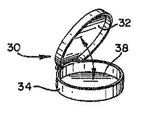
shown by embodiments 30 and 40 in FIGS. 3 and 4,
respectively. The circular plates 38, 48, 80 and 90
may~ similarly possess a flip-up lid 32 which is
pivotally attached to the circumferential sidewall 34
or~may possess an~unattached lid 42, both of which
enclose~ the media 16 contained within plates 38, 48,
80, and 90 when the lids 32;and 42 are lowered upon
; the circumferential sidewalls 34 and 44,
.~ .
~ 2~3~5 ~ j
21
respectively. In a manner similar to the rectangular
plates 18, 28, 50, 54, 60 and 70, the circular plates
38, 4B, 80 and 90 may be partitioned as~ shown in
FIGS. 6 and 7 to create wells of such shapes as
circles 92a and 92b or triangular pie-s;haped wells
82a-f and 94a-d or any combination thereof, for
example embodiment 90, FIG. 7. The shape of the
miniature culture plate and media containing wells
therein, shown in FIGS. 1 through 12, are set forth
as examples and are not meant to limit the types of
possible shapes and combinations thereof employed.
The miniature culture plate may be constructed of
plastic or such material presently employed in the
manufacture of petri plates.
The miniature plates are more economical in that
they utilize between about 2.5 to 5 milliliters of
media as opposed to the about 20 to 30 milliliters of
media required by the standard petri plates. The
miniaturized yeast identification system has been
designed to utilize a slide warmer as an alternative
mini~incubator. one such slide warmer is
commercially available from Fisher Scientific
Company. There are two requirements for use of slide
; ~ warmers: (1) an adjustable thermostat which can be
set at 37C and (2) a cover or lid for the warming
surface. ~oth the purchase cost and space needs of a
slide warmer are considerably less than a
conventional incubator.
The min~iaturized yeast identification system of
30 ~ ~ the present invention provides pre-poured,
sterilized, ready~ to use media and thereby
circumvents the~disadvantages of existing systems;
; such as the auxano~raphlc carbohydrate assimilation
~263945
22
( method which necessitates such steps as boilin~, to
dissolve the agar supporting agent and sterilization
~rior to use. A conven~ional product with~a ready-
to-use agar for chlamydospore production uses corn
meal a~ar with an emulsifying agent sold under the
name Tween* by Atl~s Chemical Company and
accompanies media for urea, nitrate and carbohydrate
utilization. The corn meal agar is inoculated and
examined at 24 to 72 hours for chlamydospore
production. The two main disadvantages of this
system are that the chlamydospore tests cannot be
performed apart from utilizing the entire pla~e which
i5 relatively expen~ive and there is a long time-to-
positivity.
In the miniaturized yeast identification system
of the present invention, the media are prepared,
poured into the miniature plates and after
solidification, the lids are closed, and the plates
aré wrapped in a plastic lined aluminum pouch. These
~20 plates may be stored up to at least 8 months at 4C
without loss of differential growth capabilities.
The miniature configuration also affects the~
performance of the media. Temperature control is
essential with these media and the smaller volume of
25~ media in the miniature plate allows rapid heat
transfer between incubation environment and the
medium. ThP sensitivity and accuracy of short term
incubations, such as~the three hour, 37D~, incubation
for~germ tube formation and subsequent shift to room
; 30 temperature for chlamydospore production on the ~OC
medium, are significantly enhanced by the
miniaturization of the system. The performance of
; the sucrose assimilation and urease tests is also
;~ * - Trade Mark
~3~345
23
enhanced. ~oth of the latter tests require a very
high oryanism to substrate ratio which is easily
attainable in the small media volume of the miniature
plate. Additionally, both tests involve a color change of
the media which is more easily visuali7ed with a medium
depth of about 1 to 4.5 millimeters than at about 5
to 15 millimeters. This incr~ased organism to
substrate ratio and more easily visualized color
change are thought to be partially responsible for
the decreased time-to-positivity of the sucrose ancl
urease tests in the miniaturized system of the
present invention, the times being 5 and 1 hours,
respectively.
Use of the SOC media in the miniaturized system
of the present invention requires a modification in
the concentration of the phenol oxidase substrate.
The preferred substrate is caffeic acid, however,
some other suitable substrate of phenol oxidase
enzymes may be employed. SuitabIe phenol oxidase
substrates include 2,3-dihydroxybenzoic acid, 3,4
dihydroxybenzoic acid (protocatechuic acid), DOPA,
3,~4-dlhydroxycinnamic acid (caffeic acid), the methyl
ester and diacetate~of caffeic acid, 3-
hydroxytryptamine, 3,4-dihydroxyphenylethanolamine
(norepinephrine), and 4-hydroxy-3,5-dimethoxycinnamic
acid. The substrate of phenol oxidase is employed as
an identification agent for Cryptococcus neoformans
since~ the reaction of the phenol oxidase enzyme of that
fungus wlt;h such. a subs~.trate produces a~ brownlsh pig-
3a ~ menta~tion of the organism which: is specific for Cryptococcus
neoformans. The original ~OC formulation, as given
in U.S. ;Pat. No. 4,144,133, issued March 13, 1979 and
~ ; entitled "~UNGAL GR~WTH MEDIA", was from about 0.005
: ` :
...,,.; . ~ .
39'~5
24
to about 0~05 weight percent caffeic acid.
Adaptation of the original SOC media to the
miniaturized system requires an increased range of
about 0.005 to about 0.5 weight percent, with a
preferred concentration of about 0.012 to about 0.12
weiyht percent and a most preferred concentration of
about 0 06 weight percent caffeic acid. This higher
concentration of caffeic acid necessitates an upward
adjustment of the pH of the medium to a neutral pH
due to the acidic nature of caffeic acid.
It was noted that chlamydospore formation, in
; certain strains of Candida albicansj was very weak on
the original SOC media, requiring a 48 to 72 hour
incubation for production. Publications by McClary,
Annals Missouri Bot. Gar., 39:137-164 (1952) and
Nickerson, Internat'l. Congress of Microbiol. Report
Proceedings, 5th Congre s, Rio de Janeiro, p. 130-131
(1950), indicated that low concentrations of ammonium
sulfate or ammonium chloride support filamentation in
~; 20 Candida species. Other work by Jansons, et al., J.
~acteriol, 104, 2:910-921 (1970); ~and, et al.,
Infect. and Immun., 11, 5:1014-1023 (1975); Mardon,~
et al.r J. Bacteriol, 100:701-707 tl969); and
Nickerson, ibid, stated that ammonium salts support
yeast growth. Twenty strains of Candida albicans
were chosen, which on occasion had given negative or
weak chlamydospore formation, and were tested for
g~erm tube and chlamydospore formation on SOC media
containing a range of ammonium ion concentrations in
the miniaturized system. All twenty strains produced
numerous genm tubes on ammonium ion containing SOC
media after three hours at 37C. The concentration
range of ammonium ions, added in the form of ammonium
~6;:~9~S
(
salts, supporting chlamydospore formation was found
to be between about 0.001 M and about 0.5 M with a
preferred concentration of about 0.005 M to about
0.05 ~ and a most preferred concentra~ion of ammonium
salt at about 0.01 M. Ammonium salts may be selected
from a non-exclusive class comprisingv ammonium
chloride, ammonium sulfate, ammonium nitrate and
ammonium citrate, vf which the preferred ammonium
salt is ammonium chloride~ Other salts such as
sodium chloride and potassium chloride were tested ~y
formulation in the SOC media but were found to be
less ef fective in promoting chlamydospore formation
than the ammonium salts.
Miniaturization of the SOC medium also required
an increase in the preferred concentration of
purified saponin fro~ the original concentration of
~; about 0.5 weight percent to about 1.0 weight percentin the impro~ed ~OC medium of the present invention.
he SOC médium used in the miniaturized yeast
identification system was prepared by adding a
powdered supporting agent such as agar~, oxgall,
purified~saponin, a~substance~for phenol oxidase such
as caffeic acid, and ammonium salt to deionized
water. The result ing mixture was then stirred and
25~ ~ heated to boiling to dissolve the ingredients and
adjusted to a final pH of about 6.5 to about 7.5.
The solution was then sterili~ed at 121C at about 15
; psi~for 15 min:utes. After cooling, the medium was
dispensed to a~depth of about 2 millimeters into 33 x
~75 x 5 mm rectangular plastic pla~tes with attached~
lip-up~;lids. The medium was al~lowed~to~solidify,
and~the plates~were wrapped in plastic lined aluminum
fo11 pouches and stored at~ 4C. ~Preparation and
:~ :
~ 263~ 5
26
~,
storage in this fashion gives a shelf life of at
least 8 months.
Generally, the improved SOC medium of the
present invsntion comprises from about 1.0 to about
5.0 weight percent agar, from about 0.25 to about
30.0 weight percent oxgall, from about 0O~ to about
5.0 weight percent purified saponin, Erom about 0.005
to about 0.5 weight percent caffeic acid, based on
the amount of water added to these components and
from about 0.001 M to about 0.5 M ammonium salts.
Preferred media can be prepared foliowing the
procedure outlined above and employing from about 1.0
to about 5.0 weight percent agqr, from about 0.5 to
about 5.0 weight percent oxgall, from about 0.1 to
about 2.5 weight percent purified saponin, from about
0.012 to about 0.12 weight percent caffeic acid, and
from about 0.005 M to about 0.05 M ammonium salts. A
most preferred medium contains about 2.0 weight
i percent agar,~about~l.0 weight percent oxgall, about
;~ 20 ~ l.O weight percent purified saponin, about 0.06 weight percent caffeic acid, and ahout 0.01
ammonium salts. ~ ~
Prior to inoculation~of the miniature SOC media-
containing plates wi~h the~yeast samples, the ysasts
~;~ 25 are yrown on a general growth medium, such as
Sabouraud dextrose agar with gentamicin, for up to
about 72 hours. Anywhere from about one to three
yeast colonies may be taken from the general growth
medium~and plated on each miniature plate. All
yeasts~were firs~t grown on a general growth media
prior to inoculation of media-containing miniature
lates. These general growth media may contain
antibiotics. ~ ~
:: : : :
:~ ~
: ~ :
... .
~.2~3~L~S
27
The miniaturized yeast identification system of
the present invention has also been adapted to
accommodate carbohydrate assimilation media.
Adaptation of these assimilation media to the
miniaturized system allows clinical technicians to
selsctively choose thoss assimilations required for a
spscific speciation of potential patho~enic fun~i and
thereby shortens the time ~f species identification
from ons week to about five to twenty four hours.
Sucrose assimilation, for ~xample, 6pscifically
differentiates Candida albicans, which assimilates
.
sucrose, from Candida stellatoidea, which does not.
When used in conjunction with such morphology tests
as germ tube and chlamydospore formation, which gives
a presumptive identification of the albicans or
stellatoidea species, the conducting of sucrose
assimiIation tests will give a conclusive species
identification. Additionally, adaptation of such
carbohydrate assimilation media as sucrose has
20 ~ dramatically shortened the t~me-to-posi.tivity for
Ca`ndida albicans and Candida stellatoidea
differentiation.
The method of the present invention shortens the
time-to-positivity for sucrose assimilation from 24
hours, and in some tests 6 days, to a period of about
5 hours. This increased efficiency is believed to be
due to at least two novel features of the present
invention. First, the procedure smployed for sucrose
assimilation utilizes a starvation step, in a
30 ~ carbohydrate depleted~media,~prior to plating of the
fungi on the assimilation media. The purpose o~ this
prestarvation step is to deplete the yeast cells of
essentially all of the internal carbohydrate pools
: : ~
: ~ :
:~ ~
:: :
~ ,. .
39~5
28
( which prevents false positive reactions. This
prestarvation is believed to increase the rate of
carbohydrate assimilation once the yeasts are plated
on carbohydrate containing media and thus decrease
the time-to-positivity. Second, the miniature plates
have a media depth of about 1 millimeter to ahout 4.5
millimeters as opposed to 5 millimeters to 15
millimeters in the standard petri plate. The
shallower depth of the miniature plates is thought to
aid in a more rapid visualization of the color change
resulting from positive carbvhydrate assimilation and
to provide the essential high organism to substrate
ratio.
In the miniaturized yeast assimilation system, a
sucrose assimilation is utilized to differentiate
between Candida albicans and Candida ~.tellatoidea.
The medium was prepared by adding a powdered
supporting agent, such as agar, a pH sensitive color
dye, such as bromcresol purple! and a yeast nitrogen
base to deionized water. The resulting mixture is
then stirred, heated to boiling to dissolve the media
components and adjusted to a pH of about 6.85 to
about 7.55 with a base, such as sodium hydroxide.
The medium is then s~erilized by heating to 121 C at
15 psi for 15 minutes. The sucrose is presterilized
and then added asceptically to the medium mixture.
The cooled sucrose-containing medium is then
dispensed in about 5 milliliter aliguots into sterile
rectangular plates with attached flip-up lids, the
plates having the approximate dimensions of 33 x 75 x
5~miIlimeters. After solidification, the lids are
closed, and the plates wrapped in plastic lined
aluminum f~il pouches~and stored at 4C. These
~' ~
,.
~ ~L2~i3~
29
plates may be stored up to at least 8 months at 4C
without dehydration or loss of color. Other su~ars,
such as maltose, galactose~ trehalose, and lactose,
may be substituted for sucrose to proYide additional
carbohydrate assimilation media. These sugars are
but non-exclusive examples of carbohydra~es which may
be subs~ituted ~or sucrose in the above-described
carbohydrate assimilation media. Additionally, it is
envisioned that a series of different carbohydrate
assimilations may be conducted simultaneousiy by
utilizing containe.rs such as those shown in FIG5. 6
through 11. Fluorescent indicators which show a
definite change in fluorescence with change in pH may
be substituted for the pH sensitive color dyes
employed in the examU12s o the present invention.
: Fluorescent indicators operable in the pH range, pH 5
to pH 8, of the carbohydrate assimilation system
:include, for example, Acid R Phosphine, Brilliant
Diazol Yellow, Cleves acid, Coumaric acid, 3,6-
:~ 20 Dioxyphthalic dinitrile, Magnesium 8-
hydroxyguinolinate, ~~Methylumbelliferone, 1-
Naph;thol-4-sulfonic acid, Orcinaurine, Patent
Phosphine, Thioflavine and Umbelliferone.
A preferred medium can be preparçd following the
procedure outlined above and employing from about one
to about five weight percent agar, from ahout 0.0005
~: to about 0.02 weight percent bromcresol purple ~ f rom
about 0.02 to about 0.7 weight percent yeast nitrogen
base, and about 0~.02 to about 1.0 percent sucrose,
: based on the amount of water added to these
components. The preferred pH of;the medium, prior-to
: sterilization is about 6.85 to about 7.55.
: :
3g4~
~,
Prior ~o inoculation of the assimilation media,
the yeasts were taken from plates containing a
general growth medium and suspended in sterile,
carbohydrate depleted media at a pH of about 7.0 to
abouk 8~5 and held at room temperatur~. Examples of
carbohydrate depleted media include sterile,
deionized ~ater or deionized water supplemented with
a yeast nitrogen base. The prestarvation period ~ay
run from about 30 minutes to 24 hours with a
1~ preferred range from about 30 minutes to about 8
hours, and a most preferred period of about one
hour. PrestarvationS were carried out in non-glass,
sealed and sterile containers. Such prestarvation
techniques have been employed in genetic and
lS biochemical studies of microorganisms generally~ but
have not been previously employed in yeast
~ssimilation methods for taxonomic purposes.
After pre~starvation, the carbohydrate deple~ed;
; medium~containing~the yeast lS vlgorously m1xed and,
20~ thereafter, at least ten mi~roliters of yeast
susp~ension is placed onto the surface of the
assimilation media. Multiple individual aliquots of
; ~yeast suspensions may be placed~on each miniature
plate. The plates are then incubated at room
~ ~ .
'5 temperature for about 4 to 6 hours and thereafter
checked for color change from purple to yellow
denoting carbohydrate assimilation.
An alternative embodiment of the miniaturized
yeast~assimilatl~on~systsm hDs been developed which
30~ combines prestarvation of the yeast inocula with
subsequent inoculation of; the prestarved yeast o~to a
serl~es of miniaturized carbohydrate agars, incubat-on
- of said inocula on said agars and post-incubation dye
. . ,
3945 ~,,?
31
or fluorescent indicator addition for detection of
carbohydrate assimilation.
In this alternative embodiment, the yeasts were
again taken from pla~es containing a general yeast
growth medium and suspended at a concentration of
McFarland ~o. 8, approximately 108 organisms, in
either the prestarvation media previously described
or 1 x 10⁻⁵M NaOH. The prestarvation period may run
from about 30 minutes to about 24 hours with a
preferred range from about 30 minutes to about 8
hours, and a most preferred prestarvation incubation
period of about one hour at a preferred temperature
of about 20 to 25C. PrestarvatiQns are, again,
uerformed in sterile, sealed, non-glass containers at
the previously described pH.
Following prestarvation, the carbohydrate
depleted medium containing the yeast is vigorously
mixed and, thereafter, at least 10 microliters of
yeast suspension is placed onto the surface of the
assimilation medium.
The carbohydrate assimilation media employed in
this alternative~embodiment were prepared by
suspending appropriate amounts of yeast nitrogen base
and agar into deionized water. The resulting mixture
was stirred with heat sufficient to dissolve the
ingredients and the pH of the suspension was adjusted
to about 7.2 with an allowable pH range of from about
6.65 to about 7.35. The mixture was then sterilized
at 121C at 15 psi for 15 minutes, cooled to about ~0
to 55C and, thereafter, an appropriate amount of
filter-sterilized carbohydrate added aseptically.
About 5 milliliter aliquots of this moIten medium was
then pipetted into s~erile rectangular plates with
~;3~ 5
32
attached flip-up lids, the plates having the
approximate dimensions of 33 x 75 x 5 millimeters.
After solidification, the lids are closed and the
plates wrapped in ~lastic lined aluminum foil pouches
and stored a~ 4C. These plates may be stored up to
at least 8 months at 4~C without dehydration. A non-
exclusive list of the type of sugars which may be
employed in this alternative embodiment includes
dextrose, galactose, sucrose, maltose, cellibiose,
trehalo5e, lactose, melibiose, and raffinose. In the
event that a single yeast inoculum is to be
simultaneously tested for its ability to assimilate
many different carbohydrates, the miniaturized plates
shown in FIGS. 6 through 11 may be employed. The
individual wells such as 72a through 72h in FIG. 10
or compartments such as 55a through 55h in FIG. 9
will each contain a different carbohydrate-containing
medium and at least 10 microliters of yeast
suspension was distributed per individual well or
compartment.
A preferred medium can be prepared following the
pro~edure outlined above and employing from about
0.02 to about 0.7 weight percent yeast nitrogen base,
from about l.O to about 5.0 weight percent agar, and
from about 0.02 to about 1.0 weight percent
carbohydrate, based on the amount of water added to
these components. The preferred pH of the medium is
from about 6.65 to about 7.35. A most preferred
medium contains about 0.067 weight percent yeast
nitrogen base, about 2.0 welght percent agar, and
about O.l weight percent carbohydrate, based on the
amount of water added to these components. The most
preferred p~ is about 7.2.
i39~5
33
(
Following inoculation of the carbohydrate-containing
medium with an appropriate aliquot of yeast suspension, the
plates are then covered with a sterile lid and incubated at
room temperature for about 6 to 24 hours. After incubation,
an appropriate amount of dye solution is added to each
assimilation test plate and assimilation detected by color
changes in the dye. Three dyes were examined for possible
use in this system, bromcresol purple, chlorophenol red and
p-nitrophenol which-give a purple to yellow, red to yellow and
~ yell~w to coloFless color change, respectively, when
carbohydrate assimilation occurs.
The dyes were prepared by dissolving an appropriate
amount of dye into water, adjusting the pH of the dye solution
to about 7.2 using 0.05N NaOH, and filter-sterilizing the
solution through a 0.22 ~m filter. A broad concentration
range-of dye solutions, from about 20 micrograms to 250
micrograms bromcresol purple or chlorophenol red per
milliliter or from about 15 micrograms to 45 micrograms
~-nitrophenol per milliliter, may be employed as stock
solutions from which an appropriate aliquot of dye~is taken
and added to the carbohydrate assimilation test plates. The
preferred concentration for the stock dye solutions is about
40 micrograms bromcresol purple dye per milliliter, 60 micro-
~rams;chlorophenol red~dye per milliliter and 20 micrograms
~-nitrophenol dye per milliliter. The most preferred dye is
bromcresol purple. ~Approximately 0.02 ml to about 1.0 ml
stock dye solution is added per about 0.1 to about 5.0
milliliters of yeast inoculated carbohydrate assimilation
test medium, respectively. ~The bromcresol purple reactlon
:
:
~ F~
,i~ :
1~3945
34
requires about 5 to 15 minutes for completion~
whereas the chlorophenvl red requires about 15 to 60
minutes for completion.
The miniaturized yeast identification system of
the present invention has also been adapted to
accommodate urea containing media. The ~}o~
species~ and occasional Trichosporon beigelii, and a
very rare Candida krusei possess the enzyme, urease,
that is necessary for hydrolysis of urea. Presence
1~ of the urease enzyme in yeast cultures i indicated
by color change'of the medium from orange to pink.
The adaptation of urea containing medium to the
miniaturized system of the present invention provides
a positive detection of the enzyme, urease, in about
one hour as opposed to the ~4 to 72 hours and 3 to 4
hours required for urease detection in presently
available urea agar slants and urea broth,
: respectively. The shortened time-to-positivity of
the present method is believed to be due to the
2~ increased organism to substrate ratio provided by the
small media volume and increased relative surface
area and by easier visualization of the color change
afforded by the shallower media depth in the
miniaturi~zed system as compared to standard petri
plates, agar slants, and broth tubes.
The miniature urease plates of the present
invention are made by preparing urea agar in
~: ~ accordance with manufacturer's instructions. The
med~ium was sterilized by heating to 12IC at about 15
psi for 15 minutes and is then dispensed in about 5
::: milliliter aliquots into the miniature plates
:descr~ibed above.~ After solidification, the plates
::: : ~ere wrapped and stored as described above so as to
..
1~3945 ~)
remain stable for at least eight months when stored
as described above. The plates were inoculated by
transferring one to three yeast colonies from a
general growth media onto the urease media. The
plates were then allowed to incubate at 35C for sne
hour.
The media adapted for use in the miniaturized
yeast identification system of the present in~ention
may be employed alone or in combination~ When
employing the partitioned plates shown in FIGS. 6
through 11, each plate may, for example, contain a
single type of medium inoculated with several
different yeasts, or each plate may contain multiple,
different differential media inoculated with the same
or multiple yeasts.
The fungal media of the present invention alone
and in combination provide a rapid screening process
for the identification of clinically important
yeasts. Specifically, the miniaturized yeast
~ identification systems described herein provide for
the relatively rapid differential identification of
::
Crv~tococcus neoformans and the two Candida species,
albicans and stellatoidea. It is further understood
that while this invention has been described in
relation to its preferred embodiments, the various
modifications thereof will not be apparent to one
skilled in the art from reading this specification
and it is intended to cover such modifications as
fall within the scope of the appended claims.
~ :~ ',,,
,
1~3~3~5 ;~ J
36
EXAMPLES
The followlng examples demonstrate the
mechanical advantages, increased accuracy and
decreased time-to-positivity of the present invention
over those previously employed for the identification
of various fungi, including Candida albicans, Candida
stellatoidea and Cryptococcus neoformans. These
_ _ .
examples are submitted for the purpose of providing a
better understanding of the present invention and are
not ~o be construed as limiting the scope thereof.
-
Exam~le 1
This example was performed in order to comparethe accuracy and sensitivity of egg white media,
fetal bovine media, two commercial systems, Flow ~BE
and A.P.I. GT Microtest, respectively, and the
miniaturized SOC media of the present invention in
detecting germ tube formation. The ease of
manipulation and microscopic examination of these
media was al50 compared.~ ~
Stock~yeast strains were subcultured on~o
Sabouraud dextrose agar and allowed to grow for up to
72 hours. The yeast colonies formed by growth of the
stock yeast on the Sabouraud dextrose agar were used
to~inoculate the test media in this comparative
study.
In both the eyg white and serum media the
substrate was added to a test tube, inoculated with
~ ~ yeast and incubated at;37C for 3 hours. Following
;~ 30 ~ ~ incubation, a drop of yeast suspension was placed on
a microscope~`slide, covered with a glass coverslip,
~ and examined under the microscope at 100X tO 400X
magnification for germ tubes. Egg white substra~e
*Trade Mark
~L~63~
was prepared by separating the egg white from the yolk
of an egg. Fetal bovine serum substrate was
purchased from Gibco. The Flow GBE tube, containing
0.1 percent weight per volume glucose and 2.6 percent
weight per volume beef extract was inoculated with
yeast and incubated as per manufacturer's directions
in a 35 to 37C incubator for 2 to 4 hoursO After
incubation, one drop of inoculated broth was placed
on a microscope slid~, covered with a glass
coverslip, and examined under the microscope at 100X
magnification for germ tubes. The A.P.I. GT
Microtest, consisting of microtubes containing 70
microliters lyophilized rabbit plasma with EDTA~ was
prepared according to manufacturer's directions,
inoculated with yeast and incubated, as per
manufacturer's instructions, in a 35 to 37C
incu~ator for 2 to 5 hours. After incubation, one
drop of yeast suspension was placed on a microscope
slide~ covered with a coverslip and examined under
the microscope at 400X magnification for the presence
of germ tubes. T~e miniaturi ed SOC plates were
prepared as previously described. One to four
colonies of yeast are picked up with a sterile cotton
swab and the bulk of the inoculum was deposit~d as a
single mound;onto the~miniaturized SOC media. From
the remaining inocuIum on the swab, a thin film of
yeast was streaked onto the medium in a dollar sign
shape and the thin film of yeast thereafter covered
~; ~ with a glass coverslip. The lid of the miniaturized
plate was closed, and the plate was incubated at 37
to 40~C for 3 hours. Following incubation, the
miniaturized plate was opened, placed under the
microscope, and the inoculum, centrally located under
*Trade~Mark
! ~ '
;
. . .
3g45 ~
the coverslip, was examined at lO~X to 400X
magnification for germ tubes.
Table 1 giv~s the comparative results on the
performance of germ tube systems. ~enty strains of
Candida albicans were tested for ge~l tube formation
and ease of microscopic examina~ion on egg white,
fetal bo~ine serum, Flow GBE, A.P.I. GT ~icrotest
and the miniaturized SOC medium of the present
invention Each strain was examined after three
hours of incubation and marked as positive (+) for
germ tube formation when ~t least eight out of ten
microscopic fields showed germ tube formation. The
strains were marked as marginal ( ) when germ tubes
were visible in only one or two out of ten
microscopic fields. Clumping was said to occur when,
despite agitation, the germinated cells remained
knotted together in masses, making it difficult to
observe, microscopically, whether there is true yerm
~ tube or pseudohyphal formation.
; ; *Trade Mark
~ :
, . ,
1~i394~
39
TABLE 1
C()MPARISON OF THE MINIAlllRIzED SOC PIATE WI~ ~ AVAIIABr.
SY~TE~ ~R GERM II~E E~RMATION BY Candida albicans
Genn nlbe Formation
No. o~
s~st~n Isc~lates No. % N~. % No. %
; 1. SOC 20 2U100 0 0 0 0
2. Egg ~ite 20 20100 0 0 0 0
.
3. Fetal bovine sen~n 2020 100 0 0 2 10
: 4. Flow GBE tube 20 1890 2 10 4 20
:: :
5. ~ API G~Mlcrotest 20 17 85 3 15~ 16 80
:
:
: ::
' ~ :
~ :
*Trade Mark ~ :
:: :: ~ : :
~:
:
.
3~3~5
C,
As can be seen from a study of Table 1, 100
percent of the strains tested formed germ tubes in
three hours on the miniaturized SOC, egg white and
fetal bovine serum media, while only 90 percent and
85 percent of the strains formed germ tubes in the
Flow GBE and ~.P.I. G ~ Microtest system,
respectively. As shown in the results displayed in
~able 1, significant clumpin ~was observed in the
serum, Flow GBE and A.P.I. G Microtest systems.
The miniaturized SOC medium is a solid support medium
on which yeast cells are dispersed in a thin film
allowing observation of single stationary ceIls. In
the other systems, the yeasts are usually in motion
in the liquid under ~he coverslip, making it
difficult to focus on any one cell.
From the standpoint of the manipulation, the
miniaturized SOC plata requires only an initial
noculum transfer, whereas the other systems require
a second transfer to a microscope slide which
increases technician time and necessitates additional
material5.
Example 2
~` This example was performed in order to establish
the effect of various concentrations of the phenol
oxidase substrate, caffeic acid, on yeast morphology
and pigment formation on SOC media in the
; min~iaturized systemO The specific morphologies
observed were germ tube, chlamydospore, blastospore,
~; 30 fllaments and pseudohyphae formation. The brown
plgment production characteristic of several
Cry~tococcus neoformans strains was also monitored.
:. :
~ ~ ~ ~ r/C
:
:
c ~.;26~945 ~
41
The test medium, the miniaturized SOC medium, of
the present example wa~ prepared as follows: 20
grams of agar, 10 grams of oxgall, 10 grams of
purified saponin, 0.54 grams of ammonium chloride,
and various amounts of caffeic acid, listed in Table
2 belo~, were added to one liter of deionized
water. The mixture thus formed was stirred and
heated to boiling to dissolve ~he ingredients and
adjusted to a final pH of about 7.2. The solution is
then sterilized by heating to about 121C at 15 psi
for 15 minutes. After cooling, the medium was
dispensed to a depth of about 2 millimeters
~approximately 5 milliliters) into sterile, miniature
plates, previously described, and allowed to
solidify.
one to four yeast colonies were transferred from
a general growth medium to the test medium, as
described in Example 1. The inocuIated plates were
incubated for 3 hours at 37C and thereafter at room
temperature.
Table 2 sets forth the results of the test
performed in order to compare the effect of various
concentrations of caffeic acid on yeast morphology
and pigment production on the miniaturized SOC
me~iumO ~ix species of Candida and four species of
Cryptococcus were plated~on the test medium and
observed at the time interval specified in Table 2.
; Mlcroscopic examination at the t~me intervals
designated in Table 2 was conducted at IOOX
magnification in the manner~described in Example 1.
Positive morphology (+) was defined as at least 8 out
of~10~ microscop~ic fieIds having shown a particular
morphology or combination of;morphologies. Negative
....
~.~63~3~5 ~ '
42
morphology (-) was defined as less than 1 or 2 out of
10 microscopic fields having shown a particular
anticipated morphology or combination thereof.
As can be seen from Table ~ below, the
miniaturized SOC media supported the characteristic
yeast morphologies and pigment production at a
relatively wide range of caf feic acid concentrations,
fro~ about 0.005 weight percent to about 0.5 weight
percent~ In pigment formation, 10 percent of the
Cryptococcus neoformans strains tested produced brown
coloration at 1~ hours on SOC with 0.006 weight
percent caffeic acid. A~ 0.6 weight percent, SOC
itself was dark brown, making the yeast's color
chanye difficult to recognize and, also, the SOC
medium became a darker brown color each day,
indicating instability. The effect of caffeic acid
concentration on general yeast morphology, as shown
in Table 2 below, establishes an upper limit for
caffeic acid. Essentially all filamentation of
Candida species was inhibited at 0.6 weight percent
eaffeic acid.
~: :
:~ : : :
35~5
43
E~t ~ ~ a. ~ 1~. CL D ~ ~,
Il ~ ~ Q Q ~ p) Pl ~ ~ ~ ~ la
~ ~ ~ ~ ~ ~- ~ ~ ~n ~ ~
Q` ~ U~ ~q U~ ~ ~-- ~ ~ l_ ~i
o ~ ~ I_ ~ U~ /- ~ ~'
~r ~ ~ ~: 8 ~ ~ ~ g.
rs ~_ ~ lD O ~ O~ ~
~ ~, ~ ~ ~ ~ tn ~ ~
5 ~- 3 ~:5 ,.. P~ ~.
i~ ~W '~ Iw ' ~ 33
~ ~ ~ ~ ~ ~ ~3 ~ cl
CO CC o~ ~ ~ W ~ ~W ~ ~ ~W .P~ ~ ~}
.
~ ~ 1
;q ~ q ; ~ + + * ~ ~ ~ ~ $ lo o ~
¦ ~
+ ;~ l o ~ ~ ~
:~ HU
o ~o: o ~ E lo ~ c~3 + .~ + ~ + ~ :+~ 1'- ~
Q ~ ~
:
~ ~ ~ ~ ~ ~ ~. ~. ~- ~- ~ a 'g .
3 3 ~ r~
,
~3~3~5
44
( Example 3
This example was performed to determine the
effects o ammonium chloride addition to SOC medium
on chlamydospore production by Candida albicans.
The test medium of the present example was
prepared as follows: 20 grams of agar, 10 grams of
oxgall, 10 grams of purified saponin, 0.6 grams
:
caffeic acid, and varying concentrations of am~onium
chloride, indicated in Table 3 below, were added to
one liter of deionized water. The mixture was then
stirred, heated to bolling, adjusted to a final pH of
about 7.2, and sterilized by heating to 121C at l5
psi for 15 minutes. After a cooling period, about 5
millilitars of cooled medium was dispensed into the
miniature plates as described in Example 1. These
plates may be stored up to 8 months when stored as
described in Example 1.
One to four yeast colonies grown on Sabouraud
~ dextrose agar were plated as described in Example 1
`~ : 20 on S~C medium containing the various concentrations
of ammoniu~ chloride listed in Table 3 below. At 1.0
M ammonium chloride, some of the medium components
precipitated making it impossible to test that
formulation. The yeast colonies so plated comprised
20 different strains of Candida albicans which had,
on occasion, given negative or weak chlamydospore
production. The plates were incubated for 3 hours at
37C and examined for germ tube formation. All 20
strains produced numerous germ tubes on all
concentrations o~æmmonium chloride listed in Table
; 3. The plates were then incubated at room
; tem~erature for 24 to 72~hours and examined for
chlamydosp~re production by the methods described in
: :~ :
:
.,: . .
~.2639~5
( Example 1 and at the time intervals indicated in
Table 3 below.
As can be seen by examination of Table 3, the
addition of ammonium chloride significantly enhanced
chlamydospore production. The optimum time f~r
determining chlamydospore production was 24 to 48 hours.
.
~: :
~::
~2G39~5 ~ ;,
46
~ABLE 3
EFE~CTS ON NH4Cl ADDITION TO SOC ON CHL~MYDOSPORE
PP~ODlfCIlON IN T~æNTy SrRAIN~ OF Candidal albicans
.
Chlamyd~:pre Production
NH4cl, Molarit:y
0 0.0~1 0.01 0.
Incubation ND. o~ % % % &
Time (hrs.) StrainsPositivePositivePositive Positive
_
`
24 20 ~5.0 80.0 100.0 50.0
: ~: 48 : 20 60.0 100.0 100.0 :~0.0
72 20 ~100.0 100.0 100.0 :100.0
:: : :
..i
' ~ :
~39~
47
~'
Exa~ le 4
This example was performed in order to test the
ability of the miniaturized carbohydrate assimilation
media of the present invention, performed by the
method of the present invention, to differentiate
between Candida albicans and Candida stellatoidea.
~he miniaturized sucrose assimilation media
employed in this example was prepared by adding 20
grams of agar, 20 milligrams of bromcresol purple,
0.67 grams yeast nitrogen ba~e and 4 milliliters of
0.5 N sodium hydroxide to one liter of deionized
water and ad~usting to a pH of about 7.2~ The media
was sterilized by heating to about 121C at 15 psi
for 15 minutes and after cooling, presterilized
sucrose is added asceptically to yield a final
sucrose concentration of 1.0 percent. The media was
then dispensed in about 5 milliliter aliquots into
the miniature plates, as described above. ,These
plates may be stored, by the method described in
xample l, up to eight months without dehydration or
loss of color.
~; Six species of Candida were first grown on
Sabouraud dextrose agar containing gentamicint as
~; previously described. Yeasts were taken from this
primary growth medium and suspended in sterile
deionized water and held for at least one hour at
room tem~erature for purp~ses of prestarvation.
After prestarvation, the suspension was vortexed and,
:
usiny a sterile pipet, one to about three drops of
yeast sus~ension was~placed onto the surface of the
mirliaturized sucrose media. The plates were then
incùbated at room temperature for 4 to 6 hours.
Following the incubation period the plates were
~: :
,.. ""~ ~ :
~3~345 ~ ~
~8
checked for color cha~ge from purple to yellow
indicating sucrose assimilation. The plates were
held a~ room temperature for 24 hours amd read a
second time.
The results of this example are sho~n in Table
4A below. Candida albicans, Candida stellatoidea,
Candida tropicalis, Candida krusel and _andida
glabrata gave the correct assimilation response on
the miniaturized sucrose assimilation plates after S
1~ hours incubation at room temperature and maintained
the correct response for 24 hours. Candida
parapsilosis, however, did not show a characteristic
positive reaction until 18 to 24 hours. From this
data, it is apparent that Candida albicans and
Candida stellatoidea can be differentiated in 5
hours.
,
~ : '
. ,
~ : ,,'
~6394~
49
lo ~
Yl ~ ~
O ~
+~I+ ~ ~
o ~ ; H
o o o o
, :~ 1~ lX 1~
~ O l_ ~ ~
0 ~~ oZI ~ ~ ~w ~
O C~ 0~ 0 ~0~ ~ ~ + ~: ,
~Q~ ; O ~ ~ : ~ : .
o o ~ ~ ~
O~ 0 o ~ ~0 ; ~d~ : .
,
: ~
'''
C ~ 39~5 '
~o
To f~rther pursue ~he concept of nniniature
assimilation for other carbohydrates besides sucrose,
identical experiments were performed where media were
prepared as described above except that 1 gxam of
each sugar listed below in Table 4B was substituted
for sucrose to yield a specific sugar containing
medium. Yeasts were inoculated, plated and grown as
described above and examined for carbohydrate
assimilation at the time indicated in Table 4B
below.
As shown in Table 4B, the proper assimilation
pattern for each species can be achieved within 24
hours. With Candida ~ , there was a
discrepancy with the positive sucrose assimilation at
5 hours and the negative results in the previous
experiment as given in Table 4A. This is a
s~ignificant example of strain variation among yeast
strains.
:
: :
'
, :
~;
: ~
: :
: ~ '
,
,.
63~
W ~ W
~
3u~
c ~ ~ ~ c ~ C D) ~ C
J ~ n 5 D~ f) n 5 ~ n ~ ~ ~
8' ~H
~ 0
& 1+ t' ~ + I ~ + ~ ~1, + + + ~ I t + + + ~ ~ H
~;~ ~3 IE~
~D o o ~l- w o l-l- ww o o w wo o ~w ~w ~ ~ : r~
,~ ,., ~ ~ ~ ~ ~ + c) .
: :oo~o OW~g~ ~0~0~ ~ Ul ~ ~
o o ~I W o o W W 0 0 : 0 0 0 0 0 0 ~ 0 n5 :
~D ~ w~ o w~lvoo wwoo~ w~~ooo ~ l 3
) o o w o~ o o o o
oo w~ o o sJ~o~ 00 0 o Ooc: o wO oO ~
O O W `~1~ 0 ~ O ~1~1 0 0 0 0 0 0 0 0 W O O O d~ I-H3
: :
O: W~ W W ~ ~ O W W WW O W W W O O ~ W WW ~1
O 00 0 ~ 00 00 0 0 0 00 00 +
O t~ O O 0 0 0 0 0 0 O O O O O O O O O O ~ .
~00000 O~ooo ooooo ooooo ~ 5
n ~`
w o~oo o w 00 00 w o o ow w oo 00 ~ l
0O 00 0 0~ 00 o~ o o o o oo oo .
00 00 0 O OO C~O 0 0 0 00 0 00 00 d~
,~
639~5
-- 52
( Example 5
This example was performed to test the ability
of miniaturized urease media to detect the presence
of the urease enzyme in various species of
ryptococcus.
The miniature urease plates in the present
example were made by preparing urea agar according ~o
manufacturer's instructions, sterilizing by heating
at 121~C at 15 psi for 15 minutes and then dispensing
the media into the plates as described in the
previous examples. Again, by storing as described in
~xample 1, the plates remain stable for up to at
least 8 months.
The plates were inoculated with four species of
Cryptococcus and six species of Candida, by
transferring, with a sterile cotton swab, two to
three yeast colonies from A general growth medium
such as Sabouraud dextrose agar. Two to three yeasts
of a single species were inoculated per plate and the
plates were incubated at 3SC for up to 24 hou~.
The results of this example are shown in Table
5. The~CrYptococcl~s species were pos~itive in one~
hour and the other yeast isolates remained negative
for 24 hours. A hydrolysis of urea releases ammonium
gas which is basic and rapidly spreads throughout the
~medium turning the medium from orange to pink. A
positive organism completely changes the medium color
in the miniature plat~ within two to three hours.
There~ore, if more than one organism is inoculated
onto the miniaturized urease platej the plate must be
read between about one and;~two hours or false
positive reactions can occur.
: .
~ ~63":3~5
53
( TAHLE S
EXAMINATION OF T~E MINIATURE UREASE PLATE
WITH VARIOUS YEhSTS
Ure~se ~E~Y---
ND. of Co~rect +
~rganism Isolates Respon~eNo. ~ No. %
Cr. neDfonmans 81 ~ 81 lOOoOO O O~O
.
Cr. albidus 20 * 20 100.0 0 0.0
Cr. laurentii 10 ~ 10 100.00 0 0.0
.. ... .
Cr. diffluens 24 + 24 100.00 0 0.0
C. albicans 122 - O 0.0 122 100.0
C, stellatoidea 21 - 0 0.0 21 100.0
C. ~ 29 - 0 0.0 29 100.0
C. rapsilosis 32 - 0 0.0 32 100.0
C. krusei 18 ~ ** 0 0.0 : 18 100.0
C. 5e~ 34 ~ - 0 0~0 34~ 100.0
*The rea~tions were read at one hour and n~gative reactians were
reconfirmed at 24 houxs.
~ **C. krusei.has been reported to xarely show a positive urease
:~ ~ : reaction, but the strains used in this experim~nt were negative.
~: :: : : : : :
:
~Z~3"3~
54
ExamPle 6
This example was performed to compare the post-
incubation addition of dye for detection of
carbohydrate assimilation with conventional
carbohydrate assimilation methods. The carbohydrate
assimilations detected by post~incubation addition of
dye were conducted in Microtiter plates and are,
hence, referred to as "Microtiter carbohydxate agar
assimilation" in the text and Tables 6A and 6B~
~elow.
The Microtiter carbohydrate assimilation media
employed in this example were prepared by adding 20
grams of agar and 0.67 grams of yeast ni.trogen base
to 950 milliliters deionized water. The mixture was
stirred with heat to dissolve the ingredients,
adjusted to a pH of about 7.2 with 0.5~ NaOH, divided
into eight equal parts, sterilized at 121C at 15 psi
for 15 minutes and, thereafter, allowed to cool to
¦: about 45 to 55C. Then, 6.25 milliliters of a two
percent, filter sterilizedl carbohydrate solution
I : containing one of each of the eight carbohydrates
listed in Table 6A :or 6B, below, was asceptically
added to on~ of each of the cooled solutions. The
two percent carbohydrate solutions were prepared by
adding 0.2 grams of carbohydrate to 10 milliliters
deionized water and adjusting to a pH of about 7.0
with ~0.05 N NaQH. After mixing, 0.15 milliliters
aliquots of the carbohydrate-containing.medium was
s~terily dispensed into the;wells of a Microtiter
plate.
~ The yeast listed in;Tables 6A and 6B, below,
:~ were taksn from~plates containing a general yeast
~ grow~h medium as:described in Example 4 and suspended
:,
~ *Tràde Mark
'
~F
,
i2~3Yi~
at a concentration of McFarland No. 8, approximately
108 organisms, in 1 x 10⁻⁵M NaO~ and held for about
one hour at room temperature for purposes of
prestarvation~ Following prestarvation, the
suspension was vortexed and 0.02 milliliter aliquots
were aseptically transferred into each medium
containing well of the Microtiter plate. The plates
were covered with a sterile lid and incubated at room
temperature for 24 hours. After incubation, 0.02
milliliter ~liquots o~ filter sterili~ed, Z0
micrograms bromcresol purple per milliliter dye
soIution was added to each well of the Microtiter
plates. The plates were read 5 to I0 minutes after
dye addition for a purple to yellow color change
lS indicative o~ positive assimilation of carbohydrate.
The results of the Microtiter carbohydrate agar
assimilations are given in Tables 6A and 6B, below,
wherein the data are compared to reports for
conventional methods given in The Yeasts by Lodder
~; 20 and the ~.P~I. 20C Strip. The conventional methods
include, Wickerham liquid, Saito agarj and
auxanographic technique.
*Trade Mark
:
:
- : : .
~2~39~5
'
ra~lnc6e ~ 3 1 + + 3 1 u
~.act.ose I I I ~ I I + +
# trehalose + + + + + + + + I ~ + + ~+1 + ~ + +
s celIibiose ~ t~ 3 1* ~ * ~+
J ~
~ maltos~ + ~ ~ + + + + + I I I I + ~ + + + I ~
¢ ~ sucrc6e +~ I ~ ++ ~+ I I I ~ ++ ++ ~+ ~,:
' : . : 3
o ,~yalactose ~ + + + + + ~ + ~ + + * + 3 1+
' ~ dextrose + + + ~ + ~ t- + + + + + ~ + + + + + ~ ~
¢ O ~ ~ O
@ ~ ~ ,0
:Z~ n ~ a~u,.
~ ~ z~ ~ a ~
3~d
:, , :
:: :
: ~ : I
~: : :
~J )
~,
~ Z~39~5
~,o oo oo oo oo oo C~O ~oo ~o C~O
raf f inose ~
. ~ 1 ~ 1 1 1 1 1 I ~ 1
oO oO 00 oo oO oo oo ~o oo
a~ o
lactose _,
~ 1 1 1 1 1 1 1 1~ 1
o~o,~ooo t~o oo a~o ~o ~o ~o
11 a~ o ,~ o ~ O ~ o15~ Q
trehalose .
~ 1~ 11' 1~
~ Oo ~o ~r- C~o oo oo 1~ ~o ~o
:r: tn O --~ ~ ~ ~~ o a~ ~o
u~ c~llibiose . _.
~ ~ ~ ~ : ~ 1 I U~1 I ~ 1 I -'I ~ I '
c ~ ~ : ~ go g g ~0 0 8 0~ 8~ ~ 0
~ ~: o ~
o ~n ~ maltose
1 ~ I ~ 1 1 J ~I ~I '`
^t~
IP ~D O C~ o o o8 0 0 0 8 0
~ sucrose
m ~ .
~ 1 ~1 ~ 1 1 1 ~
~:: o ~ : :
3 ~ ~ : ; P 0 0 ~ 0 Oo o o o O ~ 0 Oo O O a~
0 o : gala~tose : ~
z~ ::: ~ ~ I oI o
~ : ~ :: : : ~
I :8 8 g o~g o 8 oo o 8 0o o o g H
dextrose :
: ~ ~ r
h ~ ~ h h
I O ~ : ~ *
H
~;3~3~5
58
As shown in Table 6A above, there is good correlation
between the Microtiter* carbohydrate agar assimilation test
and the published results of conventional Wickerham liquid,
Saito agar and auxanographic methods. Certain carbohydrate
reactions with Cr. neoformans and Cr. albldus, however,
require examination.
As shown in Table 6~, above, there i~s an overall good
correlation between the Microtitier* carbohydrate agar
assimilation test and the A.P.I. 20C strip method with the
exceptions of raffinose assimilation for Cr. neoformans and
Cr. alb dus.
Example 7
This example was performed in order to test the effect of
an increased ratio of surface area of organisms to volume of
medium on the time-to-positivity in carbohydrate assimilation.
One way to demonstrate the effect of said increased ratio is
to hold the surface area and microbial inoculum constant while
varying the volume, herein expressed as depth o~ medium, in a
given test plate or tube.
Three different containers were used to examine the
~ effect of surface area to volume ratios on time-to-positivity:
;~ the miniaturized culture plates of the present invention,
standard l00 x 15 millimeters petri plates, and standard
; ~ 16 x 125 millimeter flat-bottom culture tubes. The
, ~ :
theoretical limits of the surface area to volume ratio
employed in a given container will be set by the dimensions
necessary to visualize the color change prompted by positi~e
carbohydrate assimilation and a minimal depth necessary to
preclude dehydration of the medium.
~ Sucrose assimilation~medium prepared as described
in Example 4, above, was aseptlcally poured
: :
* - Trade Mark
:::
~; ::: ;~: :
'''
~ ~Z~39~5 ~`~
59
into each of the containers at the depths shown in
FIG. 13.
Candida albicans, the test organism, was first
grown on Sabourand dextrose agar conta;ining
gent~micin, as previously described. Yeasts were
taken from this primary growth medium and suspended
in sterile deionized water and held at least one hour
at room temperature for purposes of`prestarvation.
Following prestarvation, the suspension was vortexed
and, using a sterile pipet, one to about three drops
of yeast suspensior was placed onto the surface of
each miniaturized plate and culture tube containing
sucrose medium and three one-drop inoculations of
yeast were placed on each 100 x 15 millimeter (mm)
petri ~late containing sucrose medium. The plates
and tubes were then incubated at room temperature for
not more than 12 hours. Each plate or tube of the
depth specified in the graph in PIG. 13, was run in
du~licate. Positive reactions, denoting sucrose
assimilation, were determined by an obvious color
change from purple to yellow at the post-inoculation
time~intervals shown in the graph in FIG. 13, given
as ~"Time-to-Positivity" in hoursO
The results of this example are shown in the
; ~ 25~ graph in FI~. 13. This example confirms that at a
;~ medium depth range of 2 to 2.5 mm, approximately 4 to
5 mi~lliliters of medium per plate, in the
miniaturized~plates (0-0), the time-to-positivity for
sucrose assimilation by Candida albicans is from
about four to five hours. Although a shift in the
: time~-to-positivity is~seen with increasing depth or
:
~; vol~ume at a constant surface area of organisms in the
miniaturized~plates, the most dramatic effect of
:
:: ~ ~ : :
.
,,
~39~
increased medium depth or volume at a constant
surface area of oryanisms is seen in the standard 100
x 15 mm petri plates (~-~). Additionally, the medium
depth in the standard petri plate is typically S mm
corresponding to 18 ~o 20 milliliters of medium per
100 x 15 mm plate which is shown to yield about a 10
hour time-to-positivity for Candida albicans in the
sucrose assimilation test. Furthermore, medium
depths of 3 mm or less in the 100 x 15 mm pe~ri
plates are not preferred due to possible problems
with dehydration during storage. The greater time-
to-positivity seen in the 16 x I25 mm flat-bottom
culture tubes (~-~) is thought to be due to problems
with oxygen tension created by the closed environment
of the culture tubes as compared to the open system
of the plates.
:
:: ;: : :
::
:
::
:
~ .
.