Note: Descriptions are shown in the official language in which they were submitted.
-1- WB57
TRANSLUMINAL THROMBECTOMY APPARATUS
-
The present invention relates to a device used to
break up blood clots or thrombi which form within
arteries. In particular, the device is particularly
well adapted l:o break up such clots or -thrombi which may
form within a coronary artery.
A large percentage of heart attacks are caused ~y
blood clots or thrombi which form within the coronary
arteries. The biochemical process which results in
thrombus formation is not fully understood. However,
in simple terms, injury to the vascular wall releases
chemicals which lead -to conversion of soluble circu-
lating fibrinogen molecules into a polymeric structure
of fibrin. The fibrin structure is insoluble and
arranges itself into a three dimensional network of
meshed strancls which entraps red blood cells. The
individual strands are approximately 0.2 microns in
diameter and the mesh size is approximately l micron.
Accordingly, five micron red blood cells are easily
trapped within the three dimensional network.
When a thrombus forms, it effectively stops the
flow of blood through the zone of formation. If the
thrombus extends across the interior diameter of an
artery, it cuts off the flow of blood through the
artery. If one of the coronarv arteries is 100%
thrombosed, the flow of blood is stopped in that artery
resulting in a shortage of oxygen carrying red blood
cells to supply the muscle (myocardium) of the heart
3C wall. ~uch a thrombosis is unnecessary to prevent loss
of blood but can be undesirably triggered within an
37
WB57
artery by- damage to the arterial wall from
atherosclerotic disease-. Thus, th~ underlying disease
of atherosclerosis may not cause acute oxyg~n deficiency
(ischemia) but can trigger acute ischemia via induced
thrombosis. Similarly, thrombosis of one of the carotid
arteries can lead to stroke because of insufficient
oxygen supply to vital nerve centers in the cranium.
Oxygen deficiency reduces or prohibits muscular
activity, can cause chest pain (angina pectoris), and
can lead t~ death of myocardium which permanently
disables the heart to so~e e~tent. If the myocardial
cell death i5 extensive, the heart ~ill be unable to
pump sufficient blood to supply the bo~y's life
sustaining needs. The extent of ischemia i affected by
many factors, including the existence of collateral
blood vessels and flow which can provide the necessary
oxygen.
Coronary artery bypass graft (CABG) surgery is a
surgical method for bypassing coronary arteries which,
because of narrowing or obstruction, are unable to
supply adequate oxygen to heart muscle. In recent
years, direct administration of chemical lysing agents
into the coronary arteries has shown to be of some
benefit to patients who have thrombosed coronary
arteries. In this procedure, a catheter is placed
immediately in front of the blockage and a drip of
streptokinase is positioned to be directed at the
upstream side of the thrombus. Streptokinase is an
enzyme which is able in time to dissolve the fibrin
molecule. This procedure can take several hours and is
not always successful in breaking up the thrombus.
~urthermore, it can lead to downstream thrombus
fragments (emboli) which can lead to blockage of smaller
d ameter branches. It would be useful to have a device
an~ method which would permit essentially instantaneous
WB57
--3--
removal of the blocking thrombus.
The present invention is a transluminal
thrombectomy apparatus. It comprises a flexible dxive
shaft having a tip affixed thereto. The tip has a
diameter which is greater than the diameter of the drive
shaft. The tip is an elongated cylinder with rounded
ends. The apparatus further comprises a flexible,
cylindrical shaft housing havin~ an inside diameter
which is greater than the outside diameter of the tip.
The shaft housing extends along and surrounds the drive
shaft. The device includes connecting means for
sealably connecting the end of the shaft housing which
is remote from the tip to an apparatus capable of
providing a fluid path through the drive shaft housing
to the end of the drive shaft housing at which the tip
is located. In addition, the device includes dr-ive
shaft connecting means at the end of the drive shaft
which extends through the shaft housing connecting means
for connecting the drive shaft to a rotating prime
mover.
Brief Description of the Drawing
In the Drawing:
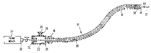
FIG. 1 is a cross-sectional pictorial view showing
the present invention set up for use;
FIG. 2 is a side view of a first embodiment of the
invention;
FIG. 3 is a side view of a second embodiment of
the invention;
FIG. 4 is a side view of a steerable embodiment of
the invention; and
FIG. 5 is a graph illustrating the proportion of
~ime the bends of the shaft housing and the drive shaft
coincide in the embodiment of FIG. 4.
357
--4--
FIG. 1 illustrates the transluminal thombectomy
apparatus 10 of the present invention. The device 10
preferably includes an elongated, cylindrical atraumatic
tip 12 having rounded ends which is rotated by a
transluminal drive shaft 14. The drive shaft 14 is
contained within a flexible shaft housing 16 which is
typically made of a plastic material. The inside
diame-ter of the shaft housing 16 is preferably selected
to be greater than the diameter of the tip 12 in order
that the tip 12 can be withdr~wn into the shaft housing
16. At the end of the shaft housing 16 remote from the
tip 12 there is preferabl~ a shaft housing connector 18
which may be used to guickly connect the shaft housing
16 to a dri~e shaft bearing block and shaft seal 20. The
shaft housing connector 18 permits rotation of the shaft
housing 16 for reasons which will be explained.
The drive shaft bearing block and shaft seal 20 is
a body which has a central cavi~y 22 and a port 24
connected thereto. The port 24 also includes a quick
connect fitting 26 in order that a fluid path exists
from the port 24 through the central cavity 22 and
through the interior lumen 28 of the shaft housing 16.
This fluid connection can be used either to withdraw
fluid through the shaft housing 16 or to dispense drugs,
such as streptokinase, through the shaft housing 16. A
fluid seal 30 which allows axial movement of the drive
shaft 14 is at one end of the drive shaft bearing block
and shaft seal 20. The end 32 of the drive shaft 14
remote from the tip 12 is connected -to a rotary prime
mover 34.
In operation, the rotating tip 12 is pushed into a
thrombus. Because the tip 12 is rotated, it is able to
cause fibrin 36 to be wound around its narrow shaft la
(See FIG. 2). This happens because of ~riction be~ween
~ s~7 ~B57
--5--
the surface of ~he rotating drive shaft 14 and because
of a localized "whirlpool" in any free liquid
surrounding the tip 12 and the shaft 14. As the fibers
36 follow about the rotating tip 12, their tension
increases, further tightening their grip on the drive
shaft 14 and eventually stripping away an interior
volume of the fibrin network. Red blood cells which
were entrapped can be released back into the circulatory
system without emboli-producing large fragments, since
the insoluble material is retained on the drive shaft 14
for later extraction from the body.
As shown in FIG~ 2, the fibrin 36 winds around
the drive shaft 14 when the rotating tip 12 and drive
shaft 14 pass through a clot. Since the fibrin 36 winds
very tightly around the drive shaft 14, and since the
fibrin 36 constitutes only about 4% of the volume of the
thrombus, it is possible to strip the clot's framework
and thereby eliminate a large volume of the clot before
the tip 12 of the thrombec~omy apparatus 10 needs to be
withdrawn from the patient for cleaning or replacement.
In most cases, the entire thrombectomy can be performed
without uslng up the fibrin storage capacity of the
thrombectomy tip 12.
As shown in FIG. 1, the tip 12 is comprised of a
generally sausage shaped radiopague piece affixed to a
cylindrical metal drive shaft 14. The rounded edges of
the front and rear of the tip 12 reduce the probability
of inadvertent perforation of vessel walls and the thin
drive shaft 14 provides torque transmission as well as
an area for coiling and storing the fibrin. In the
preferred embodiment of the invention, the tip 12 has a
diameter of about 25 mils and a length of about 80 mils,
and it is mounted on a 5 mil stainless steel drive shaft
14. As will be understood by those skilled in the art,
it could be advantageous to have tips with diameters as
WB57
-6~
small as 10 mils to negotiate very tiny arteries and
narrowed segments. In such cases, the drive shaft 14
would be proportionately smaller or compliant to the tip
12 in order to avoid ram punctuxe of an artery. While
the tip 12 of the preferred embodiment 10 is an oblong
cylinder wi~l rounded ends, a spheroidal tip 112, as
shown in FIG. 3, could also be used with some loss of
radiovisualization. In the preferred embodiment 10 of
the invention, the tip 12 is made of a radiopaque
material, such as platinum filled plastic, and it is
attached to -the drive shaft 14 by gluing or welding or
soldering. When viewed angiographically, the elongated
sausage shape of the tip 12 is quickly visualized with
its axis clearly defined. Having an elongated geometry
facilitates localization on raster scanned video
displhys.
In order to provide angiographic suxveillance of
the end of the shaft housing 16, a special end-cap 38 is
preferably affixed thereto. The end-cap 38 is also made
to be radiopaque by fabricating it from solid platinum
or a platinum alloy, or by molding, preferably from a
radiopaque-filled plastic. In the preferred embodiment,
the end-cap 38 is machined from platinum and glued to
the shaft housing 16. The end-cap 38 has been
visualized under an angiography machine along with the
platinum sausage shaped tip 12, and there is an
e~tremely clear and easily read image of the respective
components 12, 38. The end-cap 38 also serves as a
bearing surface for the drive shaft 14 and greatly
improves passage of the device 10 through a vessel since
its shoulders 40 are rounded (It is difficult to provide
good sliding characteristics with the plastic shaft
- housing 16, because the wall thickness is too small to
adequa~_ely round shoulders on it. Increasing the wall
thickness of t~e shaft housing 16 reduces the lumen
ws57
--7--
available for injection of chemicals such as contrast
agents).
Referring to FIG. 2, the drive shaft 14 of the
embodiment 10 is comprised of a 3 mil gold wire which is
helically wound around a 4 mil stainless steel axbor
(not shown). The use of gold is particularly
appropriate, since it is. highly radiopaque and exhibits
high fatigue resistance. Since flexibility is improved
by using a helical wire lay-up, it may be advantageous
in reducing perforation of the vessel wall by, in
effect, having a "softer" tip 12 for a given diameter
drive shaft 14. Deliberate roughening of a solid drive
shaft, such as the drive shaft 114 shown in the
embodiment 100 of FIG. 3, or adjusting the spacing of
the helical :Lay-up on the composite drive shaft 14 might
be desirable in some clinical applications where fibrous
material is more resistant to coiling and capture.
As described above, in the embodiment 10, the
drive shaft 14 is housed in a tubular shaft housing 16
which can be used as a conduit for infusion of fluids,
including those that aid thromboiysis. It can also be
used to moni.tor pressure or suck the thrombus or clot
onto the rotating tip 12 and up the shaft housing 16.
In order to use the device 10, the drive shaft 14
and tip 12 are rotated by a prime mover 34 at a speed
which is preferably in the range 500 - 6000 rpm for
coiling of fibrin. Higher speeds may be used, but they
are ur.necessary. If the speed is reduced too much, the
advantage of "orthogonal velocity vector displacement"
of longitudinal friction is lost. If the tip 12 is
advanced without rotation into the thrombus, it will, if
advanced very slowly, have a tendency to jerk forward
due ~o frictional fcrces on the drive shaft/housing and
frictional forces on the tip/thrombus interface.
,5 ~ithout rotation and with ste2dy longitudinal advar.~e,
B57
--8--
the dynamic coefficient of friction will govern the
force required through the catheter and into the
thrombus.
The addition of rotation to the shaft 14 and tip
12 results in tipping the friction vector away from the
longitudinal direction toward the circumferential
direction. Since the magnitude of the dynamic
coefficient of friction is normally quite constant
independent of velocity, the magnitude of the total
friction vector is essentially constant. With a
constant magnitude of friction, the more rotational
speed imparted to the drive shaft 14 and tip 12, the
more the friction vector is tipped away from the
longitudinal direction. The result is a reduction of
longitudinal force required to slide in the longitudinal
direction. This diminishes the force required to push
the catheter along the shaft housing 16 and the force
required to penetrate a thrombus. With less force
required to advance the tip 12, less distention and
bowing of the drive shaft 14 and shaft housing 16 will
result when penetrating a thrombus. Also less force
will be required by the physician at the point of
percutaneous entry. Using a representative drive shaft
14 having diameter of 0.2 mm (corresponding to . 008 1l or
8 mils) the RPM required to have a circumferential
component of velocity equal to a typical longitudinal
advance velocity of 10 mm per second is:
RPM = (02mm/s(ec) x 60 sec/min = 955 RPM
Assuming isotropic coefficients of friction for
orthogonal directions of slip, this implies a
longitudinal friction force equal t~ a circumferential
friction force or a re~-~ction of longitudinal friction
WB57
_g_
by about 30%. In this s~me example, the fibrin forms a
helix around the drive shaft with a pitch of one part in
one or 45~. This implies engagement of a large amount
of fibrin networh per single rotation and hence an
S increase in the amount of torque requixed to break the
fibrin fibers away from their radially more distant
neighbors. Operation at 4000 RPM has been shown to
reduce total torque required to a low level for a
typical advance rate of approximately 10 mm per second
and to provide a good target RPM for gene~al
applications. At 4000 RPM, the orthogonal displacement
of longitudinal friction is more than 75% in the above
example, thus reducing force required in the
longitudinal direction to less than 25% of its
non-rotating level. Of course, parts of the system with
larger radii of gyration will experience an even larger
reduction in the amount of longitudinal force reguired
vis-a-vis the non-rotating case.
The device 10 is generally operated in one of two
different modes. In the first mode, the shaft housing
16 with the tip 12 withdrawn into it is advanced to the
thrombus. The rotating tip 12 is then advanced out of
the shaft housinq 16 by advancing the drive shaft 14
axially through the seal 30.
In the second mode of operation, the tip 12 and
shaft housing 16 are advanced collectively with the tip
12 held a distance of about 5 mm in front of the shaft
housing 15.
FIG. 4 depicts a steerable embodiment 200 of the
present invention. Steerability is accomplished by
preforming the distal segment of the shaft housing 216
to have a gentle arched region 215 as shown. Rotation
of the entire shaft housing 216 by the angiographer
points the dis~al segment of the shaft housing 216 in a
given off-axis direction. Such steerability may be
~p~
W~57
--10--
coupled with a drive shaft 214 which is straight or
preformed to have its arch (not shown) potentially
overlapping that of the shaft housing 216. When such
overlap occurs, the system 200 resists rotation of the
drive shaft 214 since the two bends are in synchrony
with each other. When the system 200 is forced to
~otate, the re]ative time spent with an angular
orientation such that the bends coincide is greater than
that spent when they are in opposition. FIG. 5 provides
a qualitative picture of the proportional time spent as
the system is rotationally driven. The angiographer may
steer the system into a desired branch vessel by
rotating ~he shaft housing 216 to point in the direction
of the desired branch. A swivel connector 218
facilitates rotation of the shaft housing 216. Since
the system 200 spends more t`ime in the angular position
with preformed arches overlapping, the tip 212 will have
an enhanced probability for entering the desired branch
vessel as it is advanced, provided the steering is
correct.
While the device 10 has been described for use in
coronary thrombectomies, other uses, such as the
endoscopic removal of clots from the stomach or other
cavities via transmission through the operating channel
of the endoscope and the removal of other abnormal
intracorporeal fibrous mass or fiber reinforced mass,
are additional applications for the device.
