Note: Descriptions are shown in the official language in which they were submitted.
44
This i~vention re:Lates to medical tube device such as
cathe-ters and cannulas and more particularl~ to a medical tube
adapted for inser-tion through an incision formed in a patient.
Aortic cannulas which are often used during cardiac
!i surgery, generally have a somewhat flexible elongate tube with a
relatively rigid tip at the distal end of the cannula which is
forced through an incision in the wall of the aorta to effect
communication between the aorta and an extracorporeal circulation
system. The extracorporeal sys~em generally include artificial
heart-lining apparatus. For example, an aortic cannula can be
employed in a total bypass system in which the heart is
completely bypassed so that the heart or o-ther organs and vessels
can be operated on in a dry state.
l~i One problem associated with conventional aortic
cannulas has been that the distal tip of the cannula is rather
difficult to insert through the incision and into the aorta
without damage to -the aorta. Generally the tip is rounded and
blunt so that the slit or incision in the wall of the aorta tends
2() to be traumatized as the blunt end enters the slit and moves into
the aorta. If the lncision is made large enough to reduce trauma
during insertion of the cannula tip, then there ls of course,
greater damage to the patient due to the enlarged slit, and such
slit requires a greater number of stitches. Also, use of a
conventional aortic cannula generally produces considerable blood
leakag0 due to the forces required to enter the incision or due
to the fact that the incision is made large in an attempt to
reduce the force required to enter the aorta. In some cases,
open-ended cannulas are employed. when an open-ended cannula is
3U used the edge of the wall about the end opening tends to catch on
the edge of the inclsion and may produce damage. Some open-ended
aor-tic cannulas have angled ends for effecting a change in the
direction of blood flow from the cannula to the aorta. Such
angled end devices, however, must be manipulated for proper
3~ orientation during insertion and may cause further damage to the
-- 1 --
. ~
~LX~j7;~L~L/~
aor ta .
:I.U
1~;
2U
~!j
3U
- la -
44
The present invention provides a medical tube having an
improved distal end adapted for insertion through an incision in
a pa-tient and wherein one or more of the above-mentioned problems
or disadvantages are overcome.
The present invention also provides an improved aortic
perfusion cannula having an lmpro~ed tip whereby trauma to the
patient during insertion of the tip through an incision is mini-
mized, and the force required to pass the tip through the inci-
sion is reduced, and blood leakage is reduced.
According to the present invention there is provided anaortic cannula for ~nsertion into an incision made in an aorta of
a patient for connecting the aorta into an extra corporeal ~lood
circulation system comprising a plastic tube having a lumen
extending therethrough and proximal and distal ends, means for
connectiny the proximal end of sald tube into the extracorporeal
blood circulation system, and a generally tubular plastic tip
having a proximal end connected to the distal end of said tube,
said tip having a lumen connected with said tuba lumen and an
opening through the sidewall thereof for the flow of blood
therethrough, said tip lumen having a distal end wall closing the
distal end thereof and smoothly curving from the sidewall of said
tip lumen to the distal end of said opening, said tip having a
pair of elongate recesses in the outer surface thereof adjacent
the distal end thereof defining a generally external axially
extending ridge hetween said recesses for moving the walls of the
aorta apart at the incision, said ridge having smoothly rounded
generally axially extending edges smoothly blending into the
outer surface of the distal end of said tip, said opening being
the only opening in tha sidewall oE said tip, said ridge being
located on the side of said tip opposite the side that includes
said opening and is the only ridge on said tip, said tip having a
generally cylindrical portion proximally of the proximal end of
3S said opening, the radially outermost surfaces o~ said cylindrical
portion and of said ridge being substantially radially equidis-
-- 2 --
~ ~ ~7~
tant from the longi-tudinal axis of said tip, said tip being of a
relatively hard material such that the material maintains said
tip in a fixed shape during use, the distal end of said tip hav-
ing an outer smoothly curving surface between the distal end of
said opening and the distal end of said ridge, and a flange con-
nected to said cylindrical portion extending radially outwardly
therefrom and located a predetermined distance from the distal
end surface of said tip to limit the distance o~ insertion of
said tip into the aorta wherein said recesses are inwardly con-
cave. Suitably said recesses are substantially elliptical withtheir longer axes extending generally inwardly and toward the
distal end of said tip. Desirably said recesses are arcuate in
cross-section.
Thus, in accordance with the present invention, a medi-
cal tube device is provided which includes an elongate tube hav-
ing a distal end tip for insertion lnto an incision in a patient.
The distal end tip has an outer axially extending ridge which
aids insertion and reduces trauma to the patient.
The present invention wlll be further illus~rated by
way of the accompanying drawings, in which:-
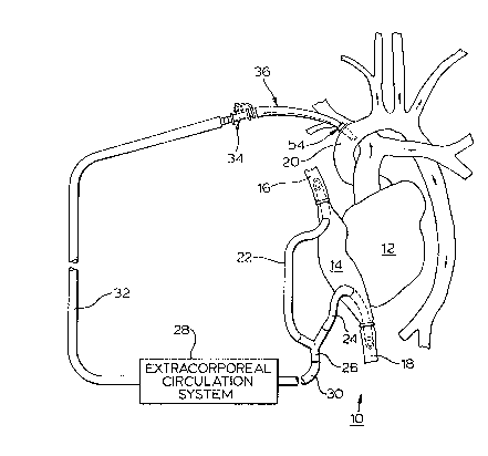
Fig. 1 is a diagrammatic illustration of a human heart
and some associated blood vessels connected into an extracorpo-
real circulation system and employing an aortic cannula device in
accordance with the present invention;
Fig. 2 is an enlarged side view of the aortic cannula
device of Fig. 1 but with a tube connector attached thereto;
Fig. 3 is an enlarged view of the distal cannula tip of
the cannula device as shown in Fig. 2 but alone;
Fig. ~ is a top view of the cannula tip shown in Fig.
3;
- 2a -
7~ 3 L~ ~
Fig. 5 is a bottom view of the cannula tip shown in
Fig. 3;
Fig. 6 is a right end view of the tip as viewed in Fig.
3;
Fig. 7 is a longitudinal cross-sectional view taken
along line 7-7 of Fig. 5 but wlth the cannula tip attached to the
distal end of the cannular tube of Fig. 2;
Fig. 8 is a cross-sectional view taken along line 8-8
of Fig. 3;
Fig. 9 is an enlarged plan vlew of a portion of the
aorta shown in Fig. 1 but prior to the insertion of the cannula;
Fig. 10 illustrates one manner of inserting the cannula
into an incision in the aorta, the cannular being shown just
before entering the incision; and
- 2b -
~ ~7~ ~
Fiy. 11 show the cannula after lt is fully inserted
into -the aorta.
Referrlng now to the drawings, and particularly to Fiy.
1, there is illustrated a portion of a surgical site 10 showing a
hear-t 12, a right a-trium 14, superior and inferior vena cavae 16
and 18 respec-tivel~, and an aorta 20 of a patient. A pair of
vena caval catheters 22 and 24 ex-tend into the a-trium 14 and into
the vena cavae 16 and 18, respectively, The vena cavae may be
tightened about the ends of the catheters 22 and 24 by suitable
string or the like. The opposite ends of the vena caval
cathe-ters 22 and 24 are connected to a Y-connec-tor 26 connected
to the inlet o~ an extracorporeal circula-tion sys-tem 28 through a
tube 30. Thus venus blood is fed into a suitable or conventional
extracorporeal system 28, for example, one that includes a blood
1~ oxygenator, blood pump, filters, bubble removing apparatus, and a
defoamer. System 28 serves as an artificial heart and lung,
changing venus blood into suitable oxygenated blood at the output
of the system ~8. A tube 32 connected to the outlet side of the
circulation system 28 is connected through a tube connector 34 to
2U an aortic cannula 36 shown inserted in-to the aorta 20 ~or
returning oxygenated blood to the arterial system of the patient.
The extracorporeal circulation system in Fig. 1, completely
bypasses the heart so that the heart or associated organs and
vessels rnay be operated on in the dry state.
The aortic cannula 36, as seen in Fig. 2, includes a
tube 38 tapering slightly from the proximal or left end 40
radially inwardly in the distal dire~tion to the right or distal
end 42. A hollow tube connector 34 has one end inserted into the
proximal end 40 of tube 38 in tight sealing frictional engagement
3~ with it. ~ removable end cap or filter member 46 is shown in
sealing engagement in the proximal end of connector 34. The
member 46 is shown having a passage 47 extending through it. A
hydrophobic filter 48 is shown covering the distal end of passage
47 while the proximal end of the passage is open to the
-- 3 --
~ 34~
a-tmosphere. The filter member 46, as will be further described,
allows air originally in the cannula 36 to be purged from the
cannula during insertion procedures but prevents blood from
Elowing through the filter. The tube connec-tor 34 is provided
with a side part 49 that is shown closed by a cap 50 which is
tethered by a resilient strap 51 having an eyelet 52 surrounding
'i the connector 34. The port 49 may be provided with a
conven-tional luer tapered inner wall (not shown) for subsequent
connection with a luer tapered syringe tip where it is desired to
withdraw a sample of blood from the patient. Also, the port 49
may be provided with conventional luer lock ears and the inner
wall of cap 50 provided with
.
2l
3U
3~
- 3a -
PATENT
7~4~ S-71 ~4
cornplemèntary luer lock thread~ so that cap S0 can be threaded onfo and of ~ ofconnector 34 as desired.
Cannula 36 ;ncludes a distal end tip 54 connected to the distal end 42 of
tube 38. Preferably, cannula tip 54 is formed or molded as a separate plastic element
and attached to the catheter 38. The catheter 38 may be made of a suitable plastic,
preferably one that is flexible enough to allow some bending but which does not
easily kink and occlude tube 38 when moderate bending forces are appiied to it, such
as during the connection of the cannula in the circulation system. Tube 38 may be
formed of a suitable plastic or rubber, for example, may be formed of a thermoplas-
tic material, such as polyvinyl chloride. The tip 54 may also be formed of a suitable
material, for example, the same material as tube 38 but preferably of a somewhatharder or more rigid material so that it can be inserted into the aorta without bending.
Tip 54 may be molded, for example, from a relatively rigid polyvinyl chloride.
Referring especially to Figs. 3-8, cannula tip 54 includes an annular collar
56 having an annular flange 58 that has a distally facing flat side 60 and may be
provided with suture slots if desired for securing the tip to the aorta. Collar 56 is
integrally connected with a distally extending, generally cylindrical portion 62 of
the tip which smoothly connects with a distal end por ~ion 64. An elongate or general-
ly eliptical opening 65 ~Fig. 4) is provided in the sidewall of the tip. As best seen in
Fig. 7, the distal end portion 42 of tube 38 is shown extending into collar 56 and
engaging an inner radially inwardly extending annular wall or land 66 on the interior
side of tip 54. The radially inner wall of the collar 56 and the outer wall of the tube
38 may be fixed together, such as by an adhesive, solvent bonding, or by other suit-
able means. Preferably, and as shown for illustration in Fig. 79 the thickness of the
sidewall of tube 38 at the distal end is substantially the same as the width of the
annular land 66 so that the tube lumen, indicated at 70, and the lumen of the cylin-
drical portion 62 of the tip, indicated at 72, are substantially the same so as to pro-
vide a smooth transition for blood flow from the tube 38 to the tip 54 where the tip
is a separate part attached to the tube.
The distal end portion 64 of tip 54 has an inner preferably smoothly curv-
ing wall 74 (Fig. 7) extending between the inner wall of the cylindrical portion 62
and the distal end of opening 65 so that blood flowing distally in lumen 70 flows into
the tip 54 and against the smoothly curving wal 1 74 and out the opening 65 withminimal turbulence even though there is a substantial angular change in the direc-
35 tion of blood flow. The wall 74 closes the distal end of the tip 54 and directs the
flow of blood out opening 65.
PATENT
7~ . S-7184
The dist~l end portior~ 6~ of -~ip S4 is provided with a smoothly contoured
ridge 8û in the outer surface of the tip formed by two smoothly curving generally
elliptical cavities or recesses 82 and 84 in the outer surface of the tip on opposite
sides and adjacent the ridge 8û. Ridge 80 smoothly blends into the cylindrical portion
S 62 of the tip as well as the distal extremity of the tip. The ridge 8û and recesses 82
and 84 have smoothly curving edges as best seen in Fig. 8, that is, the outer surface
of the tip is free of qny sharp edge. One or both of the edges of the opening 65 may
be radiused or rounded to smoothly blend with the outer surface of the tip. The
recesses 82 and 84 extend generally parallel to each other and angularly relative to
the longi~udinal axis of the tube and tip and radially inwardly toward the distal end
surface of the tip. In this way, the exterior surfaces of the tip adjacent each side of
the ridge 80 taper radially inwardly toward the distal end surface. The distal end
surface of the tip thus narrows toward the distal end surface and is rounded or sub-
stantially free of sharp edges. The radially outermost surface of the ridge 80, as
best seen in Fig. 3 and 7, is coextensive with the outer surface oF the cylindrical
portion 62 of the tip.
Preparatory to insertion, an incision or a slit 869 as shown in Fig. 9, may
be made in the aorta 20. Preferably, with the ridge 80 at the bottom of the tip, the
ridge is moved toward the slit as shown in Fig. 10, the cannula being held at an angle
to the longitudinal axis of the aorta. As the ridge enter the slit, the slit is opened
gradually or dilated until the entire tip penetrates the wall of the aorta. The cannula
is then moved into the aorta until the annular distal side 60 of the flange 58 engages
the outer surface of the aorta as shown in Fig. I l .
Upon insertion of the tip into the aorta 20, blood from the aorta flows
into the cannula 36 displacing the air in the cannula 36 and causing the air to flow
through the filter passage 47 to the atmosphere. The hydrophobic filter 48 will not
allow blood to pass through it. With the air removed from the cannula, filter 46 is
removed and the proximal end of connector 34 may be connected to the tube 32
(Fig. 1). The tube 38 may conveniently be clamped off during removal of the cap
and the connection of tube 32 to tube 38. If further air is found in the cannula 36 or
connector 34, it may be removed by removing the cap 50 to vent such air to the
atmosphere. Also, the side port 49 which may be in the form of a luer lock connector
may be used to take a blood sample by inserting a syringe into port 49 and withdrawing
a blood sample.
By providing -the longL-tudlnally extending smoothly
blendlng rldge 80, the ridge can be used as the leading edge of
the cannula during insertion into the aorta so that the forces
applied are more evenly distributed in spreading the walls of the
incision so a to reduce trauma to -the aorta and reduce blood
loss. Upon insertion of tip 5~, the slit will tend to be dilated
' and conform closely to the outer wall of the cylindrical portion
62 through the incision to reduce blood loss. Furthermore, -the
distal sidewall 60 of flange 58 can be urged against the outer
surface of the aorta such as by suturing or tying tending to
further prevent blood flow through the incision. Thus, use of
cannula 38 can effect a reduced amount of blood loss during
insertion of the cannula into the aorta as well as reduce
continued blood loss during the operation. Furthermore, there is
less trauma or damage to the patient because of the tapered shape
1~ and ridge 80 of the tip 5~ which gradually dilate the incision a
previously pointed out. By providing smoothly curving wall
sections and eliminating sharp edges, blood can flow through the
cannula with reduced hemolysis.
2~)
7~5
3~
-- 6 --
