Note: Descriptions are shown in the official language in which they were submitted.
()7~7
DI~GNOSTIC MET~OD FOR THE EVALUATION OF CLINICAL PARAME-
TERS B~ DIRECT COLLECTION OF BIOLOGICAL MATERIALS AND
DEVICE FOR ITS ACCOMPLISHMENT
This invention relates to a diagnostic method, and
to a related device for the assessment of clinical para-
meters, by direct collection of biological materials.
The device of th~ invention consists of a solid
support (of a suited nature, shape and size), carrying
fixed on its surface, by a suited technique, a pre-esta-
blished quantity of an antigen or of an antibody or, in
any case, of a substance able to inter-react with the
compound to be determined in said biological materials.
The diagnostic method of the invention consists in con-
tacting said device with the biological tisSues con-
taining the antibodies or the antigens to be analyzed. The
agglutinated product formed and fixed on the device is
then enzymatically labelled by reaction with a specific
antibody or antigen conjugated with an enzyme; after remo-
val of the labelled antigens or antibodies not specifical-
ly bound by means of washings (for instance with water),
the device is dipped in a suitable detecting solution
containing a substrate for the labelling enzyme which is
able to give a reaction useful for qualitatlve and/or
quantitative measurements.
Literature data state that the assessment of many
parameters of a clinical interest, by analysis of a biolo-
gical fluid, requires the transfer of an exactly measured
quantity into a suited container where it shall be incuba-
ted with a pre-established quantity of reagents so as to
: ~ .. :- ::-
:
... ,, , .
~2~ 57
produce a detecting reaction.
While the conventional methods involve no specificdifficulties when the bioloqical materials can be easily
collected, and are available in a relatively high quanti-
S ty, as it usually occurs in the case of urine and blood,a different situation occurs when analy~ical determinations
havé to be carried out on biological materials available
in a small or very small quantity (exudates, pus, mucus,
saliva, etc.) or of hard collection from histologically
lOaltered tissues and/or tissues in hardly accessible areas,
e.g. pustules, sy2hilomas, ulcerations, etc.
The disadvantages of said previously mentioned
techni~ue are overcome by the device and by the method
that are the object of the present invention, actually
lS enabling the direct collection of the biological material
to be examined with no resort to the measurement of the
specimen. The specimen, actually, can be collected direc-
tly on the patient, or subsequently on the specimen of
fluid or biological tissues collected according to one of
20 the usual laboratory techniques.
Further objects of the invention are provided by a
method and a device particularly suited for the diagnosis
of viruses and for the analytical determination of proge-
sterone in milk samples.
Presently, the only diagnostic methods available
for the detection of viruses in biological tissues (for
instance during viral infections or to evaluate possîble
healthy bearers in a population) are based on cell culture
procedures, which require expensive apparatuses, trained
personnel, and a long time (3 or more days) for the eva-
.,
.
., ;
,~ , ,. ,~ .
` "'; ' ; :
~7~)~7~
-- 3
luation of the results.
Said reasons make difficult timely diagnostic ana-
lysis which could be useful for the prevention of disease
spreading, for epidemiological studies and, generally, for
5 an early diagnosis of viral diseases.
Examples of viruses whose presence must be often
detected, comprise herpes viruses (type 1 and 2), Eppste-
in-Barr virus, Varicella Zoster virus, hyman T-lymphocytes
virus 3 (HTLV-3).
It is therefore highly desirable a method for the
detection of said viruses which could be carried out, even
in not specialized centers, by not highly trained or spe-
cialized personnel and without particular apparatuses such
as sterile ~ooms and so on.
The method according to the invention allows to
overcome the disadvantages of the known methods: it is in
fact particularly simple, unexpensive fast, safe and accu-
rate.
In one embodiment there is provided a device for
the qualitative diagnostic determination of a virus
selected from the group consisting of herpes virus,
Eppstein-Barr virus, Varicella Zoster virus, and Human
T-Lymphocyte virus, consisting of a handle and conical
puncture means having a pointed extremity and attached to
the handle for puncture of animal or human bodles, at least
a portion of the puncture means proximate the point of the
concial puncture means coated with a corresponding
specific antibody to the virus to be detected.
,'~t .'
:^"
.. '- .:: . : ~ ' '' :; : ...
-.. : ~ - : . . .
757
- 3a -
In another embodiment there is provided a method
for the ~uantitative diagnostic determination of a virus
selected from the group consisting of herpes virus,
Eppstein-Barr virus, Varicella Zoster virus, and Human
5 T-Lymphocyte virus, said method comprising puncturing an
animal or human body with a pointed member having a cor-
responding specific antibody to the virus to be detected
coated thereon proximate the point to cause the coated
pointed member to be contacted by a body fluid sample for
10 a time sufficient for a binding reaction to occur, removing
unfixed substances ~rom the coating remote ~rom said body,
thereafter contacting the coating with a detecting solution
containing a substrate capable of a detecting reaction with
an enzyme, and detecting the presence or absence of the
s enzyme as an indication of the presence or absence of said
virus in the sample.
Every virus can be.detected according to the method
i of the present invention provided that a specific antibody
and specific antibodies labelled with enzymes are availa-
.~0 ble. The antibody against the virus to be determined isbound on the device of the present invention. After the
agglutination reaction, the device is contacted with the
antibody labelled with the enzyme and then immersed into
the corresponding detecting solution after removal of the
enzy~e-labelled antibody not specifically bound.
In the case of herpes virus, for instance, a mono-
clonal or polyclonal anti- ~VS is absorbed on a device
having a shape suited for the direct sampling -from the
.
~, . .
. '`: :: - , ~ : :
',~
...
. ~
1~70~57
tissues (for instance a cutaneous blister).
250 Patients have been subjected to said diagnostic
method, in comparison with the known cell culture procedu-
res. The method of the invention proved to be extremely
5 reliable and in complete agreement with the previous me-
thods. The results were however obtained in only one hour
while the cell culture methods required at least three
days.
Analogously, a device suited for the detection of
10 HTLV-3, a virus responsible of the disease known as
aquiredimmuno deficiency syndrome tAIDS), can be prepared
both for the analysis of blood samples of individual sub-
jects and of blood from unknown donors.
Another important advantage of the present inven-
15 tion is that it is possible to detect the virus even in an
inactive, not replicative phase while the cell culture
methods require that the viruses can replicate, otherwise
an activation (with consequent further operative difficul-
ties) is needed.
A further preferred application and object of the
invention is a method for the detection of progesterone in
milk samples. In cow milk, high level of progesterone are
present during the luteal phase; after luteolysis the
progesterone level in the rnilk decreasesandremain to low
- 25 levels until the starting of ovulation.
Presently, these determinations are extensively
used by farmers before artificial insemination, in connec-
tion with or in alternative to treatment with prostaglan-
dine F2~ analogues for syncronization of farmers'animals.
30 In many countries, the proqesterone determined in milk
:. ,,:, :: : .: ::
. . .. - - .;
:: ,. . . .
:. ,:: .. :. . ~ . . :
-:. . :, - : :~:, .
12'7~
~_ samples, is based on RIA methods with radioactive labelled
progesterone. Also in this case the determination of pro-
gesterone according to the invention can be carried out
even by the same farmer and not-trained personnel without
5 any radioactive risk and the use of particular equipments
A~cording to the invention, the device is contac
ted with the biological material to be examined, said
device consisting essentially of a handgrip and an extre-
mity, intended for coming into contact with the biological
10 material, carrying fixed, by suited techniques, pre-esta-
blished quantities of antibodies (or antigens), specific
or partially specific for the antigene (antibody) compo-
unds to be determined.
The antigens or the antibodies, fixed on the extre-
15 mity of the device according to this invention, will con-
sequently bind, by immunochemical reaction, with the anti-
bodies or with the antigens present in the investigational
material, in a rate proportional to the quantity contained
in the material itself.
According to a preferred embodiment of the present
invention, after washing with a suited solvent or solution
in order to remove the substances not fixed by a specific
bond to the antigen or to the antibody previously fixed to
the device, a direct quantitative and/or qualitative de-
25 termination of the investigational clinical parameter,
shall be preferably accomplished resorting to already
known techniques based for example on chemical, immunoche-
mical, i~munoenzymatic, enzymatic, etc., reactions.
The shape and the size of the device of this inven-
30 tion, and particularly those of the extremity coated with
.~
~7~ 57
^ antibodies or antigens, are not by themselves critical for
the nature of this invention, being obviously conditioned
by the intended application and by the type of collection
required for the analytical determination.
For example, should the collection be made by skin
puncture, a device shall be conveniently used, characteri-
zed by a more or less pointed, but substantially conic,
shape with an extremity coated with antibodies or anti-
gens. On the other hand, should the co~ection require a
10 simple contact with a tissue or a dipping into a biologi-
cal fluid, the surface coated with the antibodies or with
the antigens may present a cylindrical, spherical, ovoi-
dal, conical or frusto-conical, etc., shape.
Should collection be made by skin puncture, final-
15 ly, a device proves particularly suited, consisting of ametal core, coated if the case with plastic material,
fitted at its extremity with a body coated with antibodies
and/or antigens, of suited size and shape (for example, a
2-5 mm diameter spherule) from which a metal tip, suited
20 for injection, juts out a few millimeters (for example
0.5-2 mm): in the course of its use, the metal tip will
cause the biological fluid to leak out (for example, a
drop of blood), and impregnate the body coated with the
antibody and/or the antigen, being with lt in an immediate
25 contact because of the minimal distance.
As suited materials for the support to be coated,
plastic materials can be used, able to bind antibodies or
antigens by adsorption, according to the present inven-
tion, such as polyethylene, polypropylene, polystyrene,
30 polyurethanes,polyvinyl chloride, BUN~-N, nylon, polyacry-
* Trade mark
~ ~7~t~
lates, polymethacrylates, polytetrafluoroethylene resins
(TEFLON( ), phenol resins, polyacrylamides, DELRIN( ),
LEXAN , cellulose derivatives, silicone derivatives,
etc.
Said materials may also coat a metal core.
The bond of the antibody or of the antigen with the
device of this invention may also be chemical should the
constructive plastic material contain amino-, hydroxy-,
carboxy-, or isocyano- functional groups suited to fix
10 protein compounds by a chemical bond. The suited plastic
material may be activated, if the case, by dipping it into
a pH g buffered solution of qlutaraldehyde, according to
an already well-known technique, or according to other
similar methods commonly known, for example, in the field
15 of enzymes immobilization.
Metal materials may also be used, when previously
subjected to such a process as to make them porous or able
in any case to adsorb antibodies and/or antigens, for
example by galvanic techniques for the deposit of colloi-
20 dal gold or other metals in a colloidal form.
Examples of coating with an antibody or an antigen
of said materials, by said methods, are described in the
US Patent No. 4,003,988, and in the following papers:
(a) IMMUNOASSAY USING ANTIGEN-ENZYME CONJUGATES
B.K. Van Weemen and A.H.W. Schuurs
Febs Letters - Vol. l5, Number 3 - June 197l -p.232-5
(b) SOLI~ P~IASE ANTIBODY ASSAY BY MEANS OF ENZYME CONJUG_
TED TO ANTI-IMMUNOGLOBULIN
P. Leinikki and Suvi Passila
J. Clin. Path., 1976, 29 - p. 1116-20
:
17S7
(c) EN~YME-LINKED IMMUNO~ORBENT ASS~Y WITII POLYVA~ENT GONO
COCCAL ANTIGEN
B.R. Brodeur, ~.E. Ashton and B.B. Diena
The Journal of Medical Microbiology - Vol.lS, No.l -
1981 - p. 1-9
(d) A MICROPLATE ENZYME-IMMUNOASSAY FOR TOXOPLASMA ANTIBO-
DY
J. Clin. Path., 1976, 29, p. 150-3
(e) ENZYMOLOGY (Immobilized Enzymes, Antigens, Antibodies
and Peptides - Preparation and Characteri2ation - Vol.
I - Ed. Howard H. Weetall - Marcel Dekker, Inc., New
York (1975).
The antibody, or the antibodies, used according to
the invention to coat the support, may be polyclonal or
15 monoclonal, or, if the case, mixture thereof, and may be
specific for one or more active sites of the antigen to be
determined; moreover, this specificity may be directed on
one or more antigens.
Both antibodies and antigens may be fixed on the
20 support with no limits of quantity: however, a quantity
suited for the intended type of analysis shall be chosen,
ranging possibly between one picogram and one millig~am
per square centimeter.
Examples of possible applications of the device and
25 of the method according to this invention, include the
direct determination of viruses and/or bacteria, the de-
termination of antibodies and/or antigens associated with
a specific pathologic condition, the determination of the
antibody titer following immunologic treatments, the de-
30 termination of drugs and their metabolites, the determina-
`"" :.'. ~ ~ :
~, : ' ' ' ` ' ' '
~L270~7~7
tion of hormones, and the determination of common clinicalparameters. The support may also be coated so as to enable
the determination or the assay of various not necessarily
related clinical parameters, e.g. the determination of
5 chorionic gonadotropin and cocaine in the urine.
Usually, the detecting reaction is properly carried
out by means of an antibody labelled with a suited enzyme,
specific for the antigen-antibody complex fixed on the
support after the specimen collection. For the purpose,the
10 device, carryi~g the antigen-antibody complex fixed thereon,
is washedwith suited buffer and/or water solutions in order
to remove the unspecifically bound antigen or antibody,
and it subsequently dipped and incubated, for a suited
time, in a solution of said antibody conjugated with an
15 enzyme.
Said antibody solutions preferably contain ncnio-
nic, ionic, and amphoteric buffering and emulsionating
agents with a concentration of enzyme-labelled antibody
ranging from O.Ol to O.OO5~ weight. The enzyme-labelled
20 antibody-antigen-antibody complex will consequently be
fixed on the support: after removing a possible unspeci-
fically bound labelled antibody, by washing with suited
buEfer and/or water solutions, dipping the support, treat-
ed as above specified, into a solution containing a suited
25 chromogenic system based on a substrate for the enzyme,
will enable a simple and direct antigenic determination of
the product of the enzymatic reaction, through colorime-
tric, spectrophotometric or other various techniques. ;
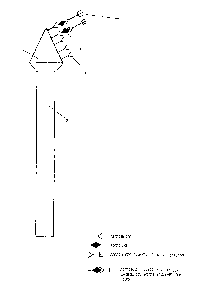
The attached figure, provided only for illustration
30 purposes and not limiting therefore the invention, repre- ~;
1270~75~7
-- 10 --
sents schematically the device of the invention.
The reference number 1 indicates the cone-shaped
support coated with the antibody or with the antigen,
intended to come into contact with the biological mate-
S rial; the stem 2 acts as a handgrip, and may be of such ashape and size as to make use quite easy and comfortable;
3, 4 and 5 indicate the antibody, the antigen and the
enzyme-labelled antibody respectively.
The device of the invention, enabling also to avoid
10 a precise deterrnination of the quantity of biological
material to be examined, also offers the advantage, within
the sensitivity limits of the method, of a higher reliabi-
lity and precision thanks also to the minimization of the
experimental errors: the analysis, moreover, become more
15 rapid and timely with no need of an expensive equipmentr
so that even a direct home-use of analytical kits can be
foreseen.
For the purpose the devices, that are the object of
this invention, shall be prepared under strict sterile
20 conditions in suited aseptic premises, and finally intro-
duced into suited sterile containers, e.g. glass test-bu-
tes sealed by sterilized stoppers.
The following not restrictive examples illustrate
some possible applications of the method and of the device
25 of the inventicn.
EXAMPLE 1
(a) Prepa ation of the device suited for the determination
of the ~erpes Virus (I~SV)
Polyvinyl chloride pointers, with a cone-shaped
30 extremity, were used as solid phase.
''~, ' ` '~ ' ` , ~,
,,. `_
'
~LZ7l~
~- The extremity of said pointers was allowed to incu-
bate with an anti-HSV-2 antiserum, diluted in a 1:300
ratio with 50 mM pH 9.6 carbonate/bicarbonate buffer. In
terms of technical procedure, the antiserum was fixed to
5 the support dipping the pointers' conical extremity into
the wells of a polyvinyl chloride microdish containing
0.15 ml of antiserum so that the whole conical area could
be dipped into the solution. The microdish was thereafter
allowed to incubate for 2 hours at 37C in a wet chamber,
10 and subsequently overnight at 4C.
At the end of the incubation, the pointers were
accurately washed with pH 7.4 saline phosphate buffer
(KH2PO4 15 mM, Na2HPO4 85 mM, NaCl 15 mM, KCl 25 mM),
containing Tween*0.05%, introduced into the wells of ano-
15 ther dish containing the same saline buffer with 0.3%bovine serum albumin, and allowed to incubate for 1 hour
at 37OC in a wet chamber. This treatment has the purpose
to saturate all possible polyvinyl chloride free sites
that, in the following steps, may also be caused to adsorb
20 the viral particles or the conjugated antiserum.
In a similar way a device for the determination of
Hepstein-Barr virus, Varicella Zoster virus and Human
T-Lymphocyte Virus-3 (HTLV) is prepared.
(b) "In Vitro" dete~mination of HSV
After an identical step, the pointers were allowed
to contact, for a short time at room temperature, a HSV
virus suspension diluted 1:5 in saline (O.9% NaCl).
~ After washing, the pointers were introduced into
glass test-tubes containing anti HSV-2 antiserum conjugat-
30 ed with peroxidase at a 1:150 dilution in saline phosphate
,
.
. .
..
*Trade mark
... .,.. ~
~71~757
- 12 -
buffer containing 0.05~ of Tween 20. This procedure was
followed by a 30-40 minutes incubation at room temperatu-
re, and by washing in saline phosphate buffer, containing
0.05~ of Tween 20, in order to prevent possible analytical
5 interferences of the conjugated antiserum unspecifically
bound with the antigen.
After the last stage of treatment, the pointers
were introduced into a chromogen solution consisting of
3.4 mg of ortho-phenylenediamine dissolvedin 10 ml of buf-
10 fer, formed by equal parts of 0.1 M citric acid and 0.2 MNa2HP04, added, just before using, with 50 mcl of a 3%
H202 solution.
The results recorded could state the good specifi-
city and sensitivity of this technique: actually, the
15 positive specimens show, through the chromogenic system,
an intense color markedly distinguishable from the almost
non-existent one o~ the negative specimens.
(c) Explanatory procedure of a ~SV determination by direct
collection
Herpes infections, Type 1, Type 2 or Types 1 and 2
occur usually with the formation of various-sized vesicles
in the genital and/or labial area; the formation of herpe-
tic vesicles in other bodily organs is more rare.
To the purpose of diagnosing an actual viral infec~
25 tion of a herpetic origin, the device, prepared according
to the procedure as per item (a), is allowed to contact
the vesicle with its lanceolated extremity, causing conse-
quently the vesicle to break.
The device is subsequently rotatedj and the fluid
30 from the broken vesicle allowed to contact the anti HSV
* Trade mark
. :- ; :. , ~ , .
. ,- ~.- :. ~ - : -, ,
~L27 [)~757
antibodies previously fixed on the pointer.
As soon as the fluid collection is terminated, the
device is introduced into an ampul containing 300 mcl of a
solution of anti HSV conjugated with peroxidase enzyme,
5 and diluted in a 30 mM phosphate buffer containing 0.05
percent of Tween 20, and 1 per cent of bovine serum albu-
min.
After a 15-minute incubation, the support is washed
with running water for about 60", and transferred into an
10 ampul containing 300 mcl of a pH 5 solution characterized
by the following percent composition (w/v~:
Tetramethylbenzidine 0.02
Citric acid 2.10
Sodium Perborate 0.07
Sodium phosphate, dibasic 3.00
Dimethylsulfoxide 20.00
Distilled water 75.00
After a 15-minute incubation, the solution, contai.-
ned in the ampul, is observed. It may be:
20 (a) colorless in the case of a negative result (absence of
HSV in the patient's vesicle);
(b) blue in the case of a positive result (presence of HSV
in the patient's vesicle).
EXAMPLE 2 ~:
25 Determination of the antistreptolysin 0 (AS0~ Titer
.
The extremity of the support, previouslycoated with
streptolysin, according to the technique used in the Exam- ::
ple 1, is allowed to stand for some seconds in contact
with the investigational serum or with a drop of whole
30 blood collected from the forefinger's tip pierced by a :
,,,, :
:: : :
, . ., . ::
~27~7~7
- 14 -
normal self-injection apparatus, e.g. Autoclik. The extre-
mity of the support is washed with water, and dipped into
a solution containing an anti-IgG antibody labelled with
an enzyme (serum alkaline phosphatase or peroxidase). The
5 labelled anti IgG - streptolysin - antistreptolysin com-
plex is consequently present on the surface and, dipped
into a solution of the substrate tpara-nitrophenylphospha-
te and azino-benzothiazolin-sulfonate), will color it more
or less intensively depending on the antibody titer of the
10 serum or of the blood.
EXAMPLE 3
Determination of morphine in the urine or in the blood
The previously indicated solid support, coated with
an antimorphine antibody according to the techni~ue speci~
15 fied in the Example i, is dipped into the investigational
urine or into a small investigational serum or blood
specimen; after an incubation of about 15 ~inutes, the
support is dipped into a solution of peroxidase-conjugated
morphine in 30 mM phosphate buffer at pH 7. After a lO-mi-
20 nute incubation time, the support is thoroughly washed,and dipped into a test solution of tetramethylbenzidine
and perborate prepared according to the Example 1 c).
The intensity of the developed color is inversely
proportional to the concentration of morphine present in
25 the investigational biological fluid.
EXAMPL~ 4
Determination of cocaine and human chorionic gonadotropin
in the urine
The polyvinyl chloride or polystyrene support, of a
30 spherical or ogival shape, is dipped ~or coating into a
* Trade mark
.
.. ..
~27(~7~;~
mixture of anti-benzoylecgonine (the major metabolite of
cocaine in man) serum and human chorionic antigonadotropin
serum.
The antisera used may be polyclonal (from rabbit
5 and goat) or monoclonal (produced both in vitro and in
vivo) or pools of both; the ratio of the two antisera in
the mixture is properly selected according to the sensiti-
vity to be attained in the determination of the two para-
meters.
The related dilutions, carried out in carbonate-di-
carbonate buffer, may range between 1:50 and 1:50,000.
The coating time ranges between 30 minutes and 72
hours at a temperature ranging between +4 - +60C.
The support, as soon as coated with the antisera
15 mixture, is saturated by applying a second coating with a-
solution of aspecificic immunoglobulins or in normal rab-
bit serum (in other cases also a bovine fetal serum may be
used), in phosphate buffer, at a concentration ranging
between 0.1 and 5 percent, under conditions of ti.me and
20 ternperature as same as the ones used to coat the antisera
mixture.
The support is subsequently washed in order to
remove a possible excess of antibodies or proteins not
fixed with the support; in this way, after washings, the
25 support is made ready for its use, intended for its dip-
ping into the investigational urine.
In the presence of one or both parameters involved
in the determination, a complex will be formed between
them and their related antibody, physically fixed on the
30 solid support.
.~
. -,, : . .. ..
:. .. .. : . ., , ~ :
:: :: " :,;''~` ' ': :. . . ~
.; ,:.. ., - : :
.
1~'7~57
~ 16 -
The support is subsequently washed, and dipped into
a mixture of serum of human chorionic anti-gonadotropin
conjugated with peroxidase, and benzoylecgonine conjugated
with beta-galactosidase: the ratio between the two label-
5 led compounds will be such as to enable a good detectionsensitivity.
After allowing a lapse of time sufficient for the
formation of the complexes tS-60 minutes), the support is
further washed, and dipped into a solution of peroxidase-
lO specific substrate (e.g., azino-benzothiazolino-sulfonate,
phenol-4-amino-antipyrine, etc.), and subsequently into a
second beta-galactosidase specific substrate (e.g., chlo-
rophenol red -beta galactoside). The first solution stains
in the presence of human chorionic gonadotropin; the color
15 of the second solution is inversely proportional to the
concentration of benzoylecgonine present in the investiga-
tional biological fluid.
EXAMPLE 5
(a) Preparation of the device suited for the determination
of progesterone in biological fluids: milk, urine,
serum and plasma
Polyvinyl chloride supports, with a round-shaped
extremity, are used as solid phase.
These supports are allowed to incubate with a solu-
25 tion, in pH 7.5 phosphate buffer, of 2.5 percent glutaral-
dehyde for a length of time ranging between 2 and 8 hours
at 37~C or at room temperature.
Subsequently, the supports are thoroughly washed
with a solution of phosphate buffer or water.
The supports, treated as above stated, are allowed
: . .
~l27~757
- 17 -
to incubate with an antiprogesterone antibody in pll 9.6
carbonate/bicarbonate buffer, at a dilution ranging bet-
ween 1:100 and 1:1000 for 2 hours at 37C, and overnight
at 4C.
At the end of the incubation, the supports are
thoroughly washed with pH 7.4 saline phosphate buffer con-
taining Tween 20 O.OS percent, and allowed to incubate in
the same pH 7.4 saline buffer containing 3 percent of
bovine serum albumin, and allowed to stand for 1 hour at
10 37C.
This treatment has the purpose to saturate possible
free sites in the polyvinyl chloride support, likely to
absorb, in the subsequent steps, the progesterone aliquot
to be determined, contained in the biological fluids, or
15 still the progesterone aliquot conjugated with the enzyme..
(b) Determination of progesterone in milk
The polyvinyl chloride support is grasped with a
hand, and dipped in to the milk specimen containing the
investigational progesterone for a length of time ranging
20 between 5' and 15'. At the end of the incubation, the
support is transferred into a test-tube containing a solu-
tion, in pH 7 phosphate buffer, of progesterone labelled
with H-R-peroxidase, and a dilution ranging between
1:1,000 and 1:1,000,000. The support is allowed to incuba-
25 te for about 10 minutes, subsequently washed thoroughlywith a phosphate buffer solution or water, and dipped into
a test solution of TMB and perborate or H202, as stated in
the Example 1 c). The intensity of the color developed is
inversely proportional to the concentration of progestero-
30 ne present in the milk specimen; said concentration can be
~ ,...
~L~71)~5~
directly assayed by a calibration scale. It is well evi-
dent that said method is entirely extensible to the deter-
mination of progesterone in other biological fluids, such
as urine, serum and plasma.
:
. : .,. : ., .