Note: Descriptions are shown in the official language in which they were submitted.
MULTIPURPOSE GASEOUS DETECTOR DEVICE
FOR ELECTRON MICROS~OPES
Background of the Invention
Scanning electron microscopes and generally instruments
employing an electron beam (probe) operate in vacuum
(pressure less than about 0.0001 mbar) and the specimens
examined by such instruments are also placed in vacuum.
Scanning a sample within a vacuum presents many problems.
Many biological specimens cannot survive in vacuum. Wet
specimens can experience evaporation of their fluid
content before an accurate image can be obtained.
Nonconducting samples can accumulate a surface charge
which obscures the details of the sample's surface and
lowers the resolution of the image obtained.
An environmental scanning electron microscope (ESEM)
which allows the examination of specimens in a gaseous
environment is described in U.S. Patent No. 4,596,928.
The purpose of the gas in the '928 patent was to act as a
conditioniny medium in order to maintain a specimen in a
liquid, wet or natural state. However, the predominant
detection mode in the ESEM has utilized various
scintillator detectors to detect backscattered electrons.
Additionally, an ESEM detection system has been described
wherein the ionization of the gaseous environment is used
L ~ 37
2--
as the detection means for all ionizing signals
(Danilatos, Micron. Microsc. Acta 14:307 -318, 1983).
osJEcTs OF THE INVENTION
An object of the present invention is to provide a more
general and multipurpose means for environmental scanning
electron microscopy.
It is also an object of the present invention to provide
means for the detection in general of all signals which
can react with a gas or mixture of gases inside an
environmental electron microscope such that the gas
itself acts as a detector.
S~mmary of the Invention
The present invention provides a scanning electron
microscope for cathodeluminescence detection of specimens
which comprises a vacuum envelope having a pressure
limiting aperture. An electron beam source is located
within the vacuum envelope and is capable of emitting an
electron beam. Focusing means are located within the
vacuum envelope and are capable of directing an electron
beam emitted by the electron beam source through the
pressure limiting aperture. Electron beam scanning means
are also located within the vacuum envelope and are
i' ~P~ 7
--3--
capable of scanning an electron beam emitted by the
electron beam source across the diameter of the pressure
limiting aperture. A sample platform means is disposed
outside the vacuum envelope and is capable of maintaining
a sample in registration with the pressure limiting
aperture such that a surface of the sample maybe exposed
to an electron beam emitted from the electron beam
source and directed through the pressure limiting
aperture so as to cause radiation to be emitted from the
sample. The scanning electron microscope of the present
invention further comprises gas containment means capable
of maintaining the sample platform means enveloped in a
gaseous medium so as to
! ~t~$~ 7
allow radiation emitted from a sample located on the
sample platform means and exposed to an electron beam
emitted from the electron beam source to come into
contact with gas molecules of the gaseous medium and
cause the gas molecules to emit photons. Detection
mean~ are provided which are capable of detecting
photons emitted from the gas molecules of the gaseous
medium.
The present invention also provides a method for micro-
scopically imaging the surface of a sample which
comprises surrounding the sample with gas molecules and
scanning the surface of the sample with an electron
beam having sufficient energy so as to cause radiation
to be emitted from the surface of the sample. Photons
which are emitted from the gas molecules which come
into contact with radiation emitted from the surface of
the sample are then detected, the photons being emitted
from the gas molecules in an amount proportional to
the amount of radiation emitted frcm the surface of the
sample. Images of the sample are then formed based on
the number of photons detected.
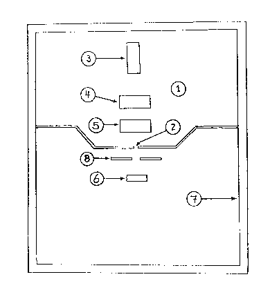
Brief Description of the Figure
Fig. 1 is a schematic cross-sectional view of a device
5 which embodies the present inverltion in a particular
form.
--6--
Detailed De8cription of the Invention
The present invention provides a scanning electron
S microscope. Referring in more particularity to Figure
l, the invention comprises a vacuum envelope l having
a pressure limiting aperture 2. An electron ~eam source
3 is located within the vacuum envelope and is capable
of emitting an electron beam. Focusing means 4 are
located within the vacuum envelope and are capable of
directing an electron beam emitted by the electron beam
source through the pressure limiting aperture. Elec-
tron beam scanning means 5 are also located within the
vacuum envelope and are capable of scanning an
electron beam emitted by the electron beam source
across the diameter of the pressure limiting aperture.
A sample platform means 6 is disposed outside the
vacuum envelope and is capable of maintaining a sample
in registration with the pressure limiting aperture
such that a surface of the sample may be exposed to an
electron beam emitted from the electron beam source
and directed through the pressure limiting aperture so
as to cause radiation to be emitted from the sample.
Within this application, "radiation" emitted from a
sample means electrons or photons emitted from the
sample.
--7--
The scanning electron .-nicroscope of the present
invention further comprises a gas containment means 7
capable of maintaining the sample platform means
enveloped in a gaseou~ medium so as to allow radiation
emitted from a sample located on the sample platform
means and exposed to an electron beam emitted from the
electron beam source to come into contact with gas
molecules of the gaseous medium and cause the gas
molecules to emit photons. Detection means 8 are
provided which are capable of detecting photons emit-
ted from the gas molecules of the gaseous medium.
In one embodiment of the invention, the wavelength ofthe photons is within the range from about lxlO-ll
meters to about 4x10⁻⁸ meters. Preferably witnin this
embodiment of the invention the detection means is a
scintillation counter or a lithium drifted silicon
detector.
In another embodiment of the invention, the wavelength
of the photons is within the range from about 4xlO-8
meters to about 7x10⁻⁷ meters. Preferably within this
embodiment of the invention the detection means is a
photomultiplier tube or a photodiode.
~o~
--8--
In yet another embodiment of the invention, the wave-
length of the photons is within the range from aoout
7x10⁻⁷ meters to about 2x10⁻⁴ ;neters. Preferably with-
in this embodiment of the invention the detection means
is a photomultiplier tube or a photodiode.
The gaseous medium may comprise a single gas or a
mixture of gases. In one embodiment of the invention
the gaseous medium comprises nitrogen. In another
embodiment of the invention the gaseous medium
comprises helium.
The present invention also provides a method for micro-
scopically imaging the surface of a sample which
comprises surrounding the sa,nple with gas molecules and
scanning the surface of the sample with an electron
beam having sufficient energy so as to cause radiation
to be emitted from the surface of the sample. Photons
which are emitted from gas molecules which come into
contact with radiation emitted from the surface of the
sample are then detected, the photons being emitted
from the gas in an amount proportional to the amount
of radiation emitt~d from the surface of the sample.
Images of the sample are then formed based on the
number of photons detected.
_9_
In one embodiment of the invention, the wavelength of the
photons is within the range from about lxlO-ll meters to
about 4xl0-8 meters. Preferably within this embodiment
of the invention the detection means is a scintillation
counter or a lithium drifted silicon detector.
In another embodiment of the invention, the wavelength of
the photons is within the range from about 4x10⁻⁸ meters
to about 7x10⁻⁷ meters. Preferably within this
embodiment of the invention the detection means is a
photomultiplier tube or a photodiode.
In yet a further embodiment of the invention, the
wavelength of the photons is within the range from about
7x10⁻⁷ meters to about 2x10⁻⁴ meters. Preferably within
this embodiment of the invention the detection means is a
photomultiplier tube or a photodiode.
The gaseous medium may comprise a single gas or a mixture
of gases. In one embodiment of the invention, the
gaseous medium comprises nitrogen. In yet another
embodiment of the invention, the gaseous medium comprises
helium.
--10--
The gas utilized in the present invention is the primary
medium, or the first and basic stage in the detection
chain of the environmental scanning electron microscope.
Additionally, the electron beam-specimen interactions
generate signals which, in turn, react wi~h the gas. The
signal-gas interactions constitute the basis for the
detection of signals. Some examples of signal-gas
interactions are: gaseous scintillation, ionization,
chemical combination, chemical disassociation, electron
attachment, photo-ionization, X-ray reactions,rotational
and vibrational collisions, collisions characterized by a
particular energy loss, etc.
This multi-purpose gaseous detector device has many
advantages over conventional detectors which can operate
only in a vacuum. The present invention provides for the
use of the gaseous environment of the specimen chamber of
the environmental scanning electron microscope as a
multi-purpose detector for the detection of high and low
energy elect:rons, for the detection of photons, including
X-ray and detection of other products from chemical
reactions. The present device allows for the examination
of specimens in air. It generates new information on
specimens in air not possible in the previous art of
--ll--
detection. It is more general and multi-purse than
previously known in the art of environmental scanning
electron microscopes and atmospheric scanning electron
microscopes.
Although the present invention has been described in
connection with various preferred embodiments thereof, it
will be apparent to one of ordinary skill in the art that
many changes and modifications may be made therein
without departing from the spirit and scope of the
present invention, which is determined by reference to
the appended claims.