Note: Descriptions are shown in the official language in which they were submitted.
1285446
41549 CAN 4A
CORNEAL HOLDER
FIELD OF THE INVENTION
The present invention relates to procedures and
means for physically handling corneal tissue that has been
obtained from a donor eye, in order to perform such
functions as storing, transporting, microscopically
evaluating, and cutting the tissue in a manner that
10 provides a portion of the corneal tissue that is suitable
for implantation in a recipient eye.
BACKGROUND ART
For purposes of possible transplantation to a
15 recipient eye, the cornea from a donor eye is typically
excised as a generally circular section of tissue that
includes a small outer rim of scleral tissue.
This corneal tissue is then generally subjected
to a variety of physical manipulations and procedures,
including storage, transport, microscopic evaluation and
lastly, "trephining" a procedure in which a circular plug,
i.e., "donor button", of the desired size is cut out from
the desired location for use as an transplant.
An excision and storage technique commonly used
in many eye banks is outlined in the chapter entitled
"Tissue Processing", by M.A. Gallagher, pp. XI-1 through
XI-4, in Eye Bank Technician Manual, Eye Bank Association
of America, Houston, TX, 1984. In this technique the
corneal tissue is cut out, i.e., excised with small
scissors in a generally circular shape having a 2-3
millimeter oute~ rim of scleral tissue. The tissue is
typically transferred to a clear vial, and stored,
free-floating, in sterile storage medium. Certain recent
developments in the formulation of storage media purport to
enable the storage of such explants for up to several days
or even weeks, see, e.g., B.F. Boyd, Highlights of
Ophthamology Letter, Vol. XIV(2): 1-16 (1986).
~285446
The corneal tissue is typically stored in the
free-floating state until just before use when it is
microscopically examined to evaluate the integrity of its
endothelial surface, e.g., by removing the tissue with a
5 forceps to a viewing chamber or viewing it directly in its
storage vial as described, e.g., in "Wide Field Specular
Microscopy of Excised Donor Corneas", C.w. Roberts et al,
Arch. Ophthalmol. 99:881-883 (1981). The condition and
appearance of the endothelial and epithelial surfaces are
10 critical factors to be determined in deciding whether a
particular corneal tissue is suitable for implantation.
Abrasions and loss of cells from the endothelial surface
are major factors for rejecting many corneal tissues for
use and can be attributed, at least in part, to damage done
15 to the tissue during and by virtue of its storage.
In an adaptation of the free-floating storage
system, Coopervision, Inc., Irvine, CA has a commercially
available storage jar called a "PRO CSVC"~which is
described as a "Corneal Storage and Viewing Chamber" for
20 use in conjunction with its microscope systems. The
chamber consists essentially of two parts, i.e., a
translucent plastic jar and a clear plastic cap.
The distinguishing feature of the jar is a circle
formed by eight plastic spikes extending upwardly from the
25 base of the jar, each being inwardly notched and then
tapered downwardly at their tips so as to provide a support
upon which corneal tissue can rest without falling between
the spikes or into the open circle defined by their center.
The cap has a circular indented portion that is
30 optically clear and configured to allow a microscope lens
to penetrate the plane of the top of the cap a distance
almost equal to the height of the cap, and to move about
therein, in order to scan and focus on the corneal tissue
when the tissue rests in the center of the chamber.
The cap indentation also serves to provide a
barrier at the top of the jar chamber, thereby restricting
the ability of the tissue to float out of its chamber. The
~ r~
i285446
--3--
tissue is nonetheless still free to move within the
confines of the chamber, such that the jar might need to be
tapped or swirled in an attempt to bring the tissue to rest
in a centered position at the base of the chamber.
As with the free-floating storage vial described
earlier, the tissue would of course, need to be physically
grasped and removed from the chamber in order to place it
carefully in a trephining device.
The basic trephining devices are simply cutting
10 blocks and corneal punches. The tissue is carefully
placed epithelial side down in a concave indentation in a
block made out of a hard inert material such as Teflon,
such that the center of the tissue is aligned with the
center of the indentation, and the tissue rests in
15 approximately its normal curvature during trephining. A
circular metal trephine blade, attached to a punch
mechanism, is then carefully aligned and oriented, in a
manner analogous to a drill press, so as to hover above or
lightly touch the tissue at the desired, generally central,
20 location. The blade is then tapped or turned down into the
tissue with sufficient force and to a sufficient distance
to cut out a plug.
Devices have been described for securely holding
corneal tissues during trephining, see e.g., U.S. Pat. Nos.
25 2,929,603, 3,058,471 and 4,077,411. U.S. Pat. No.
4,077,411 for instance, describes an apparatus having a
spring-loaded ring to secure the corneal tissue at its
edges over a semi-spherical post. The introduction of a
harmless liquid from below the tissue, through a conduit in
the post, is said to provide a cushion to resist the
downward pressure of the trephine blade.
In "Corneal Holder", Amer. J. Ophthalmol. 80(3)
Part II:551-552 (1975), there is described a holder having
a semi-spherical pedestal, a matching scleral sealing
sleeve and a retaining ring. The sleeve retains the tissue
on the pedestal at its scleral rim, and the ring locks the
sleeve to the pedestal in a manner said to trap a cushion
128~446
of air beneath the tissue, again to resist the downward
pressure of the trephine blade.
It follows that throughout the typical functions
involved from excision to implantation, the corneal tissue
5 must frequently be handled a variety of times, e.g., by
forceps, in order to place it in the storage vial upon
excision, remove it and prepare it for microscopic
evaluation, and place it and then orient it in a device or
on a block for trephining. Each physical manipulation
10 increases the chance of damage to the tissue, particularly
at its edges and on its endothelial surface, and requires
the patience, time and skill of a trained technician.
Throughout these functions, it would be highly
desirable to be able to orient the corneal button with
15 respect to its placement in the recipient eye. Kiely, et
al, "Meridional Variations of Corneal Shape", Amer. J.
Optom. Physiol. Optics, 61(10):619-626, 1984, for example,
explains that the cornea in fact consists of four
individual corneal meridians, each with its own radius of
20 curvature. If the corneal tissue is transplanted into the
recipient eye in a manner in which its radii are
incompatible with those of the recipient eye, astigmatism
can result. In order to limit this situation, surgeons
currently must generally transplant corneal buttons in an
25 unknown orientation in the eye, and then tighten and loosen
the stitches holding the transplant, in an effort to hold
the transplant in its desired configuration.
Currently practiced corneal tissue handling
procedures do not generally allow the orientation of the
30 corneal tissue or button. In fact, orientation is
essentially lost from the moment the tissue is excised and
placed to float freely in its storage medium.
Furthermore, it has been found that it may be
desirable in some situations to prevent or lessen fluid
35 absorption into the cut edges of the corneal tissue during
storage. Undue absorption can lead to a swelling of the
~2~;44~;
explant, thereby thickening it beyond use. One approach
currently used in an attempt to avoid such thickening has
been the development of modified storage media, as
described in "Minnesota System Corneal Preservation"
5 Lindstrom, et al, srit~ J. Ophthalmol., 70:47-54, 1986.
SUMMARY OF THE INVENTI!:~N
The present invention relates to a holder for
corneal tissue, amenable for use in both a corneal storage
10 system and a corneal cutting system with a minimal degree
of physical manipulation of the tissue itself. The holder
enables the tissue to be oriented in any desired position
throughout storage, thereby enabling the surgeon to cut,
orient and implant a predetermined portion, e.g, a corneal
15 button, in a manner that limits astigmatism. The holder
also facilitates the rapid and easy positioning and
alignment of a cutting device in order to make a smooth,
straight and concentric cut at the circumference of the
predetermined portion of the corneal tissue. The holder
20 also can be made to allow any desired degree of
constriction of the rim of the tissue, so as to provide a
barrier to fluid uptake in order to alleviate swelling of
the tissue.
The present invention provides a corneal holder
25 adapted to hold a corneal tissue while cutting a
predetermined portion of the tissue, the holder comprising:
a base member having a first aperture of a size
at least as large as the predetermined portion of the
tissue,
a cap member having a second aperture of a size
at least as large as the predetermined portion of the
tissue, and
attachment means associated with the base member
and the cap member for attaching the base member to the cap
35 member, the base member and the cap member thereby
cooperatively defining a compartment adapted to hold the
predetermined portion in a retained relationship in
~.285446
--6--
alignment with the first and second apertures and defining
a passageway through the first and second apertures
configured to allow a cutting device to be passed through
the passageway and into cutting contact with the
5 circumference of the predetermined portion.
The present invention also provides a corneal
storage system and a corneal cutting system comprising the
corneal holder.
BRIEF DESCRIPTION OF THE DRAWING
In the Drawing,
FIG. 1 is an exploded perspective view of one
embodiment of a corneal storage system including a corneal
holder of the invention;
FIG. 2 is an exploded perspective view of an
assembled corneal holder of FIG. 1 in a corneal cutting
system;
FIG. 3 is a cross-sectional view of the base of
the corneal holder of FIG. 1; and
FIG. 4 is a cross-sectional view of the cap for
the corneal holder of FIG. l.
DETAILED DESCRIPTION OF THE INVENTION
The corneal holder of the present invention will
25 be better understood by reference to the Drawinq.
FIG. 1 is an exploded perspective view of one
embodiment of a corneal storage system including a corneal
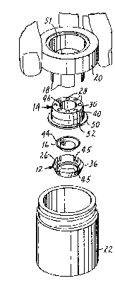
holder of the invention. In FIG. 1 there is shown a
disassembled corneal holder (10) comprising a base member
(12) and a cap member (14), between which a corneal tissue
(16) is shown for illustrative purposes. Holder (10) is
designed so that it can be picked up and heid using the
jaws (18) of jar cover (20), and thereby transferred to
storage jar (22) containing storage medium.
Base (12) has a first aperture (26) and base
attachment means to enable secure attachment with cap (14).
Cap (14) has a second aperture (30) capable of being
i285446
aligned with the first aperture (26), and cap attachment
means to enable secure attachment with base (12).
First aperture (26) and second aperture (30) are
preferably both of substantially the same size, and of a
5 size smaller than that of the tissue to be held but at
least as large as the circumference of the predetermined
portion, e.g., corneal button, to be cut.
Corneal tissue (16) as illustrated in FIG. 1 is
excised from a donor eye by methods known in the art and is
10 substantially circular in shape having a diameter, defined
by its rim (44), that is at least as large as that of the
first and second apertures, but that is generally not
larger than the largest inner diameter defined by base
sidewall (36). Typically tissue (16), if from a human
lS donor, will be on the order of 15-20 mm in diameter,
including a 2 to 3 mm rim of scleral tissue.
Corneal tissue is generally sufficiently rigid so
that it will retain its natural curved shape, e.g., if
placed with its epithelial side down in base (12), and can
20 support its own weight at its pointts) of contact. As a
result, the tissue will not typically fold over on itself
or fall through first aperture (26), but will sit in a
manner analogous to that of a watch glass covering an open
hole.
Tissue (16) as shown, is positioned with its
epithelial surface down over first aperture (26). Cover
(14) is then attached to base (12) in a manner that defines
a compartment that holds tissue (16) and that aligns first
aperture (26) with second aperture (30) so as to define a
30 passageway through the apertures that is configured to
allow a cutting device to be passed through the passageway
and into cutting contact with the circumference of the
predetermined portion of tissue (16).
Tissue (16) is preferably held in its position
in corneal holder (14) by pressure exerted at some point(s)
on tissue (16). The pressure that retains tissue (16) can
be applied at any of a variety of points. For instance,
lX85446
--8--
the inner diameter of base sidewall ~36) might be such,
with respect to the diameter of tissue ~16) that tissue
~16) is held by virtue of its own rigidity and the contact
of its rim (44) against portions of base sidewall ~36).
5 Similarly, tissue ~16) may be retained by the contact of
its rim ~44~ with the underside of cap ~14) such that
tissue ~16) is pressed against first aperture ~26) with
sufficient pressure to hold it in place.
In the embodiment illustrated in FIG. 1, tissue
10 ~16) is held by the presence of constricting means, which
constricting means is formed in part by a cylindrical
protrusion ~46)(seen more clearly in the cross-sectional
view of FIG. 4) extending downward from cap ~14) from a
point at or near second aperture (30). Cylindrical
15 protrusion (46) exerts pressure upon the periphery of
tissue (16), i.e., at points other than those within the
predetermined portion, and causes slight constriction of
tissue (16) between cylindrical protrusion (46) and first
aperture (26) when cap (14) is attached to base (12).
The amount of constriction can be varled, e.g.,
by varying the dimensions of the constricting means, and
will typically be in a range from the maximum that can be
applied to tissue ~16) without causing severe damage
thereto, to the minimum that can be applied while still
25 maintaining contact between tissue (16) and the
constricting means.
~ ase attachment means and cap attachment means
are mutually compatible so as to enable simple and secure
attachment of base (12) and cap (14). Examples of suitable
30 attachment means include pressure-, friction- or
interlocking- type arrangements such as snap-fit and
screw-fit arrangements.
A preferred attachment means is a snap-fit
arrangement wherein the sidewalls of one holder member
35 i.e., the base or the cap, are recessed, grooved, stepped
or otherwise configured to retain an oppositely configured
mating partner, upon the application of a slight engaging
force or motion.
5446
g
Shown i.n FIG. 1 for instance, is a snap-fit
arrangement in which base sidewall (36) extends upwardly
and radially outward at the periphery of base (12) on an
axis that is at an angle of approximately 80 with the
5 plane of the bottom of base (12). The inner major surface
of base sidewall (36) is substantially flat, and the outer
major surface is angled first radially outward at an angle
of approximately 67.5 from the plane of the bottom of base
(12) to a distance of approximately one-half the height of
10 the inner surface, and then radially inward to provide a
rounded lead-in guide for the inner major surface of cap
sidewall (40).
Cap sidewall (40) extends downwardly from the top
of cap (14) and has an inner major surface that contains a
15 circumferential groove (41) (seen more clearly in the
cross-sectional view of FIG. 4) of a dimension capable of
receiving and retaining the outermost portion of base
sidewall (36).
Preferably base sidewall (36) has a plurality of
20 radially located compression grooves (45), to enable base
sidewall (36) to be slightly compressed radially inward
during engagement with cap (14) and to expand again to its
~ normal shape once within the cap, thereby locking base
: sidewall (36) into cover sidewall (40).
~: 25 In the embodiment illustrated in FIG. 1, holder
and jar cover engagement means are also provided, in order
to allow jar cover (20) to pick up and transfer holder
: (10), e.g., to storage jar (22). These enqagement means
can be any means that allow holder (10) to be picked up in
;~ 30 a simple, rapid, releasable and aseptic manner. Suitable
engagement means include pressure-, friction- or
inter-locking type arrangements.
: Shown in FIG. 1 is a simple pressure-type
:~ arrangement wherein jar cover (20) is equipped with holder
engagement means in the form of a plurality of radially
located opposing resilient jaws (18) extending downwardly
from jar cover (20). As the jar cover engagement means,
the outer major surface of cap sidewall (40) is tapercd
, .
.
:
~285446
--10--
radially inward as it approaches the top of cap (14) in
order to provide a lead-in for grasping by jaws (18) of jar
cover (20). Jaws (18) are expanded slightly during
engagement with holder (10), in order to snugly hold holder
5 (10) therein.
Holder (10) preferably also includes holder
release means for removing holder (10) from secure
engagement with jar cover (20). As shown in FIG. 1 for
instance, cap (14) is provided with a flange (50) extending
10 outwardly from the bottom of cap sidewall (40). Flange
(50) can be held or secured, e.g., by the tip of a finger
or instrument, while jar cover (20) is held and pulled away
from holder (10), thereby releasing holder (10) from the
grasp of jaws (18).
Flange (50) preferably performs an additional
function in that it acts as a barrier to stop the downward
movement of jaws (18). Flange (50) also helps to insure
complete attachment of cap (14) with base (12) by virture
of the downward pressure of jaws (18) on flange (50),
20 thereby forcing cap (14) down until flange (50) rests
uniformly on a preferably flat work-station surface (not
shown).
Holder (10) preferably also includes orientation
means, e.g., notch (52) in flange (50) that enables tissue
25 (16) to be placed in holder (10) in a known radial
orientation.
Also associated with the holder shown in the
embodiment illustrated in FIG. 1 are port means, shown as a
plurality of generally circular and equally spaced holes
30 (28) through the top of cap (14), to allow fluid such as
storage medium to have continuous access into the
compartment defined by base (12) and cap (14) in order to
bathe the edges of the tissue held therein.
Base (12) and cap (14) can be made out of any
35 suitable material that is sufficiently strong, inert,
resilient and compatible for such use. Preferred materials
are plastics such as polypropylene. soth members can be
~28S446
--11--
manufactured, e.g., as one-part injection molded pieces, by
methods well known in the pertinent art.
As shown in FIG. 1, jar cover (20) includes an
optically clear viewing port (51). ~iewing port (51) is
5 indented into jar cover (20) in order to allow visual
inspection of the tissue, e.g., as described in "Wide Field
Specular Microscopy of Excised Donor Corneas", C.W. Roberts
et al., Arch. Ophthalmol. 99:881-883 (1981), and
"Examination and Photography of Donor Corneal Endothelium,
10 W.M. Bourne, Arch. Ophthalmol. 94:1799-1800 (1976). In
addition to the holder engagement means described earlier,
jar cover (20) also preferably includes means for
releasably securing jar cover (20) to storage jar (22),
preferably a screw-type or snap-fit arrangement wherein ja.
15 cover (20) is screwed or snapped onto storage jar (22).
Jar cover (20) and storage jar (22) can be made
by methods known in the art of any of a variety of
materials known for such uses. Typically, jar cover (20)
will be molded plastic, having optically clear plastic or
20 glass as viewing port (51) and resilient, e.g., plastic,
jaws. Jar (22) will typically be made of a clear or
translucent glass or plastic material, by manufacturing
techniques well known in the art.
The following procedure is typically performed in
25 order to use the corneal storage system shown in FIG. 1.
Under asceptic conditions, a cornea is excised
from a donor eye to provide tissue (16), which is
transferred by forceps to rest in a known radial
orientation with respect to notch (52), epithelial side
30 down, within base (12). Cap member (14) is picked up and
oriented above base (12) so as to approximately align first
aperture (26) and second aperture (30). Cap (14) and base
(12) are then securely attached, e.g., by a snap-fit
motion, so that tissue (16) is held between the two, such
35 that both major surfaces of the predetermined portion from
which the corneal implant is to be cut are exposed through
the first and second apertures, and such that there is
~'~85446
slight pressure constricting tissue (16) between
cylindrical protrusion (46) and base (12) at points
peripheral to the predetermined portion. soth major
surfaces of the predetermined portion, as well as the rim
5 of the tissue and the surfaces peripheral to the points of
constriction, are thereby accessible to storage medium when
placed in storage jar (22). Jar cover (20) is then grasped
and brought into a position whereby jaws (18) are brought
down onto holder (10) until jaws (18) are stopped by flange
(50), thereby releasably securing holder (10) to jar cover
(20).
The resultant jar cover-holder assembly is
transferred to storage jar (22), and jar cover (20) is
screwed onto storage jar (22). Storage jar (22) preferably
15 contains a sufficient amount of storage medium to
completely submerge tissue (16) when storage jar (22) is
stored in its usual inverted position.
Preferably fluid of the viscosity of storaye
media will be able to contact both major surfaces of tissue
20 (1~) i.e., through base aperture (26) and cover aperture
(30), and will also be able to contact the edges of tissue
(16), while holder (10) is held within storage jar (22).
Turning now to FIG. 2, there is shown a partially
exploded perspective view of one embodiment of a corneal
25 cutting system for use with the holder of the present
invention. Shown is: assembled holder (10) containing
tissue (16); base means, e.g., trephine block (62); and a
blade assembly comprising a cutting device, e.g., trephine
blade (66), and a blade holder (68).
In order to use the illustrated corneal cutting
system, holder (lO) is first placed such that flange (50)
rests in a central location on block (62). Block (62) has
a raised rim (80) that defines an inner diameter on the
surface of block (62) that is slightly larger than the
35 diameter of flange (50), such that flange (50) fits within
the raised rim, in a substantially radially immovable
position.
~.~85446
-13-
As shown in FIG. 2, the inner surface of rim (80)
has an oppositely configured protrusion (81) to mate with
notch (52) such that holder (10) can be placed on block
(62) in an orientated and secure position. The surface of
5 block (62) that faces holder (10) can have additional
features, e.g., it can be concentrically grooved or
otherwise formed, in order to provide any desired contour
for use as a cutting surface.
When holder (10) sits properly on block (62),
10 first aperture (26) is properly aligned with a
semispherical indentation (70) in the center of block (62)
that preferably has a diameter at least as large as the
predetermined portion to be cut and a radius of curvature
substantially the same as tissue (16).
Indentation (70) can be of any desired diameter
or configuration, e.g., semicircular, angular or the like.
See, e.g., "A New Punch for Corneal Transplantation", D. M.
Lieberman, Amer. J. Ophthalmol. 83:419-420 (1977), wherein
it is described that the angle and cleanness of the edges
20 of a button cut out of a corneal tissue appears to be
dependent in part on the diameter and/or configuration of
the indentation in the trephining block.
A blade assembly is then set into the top of
holder (10). The blade assembly shown in FIG. 2 has a
25 blade holder (68) and, as the cutting device, blade (66).
slade holder (68) is typically a one-part molded piece of a
sufficiently strong, inert, resilient and compatible
material such as the materials used to make holder (10),
having a cylindrical sleeve (72~ and an outwardly extending
30 sleeve flange (74). Blade holder (68) preferably has one
or more expansion grooves (76) through sleeve (72) and
sleeve flange (74) that allows blade holder (68) to be
radially expanded an amount sufficient to accept and retain
blade (66) therein. Expansion groove (76) can be used to
35 radially orient the predetermined portion of tissue as
well, by directing it towards the holder orientation means
during cutting. Llade holder (68) has an inner diameter
:
.
128544~;
-19-
and resilience capable of then snugly retaining blade (66).
When blade (66) is held in blade holder (68)
sleeve (72) defines an outer diameter slightly smaller than
the inner diameter of cylindrical protrusion t46~ of cap
(14), such that sleeve ~72) can be inserted within
cylindrical protrusion (46) in a mating relationship
whereby sleeve (72) is retained in a substantially radially
immovable position.
Sleeve (72) has a length substantially the same
10 as that of cylindrical protrusion (46). Although shown
exposed below sleeve (72), blade (66) is initially oriented
in sleeve (72) such that cutting edge (78) does not
protrude below sleeve (72).
The cutting device will typically be a
15 cylindrical metal trephine blade having a cutting edge that
defines the desired diameter of a corneal button, and will
typically be used to cut entirely through the circumference
of the predetermined portion, although variations, e.g., on
the size, dimensions and depth of the cut, are well known
20 in the pertinent art.
Sleeve flange (74) provides an easy means for
grasping blade holder (68) and is configured to rest on the
top surface of holder (10) in order to place sleeve (72)
and blade (66) within cylindrical protrusion (46).
25 Cylindrical protrusion (46) therefore serves as a guide for
sleeve (72) and, in turn, for blade (66), in order to allow
blade (66) to be brought into proper cutting contact
with the predetermined portion of tissue (16).
The corneal cutting system is then placed in an
30 appropriate corneal punch mechanism (not shown) that forces
blade (66) down through sleeve (72) such that the cutting
edge of blade (66) proceeds into and through tissue (16)
with a motion and force sufficient to cleanly cut a corneal
button having the diameter of the cutting edge of blade
35 (66).
The corneal button frequently remains within
blade (66) until a drop of sterile fluid, e.g., storage
,
. .
1285446
-15-
medium is placed from above into the inner circumference of
blade (66). The weight of the fluid is generally
sufficient to cause the button to gently drop from blade
(66), e.g., onto block (62).
The dimensions of corneal holder (10) may be
varied in a manner commensurate with the size of tissue to
be held, e.g., to accomodate tissue from different animal
sources.