Note: Descriptions are shown in the official language in which they were submitted.
1~?.9~'79~
20365-2~10
BACK&ROUND OF THE INVENTION
The present invention relates to a lithotripsy work
station which may be employed, for example, for disintegrating
kidney stones and other types of calculi.
In many conventional stations for externally applying
shock waves to a patient to disintegrate calculi, the patient
must be placed in a tub filled with water which acts as a
medium via which shock waves from a shock wave generator are
transmitted to the calculi for disintegration thereof.
It is an object of the present invention to provide a
universal lithotripsy work station which permits the following
operations to be undertaken:
~ ~k
-- 1 --
~91~9~
(1) Shock wave treatment, particularly
disintegration of renal and ureteral calculi.
(2) Urological treatments including percutaneous
nephrostomy.
(3) X-ray examinations.
The above objects are achieved in accordance with the
principles o~ the present invention in a lithotripsy work station
which includes a patient supporting table which is adjustable in
three directions, with at least one shock wave generator for
renal calculi disintegration being arranged beneath the table~
The shock wave generator can be coupled to the patient's skin via
a membrane. The shock wave generator is adjustably mounted so
that movement of the focus thereof to an isocenter is possible.
For locating renal calculi, an x-ray examination
apparatus is provided which includes an x-ray unit for
transilluminating at various angles. The x-ray apparatus has two
x-radiators, each of which emits a central ray. The plane
defined by the two central rays passes through the longitudinal
axis of the patient supporting table, with the radiators disposed
such that one radiator emits radiation in an anterior-posterior
A (a.p.)-direction~ and the second ràdiator emits radiation in the
~ c~ c.~
caudal-cranial/direction. The patient supporting table includes
means for accomodating attachment of auxiliary urological
equipment and for attaohment of an x-ray exposure means.
In one embodiment of the invention, the radiators of the
x-ray apparatus are both mounted in a common housing which is
adjustable in a vertical plane. In this embodiment, it is
possible to displace the radiators upwardly in the examination
room when they are not required for locating calculi. The
radiators can be displaced upwardly to such a degree that they do
- 2 -
~17~ ~03~5-2610
not :impede the physician in any activities which he or she may
undèxtake, particularly for percutaneous operations. The
hous:ing may include a universal connector for optional
connection to the ceiling or wall of the examination room, or
to a vertical post.
The invention may be summarized as a lithotripsy work
station comprising:
a patient support table having a longitudinal axis
for selectively positioning a patient thereon with respect to a
fixed point in space;
at least one shock wave generator for disintegrating
calculi disposed beneath said patient support table, said shock
wave generator having a membrane for coupling said shock wave
generator to the skin of said patient, and said patient support
table having an opening therein through which said shock wave
generator can at least partially extend;
means for mounting said shock wave generator for
permitting adjustment of the focus of said shock wave generator
to said fixed point in space; and an x-ray examination means
for transilluminating said patient at selected angles for
locating calculi, said x-ray examination means including two
x-ray radiators disposed above said patient support table and
each having a central ray, a central ray of one of said
radiators being emitted in a direction perpendicular to said
patient support table and the central ray of the other radiator
being emitted in an oblique direction with respect to said
patient support table, with both of said central rays
proceeding in a common plane which includes said longitudinal
axis of said patient support table and intersecting in said
fixed point in space.
-- 3 --
1~`.9~7~ 20365-2610
DESCRIPTION OF THE DRAWINGS
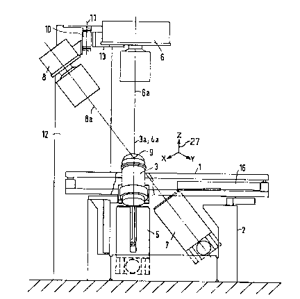
FIGURE l is a side elevational view of a lithotripsy
worX station constructed in accordance with the principles of
the present invention.
FIGURE 2 is an end elevational view of the
lithotripsy work station shown in FIGURE l.
FIGURE 3 is a side elevational view of a lithotripsy
work station constructed in accordance with the principles of
the present invention with a modified positioning of the x-ray
radiator.
FIGURE 4 is an end view of the lithotripsy work
station shown in FIGURE 3.
FIGURE 5 is a side view of another embodiment of a
lithotripsy work station constructed in accordance with the
principles of the present invention.
FIGURE 6 is an end view of the lithotripsy work
station shown in Figure 5.
FIGURE 7 is a side view of a lithotripsy work station
constructed in accordance with the principles of the present
invention with another modification of the x-radiator
mounting.
FIGURE 8 i9 an end view of the lithotripsy work
station shown in Figure 7.
DESCRIPTION OF THE PREFERRED EMBODIMENTS
A lithotripsy work station constructed in accordance
with the principles of the present invention in a first
embodiment is shown in Figures l and 2. The station includes a
patient supporting table l adjustable along three perpendicular
axes schematically indicated by the coordinate system 27, in a
-- 4 --
.
20365-2610
manner known to ~hose skilled in the art as s~own, for example,
in U.S. Patent No. 3,302,022. The table 1 is disposed on a
base 2 and beneath which two shock wave generators 3 and 4 are
arranged. The shock wave generators 3 and 4 can best be seen
in Figure 2; and Figure 1 the shock wave generator 4 is covered
by an x-ray image intensifier 5. Together with an x-ray
radiator 6, the x-ray image intensifier 5 forms a first x-ray
system, the x-ray radiator 6 emitting radiation perpendicular
to the patient supporting table 1 in an a.p.-direction.
A second x-ray system is formed by another x-ray
image intensifier 7 and another x-ray radiator 8. The x-ray
radiator 8 emits radiation in the caudial-cranial direction.
As a result of the different directions of the
central rays 6a and 8a of the respective x-ray systems, renal
calculi to be disintegrated can be located. The central rays
6a to 8a intersect at an isocenter 9, (i.e. at a fixed point in
space) to which the renal calculus to be disintegrated is
displaced by appropriate movement of the patient supporting
table 1, with a patient thereon.
'rhe plane defined by the two central rays 6a and 8a
proceeds through the longitudinal axis of the patient
supporting table 1, so that the lateral space requirement for
the x-ray units is small.
'rhe x-ray radiator 8 is secured to an arm 10 which is
mounted to a post 12 so as to be pivotable around a vertical
axis 11. It is thus possible to pivot the x-ray radiator 8 out
of the work area, as needed.
-- 5 --
~ 2~3~5-2~10
The x-ray radiator 6 is also connected to the post
12, by means of an arm 13 which permits the x-ray radiator ~ to
~e displaced at right angles relative to the longitudinal axis
of the patient supporting table 1. The x-ray radiator 6 thus
can also be moved out of the work area, as needed.
The shock wave generators 3 and 4 are mounted on a
common support 15, best seen in Figure 2, which is pivotable
around an axis 14 extending parallel to the longitudinal axis
of the table~ The shock wave generators 3 and 4 are mounted on
the support 15 such that their respective axes 3a and 4a
intersect at an angle. Each shock wave generator 3 and 4 is
adjustable in the direction of its respective axis 3a and 4a.
For shock-wave treatment of a patient, the patient is
seated on the patient supporting table 1 and the calculus to be
disintegrated is moved into the isocenter 9 under x-ray
supervision. Subsequently the axis 3a or 4a of one of the
shock wave generators 3 or 4 is directed to the isocenter, and
that shock wave generator i9 moved toward the patient through
an opening in the patient supporting table 1 until a membrane
of the shock wave generator presses against the skin of the
patient. The shock wave treatment can subsequently ensue.
For the introduction of an x-ray film cassette, the
support 15 can be used to move the shock wave generators 3 and
4 to the position shown in Figure 4, permitting an x-ray film
cassette 16 to be adjusted to an exposure position shown in
Figure 3. In Figure 1, the x-ray cassette 16 is shown in a
preparatory or standby position, in which the space under the
calculus to be distintegrated is maintained free for
application of one of the shock generators 3 or 4. The shock
wave generators 3 and 4 are respectively allocated to the two
kidneys.
- 5a -
,~
The patient supporting table 1 has means for attaching
urological auxiliary equipment, for example containers, so that
standard urological routines can be undertaken. The lithotripsy
work station disclosed herein can be used as a standard x-ray
work station for transillumination and exposure.
It is also possible to omit the pivoting capability of
the support lS if the support 15 is arranged such that the
longitudinal axes of the two shock wave generators 3 and 4
intersect at the isocenter 9.
Instead of two x-ray systems for transilluminating the
patient at different angles, only one such system, having one or
~wo image intensifiers, may be used if mounted in an
appropriately adjustable manner. It is also possible to provide
a single stationary x-ray system including an image intensifier
if a stereo x-ray tube is employed.
Coupling of the patient to the shock wave generators 3
and 4 via a water-filled tub is not required as a result of the
use of shoclc wave generators 3 and 4 designed as individual units
to be applied directly to the surface of the patient.
In the embodiment shown in Figures ~ and 6, components
already described in connection with Figures 1 through 4 are
identified with the same reference symbols. In this embodiment,
the patient supporting table la is somewhat shorter than the
patient supporting table 1 shown in the previous embodiments.
The shock wave generators 3 and 4 are adjustable only along the
directions of the two axes 3a and 4a, which intersect at the
isocenter 9. Instead, the base 2a occupies a slightly greater
width than the previously-described base 2.
~917~1
Also in the embodiment of Figures 5 and 6, the x-ray
radiators 6 and 8 are mounted in a common housing 20, which is
adjustable in a vertical plane. ~or this purpose, the x-ray
radiator 6 is adjustable in the direction of the central ray 21,
wherea~ the x-ray radiator 8 is secured to an arm 22 which is
pivotable about an axis 23~ The respective positions of the x-
ray radiators 6 and 8 within the housing 20, when they are not
being use~, is indicated with broken lines in Figure 5. The
housing 20 is provided with a universal connector 24 which may,
for example, be secured to the ceiling 25 of the examination
room.
For locating calculi, the radiators 6 and 8 assume the
positions shown with solid lines in Figures ~ and 6. When they
are no longer required, the radiators are moved into the housing
20 and do not interfere with further treatment, for example
during percutaneous operations ~percutaneous nephrostomy), so
that free access to the patient is insured.
Another modification for mounting the x-ray radiators is
shown in Figures 7 and 8. In this embodiment, the universal
connector 24 is secured to an arm 26 which is connected to a
column or post 12a, which is inturn supported on the floor of the
examination room. Alternatively, the arm 26 may be secured to a
vertical wall of the examination room.
Although modifications and changes may be suggested by
those s~illed in the art it is the intention of the inventors to
embody withln the patent warranted hereon all changes and
modifications as reasonably and properly come within the scope of
their contribution to the art.
