Note: Descriptions are shown in the official language in which they were submitted.
BAC KGROUN D OF TH E I NV ENT I ON
2 The present invention relates to devices useful for the
3 detection of air embolisms in fluid lines. More particularly,
4 the present invention relates to an untrasonic device which is
engageable with a flexible tube to determine when air, rather
6 than fluid, is flowing through the tube. The present invention
7 is particularly, but not exclusively, useful in the health care
field for detecting air-in-line conditions in an IV tube
9 through which medical solutions are being administered.
DESCRIPTION OF THE PRIOR ART
12 Intravenous (IV) drug delivery systems have been used in
1:~ numerou~ meuicai procedures for many years. lheir efficacy is
14 widely accepted. Always, however, whenever medical solutions
are administered intravenously there is the danger that a large
16 bolus of air may also be inadvertently administered. The
17 potentially fatal consequences of such an accident are well
1~3 known.
19 As might be expected, whenever a mechanical device is used
in an IV administration system to assist the infusion of
~1 medical solutions to a patient, the possibility of an air leak
22 or some other inadvertent introduction of air into the IV line
.1 is increased. Accordingly, great effort is taken in the design
2~ o~ IV administration systems to insure that the possibility of
such an accident i9 made extremely unlikely. 'rhus, air-in-line
\(,
~ -2-
~z9~
l detectors are of great importance to the safe operation of a
2 medical infusion device.
~3 The mere incorporation of an air-in-line detector into the
4 system of a medical infusion device is not sufficient by
itself. For any medical device to operate efficiently there
6 must be some means for making periodic safety checks of the
7 system. This is particularly important where air-in-line
8 detectors are concerned. Operation of the system when there is
9 air in the fluid line can, as previously mentioned, have
catastrophic consequences. Accordingly, it is desirable that
11 an air-in-line detector be able to periodically test itself to
12 ensure its proper operation.
1~ S~eL~i devi~es hav~ beel1 proposed for air-in-iine
14 detectors. Typically such devices have employed either an
optical system which depends on the light transmissive
16 characteristics of fluids or an ultrasonic system which depends
17 on the ultrasonic energy transmissive characteristic of
I8 fluids~ The present invention is concerned with ultrasonic
19 detection of air embolisms in an IV fluid infusion tube.
A major advantage of using ultrasonic technology for the
21 detection of air in a fluid line is the inconsequential effect
22 of the fluid's opacity. Unlike opt1cal systems which are
23 greatly affected by fluid opacity, ultrasonic systems can
24 substantially ignore this variable. On the other hand, unlike
optical systems which are operative merely by properly aligning
26 the system's elements relative to the fluid tube, an ultrasonic
:: :
~ -3-
;~ ~
1 system, in addition to proper alignment of the elements, must
2 also establish proper impedence matching. Essentially, this
3 means that a direct ultrasonlc energy path must be created
4 through the fluid tube between the transmitter and receiver.
Such a path requires effective contact between all aligned
6 elements in the system. Further, in order to obtain valid
7 indications of the air-in-line condition, alternate paths which
8 are able to transmit ultrasonic energy between the transmitter
9 and receiver and around the fluid tube m~st somehow be
obstructed.
Il Various devices have been proposed for ultrasonic air-in-
12 line devices which address these problems. For example, in an
1~ attemp~ to achieve proper impedence matcning, the device
14 disclosed in U.S. Patent No. 4,418,565 to St. John incorporates
an elastomeric material positioned between each transducer and
16 the tube to make direct contact therebetween. U.S. Patent No.
17 3,97g,681 to Nanery incorporates sound pipes between the
1~ transducer and tube for this same purpose. Further, since the
19 St. John patent discloses a single base for supporting both the
~ transmitter and the receiver, it also discloses a slot formed
~21 ~ therebetween to hinder the propagation of ultrasonic energy by
22 a route other than through the elastomer members which are
23 positioned between the receiver and the transmitter.
As an advancement from the prior disclosed devices, the
~ present invention recognizes that enhanced impedence matching
26 can be obtained by direct contact between the ultrasonic
,~
-4-
~:
1~
l transducers and the fluid tube. Thus, the need for
2 intermediate structure such as an elastomeric material or sound
3 pipes is obviated. More particularly, the present invention
4 recogni~es that an ultrasonic transqucer having a convex
S surface which is in direct contact with the fluid tube can be
6 positioned to cause a slight indentation of the tube. This
7 indentation accomplishes a dual purpose. First, it provides
8 good coupling at the flushed interface between transducer and
9 fluid tube which is necessary for proper operation of an
ultrasonic system. Second, the indentation results from an
11 interface fit between the tube and the transducer which holds
12 the tube in a fixed position relative to the ultrasonic
13 ~crar,sd~c~rs.
14 Accordingly, it is an object of the present invention to
provide an ultrasonic air-in-line detector for use with a
16 flexible flui~ tube which provides good ultrasonic coupling by
17 positioning the ultrasonic transducers, both transmitter and
18 receiver, into direct contact with the fluid tube. It is
19 another object of the present invention to provide a housing
for the ultrasonic transducers which will create a direct
21 ~ ultrasonic path through the fluid tube and hinder the
2 ~propagation of ultrasonic energy by other routes. Still
23 another object of the present invention is to provide self
~ :
24 testing electronic circuitry which will monitor the operation
of the air-in-line detector and ~de~ermine when an inoperative
26~ ~ condition exists. Yet another object of the present invention
; -5-
.. . ,- - :
~,
~Z~ 92
l is to provide a cost effective ultrasonic air-in-line detector
2 which is easily manufactured and easy to use.
4 SUMMARY OF THE INVENTION
The preferred embodiment of the ultrasonic air-in-line
6 detector for the present invention used to detect air in a
7 flexible fluid tube comprises a generally U-shaped base which
8 forms a cavity between the branches of the U. An ultrasonic
9 transmitter is mounted on one branch of the U with its
assoeiated convex lens protrudiny into the eavity. Likewise,
11 an ultrasonie receiver is mounted on the opposing branch of the
12 U with its assoeiated convex lens protruding into the cavity.
1~ ~ pedes~al a~ach~d at the base of trie u disO protLudes in~o
14 the cavity. The pedestal helps stablize the fluid tube when
the tube is placed into the cavity and held between the lenses
,,
16 o'f the ultrasonic receiver and ultrasonic transmitter. With
17 the tube so positioned in the cavity, a second pedestal can be
1$ moved into contaet with the tube at a point which is
19 substantially diametrically opposite from the point at which
the first pedes~al eontacts the tube. Thus, the tube is
21 stationarily held in the eavity between the pedestals and
22 between the convex lenses of the ultrasonie transducer. The
23 lenses con~act the tube at substantially right angles to the
24 line on whieh the pedestals contaet the tube.
The ultrasonie air-in line deteetor of the present
26 invention mây also inelude eleetronie eireuitry whieh is self-
`; ~
-6-
:.,,, , ". , ~
':'' ' ' '
~2~
l testing. This self-test is accomplished by periodically
2 stopping the input to the ultrasonic transmitter and
3 recognizing that a transmitter-off condition is substantially
4 equivalent to an air-in-line condition. Accordingly, when the
receiver cannot distinguish between a transmitter-on condition
6 and a transmitter-off condition, either there is an air-in-line
7 condition or the circuitry of the system is faulty.
8 The novel features of this invention as well as the
9 invention itself both as to its organization and operation will
be best understood from the accompanying drawings taken in
11 conjunction with the accompanying description in which similar
12 reference characters refer to similar parts and in which:
13
14 BRIEF DESCRIPTIO~ OF THE DRAWINGS
_ _ .
Figure l is a front elevational view of the present
16 invention in operative association with other elements of an IV
i7 infusion system;
18 Figure 2 is a perspective view of the detector of the
19 present invention;
Figure 3 is a cross-sectional view of the detector as seen
,21 along the line 3-3 in Figure 2;
22 Figure 4 is a cross-sectional view of the detector as seen
23 in Figure 3 with a fluid tube mounted thereon and restrained
;~ 24 therein;
26
-7-
, . .
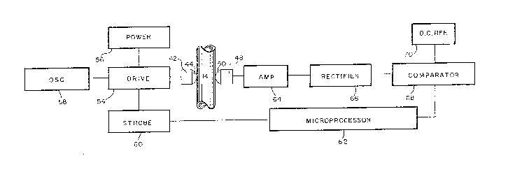
1Figure 5 is a block diagram of the electronic componentry
~used for the present invention; and
3Figures 6~, B and C are graphs of the outputs of selected
electronic components of the present invention.
6DESCRIPTION OF THE PREFERRED EMBODIMENT
_ _
7Referring initially to Figure 1 the air-in-line detector
8 of the present invention, generally designated 10, is shown in
9 operative association with an intravenous (IV) administration
system. Specifically, the detector 10 is shown mounted on a
Il medical device 12. While the detector 10 of the present
12 invention, may be used with~ any IV infusion device, it is
i5 ~aL tiCUia~li w-èil S~ ed f~r ~peratioll wi~h a ue~i~e SuCi~ dS
14the one disclosed in U.S. Patent No. 4,617,014 to Cannon et
al. As shown in Figure 1, such a device 12 is operatively
16 connected to cooperate with an IV tube 14 for the purpose of
17 pumping or controlling the flow of fluid through the tube 14.
18 Typically, in medical procedures, a fluid source 16 is
19 connected in fluid communication with tube 14 and hung from an
IV pole 18. The system is then assembled for the infusion of
21 medical solutions from source 16 to a pati.ent 20.
22~Reference to Figure 2 shows that detector 10 has a
23substantially U-shaped base 22 with two oppositely extending
24branches 24, 26. A cavlty 28 is formed between branches 24, 26
and a pedestal 30, which is attached to base 22 at the bottom
, ~
,~26 : of the U, protrudes into cavity 28. As will be readily
:~'.
: -8-
:
.~ ' ~ ` ' ' ' ' '
. . . ~
`\
~2~
1 appreciated by the slcilled artesan, base 22 with branches 24,
2 26 and pedestal 30 can be of unitary construction and
3 manufactured by processes well known in the pertinent art, such
4 as by injection molding. Figure 2 also shows a door 32 which
is associated with base 22 by means such as the hinge 34. It
6 is to be understood that door 32 need not be directly hinged
7 onto base 22. Instead, and preferably, door 32 may be hingedly
8 attached to device 12. In either case, it is important that
9 the pedestal 36, which is mounted on door 32, be moveable into
a position wherein pedestal 36 protrudes into the cavity 28
11 between b~anches 24, 26. It will be appreciated by the skilled
12 artesan that door 32 and pedestal 36 can be of unitary
3 -onstLuc~Lc.. a..d ma..ufac~ured by a ~rocess s~c" as injec~ ol.
14 molding.
15 Figure 3 is a cross-sectioned view of detector 10 which
16 shows the interaction of its components. As shown, branch 24
of detector 10 is formed with a housing 38. Likewise, branch
26 is formed with a housing 40. A piezo-electric crystal 42 is
; mounted in housing 38 and an acoustic lens 44, which is
preferably made of an epoxy material, is attached to crystal 42
1 by any appropriate means, such as by an epoxy adhesive well
2 known in the pertinent art. Also, for purposes of the present
23 invention, crystal 42 can be made of any well known piezo
2~ ceramic material. Further, in the preferred embodiment, lens
44 is formed into a spherical convex shape with a relative
26 curvature substantiall~ as indicated in Figure 3. Wiring 46 is
~,
~ : _g_
'`~ ~''' ` ` '''
~ \
~ 9~:
l provided to electrically connect piezo-electric crystal 42
2 with appropriate electronic components. For purposes of
3 further discussion, the combination of crystal 42 and lens 44
4 will hereafter be sometimes generally referred to collectively
as the ultrasonic transmitter.
6 An ultrasonic receiver is constructed in a manner similar
7 to the construction of the transmitter and will include a
8 piezo-electric crystal 48 which is mounted in the housing 40 of
9 branch 26. An acoustic lens 50 is attached to crystal 48 and
wiring 52 is provided to electrically connect crystal 48 with
11 appropriate electronic componentry. Like the components of the
12 transmitter, the receiver components are made of materials well
1~ krlOWn ii- t~ in~rlt aL t. Specific~iiy, cry~tal 4~ ade
14 of a piezo-ceramic and the spherically shaped convex lens 50 is
made of an epoxy material which is attached to crystal 48 by an
16 epoxy adhesive.
As an alternative to the epoxy material used for acoustic
i8 lenses 44 and 50, a polycarbonate material may be used. This
19 could lead to a design in which the lenses 44 and 50 are
integrally molded into the base 22. With this configuration
21 piezo-electric crystals 42 and 48 can be respectively epoxy
22 bonded in a manner well known in the relevant art, to ]enses 44
'3 and 50.
!4 The operative placement of tube 14 into detector 10 is
!5 best seen in Figure 4 where it can be appreciated that tube 14
~6 is positioned in cavity 28 with lens 44 substantially
-10-
.. , . ~ ., . ,, ,~, ~ , .. . .
1 diametrically opposed to lens 50. Also, tube 14 is positioned
2 to remain in contact with pedestal 30 when door 32 is moved
into position to bring pedestal 36 into contact with tube 14.
4 As shown in Figure 4, lens 44 can be prepositioned relative to
lens 50 to pinch tube 1~ therebetween when the tube 14 is
6 placed into ca~ity 28. This pinching action causes
7 indentations of tube 14 at the respective interfaces of tube 14
8 with lens 44 and 50 to establish good acoustic coupling for the
9 detector 10. Also, this cooperation of structure provides an
interference fit between the tube 14 and detector 10 which will
Il help hold tube 14 on detector 10.
12 The acoustical coupling of lenses 44 and 50 with tube 14
1~ is ~llh~iice~ by the dction or peaestais 30 and 36 on tuDe 14.
14 As seen in Figure 4, when pedestals 30 and 36 both make contact
with tube 14 they, like lenses 44 and 50, tend to pinch tube
16 14. This action causes a slight deformation of tube 14 which
17 urges tube 14 into more immediate contact with lenses 44 and
18 50. As will be appreciated by the skilled artesan, there are
19 competing concerns at play in this cooperation. On the one
hand, there is the desire to create the maximum beneficial
21 acoustic coupling attainable for detector 10. This re~uires
: :
~; ~ 22 intimate contact of lenses 44 and 50 with tube 14. On the
~ 23 other hand, there is the desire to minimize distortion of tube
:; 24~ 14. Thls means that the pinching action of detector 10 on tube
14 must be limited to some extent. The balance of these
26 ~apparently competing interests is best attained by a proper
: ~
:
~ æ
l geometrical arrangement oE the components of detector 10 needed
2 to accomplish its intended operational specifications.
3 The preferred electronic componentry necessary for
4 activation of detector 10 is schematically shown in Figure 5.
It will be understood by the skilled artesan that all
6 electronic components disclosed for this system are well known
7 in the pertinent art and are commercially available. As shown
8 in Figure 5, tube 14 is placed in operative engagement with
9 piezo-electric crystals 42, 48 through the mechanical coupling
of lenses 44, 50 with tube 14 in a manner previously
Il discussed. From the block diagram drawing of Figure 5, it can
l2 be seen that piezo-electric crystal 42 serves as an ultrasonic
1~ transmi~t~r ~ gen~rates uitrasoulld eneryy according ~o
14 input received from drive 54. The output from drive 54, which
is input for crystal 42, is a step signal generated by the
16 interconnection at drive 54 of power source 56 with oscillator
17 58 and strobe 60. Specifically, power source 56 supplies
18 electrical power for the system while oscillator 58 causes
19 drive 54 to generate a sinusoidal output at the resonant
frequency of crystal 42. Simultaneously, strobe 60 causes
2l drive 54 to turn ON or OFF at predetermined intervals. The
22 result is a step input to crystal 42 that alternates between an
23 OFF condition wherein there is no excitation of crystal 42 and
24 an ON condition wherein crystal 42 is excited at its resonant
~re~uency to generate ultrasound energy. Preferably, strobe 60
i :~
~ ~ 26 is operated by microprocessor 62 to cause switching between the
'
~ -12-
, . "=, . . . . .
1 ON and OFF condition approximately every nin~ milliseconds.
2 Thus, drive 54 generates a stepped output having an eighteen
3 millisecond cycle.
4 On the receiver side of detector 10, piezo-electric
crystal 48 is mechanically coupled with tube 14 through lens 50
6 to receive signals eminating from crystal 42, In the
7 electrical circuitry of detector 10, piezo-electric crystal 48
8 is electrically connected to an amplifier 64 and the output
9 from amplifier 64 is fed directly to filter/rectifier 66. At
filter/rectifier 66, this output is substantially changed from
Il a sinusoidal signal to an amplitude modulated signal. The
12 comparator 68 then takes the output from filter/rectifier 66
and ~o.l,par~ it ~ a d.c. L~fer~nce voltage from reîeLence 7u
l4 to establish a digital output from comparator 68 which is
lS passed to microprocessor 62.
16 Microprocessor 62 analyzes the digital output from
comparator 68 to determine whether the device 12 is safely
3 operating without air in tube 14. This determination is made
~9 according to an algorithm which accounts for the rate of fluid
flow through tube 14 in its analysis in order to ignore very
21 small air bubbles, i.e. bubbles of less than approximately
2Z fifty microliters, which are of no real medical concern. Also,
23 microprocessor~62 provides input to strobe 60 to regulate its
operation. With these connections, microprocessor 62 is able
to analyze the output of detector 10 coming from comparator 68
.,
26
, ~ ~ ~
-13-
~ ~6~I92
l in relation with the input to detector 10 beginning at strobe
2 60.
4 OPERATION
S In its operation detector 10 is activated by power from
6 source 56. IV tube 14 is operatively associated with detector
7 10 when it is inserted into cavity 28. This insertion brings.
8 convex-shaped acoustic lenses 44 and 50 into direct mechanical
9 coupling with tube 14 and generates an interference fit
therebetween which helps hold tube 14 in cavity 28. Door 32 is
11 then closed onto base 22 to bring pedestal 36 into contact with
12 tube 14. This action provides a pinching engagement of tube 14
~etw2en t.c dlamet~ a.iy opposed pedestals 3û and ~ in
14 addition to the pinching engagement of tube 14 between the
diametrically opposed lenses 44 and 50.
16 Operation of device 12 is intended to cause fluid flow
17 through tube 14. Accordingly, microprocessor 62 must be
18 capable of interpreting the output it receives from comparator
19 68. As is well known by the skilled artesan, the signal
received by acoustic lens 50, which eventually establishes the
21 output from comparator 68, will vary depending on whether there
22 is fluid or air in tube 14. This, of course, results from
23 impedence matching and the fact that fluid will transmit
2~ ultrasound energy very well whereas air will notO Thus, it is
~,;
~ S ~ theoretically sufficient if microprocessor 62 ls merely able to
::
~ 26 distinguish between the two resultant signals. Unfortunately,
' ~ :
-14-
:
;
~2~6~
1 too many adverse possibilities exist for such a simple system
2 to be reliable. As a consequence the present invention
3 incorporates a self-testing feature in its operation.
4 It happens that an ultrasonic receiver, such as piezo-
electric crystal 48, which receives siynals through a fluid
6 tube from a diametrically opposed transmitter does not clearly
7 distinguish between a transmitter OFF condition and an air-in-
8 line condition. On the other hand, a distinction between a
9 fluid-in-line condition and an air-in-line condition is
relatively easily made. Microprocessor 62 uses these
11 comparative distinctions to advantage. Briefly, since piezo-
12 electric crystal 42 is alternatively excited into ON and OFF
i3 c-cndi~icns accvrdlng to cycies es~ablislied by s.~o~ o~
14 follows that piezo-electric crystal 48 will receive respective
alternating ON and OFF signals if there is fluid in the tube to
16 transmit the signals. The result is that microprocessor 62
17 receives an alternating output from comparator 62 for the
18 fluid-in-line condition. When an air-in-line condition exists,
19 however, despite the fact crystal 42 is still alternatively
excited into ON and OFF conditions, crystal ~8 no longer
21 receives the ON condition signal. Instead, crystal 48 receives
22 the OFF condition signal and the air-in-line signal. As stated
23 above, crystal 48 cannot significantly distinguish between
24 these two signals. The result is that microprocessor 62
receives an essentially steady output signal from comparator 68
26 for the air-in-line condition.
-15-
: ~ ~ :
l A representation of the signals used by microprocessor 62
2 for its logic is presented by the graphs shown in Figures 6A,
3 6B, and 6C. These graphs are aligned for respective conditions
4 at any particular time. Specifically, Figure 6A depicts the
output 72 of strobe 60 over a determinable time frame. In
6 Figure 6A it can be seen that time tl represents an ON
~ condition for strobe 60 just before it changes to an OFF
8 condition. At time t2 strobe 60 is in the OFF condition and is
9 about to switch back to the ON condition. The condition of
strobe 60 at t3 is similar to its condition at tl. Recognize
11 that output 72 could also represent the output of piezo-
12 electric crystal 42. Thus, essentially, output 72 represents
13 _hc ~ nd ~FF _ ~a ~cc C- the ul.rasonic trans~ cL.
14 Figure 6B depicts the output 74 of comparator 68 under a
fluid-in-line condition. As discussed above~ when there is
fluid in the line, the output 74 of comparator 68 should
generally track what is input to crystal 42, i.e. the output 72
of strobe 60. As a practical matter, there is some delay
between the time when strobe 60 turns OFF and the time at which
the output 74 of comparator 68 responds. Thus, there is a lag
time 78 which must be compensated for. Consequéntly,
2 microprocessor 62 is programmed to monitor outputs 72 and 74 at
-:
>3 time tl, t2, t3 et. seq in order to avoid a confusing signal
24 such as would be received during time intervals within the time
lag 78.
26
-16-
~ .
~ 2
l When there is an air-in-line condition, comparator 68 is
2 no longer able to track the output 72 from strobe 60. The
3 result is that for the air-in-line condition, output 76 from
4 comparator 68 is essentially a constant as indicated in Figure
6C. It should also be recognized that when detector l0 has
6 faulty circuitry, the output 76 of comparator 68 will also be a
7 constant. Thus, with an alternating output 72 from strobe 60,
8 the output 7~ of comparator 68 should also alternate
9 substantially as shown in Figure 6B if there is fluid in tube
14, i.e. a normal operation. On the other hand, according to
11 the logic of microprocessor 62, a constant high or low output
12 76 from comparator 68 indicates an abnormal condition which
must u~ a~nded ~o. Micr~proc~ssor 62 edn ~e progLammed to
14 provide an alarm signal in abnormal conditions which can be
used to cease operation of the device 12.
16 While the particular ultrasonic air-in-line detector as
? herein shown and disclosed in detail is fully capable of
18 ~ obtaining the objects and providing the advantages herein
19 before stated, it is to be understood that it is merely
illustrative of the presently preferred embodiment of the
21 invention and that no limitations are intended to the details
22 of construction or design herein shown other than as defined in
23 the appended claims.
24
: ~
~ 26
` ~':
~ -17-
'
~,,, ,,~ . , . ~
.
.