Note: Descriptions are shown in the official language in which they were submitted.
:I Z~77~i3
60557-3448
Field of the E__ ention
This invention relates to medical catheters and in one
aspect to a medical catheter suitable for draining fluid from a
vena cava and fro~ a right atrium of a human heart into
extracorporeal life support equipment.
Backqround_Art
Extracorporeal life support equipment is used during
extracorporeal cardiopulmonary bypass to mechanically perform the
functions normally performed by the heart and lungs. The venous
blood, which is depleted in oxygen and rich in carbon dioxide, is
mechanically removed from the patient via medical catheters and
connectlng tubing and pumped to oxygenating apparatus. The
oxygenated blood is later returned to the patient's arterial
system via further medical tubes and medical catheters.
The medical catheters used to drain the venous blood are
generally known as venous return catheters. United States Patent
Nos. 4,639,252 and 4,129,129 describe such catheters to be of a
single or of a dual drainage construction. The dual drainage
construction inc].udes drainage openings at thè distal end and also
along the catheter's length proxlmal to the dis~al end. This is
known to many tin~es ellminate the need for a second catheter
requlring a second entry lncision or wound in the wall of the
heart. Dual-drainage catheters are typically inserted through the
12~ i3
--2--
right atrium and into the inferior vena cava with the
proximal drainage openings positioned within the right
atrium. This placement permits blood to be drained
simultaneously from the vena cava in which the
dual-drainage catheter is placed and from the right atrium.
As noted in the foregoing patents, it is
exceedingly important that adequate volumes of blood be
drained from the patient during cardiopulmonary bypass so
that the extracorporeal life support equipment can keep up
with the patient's need for oxygen and can adequately
remove excess carbon dioxide. Insufficient quantities of
oxygen can lead to serious tissue damage.
As pointed out in U.S. Patent No. 4,639,252, some
surgical procedures require manipulation or movement of the
heart. Since the inferior vena cava is substantially
anchored ln place, manipulation of the heart frequently
lncreases the angle of bend in the portion of the catheter
situated at the juncture between the inferior vena cava and
the right atrium. Not uncommonly, according to this
patent, the increased degree of bending causes the catheter
to become kinked. This is said to restrict or even
interrupt blood drainage from the inferior vena cava.
The dual-drainage catheter described in U.S.
Patent No. 4,639,252 is reinforced in the area of the
proximal drainage openings to minimize such kinking. This
reinforcement is described as a reinforcing member 24 in
the form of a layer of 90 Shore A durometer polyvinyl
chloride material having a thickness of about 1 millimeter
with the proximal drainage openings being punched through
this layer.
Summary of the Invention
The present invention provides a kink-resistant,
dual-drainage, venous return catheter without the need for
a separate layer of reinforcement. This simplified
catheter construction can permit an adequate venous blood
flow with a relatively smaller cross-sectional flow area.
.. : .
~2~S3
60557-3448
This, in turn, permits a smaller wound to be made in the heart.
Specifically, the invention is a kink-resistant, dual-
drainage medical catheter suitable for draining fluid from a vena
cava and from a right atrium of a human heart into extracorporeal
life support equipment, said catheter comprising, A. a first
cross-sectional area catheter portion dlmensioned to be recei~ed
within said vena cava and having at least one inlet opening; B. a
second, larger cross-sectional area catheter portion having an
outlet opening; and C. a tapered transition catheter portion
between sald first and second catheter portions in a fluid
communication wlth said first and second catheter portions, said
transition catheter portion having a plurality of inlet openings
at least a portion of which are peripherally chamfered on the
in6ide generally away from the first cross-sectional area catheter
portlon.
The medlcal catheter is preferably injection molded from
a plastic material.
In one embodiment, the transition catheter portion
comprises ~1) means ad~acent the first cross-sectlonal area
catheter portlon for stinting the second, larger cross-sectional
area catheter portion so that ~he second catheter portion cannot
enter the vena cava, (2) a plurality of inlet openings adjacent
the stintiny means and (3) a plurality of generally
longltudinally-allgned, reinforcing channels disposed along the
inside of the transition catheter portion. These channels each
have one end connected to the inlet openings, and their other ends
connected to the second, larger cross-sectional area catheter
~ 3
12~53
60557-3448
portlon. Fluid entering the inlet openings of the transition
catheter portion is channeled into the second, larger cross-
sectional area catheter portion together with fluid entering the
inlet opening of the first cross-sectional area catheter portion.
In a preferred embodiment, the first cross-sectional
area catheter portion generally comprises a cylinder, and the
stinting means generally comprises a diameter-increasing
protrusion. Also in the preferred embodiment, the inlet openings
of the transition catheter portion comprise a plurality of
generally triangularly~shaped openings disposed about the
transition catheter portion adjacent the diameter-increaslng
protruslon and a plurality of generally elongated openings
disposed about the transition catheter portion adjacent the
~`~' 3a
~Z~3
--4--
triangularly-shaped openings. All of these openings are
preferably peripherally chamfered on the inside, generally
away from the first cross-sectional area catheter portion
and on the outside generally towards and away from the
first cross-sectional area catheter portion.
3rief Description of the Drawing
The invention is illustrated in the accompanying
drawing wherein like numbers refer to like parts.
Figure 1 is a perspective view of a preferred
embodiment of the medical catheter of the present
invention.
Figure 2 is a plan view of the distal end of the
medical catheter of Figure 1.
Figure 3 is an enlarged, sectional view of the
medical catheter of Figure 1 taken approximately along the
line 3-3 of Figure 2 with portions broken away.
Figure 4 is an enlarged, end view of the proximal
end of the medical catheter of Figure 1.
Figure 5 is an enlarged, cross-sectional view of
the medical-catheter of Figure 1 taken approximately along
the line 5-5 of Figure 2.
Figure 6 is an enlarged, cross-sectional view of
the medical catheter of Figure 1 taken approximately along
the line 6-6 of Figure 2.
Figure 7 is a perspective view of an alternate
embodiment of the medical catheter of the present
invention.
Figure 8 is an enlarged, cross-sectional view of
the medical catheter of Figure 7, with portions broken
away, taken approximately along the line 8-8 of Figure 7.
Detailed Desc_iption
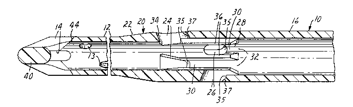
Referring to the figures of the drawing, there is
shown in Figure 1 a perspective view of a preferred
embodiment of the medical catheter 10 of the present
invention. The medical catheter 10 is generally comprised
~lZ~f7S3
--5--
of a first cross-sectional area catheter portion 12 having
an inlet opening or openings 13 and 14 through a
cone-shaped distal end portion 40, a second, larger
cross-sectional area catheter portion 16 having an outlet
opening 18 and a transition catheter portion 20 between the
first and second catheter portions 12 and 16. The
transition catheter portion 20 is in fluid communication
with the first and second catheter portions 12 and 16.
The first cross-sectional area catheter portion
,10 12 is preferably dimensioned to be received within a vena
cava, preferably the inferior vena cava, not shown. The
second, larger cross-sectional area catheter portion is
preferably dimensioned not to be received within the vena
cava; that is, this second catheter portion is preferably
too large to be received within the vena cava.
As perhaps best shown in Figure 3, the transition
catheter portion 20 is generally comprised of a means 22
ad~acent the first cross-sectional area catheter portion 12
for stinting the second, larger cross-sectional area
catheter portion 1~ so that the second catheter portion 16
cannot enter the vena cava even if appropriately
dimensioned, a plurality of inlet openings 24 and 26
ad~acent the stinting means 22 and a plurality of generally
longitudinally-aligned, reinforcing channels 28 disposed
along the inside of the transition catheter portion 20 as
shown in Figures 3, 4 and 5. The first cross-sectional
area catheter portion 12 preferably generally comprises a
cylinder, and the stinting means 22 generally comprises a
diameter-increasing protrusion.
The reinforcing channels 28 each have one end 30
connected to the inlet openings 24 and 26 of the transition
catheter portion 20 and the other ends 32 are connected to
the second, larger cross-sectional area catheter portion
16. Fluid entering the inlet openings 24 and 26 is
channeled into the second, larger cross-sectional area
catheter portion 16 by the channels 28 together with fluid
entering the inlet opening 14 of the first cross-sectional
area catheter portion 12 with a minimum of interference
~L2~753
--6--
between these fluid flows, thereby reducing fluid pressure
drop across the medical catheter 10.
In the preferred embodiment, the inlet openings
24 and 26 comprise a plurality of generally triangularly-
shaped openings 24 disposed about the transition catheter
portion 20 adjacent the diameter-increasing protrusion 22
with one side of each triangle generally transverse to the
length of the medical catheter 10 and a plurality of
generally elongated openings 26 disposed about the
transition catheter portion 20 adjacent the
triangular~y-spaced openings 24. As shown in Figure 3,
these openings 24 and 26 are preferabl~ peripherally
chamfered on the outside and on the inside. Most
preferably, openings 24 and 26 are peripherally chamfered
on the outside generally towards and away from the first
cro8s-sectional area catheter portion 12 with
longitudinally-aligned chamfers 34 and 37 and on the inside
generally away from the first cross-sectional area catheter
portion 12 with longitudinally-aligned chamfers 35. The
chamfers 35 are preferably between 30 and 70 with respect
to the length of the medical catheter 10 and most- .
preferably are about 45. The chamfers 34 and 37 reduce
tissue trauma upon catheter insertion and withdrawal from
the wound in the heart and reduce fluid pressure drop
across the medical catheter 10. These chamfers 34 and 37
together with the chamfers 35 and the channels 28
significantly reduce the pressure drop across the medical
catheter 10 as compared to an otherwise substantially
identical medical catheter 10 without these chamfers 34, 35
and 37 and channels 28. This reduced pressure drop
translates into a higher fluid flow rate for a given
medical catheter 10 or can be capitalized upon to downsize
the catheter 10. The later has the advantage of providing
an adequate venous blood flow with a relatively smaller
cross-sectional flow area which, in turn, permits a smaller
wound to be made in the heart.
~2r37~3
--7--
The channels 28 facilitate the fluid flow from
the openings 24 and 26 to the second, larger
cross-sectional area catheter portion 16 and also rei-nforce
the transition catheter portion 20 in the area of the
openings 24 and 26 to resist kinking of the medical
catheter 10 in this area in the event that the catheter 10
is bent in use. As noted earlier, U.S. Patent No.
4,639,252 describes some surgical procedures as including
bending dual-drainage catheters when manipulating or moving
the heart. ,
The channels 28 are comprised of a plurality of
longitudinally-aligned, reinforcing rails 36 with each rail
36 forming a common wall between a channel 28 connected to
a triangularly-shaped opening 24 and a channel 28 connected
to an elongated opening 26. In this configuration, as
perhaps best shown in Figure 5, the openings 24 and 26 are
generally equidistantly disposed about the circumference of
the transition catheter portion 20 in an alternating
fa6hion with one triangularly-shaped opening 24 and
connected channel 28 generally between two elongated
openings 26 and connected channels 28, and vice versa.
As perhaps best shown in Figure 1, the medical
catheter 10 of the present invention is in this embodiment
preferably of a one-piece construction. sy contrast, the
embodiment of Figures 7 and 8 is of a three-piece
construction with two of the pieces joined at a connector
38. This connector 38 is preferably of a telescoping
nature with the pieces ~oined by conventional means such as
cement or solvent bonding or radio f requency welding. This
three-piece construction facilitates manufacturing where,
as in the alternate embodiment of Figures 7 and 8, one
piece has embedded therein in conventional fashion a
helical coil 50 of reinforcing wire whereas the remainder
of this embodiment is not wire reinforced.
The medical catheter lO of Figure l is preferably
completely comprised, via conventional injection molding,
of a medical grade polyvinyl chloride having a hardness in
~2~*~;3
--8--
the range of 60 to 90 Shore A durometer and preferably
about 75 Shore ~ durometer. This includes the distal end
portion 40 having the inlet openings 14 which are
preferably elongated and provides the medical catheter 10
with a relatively soft end. As perhaps best shown in
Figure 2, this soft end portion 40 is reinforced at the
joining of the portions of the distal end portion 40
between the openings 14 to form the distal end, and a
portion of the inlet openings 14 are preferably
peripherally chamfered on the outside. Most preferably,
the. openings 14 are peripherally chamfered on the outside
generally away from the transition catheter portion 20 with
longitudinally-aligned chamfers 44. This reinforcement and
chamfering reduces the likelihood of the soft end portion
40 kinking, collapsing or further damaging the already
wounded heart tissue while at the same time further
facilitating blood flow by further reducing the pressure
drop across the medical catheter 10.
From the foregoing, it will be apparent that
various modifications and changes may be made by those
skilled in the art without departing from the scope and the
spirit of the invention. Because these modifications and
changes may be made by one skilled in the art and without
departing from the scope and spirit of the invention, all
matters shown and described are to be interpreted as
illustrative and not in a limiting sense.