Note: Descriptions are shown in the official language in which they were submitted.
~ ~30~689 F.6055
538-942
METHOD FOR PERFORMING
OPHTHALMIC LASER SURGERY
BACKGROUND OF THE INVENTION
The invention relates to that aspect of
ophthalmological. surgery which is concerned with
operations upon the external surface of the cornea.
Operations of the character lndicated include corneal
transplants and keratotomies; such operations have
. traditionally required skilled manipulation of a cutting
~ instrument. But, however keen the cutting edge, the mere
. entry of the edge into the surface of the cornea
;` necessarily means a wedge-like lateral pressure against
body`cells displaced by the entry, on both sides of the
entry. Such lateral pressue is damaging to several
layers of cells on both sides of the entry, to the extent
: impairing the ability of the wound to heal, and resulting
in the formation of.scar tissue.
My original Canadian patent application No. 464,792,
filed October 8th, 1984, includes a back~round discussion
of the effects of various av.ailable wavelengths of laser
radiation in ophthalmologic surgery and, in particular,
surgery performed on the anterior surface of the cornea.
It is explained that radiation at ultraviolet wavelengths
~ is desirable by reason of its high photon energy. This
energy-is greatly effective on impact with tissue, in
33 that molecules of tissue are decomposed on photon impact,
,. ~3
~30~3689
resulting in tissue ablation by photodecomposition.
Molecl~le~ at the ;rradiated surface are broken into
smaller volatile fragments without heating the remaining
substrate; the mechanism of the ablation is photo-
chemical, i.e., the direct breaking of intra-molecular
bonds. Photothermal and/or photocoagulation effects are
neither characteristic nor observable in ablations at
ultr,aviolet wavelengths, and cell damage adjacent the
photodecomposed ablation is insignificant. The order of
magnitude of this ablative process, in the case of
radiation exposure at ultraviolet wavelengths (in the
range of about 400 nm or less)/ is that an energy density
of l joule/cm2 incises to a depth of l micron (1~ ).
Said original patent application discloses a technique of
scanning a laser beam over the anterior surface of a
cornea in such a controlled pattern as to sculpture said
surface, imparting a new curvature to said surface,
whereby to achieve optical correction of an optically
deficient eye. But the scanner and scanner control to
perform the technique are relatively complex and
e~pensive.
BRIEF STATEMENT OF THE INVENTION
It is an ob]ect of the invention to proviae an
improved apparatus and technique for surgically operating
upon the outer surface of the cornea.
Another object of the invention is to simplify and
reduce the cost of apparatus and technique for surgically
modifying optical properties of the eye through surgical
procedure on the outer surface of the cornea.
It is a specific object to achieve the above objects
with surgical techniques and apparatus for reducing a
myopic, for reducing a hyperopic, and/or for reducing an
astigmatic condition of an eye.
Another specific object is to provide an improved
surgical technique in performing corneal-transplant
operations.
A still further specific object is to achieve the
above objects with automatic means for safely applying
ultraviolet irradiation in surgical procedures on the
cornea.
1;~0~689
50538-g42
It is also an object to achieve the above objects
without use of scanning techniques or apparatus.
The invention achieves these objects with apparatus
which effectively fixes the position of an eye with respect to
a non-scanning laser characterized by ultraviolet radiation, at
an energy level capable of achieving controlled ablative
photodecomposition of the cornea, namely, of the epithelium,
Bowman's membrane, and stroma levels of the cornea. Irradiated
flux density and exposure time are so controlled as to achieve
desired depth of the ablation. As distinguished from the
scanning procedure described in said application ~o. 464,792, a
~ sculpturing action results from controlled change of projected
laser-spot size, in the course of a given treatment, wherein
spot size ranges from a maximum which covers the entire area to
be treated for curvature correction, down to a predetermined
minimum tolerable size~ In one embodiment, a zoom lens in the
optical path o~ projection ls the means of changing spot size,
and in another embodiment an indexable mas]c or mirror is
employed; in both cases, the weighted allocation of time as
function of spot size is such as to achieve a desired ultimate
optically correc~ed cornea, from prior ascertainment of an
optically deficient corneal curvature. Spot-size control is
not only disclosed for effecting spherical-curvature
correction, but also for cylindrical correction in reduction of
astigmatism; still ~urther use is described in connection with
a corneal-transplant procedure.
Thus, according to one aspect, the invention provides
sculpture apparatus for cur~ature-correcting operation upon the
anteri~r surface of the cornea of an eye, Gomprising laser
means producing an output ~eam in the ultravlolet portion of
the electromagnetic spectrum, reflector means for variably
limiting the area of said beam at focus on the cornea, said
13006~39
60538-942
reflector means including actuating means for varying the
reflector area thereo~, the range of re~lector-area variation
at lea~t in~luding a maximum curvature-correcting area to be
ablated and being symmetrical with respect to a beam-projec~ion
~; axis which coincides with the optical axis of the eye, the
intensity of laser-spot projection being limited per unit time
to ablate but a fraction of a predetermined maximum depth of
ablation into the stroma region of the cornea, and means
including a microprocessor with coordinating control
connections to said laser means and to said actuating means,
for so correlating laser-beam impingement at the cornea with
variation of reflected-spot area as to e~fect a diopter change
at the cornea.
Accordlng to another aspect, the invention provides
sculpture apparatus for operation upon the external surface of
the cornea of an eye of a patient, comprlsing laser means
producing an output beam in the ultraviolet portion of the
electromagnetic spectrum, masking means for variably limiting
the area of said beam at focus on the cornea, said masking
means including actuating means for varying the masked area
thereof, the range of mask-area variation being within a
maximum area to be abiated and being symmetrical with respect
to a beam projection axis which coincides with the optical axis
of the eye, the intensity of laser-spot projection being
limited per unit time to ablate but a fraction of a
predetermined maximum ablation into ~he stroma region of the
cornea, said masking means being operative to provide cornea
exposure at said maximum area and at a plurality of similarly
shaped but lesser areas, said areas being circularly annular,
and characterized by varying inner diameter, said areas being
further defined by constant outer diameter for an area within
-3a-
- ~300~i89
60538-~42
which a hyperopia correcting curvature change is to be
effected; said last-mentioned area being less than said maximum
area thereby defining an annular area of laser-beam projection
outside said area of curvature change, said masking means being
further operative wi~hin said annular area to provide cornea
exposure at a succession of circularly annular areas contiguous
to the area o~ curvature change and of varying outer diameter,
and means including a microprocessor with coordinating control
connections to said laser means and to said actuating means,
whereby laser-beam impingement at the cornea may be so
correlated with variation of masked-spot area as to effect a
hyperopia-correcting diopter change at the cornea, together
with a smoothed surrounding annulus of transition to adjacent
unexposed corneal tissue.
: DETAILED DESCRIPTION
The invention will be illustratively described in
detail, in conjunction with the accompanying drawings, in
which,
Figure 1 is a schematic diagram in perspective, to
show the general arranyement of operative components of the
invention,
Figure 2 is a simplified view in longitudinal
section, showing an eye-retaining fixture used with the
apparatus of Figure l;
Figures 3, 4 and 5 are simplified diagrams to
illustrate the nature of ablative corneal sculpture, performed
with appara~us as in Figure 1, for the case of correcting a
myopia condition;
-3b-
~ ~006~3~
60538-942
Fig. 6 is a simplified diagram schematically showing
operative components of another embodiment of the invention;
Fig. 7 is a plan view of an inde~ible mask usable in
the embodiment of Fig. 6;
Fig. 8 is a diagram similar to Fig. 6, to show a modi-
fication;
; Fig. 9 is a fragmentary plan view of an indexible mask
usable in the modification of Fig. 8;
Figs. 10 and 11 are simplified diagrams to illustrate
use of the invention, for the case of correcting a hyperopia
condition;
Fig. 12, 13 and 14 are simplified diagrams to illus-
trate use of the invention to achieve a Fresnel-type optically
corrective contour at the anterior surface of the cornea;
Figs. 15 and 16 respectively illustrate components and
features of an embodiment of the invention to achieve correction
of an astigmatic eye;
; Figs. 17 and 18 are simplified diagrams to illustrate
use of the invention in connection with a corneal-transplant
operation;
Figs. 19 and 20 are simplified diagrams to illustrate
two different alternatives for the embodiment of Figs. 15 and
16,
Figs. 21 to 26 correspond to Figs. 6, 7, 8, 9, 11 and
14, respectively, in illustration of a further aspect of the
invention;
Figs. 27 and 28 are graphical diagrams to illustrate a
principle of reflector design;
; Figs. 29 and 30 are diagrams similar to Figs. 10 and
11, respectively, to illustrate a special-purpose refinement of
the inven-tion;
~\~
~3~6as
60538-942
Fig. 31 is a schematic diagram to illustrate an
alternative for Fig. 30; and
Figs. 32 and 33 are similar diagrams illustrating
different special-purpose refinements of the invention.
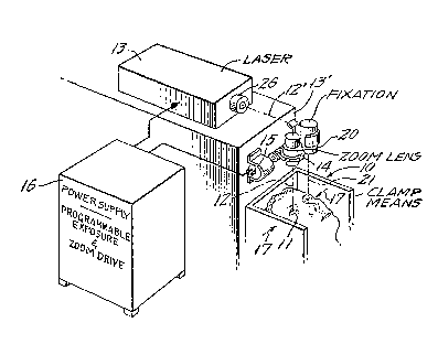
In Fig. 1, clamp means 10 is shown for fixed reten-
tion of the head of a patient (reclined, face up~ such that the
eye 11 to be operated upon is fixedly aligned with a downwardly
folded portion 12 of the central axis 12' of beam output from a
stationary laser device 13, supported by a table or other base
13'. The optical system of laser-beam projection to eye 11
includes zoom-lens means 14 having a reversible motor drive 15,
whereby laser-spot size at eye 11 can be caused to vary from a
predetermined minimum, to a maximum of 3 or 3.5-mm radius,
corresponding to the corneal frontal area to be subjected to
laser action. A cabinet 16 is shown by legend to include a
power supply for the laser, and cabinet 16 is also shown (by
legend) to include programmable microprocessor means for con-
trolling exposure and beam (spot) size on axis 12, as will
later become more clear.
Clamp means 10 preferably includes means, symbolized
at 17, to stabilize the patient's head via opposed engagements
at the region of his temples, and an eye-retaining fixture (18,
in Fig. 2) peripherally engages eye 11 at the corneal-scleral
area. Also preferably, an optical-fixation device 20 is
adjustably fixed, as to the table or base 13'. Illustratively,
device 20 includes a sighting reticle and lens, whereby the eye
11' not being operated upon can view the reticle as if at
infinity, the sighting alignment 21 for device 20 is parallel
to the axis 12, and it will be understood that adjustable means
(not shown) may provide an adjustable offset, as needed for
~ 5 -
~3006~9 60538-942
accommodation of the patient's inter-pupilary distance and to
adapt to the particular mounted oEfset of device 20 from axis
12. For an operation on the other eye 11', the eye 11 will be
available for similar fixation, in conjunction with another
fixation device (not shown) and associated adjustably offsetting
means; alternatively, the fixation device 30 may be adjustably
mounted at correct offset on the opposite side of scanner 14.
For purposes of operating on eye 11', clamp means 10 will have
been indexed laterally with respect to laser 13 to the extent
aligning axis 12 with the eye (11') then to be operated upon,
thereby positioning eye 11 for use oE the fixation device.
The eye-retaining fixture 18 oE Fig. 2 is seen to
comprise a hollow annulus, having a convergent axial-end wall 23
of air-permeable material contoured to engage and retain the eye
via a scleral-corneal region. A side-port
:1 5a
v
~3~89
connection 24 to a vacuum pump enables retention of eye
en~agem~nt to wall 23, and outward lug or flange means 25
enables rigid aligned and spaced connection of fixture 18
to laser 13 and its scanner 14 via means suggested by
legend in Fig. 2, such means being omitted from Fig. 1
for reasons of more simplified showing.
The laser selected for use at 13 preferably emits in
the ultraviolet, namely, at wavelengths of less than
substantially 400 nanometers. Such emissions for gas
lasers are characteristically at 351-nm for
xenon-fluoride lasers,-337-nm for nitrogen lasers, 308-nm
` for xenon-chloride lasers, 248-nm for krypton-fluoridelasers, 193-nm for argon-fluoride lasers, and 157-nm for
fluorine lasers; and within this range, frequency-
doubling techniques applied to other lasers, including
crystal lasers, provide further alternative sources.
One of the existing commercial excimer-laser products
of Lambda Physik GmbH, Gottingen, Germany, for example
their Model EMG 103 operating with argon-Eluoride, is
satisfactory for use as laser 13; for this product,
maximum energy per pulse is 200 milli-joules, with a
pulse-repetition rate of 200 per second, 3 X 10⁵ shots
(pulses) being available from a single charge of the
involved gas, before reducing to 50 percent of specified
power at this repetition rate, it being noted that full
rated power is not necessarily required in use of the
present invention. Pulse width is about 15 nanoseconds,
and typical beam dirnensions are rectangular; as shown,
however, the opening in a mask 26 reduces the laser beam
to a circular section, and it will be understood that the
optical elements o~ lens 14 are of quartz, calcium
fluoride, magnesium fluoride, or otherwise as suitable
for laser-beam accommodation.
; Fig. 3 is an attempt to depict the action of laser
output as modified by the setting of zoom lens 14, it
having already been indicated that, through the action of
lens 14, spot size at eye 11 can be caused to vary from a
- minimum diameter at 28 to a maximum diameter at 29. The
diagram shows a plurality of intermediate circular spot
sizes, but it will be understood that since the zoom
adjustment of lens 14 is continuously variable, there is
~ 3 O ~ ~ 8 g 60538-942
no need to presuppose discrete circular spots of different
diameter, except for the fact that in the course of a continu-
ous variation in zoom adjustment the intermittent delivery of
laser pulses will mean that each pulse is projected at a
slightly different spot size.
Figs. 4 and 5 are illustrative of use of the inven-
tion in an optically corrective ablation of the anterior sur-
face 30 of eye 11, wherein a myopia problem is to be solved,
meaning that the curvature of surface 30 is of too-short radius
to establish focus at the retina, for the case of distant
objects. On the other hand, the dashed line 31 represents the
; ultimate curvature to which the anterior surface of the cornea
should be modified -to achieve a diopter-reducing corrective
effect. To achieve the curve 31, the minimum desired photo-
decomposition is at the outer boundary 29, and the maximum is
at the center. This is achievable by programming the micro-
processor to progressively change the projected spot size
(through driven adjustment of lens 14) in the course of a pre-
determined succession of laser pulses. The result is the same
whether spot size is caused to expand from minimum (28) to
maximum (29) or to reduce from maximum (29) to minimum (28).
Of course, for each laser pulse or "shot", ablative penetration
into the cornea will be a function of delivered energy density,
and therefore the number of pulses needed to achieve a given
increment of ablative penetration will be greater, the larger
the diameter of the projected spot.
Fig. 5 is a very much simplified diagram to represent
the progressive ablative effect of a succession of laser-spot
projections at successively reducing diameters Dl, D2, D3 ...
Dn. The least resulting energy density is at the largest
~ ,
~L300689
60538-942
diameter D1, which can be assumed to have made the least pene-
tration, although such penetration will have been uniform over
the entire spot area for diameter D1. An incrementally greater
energy density results at the next step D2 of diameter reduc-
tion, in which event penetration has become cumulative with
that of the first shot, over the area common to both shots,
The cumulative penetration effect continues for shots of
successively reduced diameter, so that a new, larger-radius
curvature emerges from a pattern of stepped reduction in pro-
jected spot size. However, for a sufficiently great number oflaser pulses (and hence, potentially discrete steps), indivi-
dual steps cease to appear discrete, and a sufficiently smooth
new spherical anterior surface characterizes the cornea. This
is particularLy so after a post-operative period of about two
days, by which time a thin epithelial layer will have spread
into smooth and protective coverage of the newly charac-terized
surace.
The foregoing discussion in connection with Figs. 1
to 5 presupposes a pulsed laser, exemplified by an excimer
laser. But other lasers are known to emit at presently suit-
able energy levels and at ultraviolet wavelengths of present
utility, and these other lasers will emit continuously for
periods of controlled duration~ For example, an organic-dye
laser utilizing the proper organic dye can be made to produce
laser emission in the region of 380-nm when pumped by ultra-
violet laser sources such as a continuous-wave frequency-quad-
rupled neodymium-YAG laser operating at 266-nm; in this case,
the organic-dye laser emission at 380-nm can be frequency-
doubled by a proper non-linear crystal such as a potassium-
deuterium-phosphate (KDP) crystal or a potassium-titanium-
phosphate (KTP) crystal to an emission wavelength at l90-nm.
~,~
` ~0~)~8~
60538-942
The showing of Figs. 1 to 5 will thus be understood to illust-
rate the further case wherein ultraviolet laser radiation on
axis 12 is of continuous-wave nature, for a treatment duration
predetermined by programming at 16, and wherein the programming
at 16 further continuously drives the zoom-lens 14 to provide
that time-variation of projected spot size as has been pre-
determined to achieve a myopia-correcting change in curvature,
from curve 30 to curve 31, in the course of the treatment dura-
tion. And this result is achieved whether spot size (at the
eye ll) is caused to expand continuously from minimum (28) to
maximum (2~) or to reduce continuously from maximum (29) to
minimum (28).
In the embodiment of Figs. 6 and 7, a masking
technique is employed, in place of the zoom-lens technique of
Fig. l, to achieve a similar myopia-correcting curvature change
in the anterior surface of the cornea. Such masking could
proceed continuously with a suitably programmed variable iris
diaphragm in place of lens 14, but in the form shown, a single
precision masking plate 35 is employed. The masking plate 35
is rectangular and is mounted (by means not shown) for indexed
unit displacement in each or both of two orthogonal axes X-Y.
For each of the grid-like layouts of mask openings provided on
plate 35, the size of the involved circular opening increment-
ally changes. Thus, for a first row of mask openings beginning
and ending with openings 36 and 36', respectively, the openings
are of progressively reducing diameter; in the next-adjacent
row, beginning and ending with openings 37 and 37',
respectively, the openings continue with progressively reducing
diameter; in the third row, the progression continues to reduce
from opening 38 to opening 38', and the final row reduces still
`:~
~3~0~89 60538-942
further from 39 to the smallest opening 39'. An X-Y coordinate
index drive 40 will be understood to provide correct X and/or Y
successive displacements of masking plate 35 under control of
microprocessor means 41 having programmable means ~or alloca-
ting numbers of excimer-laser "shots" (or, in the case of a CW
laser, for allocating variously controlled pulse duration) at
particular succeeding mask-opening sizes, whereby to effect a
given desired ablative "sculpture" which will predictably and
correctively change optical performance of the eye (11). As
shown, optical-transducer elements in pairs 41-41' and 42-42'
straddle each mask opening as it is indexed into the laser-
projection axis 12; these transducer elements sense registry
with grid lines, such as x-positioning grid linès 43-43' on
opposite sides o~ a given mask opening 37" (Fig. 7) and ortho-
gonally related y-positioning grid lines 44-44' on opposite
sides o~ the same mask opening 37", whereby such registry may
be certified to the microprocessor 41, for interlock purposes,
to achieve correct mask-opening positioning on axis 12 before
firing the next laser pulse, the latter being symbolized by a
synchroniæing connection 45.
In the arrangement of Figs. 8 and 9, myopia-
correcting sculpture relies on indexed shifting from one
to another of successive different-area masX openings, via
incremental angular indexing displacement of a masking disc 50
(about an indexing axis 50'); disc 50 has a peripherally
distributed succession of mask openings, ranging ~rom the
largest opening 51 to the smallest opening 52. A radial mark,
as at 53 for opening 51, identifies the angle at which the
given opening is correctly indexed into position on the laser-
projection axis 12. Disc 50 is shown mounted to an annular
- 10 _
~3~689 60538-942
ring 54 which will be understood to be counterbored for central
and keyed location of disc 50, and ring 54 is edge-driven by
suitable means 55 under control of a rotary-drive signal gener-
ator 56. Again, a programmable microprocessor 57 is respon-
sible for controlling the rotary-index drive 55-56 for prede-
termined allocation of laser pulses to given mask openings, to
achieve the desired cornea-profile correction, with laser-pulse
synchronization via lines 58, as an optical transducer 59
tracks registry with the particular radial-marker line for each
given mask-opening area.
Figs. lO and ll illustrate that the device of Fig. 8
is equally adapatable to making corrective sculpture of the
cornea 60 of a far-sighted (hyperopic) eye, meaning that the
anterior curvature is to be increased, as to achieve a new
profile 61 (Fig. 10). This is illustratively done by substitu-
ting a different masking disc 62 for the disc 50 of Fig. 8. In
the disc 62, for each of the angular mark locations (as at 63),
a basic opening limit, e.g., of 3,5-mm radius, is the outer
edge of each of an angularly distributed succession of annulus
openings, produced by a central opaque masking spot of pro-
gressively changing diameter. Thus, for the smallest annular
mask area 63' (which applies at radial mark 63), the central
opaque spot is a circle of nearly the diameter of the basic
limiting opening, to produce a first, or thinnest annulus 63'.
At the next mark 64, the outer diameter of a slightly thicker
annulus 64' is determined by a central opaque spot of slightly
lesser area. The progression continues, at increments of equal
angle (about the index axis of disc 62), until reaching the
largest annular opening 65' at angular location 65, where the
central opaque masking circle is of least diameter. In use of
-- 11 --
,~
~ 89 60538-942
the mask 62 in conjunction with the positioning and control
apparatus of Fig. 8, the microprocessor 57 will be understood
to so allocate laser pulses to particular sizes of annular mask
openings that greatest cumulative ablative penetration of the
; cornea is at larger radii, while least penetration is at
smaller radii, resulting in the corrected ultimate profile 61
of decreased radius.
The arrangement of Figs. 12, 13 and 14 illustrates
that above-discussed principles of the invention are further
applicable to corrective sculpture of the cornea to achieve a
Fresnel-type distribution of the desired ultimate curvature,
which can be either hyperopia-correcting or, as shown, myopia-
correcting. Such an operation (i.e., Fresnel-type) would be
; used when, in the surgeon's considered judgment, a single
smoothly developed corrected curvature would entail excessive
removal of tissue at the involved region of necessarily deepest
cut. To avoid too deep a cut, Figs. 12 and 13 illustrate that
an ultimately reduced-curvature surface, as at 31 in Fig. 4
(dashed line 71 in E'ig. 13), is achieved in annular increments
within the field bounded at 70. In the outer one of these
annuli (72), the curvature and depth of cut are precisely as
would have applied to generate the continuous curve 71 (i.e.,
without Fresnel steps). But the intermediate annular area 73
effectively achieves a continuation of curve 71 with much less
volume of corneal excision. Finally, the inner circular area
74 effectively completes curve 71, with minimal removal of
corneal tissue~
The removal of tissue at the center is denoted ~74
for the Fresnel cut 74 of Figs. 12 and 13 and, co~paratively,
is but a small fraction of the maximum removal depth ~71 which
- 12 -
~006~9 60538-942
would have been needed to achieve the same optical correction
with the smoothly developed corrected single-curvature surface
71. Fig. 14 illustrates an indexible rotary masking disc 75 of
a type compatible with the system of Fig. 8, in substitution
for the disc 50 of Fig. 8, to achieve Fresnel-type cuts of the
nature described for different annuli 72, 73, 74. Beginning
with th~ largest area of mask annulus 76 (at location 76') and
proceeding for a first 120 sector of disc 75, the succession
of annular mask openings will be understood to progress with
decreasing radius, by reason of a constant-area central mask
spot, in the context of a progressively shrinking outer-circle
; diameter. The programmable means 57 (of Fig. 8) will be
understood to Eunction as a control for allocation of laser-
pulse shots, using a programmed distribution of the annular
mask openings of this first sector, for achievement of ~he
curvature 71 within outer annulus 72. A similar succession of
annular mask openings will be understood to be similarly
accessible via a second sector (not shown) of mask disc 75, in
establishing the curvature 71' within the intermediate annulus
73. And finally, the curvature 71" is established within the
inner circular area 74 by programmed projection of laser shots
on axis 12, through an indexibly available succession of
progressively shrinking circular openings, ~eginning with a
mask-opening diameter of largest (circle-74) area, and reducing
throughout the third sector to the smallest opening 78 at
location 78', adjacent the location 76' (of the first sector).
The diagrams of Figs. 15 and 16 are illustrative of
the variable aperture or indexible-mask technique of the inven-
tion in the development of corrections for astigmatism, by
~3~689 60538-942
ablative laser pulsing with a rectangular beam section wherein
the width of the section is changed to create a cylindrical
profile of cumulative ablative penetration. This can be done
by masking the laser beam with a slit or diaphragm of variable
width, and with the ability to selectively rotate the orienta-
tion at which the major dimension of the slit is positioned
(i.e,, based on prior measurement of the angle and of the
cylindrical diopter strength of the particular eye's astigma-
tism; however, in the form shown in Fig. 15, the mask is an
elongate strip 80 having a succession of rectangular slit open-
ings of progressively different width. In the fragmentary
showing of Fig. 16, these openings proceed from a largest area
opening 81 to a smallest area opening 81', and the central axis
of symmetry of each of these openings is identified wi-th a
mark, as at 82 for opening 81; preferably, all such marks are
at equal spacing.
Strip 80 is a slide guided by means 83 forming part
of a rotatable mask-supporting disc or ring 84; and guide means
83 locates the longitudinal axis 86 of slot symmetry on a dia-
meter of ring 84. Manually operable means 85 has edge-drive
coupling to ring 84 to enable selective angular orientation of
strip 80 (about the laser-projection axis 12), as by observa-
tion via a fixed indicator mark 87 against azimuth edge mark-
ings on ring 84. A bidirectional slide-drive signal generator
88 is under control of a microprocessor 89 to coordinate slide
(80) positioning with laser-pulse control, suitably synchron-
ized by optical-transducer (90) tracking of the mark (82)
applicable to the particular index~d mask opening, whereby each
mask opening can be assuredly on the axis 12 of laser-beam
projection.
~ 14
., -~ ,~. .
13~0~9 60538-942
In use of the invention for laser surgery upon an eye
having need for both astigmatic and spherical correction, it
is preferred that the astigmatic correction, described in
connection with Figs. 15 and 16, be the first of two proced-
ures. This is considered advantageous because astigmatic
errors are generally not as severe as spherical errors, so that
fewer diopters of cylindrical curvature ablation will be in-
volved than for the subsequent spherical-correction procedure.
Furthermore, to have eliminated or substantially eliminated the
astigmatism in a first procedure is to have constituted the
anterior surface of the cornea to an essentially spherical
surface, which (be it myopic or hyperopic in nature) is more
assuredly correctively sculpted to the desired profile (also
spherical) for emmetropic vision, particularly where, as in the
case of this invention, all ablative-laser shots (whatever the
currently operative mask opening) are effectively centered on
the optical axis of the involved eye.
Quite aside from the variable-depth character of the
removal of corneal tissue (Figs. 4 and lO), the invention also
lends itself to uniform-depth removals, over a single entire
area of the cornea, in preparation for reception of a corneal
transplant. In Figs. 17 and 18, the cornea of an eye ll is
subjected to a succession of laser pulses which have been mask-
ed to the same area, of diameter D, e.g., 7-mm; the succession
of pulsed laser shots will in such case be seen to produce a
carved base or recessed-floor curvature 95 for reception and
location of an implanted corneal transplant. Alternatively, in
Figs. 17 and 18, the cornea of eye ll may be subjected to
steady (CW) laser exposure of such intensity as to ablate ~a)
via the same mask on constant diameter D and (b) at a rate of
- 15 -
,~,
13~0689 60538-942
ablative penetration for which a given duration ~exposure time)
of laser-beam projection will achieve the desired depth of
penetration.
Further with respect to a corneal-transplant proce-
dure, the described apparatus will be seen to be further use-
ful, as in preparation of the corneal insert to be implanted at
and within the recess 95. A donated eye may be reversibly held
to a fixture as described at 18 in Fig. 2; by "reversible" it
is meant that, depending upon the manner of mounting flange 25,
either the epithelium or the endothelium of the donated eye may
be mounted for upward exposure to the laser beam 12, it being
understood that for the latter situation with the donated eye,
- iris and other regions not needed for corneal-scleral mounting
and for corneal operation will have been initially removed. A
preferred procedure is first to so expose to laser action the
concave inner side of the donated cornea; such action is to an
extent (achieved by timed CW exposure, or by multiple pulsed-
laser shots, of a full circular field exceeding the diameter of
recess 95) sufficient to remove tissue at least to a uniform
depth within the donated stroma, whereupon the mounting of
fixture 1~3 (and its partially machined corneal workpiece) is
reversed, to expose to laser action the convex outer side of
the donated cornea. Laser action on the outer side consists of
two steps; first, timed CW exposure multiple laser pulses of
the full circular field (exceeding the diameter of recess 95)
thereby excising at least the epithelium and to a depth which
preferably achieves a transplant thickness Tl exceeding the
depth T2 f recess 95; second a scanner (not shown, but of the
type disclosed in Canadian Patent No. 1,243,732 is operated
in a line~cutting mode wherein successive laser pulses
~ 15a ~
J
~3006~9 60538-942
sequentially advance along the circumference of a circle de-
signed for precise acceptance in the circular recess 95, until
full severence of the circular cut-out, which then becornes the
prepared transplant. Upon implanting, donated stroma is placed
in full endothelium-free contact with the patient's prepared
stroma, and the implant may be sutured. Later, upon removal of
sutures, the outer surface of the eye 11 and its transplant 96
will have the appearance shown in Fig. 18, wherein the trans-
plant projects beyond adJacent areas of the patient's cornea,
and this projecting surface of the transplant may be reduced by
above-described laser sculpting to a finish contour 97 of pre-
ferably flush marginal conformance with non-sculptured adjacent
tissue of the patient's eye. It will be further understood
that, subject to the surgeon's decision, such a finishing cut
may be to a curvature which does or does not effect a predeter-
mined change in optical performance of the eye.
It will be seen that the described methods and appar-
; atus achieve all stated objects and provide readily controlled
procedure for correcting eye abnormalities attributable to
cornea curvature. The ablative penetration of laser-beam
action may be kept to a relatively harmless fraction of the
thickness of the cornea, and whatever the depth of invasion, a
natural body process provides protective epithelium coverage of
the sculpted region, within a few days after an operation. The
programmable coordination of laser-beam size and shape (circu-
lar, annular, or rectangular) in conjunction with numbers of
pulses at given sizes and shapes will produce predictable and
controlled changes in curvature, whereby cylindrical errors
and/or spherical errors may be eliminated or substantially
reduced, to the enhanced comfort and convenience of the
patient.
- 15b -
e
~3~0689 60538-942
While the invention has been described in detail for
various illustrative embodiments and modes, it will be under-
stood that modifications may be made without departing from the
spirit and scope of the invention. For example, what has been
described above as manual means 85 to preset the angle at which
astigmatic correction is to be achieved, may in fact be an
automatically driven setting of the astigmatic-correction
-.~
~ - 15c -
- ~300~89
angle, wherein -the angle-input data for making the
automatic drive is produced by a diagnostic system or
method as described in my copending Canadian patent application,
No. 510,018, filed May 27th, 1986.
Also, by way of example, achievement of cylindrical
sculpting in reduction of astigmatism does not
necessarily require the indexible-slot technique of Figs.
15 and 16. As a first alternative (Fig. 19), the
variation in slot width may be achieved electro-
mechanically, via microprocessor control of means 100 to
differentially drive opposite side plates 101-102 of a
variable-width opening which is always centered on the
axis of the projected laser beam 12, plates 101-102 being
slidably ~ounted to an annular base 104 which is
adjustable in rotation to the angle for which astigmatism
is to be reduced (as suggested by a double arrow 103).
As a second alternative (Fig. 20), a cylindrical-lens
zoom system 105 is motor-driven by microprocessor output
(as suggested by double arrow 106) to establish a shaping
of the projected laser beam 12 to a line o variable
width, and said line is settable to the angle for which
astigmatism is to be reduced, as by edge-drive means 107
to the rim 108 of an annular mount for zoom system 105.
Figs. 21 to 26 are illustrative of a different aspect
of the invention wherein the variously described
sequences of spot shaping to achieve laser-ablated
corneal-curvature change are produced by reflection
techniques. And because the identification of parts in
these figures corresponds with parts in Figs. 6, 7, 8, 9,
11 and 14, the same numbers are used, as applicable, in a
100-series.
In the embodiment of Figs. 21 and 22, a transparent
plate 135, as of quartz, is characterized by a succession
of elliptical reflection areas, oriented with their major
axes parallel and respectively centered on each of the
two-dimensionally (X-Y) indexible positions of plate
135. For each of the grid-like layouts of elliptical
reflecting areas on plate 135, the size of the involved
ellipse incrementally changes. Thus, for a first row of
reflective ellipses beginning and ending with areas 136
and 136', respectively, the areas are progressively
reducing; in the next-adjacent row, beginning and ending
- 16 -
~300~;89
with areas 137 and 137', respectively, the reflecting
ellipses continue their progressive reduction; in the
third row, ~the progression continues to reduce from area
138 to area 138'; and the final row reduces still further
`~ 5 from 139 to the smallest, 139'. Support for indexing
` displacement of plate 135 will be understood to position
the reflective side thereof in inclined facing relation
to the laser-output beam alignment 12', the inclination
;~ being preferably such that the major axis of each of the
ellipses is at 45 to alignment 12' when the center of
the particular ellipse has been indexed for intersection
with the alignment 12'; at the same time, the minor axis
of each ellipse is at 90 to alignment 12' when the
center of the particular ellipse has been indexed for
intersection with alignment 12', and the major/minor
axis-span relation is .~:1. This preferred relation
determines that for each ellipse-index position, the
reflection 12 of the laser beam will be at 90 to the
alignment 12' and that this reflection will be a circle
of diameter ec~ual to the minor-axis span of the involved
ellipse. The X-Y coordinate index drive 140 and the
microprocessor 141 perform as described for Figs. 6 and
7, and optically readable grid lines on plate 135
(between the reflective ellipses) enable optical-
transducer pairs 141-141' and 142-142' to assure precise
positioning of each reflecting ellipse, centered on axis
12', before firing the next laser pulse
The automated running of the Fig. 21 device, in the
full two-coordinate program of indexing plate 135, will
be seen to deliver the greatest density of ablating
energy in the central part of the total circular corneal
area which is operated upon, with such density decreasing
as a function of increasing radius from the optical axis
of the eye. The curvature change is therefore of
myopia-correcting nature.
The embodiment of Figs. 23 and 24 has its corres-
pondence to Figs. 8 and 9, and thus the circumferentially
distributed pattern of reflecting ellipses is on an
indexible circular plate or disc 150, plate lS0 being
suitably transparent and of quartz. Preferably, the
centers of all ellipses are on one 9eometrical circle
~L~0~689
about the index axis 150', and the index axis 150' is
oriented to bisect t.he right-angle relation between laser
axis 12' and the (reflected) projection axis 12 to the
eye, axis 12 being aligned with the optical axis of the
eye 11; also preferably, the major axis of each of the
ellipses is oriented radially of the indexing center of
plate 150, and, again, the major/minor axis relation of
all ellipses is ~ :1. The automated running of the
ro~ary-indexed Fig. 23/24 arrangement will be seen to
produce the same cornea-ablating result as the
orthogonally indexed Fig. 21/22 arrangement, so that the
result is again myopia-correcting.
The fragmentary showing of Fig. 25 illustrates that
upon substitution of a different circular reflecting
plate 162 (in place of plate 150 of Fig. 24), the
microprocessor programming of rotary indexing and of
laser pulsing will produce a hyperopia-correcting change
in cornea curvature, of the nature shown in Fig. 10. The
reflecting ellipses of Fig. 25 are in an angularly spaced
succession of elliptical annuli of constant outer
periphery; the succession ranges from the radially
thinnest ellipse 163' at index location 163, to the
radially thickest ellipse 165~ at index location 165. In
other words, the succession of reflecting ellipses of
Fig. 25 accounts for annular projection of a constant
outer diameter and of a varying inner dia~eter,
throughout a single indexed rotation of plate 162,
accounting for maximurn ablating penetration of the cornea
at the outer diameter, and progressively reduced ablating
penetration as a function of decreasing radius about the
; optical axis of eye 11. For all ellipses, the
major/minor axis ratio is ~ 1, in view of the 45
incidence of the laser beam on each indexed elliptical
reflector.
The arransement of Fig. 26, taken with Figs. 12 and
13 is illustrative of the application of the reflection
principles of Figs. 24 and 25 to corrective sculpture of
the cornea to achieve a Fresnel-type distribution of the
desired ultimate curvature, which, as for Fig. 15, can he
either hyperopia-correcting or, as shown, myopia-
correcting. To avoid too deep a cut of ablative
- li3 --
~300~9 60538-94~
penetration, the ultimate reduced-curvature surface, as at 31
in Fig. 4 (dashed line 71 in Fig. 13), is achieved in annular
increments within the circular area bounded at 70, and the
curvature 71 is produced at steps 72-73-74.
As shown in Fig. 26, a transparent plate 175 serves
as a replacement for plate 150 in Fig. 23 and is provided with
an angularly stepped progression of reflecting elliptical annu-
li, beginning with the largest and thickest elliptical annulus
176 at location 176', and proceeding clockwise to the next
elliptical annulus of incrementally smaller size and thickness,
based on an inner limiting ellipse 177 of constant size. For
the three-step profile 72-73-74 shown, the reflecting ellipti-
cal annuli based on the same inner limiting ellipse 177, are
distributed over a first 120 sector of disc 175, with the
outer elliptical periphery progressively shrinking to a final
radially thin ellipse (not shown); and the programmable means
57 (of Fig. 8) will be understood to function as a control for
allocation of laser-pulse shots, using a programmed distribu-
tion of the first-sector elliptical reflectors, for ablative
achievement of the curvature 71 within outer annulus 72. A
similar succession of reflecting elliptical annuli will be
understood to be similarly indexible over a second 120 sector
(not shown) of disc 175, in establishing the curvature 71
within the intermediate annulus 73. And ~inally, the curvature
71" is established within the inner circular area 74 by pro-
grammed projection of laser shots on axis 12', through an
indexibly available succession of progressively shrinking
elliptical areas, beginning with an ellipse of largest minor-
axis span (not shown, but equal to the diameter of the central
circular area 74), and reducing throughout the third 120
19 -
,.,.,.~ .
- ~00~89 60538-942
sector to the smallest reflecting ellipse 178 at location 178',
ad~acent location 176' of the first sector.
A full rotation of disc 175, in the context of
suitably programmed pulsed-laser delivery on alignment 12',
thus creates the Fresnel steps 72-73-74 in succession. But it
will be understood that by using the highly precise photo-
reduction and metal-disposition techniques available from
micro-circuit technology, each indexed step of a single disc
(not shown) may be instrumental in the progressive formation of
all annular components of a full Fresnel-type ablation pattern.
To create reflecting elliptical patterns to achieve this
result, Fig. 27 outlines the course of minor-axis size varia-
tion for all involved reflecting ellipses, for the case of
myopia-correction, and Fig. 28 similarly outlines the course of
minor-axis size variation for all involved reflecting ellipses,
for the case of hyperopia correction.
In Fig. 27, it is seen that by dividing the full 360
angular extent of a given circular disc (utilizable in place of
disc 150 in Fig. 23) into the desired number (n) of indexible
steps at 360 spacing, and by drawing an ordinate line (e.g.,
at 120) for each such increment of azimuthal distribution,
intercepts (e.g., a-b-c-d-e, for location 120) are obtained for
each of five loci, establishing the requisite minor-axis span
for each of the involved plural reflecting ellipses at each
particular index location. The result for Fig. 27 relation-
ships is myopia-reducing because all outer perimeters (for
areas 72-73-74) vary, while inner perimeters remain constant.
On the other hand, the result for Fig. 28 relationships is
hyperopia-reducing because all inner perimeters (for areas
72'-73'-74', not otherwise shown) vary, while outer perimeters
:
~ - 2Q -
~3~689 60538-942
remain constant, noting intercepts a'-b'-c'-d'-e'-f' for loca-
tion 121.
All discussion thus far, for laser projection via
indexed reflective areas, has been concerned with essentially
spherical curvature correction, treating the myopic or the
hyperopic situation, as the case may be. It should, however,
also be apparent that similar principles are applicable to
astigmatism ~orrection, in which case the pattern of progress-
ively indexed reflecting areas is rectangular, of progr~ssively
varying width, symmetrically developed on opposite sides of the
central elongate axis of the most narrow rectangular pattern in
the progression. The drawing of Fig. 16 may thus be considered
illustrative of such a pattern development, wherein the index-
ible strip 80 is a transparent plate (as of quartz) and the
series of
-- 2aa --
.. .
, .., --,,
- 1~00689
rectangles 81 to 81' is reflecting and at equal
centerline-to-centerline spacing, with indexing from one
centerline (82) to the next, and with the laser-beam axis
12' directed at intersection with the central alignment
86 for each indexed position. It is realized that when
strip 80 is supported on guide ring 84 and in an inclined
plane as discussed for disc 150 in Fig. 23, the angular
orientation of ring 8~ (by setting adjustment at 85) will
account for a range of width variation in rectangular
spots incident at the eye, but the desired cumulative
ablation can still be achieved at the eye for any and all
`~:
selected angular orientations, by entering a suitable
angularity correction into the microprocessor, the
correction being a simple trigonometric function of
orientation angle.
; For the above-described reflective uses of the
invention, it is to be understood that the individual
patterns of reflection are operative upon a portion only
of the laser-beam sectional area (on alignment 12'), and
that, whether the reflective patterns are mounted to or
formed upon a transparent plate (as of quartz) or are
otherwise mounted, the portion of any given shot of
laser-beam output that is not reflected will be further
transmitted on essentially the alignment 12'. This
further transmitted energy is not used for the surgery
and may be trapped and dissipated by suitable means (not
shown).
For the various embodiments thus far described in
application to hyperopia-correction, Fig. 10 is
illustrative of the fact that penetration into the cornea
is deepest at the radially outer limit of the optically
corrected area tsurface 61), thus leaving a relatively
sharp circular edge, of depth proportional to the
;~ magnitude of diopter correction achieved. Such a sharp
edge presents a problem for epithelial regrowth over the
area (61) of surgery, in that epithelial regrowth is
optimum for essentially continuous surfaces, i.e., when
uninterrupted by sharp edges or by sharp discontinu-
ities. To a;void such a sharp-edge development, the
projected laser beam 12 should be of sectional area
larger than that over which hyperopia-curvature
correction is to be achieved, thus providing for an outer
- 21 -
~L30~6~9
profile-smoothing annulus contiguous to and surrounding
the circle of curvature-correction. In Fig. 29, the
optically corrected surface 61 is identified as having an
outer radius of curvature correction RCc, the diameter
being shown as 2RCc; and the outer profile-smoothing
annulus is shown to be of radial thickness a R, so that
the full area of the laser-beam section is of diameter
2(R~C + ~R). The smoothing action to be described
accounts for the gently sloping transitional profile
achieved in the annulus ~ R, as suggested in Fig. 29 by
dashed lines connecting the curvature-correcting ablated
area 61 to the outer corneal area not subjected to
ablation.
More particularly, Fig. 30 shows an indexible rotary
masking disc 162' of the nature described in connection
with Fig. 11 but incorporating the profile-smoothing
feature of Fig. 29. Dimensional legends in Fig. 30 show
that, at a first index position 163, the masking is such
that the projected laser spot is a thin annulus wherein
the inner diameter is determined by the outer diameter of
the central masking circle 163'; this outer masking
diameter is labeled 2RCc, meaning twice the radius of
the corrected-curvature surface 61 (Fig. 29), and in
successively indexed positions 164-165, the central
2S masking circle (164'-165') exhibits progressively
shrinking diameter, respectively identified 2RCc' and
2RCc''. Also, for these successively indexed positions
(164-165), the outer diameter of the projected annular
beam 12 exhibits progressive shrinkage, in a first
decrement to the diameter 2(RCc + /~R'), and in the
next decrement to the diameter 2(RCc +.~ R"). These
progressive shrinkages continue in successive decrements,
with each indexing displacement of disc 162, culminating
at a final index position 166, with the smallest central
masking circle 166', and with the most-reduced outer
diameter (2R + G~R, where n is the number of index
cc n
positions) equal or substantially equal to the outer
` diameter (2RCc) of the area 61 of h~peropia-corrected
curvature.
With a sufficient number n of index positions for
disc 162, the cumulative penetration of the cornea
develops a smooth profile of the optically corrected
surface 61, as well as a smooth profile of the outer
44 transitional annulus.
- 22 -
~ 3C1~689
Fig. 31 provides a schematic description of another
means whereby the described smoothing annulus A R may be
achieved, without reliance on successively indexed
masks. The beam projected from laser 13 is expanded by
means 170 so as to project a collimated beam of enlarged
section. Fixedly and centrally mounted within the
enlarged beam section is a mask device 171 which can be
controlled by drive means 172 to exhibit a range of
varying outer diameters, illustratively corresponding to
the range of diameters for the successive central masks
153' to 166' in Fig. 3~. The difference in Fig. 31 is
that this outer-diameter progression is smoothly
continuous, and mechanical structure at 171 to achieve
this result may be adopted from an umbrella, the outer
surface of which is preferably reflecting in nature, so
that reflected laser-beam energy may be deflected to a
surrounding annular absorber 173. A zoom lens 174
focused on the expandable skirt of the umbrella/reflector
device 171 is reversibly driven by motor means 175 and a
drive circuit 176, so as to progressively change the
outer diameter of beam output 12'/12 over the range
2 ~ R, while the reversible mask-expansion drive 176 is
changing the skirt diameter of the umbrella/reflector
device 171. A microprocessor 177 is shown connected for
coordinating control of laser 13, of the mask drive 177,
and o the zoom drive 176.
It will be understood that in discussion of changing
diameters and exposures in the various embodiments of
this invention, the nature of the change has for the most
part been presented as linear, e.g., as in Figs. 27 and
28. However, for the case of uniform ablative depth
penetration (of cornea tissue) per unit time and at a
given flux density of laser-beam projection, -the relation
of diameter change to exposure time is more akin to a
square-law function, hence, quasi-parabolic. Figs. 32
are presented to show that the relation is
quasi-parabolic whether the curvature correction is to
reduce or elirninate a myopia (Fig. 32) condition, or to
reduce or eliminate a hyperopia (Fig. 33) condition.
In each of Figs. 32 and 33, relative exposure
(cumulative flux density of laser-beam impact at the
cornea) is displayed as a function of radius, out to
~3~689
RCc, the outer radius of curvature correction. In the
myopia-correcting case (Fig. 32), maximum exposure is at
the center (eye axis), and cumulative exposure decreases
to a minimum (effectively zero) at the radius RCc. In
the hyperopia-correcting case, maximum exposure is at the
radius RCc, and cumulative exposure decreases to a
minimum (effectively zero) at the center; also, it will
be noted that in the hyperopia-correcting case, there is
a smooth transition from maximum to minimum cumulative
exposure in the outer annulus f~R.
It will be further understood that the linear
reduction in cumulative exposure shown in Fig. 33 for the
annulus ~R will account for minimum slope at all points
within the annulus, meaning that for deepest surgical
; 15 penetration of the cornea (e.g., 100 microns, for a
lO-diopter correction over a 5-mm diameter circle of
; curvature correction), a linear characteristic is best;
but for lesser penetrations such as for diopter
corrections up to 5 diopters, a non-linear relationship
(as suggested by the dashed line spanning AR in Fig. 33)
enables provision (within the radial span ~R) oE
continuously smooth curvature transition, from the radius
RCc of maximum penetration and radially outward to the
24 untreated adjacent original profile of the cornea.
- 2~1 -