Note: Descriptions are shown in the official language in which they were submitted.
f ~.3~ ..3
36936
BACKGROVND OF IHE INVENTION
Many tests have been devised to determine the interaction of various
agents and tissues of humans and animals.
Tissue as used herein comprises any ~roup or layer of cells which
together perform one or more certain functions. Tissue includes, but is
not limited to, epithelial tissue, connecti~e tissue, cartilage, bon0,
blood, organs, glands and blood vessels.
The effects of chemicals found in the environment, both on man and
animals, is of widespread concern. The effects of new drugs, both
veterinarian and human, are routinely tested in accordance with federal
regulations. Chemical companies, petroleum and paint companies,
pharmaceutical companies, and cosmetic companies use test systems to
assay the reaction of skin to the substances they use and produce. In
atdit~on, biometical laboratories in pharmaceutical companies,
hospitals, ant universities use test systems for the study of disease
mechanisms ant for the evaluation of treatment procedures.
,~ ,, jj . . ~
~t present, systems for tetermining the interaction of tissues and
a8ents inclute (i) experimental animals (mainly rotents and rabbits),
(ii) monolayer cultures of human cells, (iii) tissue slices or organs
-1-
.
'
,
.
-: ~1.3~5~
from cadavers, and (iv) mathematical models developed to si~ulate
biological responses. Each of these test systems has its advantages and
its shortcomings.
Experimental animals (excluding human sub~ects) have been widely
used. Because the cells and tissues of these animals are different from
those of humans the use of experimental animals to determine the effect
of various agents on man is limited. Furthermore, experimental animals
are expensive to maintain, and there are ethical considerations
associated with the use of animals for such purposes.
Cultures of cells are highly reproducible, inexpensive, well
standardized test systems, but they do not mimic the state of cells and
tissues in the organism. As a consequence, the biosynthetic activities
and physiologic functions expressed by cells grown in monolayer cultures
are markedly different from those in the organism, thus yielding
misleating test findings.
Tissue slices from cadavers can provide both the complexity as well
as the normal biosynthetic output and cell propert~es needed for a test
system capable of mimicking human responses; however, they are
moribunt. Some cells are alive, others are tying, and many are already
tead. This limits their usefulness since, for example, in a toxicity
assay, it may be difficult to distinguish between the effects of the
test substance and the natural degenerative changes occurring in the
cadaver.
~1 3~5~L3
Mathematical models are useful when responses are well understood
and predictable and when the full range of variables is defined, but
they are not appropriate for testing new substances.
From the point of view of human health protection, the ultimate test
organism is of course the hu~an; however, human testing is subject to
stringent limitations. Animals are widely used in testing because they
can be dissected and probed invasively, and because they can be used for
substances known to be toxic to humans; however, as mentioned previously
their responses do not necessarily reflect human responses.
The skin, a very i~portant tissue, is the principal barrier between
the organism's internal milieu and the chemical and physical world
without. It is thus subject to the ravages of the environment. It is
exposed to agents, such as, chemicals and antigenic substances, in the
workplace, in the home, and in the atmosphere generally. Medicaments
are applied to the skin both for the treatment of systemic conditions by
topical therapy, as well as for the mana~ement of wounds and numerous
!
disorters that afflict the skin itself. The skin i9 treated
cosmetically to improve its appearance and sometimes its healt~. Today
there is broat concern with the necessity of establishing safe practices
to protect the individual against the effects of intrusive and in~urious
substances that come into contact with the skin and to evaluate the
effects of cosmetic and remedial emollients that are applied to it.
Thus, it is not surprising that many tests have been devised to
determine the interaction of various substances and human skin.
/
~, ,
. .
,.,
~.3~S~D~3
Skin testing on humans is limited primarily to tests of a nbenign"
character dealing with sensitization. For example, when human sub~ects
are used to evaluate the effect of test substances on the skin, the skin
responses monitored are usually erythema and edema. These are gross
manifestations of complex processes that have well defined
immunochemical, biochemical, and physiolo~ical counterparts at the
cellular level. To analyze for such effects requires invasive
procedures that are frequently inappropriate.
Although excised cadaver skin has been used for skin testing, it is
not readily available and it rapidly becomes moribund. As it
degenerates, the skin loses its capacity to respond normally, that is,
to emit slgnals or to metabolize foreign substances. Thus, it becomes
impossible to distinguish between effects due to the substance being
tested and those due to autolysis and deter~oration of the organ in
, tro. See, e.g., Bronaugh and Stewart, J. Pha~ Sci. 74: 64-67 (1985)
and Franz, J. Invest. Dermatol.: 190-195 (1975).
The hirsute skin of experimental animals differs fundamentally from
the skin of humans in its morphology, its physical properties, and its
r-action~ to allergenic and other stimuli. For example, the rates of
percutaneous absorption of animal skin differ considerably from those of
human skin. Although, animals w~ll continue to be used to determine
LD50 values and the responses to toxic substances of internal organ
yst-=o, eor =-ny oehor eoxioiey eudi-s altern-tivos to ni=-l testing
~'
o
.,
~.3~5~3
are being sought, both for ethical reasons as well as for the
development of more effective tests (See, e.~., Alternatives to Animal
Use in Research, Testing, and Education. Office of Technology
Assessment. Washington, DC (1985)).
Although cell cultures have many uses as test systems, it has been
rigorously shown that the cells grown in monolayer cultures exhibit
neither the same biosynthetic repertoir~ nor the same permeability
propert~es as cells in the organlsm, nor are they organized or
differentiated in the same manner as cells in a tissue or organ.
Thus, alternatives to animal testing and cell culture test systems
are being sought. Equivalents of tissue that reproduce in vitro many of
the physical and biological characteristics of natural tissues would be
useful for the study of the tissue cell biology, physiology and
pathology.
'
SUMMARY OF THE INVENTION
The present invention provides methods of, apparatus for, and kits
for dHtermining the interaction of tissue and at least one agent by use
of at least one tissue equivalent.
Tissue/agent interactions which may be determined in accordance with
the present invention, include but axe not limited to, the passage of
the agent into or through the tissue equivalent; the production or
release of one or more substances by the tissue equivalent; and a change
,- ,
in permeability, proliferation, differentiation, or configuration of
cells of the tissue equivalent. In some embodiments the interaction of
the tissue and the agent se~ves to protect the tissue. The agents
tested include, but are ~ot limited to, various stimuli, e.g., light,
physical injury, and various substances, e.g., chemicals, cosmetics,
pharmaceuticals and tissue protective agents.
Various types of tissue equivalents may be used in the practice of
the present invention and include, but are not limited to, epithelial,
connective, cartilage, bone, organ, gland and blood vessel t~ssue
equivalents. The composition and configuration of the tissue equlvalént
will be selected in light of ~he nature of the interaction studied and
limitations of the assay procedure used to determine the interaction. A
tissue equivalent may be cast in any desired configuration.
One method accordin~ to the present invention of deternining the
lnteraction of tissue and at least one a6ent by use of at least one
tissue equivalent comprises the steps of:
a. contacting the agent with a tissue equivalent, wherein the
tissue equivalent is ad~acent to a liquid phase; and ,
b. datermining the interaction of the tlssue equivalent and the
agent by analyzing at least one of (i) the tissue equivalent,
(ii) sn intracellular fluid of the tissue equivalent, or (iii)
the liquid phase. -6-
.
In other methods psovided by the present invention, at lea~t one
tubular tissue equivalent is used to determine the interaction of tissue
and at least one agent, ehe method co~prising:
a. contacting the agent with the lumen or abluminal surface of the
tubular tissue equivalent, such contact being effected by
providing a liquid phase adjacent to the lumen or the abluminal
surface of the tubular tissue equivalent, and introducing the
agent into the liquid phase; and
b. determining the interaction of the tubular tissue equivalent
and the agent by analyzing at least one of (i) the tubular
tissue equivalent, (ii) the intracellular fluid of the tubular
tissue equivalent, or (iii) the liquid phase.
Preferred tubular tissue equivalents include skin, blood vessels and
glands.
One apparatus according to the present invention for tetermining the
j
interaction of tissue and at least one agent by use of at least one
tissue equivalent, comprises a container for ths tissue equivalent, the
,............ . .
container comprising:
~i) means for positioning the tissue equivalent in the container,
,~ .
whereby the tissue equivalent defines at least one region in
the container;
(ii) at least one port; and
(iii) means for closing the container.
13~5~ f
When the tissue equivalent is included in apparatus of the present
invention, the tissue equivalent i9 preferably positioned in the
container so that it defines at least two re~ions in the container.
In some embodiments the tissue equivalent defines an upper and a
lower region in the container. In yet other embodiments wherein the
tissue equivalent is a tubular tissue equivalent, the tubular tissue
equivalent is positioned so that it defines an inner and an outer region
in the container. In some embodiments, the container is further
provided with one or more liquid phases.
Various means for positioning a tissue equivalent in a container are
taught by the present invention. In some embodiments of the present
invention, the means for positioning a tissue equivalent in the
container is disposed in the container and comprises a permeable member.
In yet other embodiments of apparatus according to the present
in~ention, the tissue equivalent is a tubular tissue equivalent, and the
means fos positioning the tubular tissue equivalent in the container
comprises (i) means for attaching the tubular tissue equivalent to the
moans for positioning the tubular tissue equivalent in the container,
~ii) moans for limiting the longitudinal contraction of the tubular
,,
;~ tissue equivalent, and (iii) means for allowing selected materials to
pass bet~een the tubular tissue equivalent and at least one liquid
phase. The means for positioning the tissue equivalent may also serve
as a support member for the tissue equivalent. Furthermore, in some
~.31DS~3
embodiments, the tissue equivalent ~ay be cast on the means for
positioning the tissue equivalent if desired.
The present invention includes methods of determining the
interaction of tissue and at least one agent with the aid of ~n
apparatus for determining the interaction of tissue and at least one
- agent by use of at least one tissue equivalent, the apparatus comprising
a container for the tissue equivalent, the container comprising:
: (a) means for positioning the tisRue equivalent in the container,
whereby the tissue equivalent defines at least one region in
the container;
(b) at least one port; and
(c) a means for closing the container; and
the method comprising the steps of:
(a) contacting the agent with the tissue equivalent; and
(b) tetermining the interaction of the tissue oquivalent ant the
agont by analyzing at least one of (1) the ti~sue equivalent,
~- ~ii) an intracellular nuit of the tissue oquivalent, or (iii)... , .;, . ~ ,
the liquit pha~e.
Apparatw of the pro3ent invention may be lncorporatet into kits
,., which compri~o, in combination:
(a) an apparatus for determining the interaction of tissue ant at
least one agent by use of at least one tissue equlvalent, the
apparatus comprising a container for the tissue equivalent, the
container comprising:
,j""~,"" ,~ " ........ ... .. .
,~ ' ' '.
, ' .
( ~.3~5''3~3
(i) means for positionins the tissue equivalent in the
container, whereby the tissue equivalent defines at
least one region in the container; and
~ii) at least one port; and
(b) a tissue equivalent.
In preferred embodimPnts, the tissue equivalent is positioned so that lt
defines at least two regions. The apparatus of such kits may be further
provided with one or more liquid phases. In preferred em~odiments of
kits of the present invention, the apparatus is provided with two or
more individual containers. In other emBodiments of kits, the
containers are interconnected so that the liquid phase is common to each
tissue equivalent. One or more reagents for use in determining the
interaction of the tissue equivalent and the agent are optionally
included in the kits of the instant invention.
: . '
BRIEF ~ RI~IION OF THE DRAWINGS
Flg. LA is a cross-section throu p the center of one apparatus
' according to the present imention.
; Fig. lB is a diagrammatical view of ano~her apparatus of the present
invention.
Fig. 2 is a cross-section through the center of a container
accordin~ to ths present invention.
-10-
Fig. 3 is an isometric view of a means for positioning a tubular
- tissue equivalent together with a cover means both in accordance with
the present invention.
Fig. 4A is a side view of another embodiment of an apparatus
provided by the present invention.
Fig. 4B is a diagrammatical view of Fig. 4A.
; Fig. 4C is a schematic view of one apparatus provided by the present
invention, the apparatus being incorporated in a circulatory loop.
DETAILED DESCRIPTION OF THE INVENTION
The present invention provides apparatus for, methods of, and kits
for determining the interaction of tissues and one or more agents by use
of tissue equivalents. The apparatus, methods and kits provided by the
present invention will be illustrated for human skin tissue equivalents
- and bloot vessel tissue equivalents. ~owever, other tissue equivalent~
such as glant and bone are equally suitable for use in such methods,
apparatus ant kits.
As previously mentioned, tissue is used in this application in the
usual biological sense, i.e., tissue a~ used herein compri3es any group
or layer of cells which together perform one or more certain functions.
A~ent as used herein includes, but is not limited to, various
substances such as chemicals, cosmetics, pharmaceuticals, stimuli, e.g.,
light or physical injury, and tissue protective agents.
~3~
The tissue/agent interactions deter~ined in accordance with the
present invention include the myriad of interactions normal tissues are
subject to. These inceractions include, but are not limit2d to, the
rate and extent of penetration of agents into or through the tissue,
changes in tissue permeability, the release of one or more substances by
a tissue into its intracellular tissue fluids, the effecL on tissue
metabolism or cell proliferation or differentiation, and reDrganozation
of the cells of the tissue. In ~ome embodiments of the present
invention, the interaction determined comprises the effect of the tissue
on the agent, e.g., where a tissue breaks down an agent. In other
embodiments, the agent is a nutrient or precursor and the interaction is
the synthesis or production of a substance. In yet other embodiments,
the interaction determined may be the protection conferred on a tissue
by one or more agents. In yet other embodiments of the present
ir~ention, more than one agent is used to determine the agen~/tissue
interaction, e.g., the interaction of a tissue and a first agent ~ay be
dotermined by u8e of a second agent. For example, a tissue equivalcnt
i8 contacted with a first agent. A second agent is subsequently -'
contacted with the tissue equivalent, and the first agcnt prevents or
reduceq the interaction of the second agent ant the tissue equivalent.
Tissue equivalent, as uscd herein, shall include, but is not limited
to, epithelial tissue, connective tissue, cartllage, bone, blood,
organs, glands and blood vessels comprising living cells and
-12-
~3~ 3
extracellular matrix molecules, principally collagen. See, for example,
U.S. Patent Nos. 4,485,096; 4,485,097; 4,539,716; 4,546,500; and
4,604,346. Tissue equivalents for use in accordance with the present
invention may optionally be provided with components not typically found
in normal tissue.
Tissue equivalents for use in the present invention are populated
with cells that can remain alive for long periods and can be produced in
quantity with the assurance that all units fabricated will be
essentially uniform. Such tissue equivalents include, but are not
limited to, skin tissue equivalents, organ tissue equivalents, gland
tissue equivalents and bone tissue equivalents. Cells in the tissue
equivalents used in accordance with the present invention resemble those
of normal tissue in their structural arrangement, in their biosynthetic
output, and in their permeability. It should be understood that tissue
equivalents for use in the present invention need not be human but may
be those of any animal as desired.
In some embodiments of the present invention it is desirable to
provide tissue equivalents with protective means, e.g., by disposing a
removable means for protecting the tissue equivalent on exposed surfaces
thereof. A thin, flexible film, e.g., of a plastic, is acceptable for
such applications.
Human skin tissue equivalents used in the practice of the present
invention permit the growth of normal human epidermal cells that
-13-
`.~,.
/
1;~ 3
differentiate fully producing a normal stratum corneum and a complete
basal lamina which have not, to date, been obtained by routine culture
methods. Such skin ~issue equivalents have been extensively used as a
permanent skin replacement ln animal experiments and recently in initial
human trials in France; the morphological appearance of such skin tissue
equivalent is normal, its constituent cells persist after grafting as
shown by genetic marking, and its functional perfosmance has been
demonstrated. See, e.g., Science, 21: 1052-1054 (1981); J. Invest.
Dermatol, 81: 2s-lOs (1983).
Skin tissue eqivalent fabricated in vitro bears a close resemblance
to natural skin. It consists of a multilayered epidermis with weli
developed basa~ cells joined to the dermal layer by a fully structured
basal lamina. The dermal layer is a collagen matrix in which dermal
fibroblasts are distributed. Cells in the three-dimensional collagen
matrix schieve a state of differentiation in many respects similar to
that which prevails in vivo. For example, fibroblasts are synthetically
active and enrich the matrix i~ vitro with collagen, as well as with a
number of other molecular species, and exhibit permeability properties
typical of a cell i~ vivo. See, e.g., Çollaeen Rel, Res. 4: 351-364
(1984). The effects of steroids on the capacity of human and rat
fibroblasts to contract tissue equivalent lattices has been evaluated
(J. Invest. Dermatol. 82: 341-344, 1984). A skin tissue equivalent
model has been used to fabricate tissues with psoriatic and normal cells
-14-
~ E)5~3
for the study of the disease psoriasis (Science, 230: 669-672, 1985).
Recently it has been shown that skin tissue equivalents can be pigmented
by inclusion of melanocytes that donate pigment to keratinocytes and
that the process is speeded up n vitro by W radiation (J. Invest.
Dermatol. 87: 642-647, 1986).
Human blood vessel tissue equivalents for use in the present
invention are multilayered tubes constructed from extracellular matrix
molecules and cultured vascular cells. See, e.g., Science 231: 397-400,
/ 5~ ~
1986; United States Patent Nos. 4,539,716 and 4,456,500). They resemble
human blood vessels in structure and function and are used in the
methods, apparatus and kits of the present invention for n vitro and ex
~i~Q bio-tests for the study of normal human vascular physiology and of
blood-surface interactions, as well as the study of pathological
processes and their amelioration.
In one embodiment of the present invention the blood ~essel tissue
equivalent is l~ned with a monolayer of endothelial cells which produce
a basal lamina ~a vitro. Together, the endothelial cells snd basal
lamina constitute the intima of such blood vossel tissue equivalents.
The middle layer consists of smooth muscle cells in a collagen lattice,
ant constitutes the media of the blood vessel tissue equivalents. The
smooth muscle cells contribute collagen, elastin, and other molecules to
the matrix. In some embodiments, other extracellular matrix components
such as hyaluronic acid are optionally added for particular
-15-
,
"
~ ~.
5~
applications. The ou~er layer of the blood vessel tissue equivalent is
fabricated from ad~entitial fibroblasts in a collagen lattice and
constitutes the adventitia of the blood vessel tissue equivalent. A
support member, e.g., a synthetic mesh, may also be optionally included
in the blood vessel tissue equivalent, typically in the wall between the
media and adventitia, to strengthen the blood vessel tissue equivalent.
A removable, protective impermeable member, e.g., a plastic sleeve
ad~acent the abluminal surface may also be optionally provided.
It should be understood that the order of the layers in the blood
vessel tissue equivalents for use in accordance with the present
lnvention may be organized in the reverse order of that typically found
in a natural blood vessel. For example, the endothelial cells and basal
lamina which constitute the-intima of normal blood vessels can be
located so that they are on the outside of a tubular blood vessel tissue
equivalent. The middle laysr of such a blood vessel tissue equivalent
consists of smooth muscle cells and a collagen lattice, thereby
constituting the media of the blood vessel tissue equivalent. The inner
layer of such a revsrse order blood vessel tissue equivalent is
$abricated from adventitial fibroblacts in a collagen lattice and forms
the layer that would constitute the adventitia of a normal blood vessel.
Blood vess~l tissue equivalents for use in the present invention can
be made for different types of blood vessels by usin~ cells cultured
from the appropriate sources. Arterial blood vessel tissue equivalents
-16-
~3~ ;3
further comprise cells cultured from the corresponding layers of an
artery. Capillary blood vessel equivalents further comprise capillary
endothelial cells and pericytes in place of the adventitial
fibroblasts. Venous blood vessel tissue equivalents further comprise
cells cultured from veins and are fabricated with thinner outer layers
than arterial blood vessel tissue equivalents. For the studies of
certain diseases in accordance with the present invention, cells
cultured from patients with the part~cular disease are incorpor~ted into
the blood vessel tissue equivalent.
The configuration of apparatus according to the present invention
will depend upon the tissue equivalent used as well as the nature of the
interaction to be determined. Tissue equivalents for use in the present
invention are generally cast as a flat sheet, a hollow tube or a network
of hollow tubes~ However, they can be cast in any desired shape. For
example, $n 80me embodiments of the present invention, it is desirable
to change the natural geometry of the tissue equivalent. For example,
~kin tissue equivalent may be cast as a cylinder rather than as a sheet
and the layers of blood vessel tissue equivalent may be cast in the
reverso of the order of natural blood vessels.
The present invention provides apparatus for determining the
interaction of tissue and at least one agent by use of at least one
tissue equivalent, the apparatus comprising a container for the tissue
quivalent, eho con~a~ner co=prlslng ~ oans for posi~ioning ~hc
,
~3~5~ 3
tissue equivalent in the container, whereby the t$ssue aquivalent
defines at least one region in the container; (ii) at least one port;
and (iii) means for closing ehe container. In preferred embodiments of
the present invention the tissue equivalent defines at least two regions
in the container. Some embodiments of the instant invention further
comprise cover means for closing the container~ In some such
embodiments the cover means is removably sealable to the container.
- This invention also includes within its scope apparatus further
comprising a tissue aquivalent. In some embodiments the tissue
equivalent defines an upper and lower region in the container, the lower
region further comprising a liquid wherein one surface of the tissue
equivalent is ad;acent to the liquid. In some such e~odiments of the
present invention, the upper region is further provided with a second
liquid phase. The present invention also includes within its scope
apparatus further co~prising a ~ubular tissue equivalent, wherein the
tubular tissue equivalent defines an inner and outer region in the
contalner, the inner region comprising a iirst liquid phase and the
outer region comprising a second liquid phase.
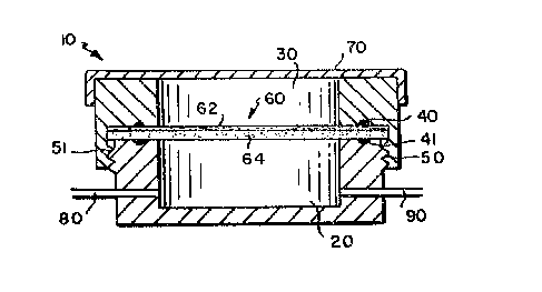
Referring to the trawings, Fig. LA illustrates one embodiment of an
apparatus according to the present invention for determining the
interaction of skin and one or more agents by use of skin tissue
equivalents, the apparatus comprising a container 10, the container
providing means for positioning the skin tissue equivalent 60, a lower
-18-
13~S~
chamber 20 and an upper chamber 30, the chambers being sealably
connected by means of an upper gasket means 40, a lower gasket means 41
and connecting means 50, 51. This embodiment is provided with a skin
tissue equivalent 60, removably positioned between the lower chamber 20
and the upper chamber 30. The skin tissue equivalent 60 comprises two
A layers, layer 62 comprising an epidermal layer, layer 64 comprising an
er,n ~/
elY~hrr=~ layer. In this embodiment the container 10 is provided with
~- cover means 70 which is placed over the upper chamber 30, and the lower
chamber is provided with ports 80, 90.
In the embodiment show in Fig. lA, the container is provided with
sealably connected lower and upper chambers 20 and 30 to provide means
for postioning the skin tissue equivalent 60 in the container 10. Other
means may be used for positioning a tissue equivalent in the container
10 such as positioning the tissue equivalent in the container by means
of a permeable support member tisposed in the container, wherein the
tissue equivalent attaches to or is cast on the permeable support
member. One such embodiment of the present invention is shown in Fig.
lB. Element~ similar to those in other described embodiments are .
inticated by the sama numeral. This embodiment comprises a container 10
t defining a hold~r for a skin tissue equivalent 60, container 10 having
disposed therein a permeable support member 65, the support member 65
providing means for positioning the skin tissue equivalent 60 in the
container 10. This embodiment further comprises a skin tissue
-19-
~: ~.3~5~
equivalent 60, urther defining a lower chamber 20 and an upper chamber
30. In this embodiment, the container 10 is provided with a cover means
for closing the container 70, which is placed over the upper chamber 30,
and the lower chamber is provided with ports 80, 90.
Figs. 2 and 3 show yet another apparatus according to the present
invention. Elements similar to those in other described embodiments are
indicated by the same numeral. The apparatus comprlses a cylindrical
cDntainer 10 for a tubular tissue equivalent, ths container having two
ports, 81, 82, at the bottom, a hollow mandrel 100, the upper end of the
mandrel being provided with a port 71, the lower end of the mandrel
being open, and a cover means 70 for closing the container 10, the cover
means being sealably connectable to the mandrel 100. The inner surface
12 of the container 10 comprises an inert, non-wettable material. The
container 10 is threaded to accept the cover 70 for removably sealing
the container 10. The cover means 70 is provided with a port 72. The
port 72 may also serve as a vent during filling or a separate vent 73
may be provided. The container lO is further provided, at the bottom
thereof, with a means (not shown) for sealing the mandrel 100 to the
base of the container. In some embodiment~, the bottom of the mandrel
100 is provided with an inlet or outl0t means which is provided with
means for sealing the bottom of the container.
The mandrel 100 shown in Fig. 3 comprises a number of regions:
region 105 comprising a means for sealably covering the container;
-20-
~3~ 3 (
regions 101 and 10~ comprising means for the cells of the tubular tissue
equivalent to adhere to; regions 103 and 107 comprising means for
limiting the longitudinal contraction of the tubular tissue equivalent;
region 104 comprising a liquid permeable means for allowing selected
materials to pass through the tubular tissue equivalent and, in some
embodiments for supporting the tissue equivalent; and region 109
comprising means (not shown) for removably sealing the mandrel 100 to
- the base 1~ of the container 10 ~e.g., groove fos 0-ring or screw
threads).
The container 10 and cover means 70 may be made of any deslred
material which does not react with or have an undesirable effect on the
components of the assay, including the tissue equivalent. In some
embodiments, it is desirable that the container 10 be made so that the
tissue equivalent is visible through the container, e.g., through the
walls of the container or through 8 window in the container. Preferrcd
materials for the container include glass, polycarbonate, polystyrene,
TEFL0 ~ and stainless steel. In yet other embodi~ents, the inside 12
,
oi' the container 10 comprises an inert, non-wettable surface such as
IEFLO ~, poly¢arbonate or stainless steel or the inside 12 is coatet
,.~
to make it non-wettable.
The container may be of any shape and volume which will accomodate
the size and shape of the desired tissue equivalent. The dimensions of
the container will again depend upon the size and shape of the desired
( ~3~)5~3~3 (
. . .
tissue equivalent and the desired assay volumes. For example, a
container ha~ing an outer diameter of about 25mm and a volu~e of about
5ml is useful in practicing the present invention. In some embodiments
of the present invention, multiple containers will be provided ~n a base
or holder.
In embodiments wherein a permeable support member 65 is provided for
positioning the tissue equivalent 60 in the container 10 the permeable
support member 65 preferably comprises a membrane or a mesh. Preferred
materials include polypropylene, nylon, and polycarbonate.
Ihe gasket means 40, 41 may be made of any material which is inert
to the conditions of the assay and the tissue equivalent being used, as
well as provides a good seal between the upper And lower chambers 20,
30. Preferred materials include silicone and TEFL0 ~.
In the embodiment shown in Figure 1, the upper and lower chambers
20, 30 are attached by screw means S0, 51. It will be appreciated by
those of ordinary skill in the art that any suitable connecting means
~ay be uset to sealably attach the upper and lower chambers.
The apparatus shown in Figs. LA snd lB are provided with ports so
that a liquid phase may be passed into ant out of the lower chamber 20.
The desired number of ports for the lower chamber will depend upon the
assay being conducted.
In the embodiment shown in Fig. 3, the cover means 70 for the
container 10 is provided with a valve 71, a port 72 and a vent 73. In
-22-
some embodiments, a vent is not provided, and a port 72 may serve both
as a port snd as a vent. The port may be used, e.g., for epidermalizing
the tissue equivalent or applying test agents. It must be understood
that although the apparatus shown in the Figures are provided with cover
means 70 for closing the container 10, alternative means for closing the
container 10 are acceptable. In alternative embodiments, access to the
inside of the container or a tissue equivalent positioned therein is
achieved by means of ports or other openings or passages appropriately
disposed on the container. These openings or passages will be provided
with valve means or other means for closure. In yet other embodiments
the means for closing the container may be sealed or fused to the
container, e.g., as by heat sealing. In such embodiments access to the
container is achieved by removing the sealed or fused means for closing
the container.
In the embodiment shown in~Figs. 2 and 3, the mandrel 100 provides a
means for positioning the tissue equivalent in the container 10. In
~ome embodiments of the present invention, the tissue equivalent is cast
on the mandrel 100 after the mandrel 100 has been disposed in the
container 10.
As has been described above, the mandrel 100 is comprised of a
number of regions. Preferred materials for regions 105 and 109 are
rigid, nonporous and inert, and include plastics such as polycarbonate
or polystyrene, TEFL0 ~ glass or stainless steel or combinations
-23-
. , . ,, , . _ . , , _ _ _ . _ . _ . _ . .. , .. . _ . . , .. _ .
.. .. .. .. .. . . . . . .. .
13~
thereof. Regions 101 and 108 comprise means for the cells of the tissue
equivalent to adhere to and in some embodiments also provide a seal
between the tissue equivalent and the mandrel 100. Preferred materials
for regions 101 and 108 include glow dischar~e treated plastic, collagen
or fibronectin-coated glass or plastic. Regions 103 and 107 provide
means for limiting the longitudinal contraction of the tissue
equivalent. Regions 103 and 107 preferably are highly textured.
Texture can be provided, e.g., by providing the region with holes or
with numerous fine projections. Preferred materials include, but are
not limited to, VELCR ~, textured stainless steel, e.g., wire cloth,
textured plastics, textured TEFL0 ~ and polyurethane foam. Preferred
materials for region 104 include materials which are permeable ~o small
molecules such as nutrients, but are of limited permeability to larger
molecules such as collagen and especially to collagen fibrils. Such
liquid permeable means include strong but inert materials such as nylon
or polypropylene. Pore sizes of about 0.2 to 5~m are useful; pores
of 0.5 to 3~m are preferred. In some embodiments support means is
provided for the membrane. Preferred materials include a framework or a
screen oi' a rigid and inert material, such as stainless steel, plastics
including polycarbonate or polystyrene, TEFL0 ~.
Apparatus according to the present invention may be provided with
means for controlling the flow oi the liquid phase(s) into and out of
the apparatus. Means for controlling the flow of a liquid are well
-24-
- ( ~3~
known to those skilled in the art. For example, means for sampling,
circulating, exchanging or feeding the liquid phase(s) ad~acent to the
tissue equivalent may be provided. In the embodiment shown in Fig. lA,
the liquid phase in chamber 20 may be moved through the apparatus by
attaching ports 80 and 90 to any appropriate circulatory means known to
those skilled in the art.
The present invention provides methods of determining the
interaction of tissue with at least one agent by use of at least one
tissue equivalent, the method comprising the steps of: (a~ contacting
the agent with a tissue equivalent, wherein the tissue equivalent is
adjacent to a liquid phase; and (b) determining the interaction of the
agent with the tissue equivalent by analyzing at least one of (i) the
tissue equivalent, Sii) an intracellular fluid of the tissue equivalent,
or (iii) the liquid phase.
In some embodiments of the present invention, the interaction of
skin and at least one agent is determined by the use of at least one
skin tissue equivalent, the skin tissue equivalent having an epidermal
and a dermal layer, the method comprising the steps of: (a) contacting
the agent with the epidermal layer of the skin tissue equivalent,
wherein the dermal layer of the skin tissue equivalent i5 ad~acent to a
liquid phase; and (b) determining the effect of the agent on the skin
tissue equivalent by analyzing at least one of (i) the skin tissue
equivalent, (ii) an intracellular fluid of the skin tissue equivalent,
or (iii) the liquid phase.
-25-
.. , . . ..... . . . . . _ _ _ . . _ ... _ ... . , . _ _ . . . .
.. _ _ . . . _.. . _ . . ...... , , , _ ,,
: ~3~)5~ ~
Apparatus, methods and Xits based on human skin tissue equivalent
are provided by the present invention for use in determining the
interaction of skin and various agents, including, but not limited to,
the measurement of:
1) the rate and extent of penetration of substances into and through
the skin ~Skin Absorption Test);
2) the interaction of agents reflected by changes in cell
permeability (Skin Cell Permeability Test);
, .
3) the responses of skin cells to agents that provoke or promote the
release of various regulatory or signaling molecules ~nto the
intracellular tissue fluids (Skin Chemical Response Test); and
4) the responses of skin cells together with specialized immune
cells to substances that are true allergens (Skin
Immunoreactivity Test).
Such apparatus, methods and kits provide means for quantifying the
. interaction of human skin tissue equivalent to agents. The results of
such tests should reflect the response of natural human skin more
closely than a corresponding test conducted with animal or cadaver skin.
In some methods according to the present invention, the epidermis ii
exposed to a gaseous atmosphere, while the dermis is bathed by a sterile
tissue culture fluid freely exchangeable with the tissue fluids of the
skin tissue equivalent. The skin tissue equivalent is positioned
between a gaseous phase (e.g., air) and a fluid phase (e.g., culture
-26-
..
.. , ., . , . , , , . . , ..
_ .... ... .. . , . . , . , _
.~, .
~ 3i[~ 3
medium) (See, e.g., Fig. 1). In such embodiments, the dermal layer of
the skin tissue equivalent is ad~acent to, typically in contact with,
the fluid phase that serves as a nutrient medium simulating the "milieu
interieur" while the corr.ified epidermal layer contacts the gaseous
phase simulating the environ~ent. The skin tissue equivalent provides a
fluid-tight seal between the two phases; the test substance is applied
to its epidermal surface in an appropriate vehicle. The skin tissue
equivalent itself, the tissue fluids thereof and the culture medium are
each available for analysis.
The composition of skin tissue equivalents for use in tests
according to the present invention will vary depending upon the nature
of the test to be conducted.
The present invention provides apparatus, methods and kits for use
in skin absorption tests which permit a determination of the amount of
an agent that traverses the skin during a predetermined period of time.
With the exception of the initial phase of absorption, appendageal
absorption has generally been found to play a minor role. Thus standard
skin tissue equivalents, although lacking appendageal openings such as
hair follicles and sweat glands, are expected to be suitable for most
steady-state absorption studies. However, in some embodiments of the
present invention, it may be desirable to fabricate skin tissue
equivalents with a number of cell-lined pores that provide a physiologic
ratio of appendageal to non-appendageal percutaneous absorption. Long
. . . _
3~3
term testing using skin tissue aquivalents is possible because skin
tissue equivalents can be maintained in a viable state for many months
or even longer.
Skin absorption tests according to the present invention may include
measuring the amount of an agent in the liquid phase adjacent to the
dermal component of the skin equivalent, or directly in the skin tissue
equivalent itself or lntracellular fluids thereof since lipid soluble
substances, for example, tend to become entrapped in skin. Agents used
in such tests are capable of being detected in some way, e.g., labeled
with radioactivity, or are measured by direct assay, using methodologies
known to those skilled in the art.
Some methods according to the present invention measure the kinetics
of skin penetration by the agent(s) under test. In some such
embodiments, an apparatus according to the present invention is provided
with multiple containers in a holder therefor, mounted into a ~ig and
coupled to a reservoir for feeding each sample skin tissue equivalent
periodically or continuously with, e.~., a nutrient solution to bathe
the dermis. The pumped or gravity-feed output allows for fraction
collecting so that flow through as a function of time under conditions
simulating blood flow is measurable. The data is used to calculate the
permeability constant or other relevant parameters, such as, the time
for initial permeation or the percentage absorbed of the agent. Such
methods also provide an optional calibration step that uses tritiated
-28-
.
~3i ?5~3
water before and after testing penetration so that the effects of the
agent on the barrier properties of the skin tissue equivalent are
determined.
The present invention also provides apparatus, methods and kits for
use in skin cell permeability tssts to determine changes in cell
permeability brought about by the interaction of skin and agents such as
chemicals, cosmetics or drugs. The release by cells of strictly
intracellular proteins may serve as an indicator of cell damage due to
the agent under test. Dose response data is derived by testing multiple
skin tissue equivalents using apparatus, methods, and kits in accordance
with the present invention, each skin tissue equivalent being positioned
in an individual container which is provided with its own fluid phase,
the contents of which are available for assay. The release of a
cytoplasmic protein such as LDH is measured, for example,
chromogenically. Release of at least one cytoplasmic and one lysosomal
enzyme is measured by use of appropriate assay techniques well known to
those skilled in the art. For example, various protein-binding
methodologies including radioimmunoassay (RIA), enzyMe immunoassay
~IA), enzyme-linked immunoassay (ELISA), and fluorescent immunoassay
are useful. The skin cell permeability test of the present invention is
especially useful for discriminating between irritant and corrosive
agents since, in general, the former are expected to give negative
results, and the latter, positive results.
~ -29-
` ~3~ 3 ~
The present invention also provides apparatu-q, methods and kits for
US8 in skin chemical response tests for the measurement of physiological
responses of skin cells to agents that induce edema and erythema or
inflammation. The test is designed to quantitate complsx tissue
reactions resulting from multiple chemical triggers. Chemicals released
from cells into the tissue fluids in response to the dgent(s) under
consideration are assayed. These include, for example, prostaglandin
E2, prostacyclin and other signaling factors of arachidonic acid
derivation as well as Interleukin I that can amplify the response by
stimulating fibroblasts of the dermis to secrete prostacyclin. Multiple
skin equivalent samples provide data for dose response curves.
The degree of release is quantitated using techniques known to those
skilled in the art. By testihg known contact irritants, release of one
or more of these mediators is correlated with the classical, though
difficult to quantitate, responses of erythema and edema. While the
keratinGcytes of the skin tissue equivalents for use in methods provided ~ ;
by the present invention are expected to be the principal emitters of
proqtaglandln E2, in some embodiments capillary endothelial cells are :
, ..... . .
optionally included in the dermis of the skin tissue equivalent to
... .
~'i provide a response source for prostacyclin. Although both factors have
relatively short half lives, they can be measured by assaying their
degradation products using methods known to those skilled in the art.
Apparatus, methots and kits for use in skin immunoreactivity tests
are also provided by the present invention to measure responses to
-30-
.. , .... , .. , . , . .. , .. , .. ,.. .. . ::___ _ _. ._ _ .. ..... _ .. .. _ .
.... _ _ .. ..... , _ .. _ . . .. _. . . -
.
various agents which emanate from in~une cells incorporated into the
skin tissue equivalent, for example, an agent capable of causing contact
sensit~vity by forming covalent bonds with proteins. The i~munogen will
often be a complex of selr-protein acting as "carrier" and the contact
sensitizer acting as hapten.
In some embodiments of skin immunoreactivity tests according to the
present invention, the macrophage-like Langerhans cells (LC) of normal
~ n ~ e~
: ~L3 skin which process imu~i~ complexes for presentation to other immune
cells are incorporated in the epidermis of the skin tissue equivalent to
provide the basis for the first step in the immune reaction chain
involving the skin. Measurement of the migration of activated LC out of
the epidermis of the skin tiss~-e equivalent and of the protein-hapten
complex are suitable assays of allergenicity. The movement of cells may
be followed by immunofluorescence or other methods known to those
skilled in the art and may be correlated with allergenicity by using
substances of known allergenicity. In other embodiments, macrophages
re optionally included in the sXin tissue equivalent to provide another
source of the assayable lymphokine, IL-l, secreted by the cells in
r0sponse to substancss that initiate humoral immune reactions. In yet
other embodiments, subsets of sensitized T cells that will respond to
particular classe~ of immunogens, together with mast cells that release
strong signals such as histamine, are optionally incorporated in the
skin tissue equivalent to provide immune signals, easily assayed because
of their degree.of amplification.
-31-
., . _ . . .. __ __ , _ . . _ _ .. . .. _ .. .. . _ , .
3 (
-- Mediators are collected from the culture fluid in contact with the
dermis or from the tissues themselves for assay. ~ith the skin
irritation test and ~kin chemical reaction test, the user will be able
to discriminate between irritants and allergens.
The apparatus and methods of the present invention may be used in
the production of kits for determining the interaction of tissue with at
least one agent by use of one or more tissue equivalents. One kit
- according to the present invention comprises the following in
combination:
(a) an apparatus for determining the interaction of tissue and at
least one agent by use of at least one tissue equivalent, the
. ~ ~ss,u~
apparatus comprising a container ~or the ticuoo equivalent, the
container comprising:
(i) means for positioning the tissue equivalent in the
container, whereby the tissue equivalent defines at
least one region in the container;
(ii) -at least one port; and
(iii) means for closing the container; and
(b) a tissu,3 equivalent.
. , .
' In preferred embodiments, the tissue equivalent defines at least two
regions.
In some embodiments of kits according to the present in~ention, the
tissue equivalent defines an upper and a lower reglon in the container.
-32-
, .. .. . .
~3~ 3
In yet other embodiments wherein the tissue equivalent i9 a tubular
tissue equivalent, the tubular tissue equival0nt defines an inner and
outer region in the container. In some such embodiments, the contalner
is provided one or more liquid phases.
In preferred embodiments of kits according to the present invention,
the apparatus is provided with two or more individual containers. In
some such embodiments, the apparatus may be provided with means for
hooking to a manifold in order to sample or perfuse individual
containers. In yet other embodiments, the containers are interconnected
so that the liquid phase adjacent to the tissue equivalents is common to
all the tissue equivalent samples. In such kits, the tissue equivalent
in each container is accessible for contact with the agent(s) and the
liquid phase in each container is accessible fox analysis.
Such kits further optionally comprise one or more agents as well as
one or more of the reagents necessary for tstermining the interaction of
the agent and the tissue equivalent. Various assay techniques known to
thoss skilled in the art are used to deter~ine the interaction of the
tissue equivalent and the agent and, include, but are not limited to,
histological analysis, mass spectrometry, magnetic resonance imaging,
ultrasonic imaging, radioactive tracer methodologies, radioimmunoassay,
enzyme immunoassay, enzyme-linked immunoassay, and fluorescent
immunoassay.
... , . . .. , .. .. _ .. . .
~3~ 3 "
The present invention provides also kits for determining the
interaction of skin tissue with at least one agent. In one embodiment,
such skin tissue equivalent based kits comprise in combination:
(a~ an apparatus for determining the interaction of skin tissue and
at least one agent by use of at least one skin tissue
equivalent, the apparatus comprising a container for the skin
tissue equivalent, the container comprising:
(i) means for positioning the skin tissue equivalent in the
container, whereby the skin tissue equivalent defines an
upper and a lower region in the container, the lower
region further comprising a liquid phase; and
(ii) at least one port; and
(iii) means for closing the container; and
(b) a skin tissue equivalent having an epidermal and a dermal
layer, the dermal layer being adjacent to the liquid phase.
In other embodiments, a second liquid phase ls disposed between the skin
tissue equivalent and the means for closing the container. In such
embodiments, it may be desirable to remove the second liquid phase
before contacting the agent and the skin tissue equivalent. In yet
other embodiments the apparatus is provided with two or more individual
containers or two or more interconnected containers.
Other embodiments of apparatus according to the present invention,
e.g., embodiments comprising tubular tissue equivalents, such as tubular
skin tissue equivalents, may be included in kits provided by the present
invention.
-34-
,, ._ . .. ,, .--.. . .
~.3~ 3 f
In some embodiments of the present invention, it may be desirable to
determine the interaction of an agent wLth different types of tissue.
For example, the interaction of liver tissue and an agent which has
pene~rated the skin could be determined by first passing the agent
through a skin tissue equivalent in accordance with the present
invention, collecting the agent or its breakdown products, and then
contacting the collected agent or breakdown products, with a llver
tissue equivalent in accordance with the present invention.
Apparatus, methods and kits based on human blood vessel tissue
equivalents are also provided by the present invention for use in
determining the interaction of the blood vessel tissue equivalent and at
least one agent. Such interactions include but are not limited to the
effect(s) on vascular physiology, blood-surface interactions and
pathological processes.
In general, a blood Yessel tissue equivalent based test system
comprises the appropriate type of blood vessel tissue equivalent
incorporatet into a circulatory loop. The circulatory loop is
complstely mechanical (pump, tubing, reservoir, etc.) or biological with ~;
the bloot vessel tissue equivalent incorporated ex vivo as in an
aterio-venous shunt. The circulating fluit is culture metium, culture
~, , .
- medium containing blood elements such as platelets, heparinized blood,
or ~as in the ex vivo loop), untreatet blood. Other cells such as
macrophages are optionally added to the blood vessel tissue equivalent
or the circulating fluid as desired.
-35-
, . . : ., ., . , ... ., _ ., . . . _ _ _ _,,_ _ ._ . . .. .... _ _ . .. , . _
... . . . ... . .
~3~5~
Figs. 4A and 4B show one e~bodiment of an apparatus in accordance
with the present invention for determining the effect of one or more
agents on blood vessels by use of blood vessel tissue equivalents, the
apparatus comprising a container 10 for the blood vessel tissue
equivalent, a cover means 70 for sealable connection thereto, the cover
means 70 being provided with two ports 14, 16. The container 10 is
further provided with two valved cannulae 15, 17 to provide means for
positioning the blood vessel tissue equivalent in the container 10.
Fig 4C is a schematic illustration of the container 10 shown in
Figs. 4A and 4B Lncorporated into a mock circulatory loop which
comprises tubing 120, a reservoir 121, a pulsatile pump 122, the
circulatory loop being provided with a port 123. The circulatory loop
also comprises a pressure transducer (not shown) connected to an
ampliiier and chart recorder (not shown). The reservoir 121 is provided
with a means (not shown) for bubbling gas through the circulating fluid
to provide gas exchange.
. , , i
~- The same general materials msy be used for constructing the
embodiment shown in Figs. 4A and 4B as are described for use in
constructing other embodiments described herein. As in other
~; embodiments, the cover means for the container is optional, access to
the contalner and tissue equivalent being provided via ports or other
appropriate means.
Preferred materials for the tubing include medical grade tygon,
teflon, silicon, stainless steel or other inert tubing.
-36-
... . .... . ..
,
,~,, ., ", ,
~ 3 $y~fe~ns
Two examples of blood vessel equivalent based test syctssmc are the
atherogenesis te.st system and the metastasis bio-test System.
The atherogenesis test system is designed for the study of the
formation and amelioration of atherosclerotic plaque in vitro. It
consists of a mock circulatory loop, illustrated in Fig. 4C, comprising
tubing 120, a blood vessel tissue equivalent 68 made with human arterial
cells in a container 10, a pulsatile pump 122, a pressure transducer
(now shown), and a fluid reservoir 121. The circulating fluid is
hyperlipidemic serum or culture medium supplemented with known
atherogenic factors such as low density lipoproteins. Macrophages are
optionally included as precursors to foam cells. The blood vessel
equivalent is subjected to local mechanical or other type of injury to
induce plaque initiation (Ross & Glomset, 1976). The sys~em allows the
experimenter to manipulate the circulating fluid, the pressure and
pressure changes, the constitutents o the artery equivalent, etc. to
determine their role in atherogenesis and their response to
pharmacological treatments.
The metastasis test system is designed for the study of the passage
of cancerous cells across cap$11ary walls. Most tumor metastases are
blood-borne so the various steps in metastasis, includine invasion of
the vessel, circulation, attachment, and diapedesis are likely sites for
general antitumor agents to act. A circulatory loop similar to that
shown in Fig. 4C, is used. The metastasis bio~test system comprises a
~L3~ 3
low pressure, low flow rate mock circulatory loop containing a capillary
equivalent (either as a single tube lined with capillary endothelial
cells or as a capillary network grown within a collagen lattice), and
ports for adding transformed cells to either the luminal or abluminal
fluid compartments of the blood vessel tissue equivalent. The time
course and steps in the metastatic process, as well as their response to
pharmacologic agents are easily studied in this in vitro system.
The present invention also includes kits for determining the
interaction of blood vessel tisuse equivalents and at least one agent~
similar to those kits descr~bed hereinabove.
The invention will be further understood with reference to the
following examples, which are purely exemplary in nature, and are not
meant to be utilized to limit the scope of the invention.
~ EXAMPLE 1 -- SKIN ABSORPTION TEST
Skin tissue equivalentts~ cast as a sheet used to test percutaneous
absorption of benzoic acid.
1) A dermal equivalent i8 cast with human dermal fibroblasts using the
method of Patent No. 4,485,096. A suitable size for casting is a
total volume of 15 ml. in a Petri dish 100 mm in diameter.
,~i
2) The dermal equivalent is seeded with human epidermal cells. Small
droplets of a cell suspension, made with cultured epidermal cells or
freshly dissociated epidermis, are applied to the surface of the
~ . .. .
;
dermal equivalent (See, e.g., Patent No. 4,485,096). Alternatively,
- pieces (strips, punches, or other shapes) of skin or of skin tissue
equivalent are applied to the surface of the dermal equivalent (See,
e.g., Patent No. 4,604,346). The seeded dermal equivalent is
cultured to allow a complete epidermal layer to form.
3) The lower half of the test chamber is filled with fluid (typically
culture medium or buffered salt solution). The completed skin
tissue equivalent is transferred to an apparatus in accordance with
the present invention such as shown in Figs. lA or lB, chamber so
that the entire lower chamber and gasket are covered. The upper
gasket is placed on upper surface of the skin tissue equivalent.
The diameter of the chamber is about 25 m~. ~he upper half of the
test chamber is screwed on tightly enough to make an effective seal
without crushing the skin tissue equivalent.
4) Any excess fluid is aspirated from the upper surface of the skin
tissue equivalent. Flow of buffered salt solution or other
collection fluid is started through the lower chamber and collected
ln a fraction collector. -
5) The agent(s~ may be applied, its passage throu~h skin tissueequivalent assayed, and time course and absorption calculated as in
J. Pharm. Sci., 74, 65-67 (1985). The test sample is applied to the
upper surface of the skin tissue equivalent. For example, 114C]
-39-
....
,, . , . . , . .. , , , ., . , . . , .. _ , _ _ . . ~ . . . . ., _ , ..
~ 3
.:
sen~oic acid in petrolatum as a vehicle (suitable concentrations are
10, 100, and l,OOO ng/mg) with 25 ~g of vehicle applied per cm2 of
skin tissue equivalent.
6) Sa~ples of the effluent are collected and counted by standard
methods known in the art in a liquid scintillation counter.
Alternatively, the skin tissue equivalent sample itself may be
washed (with buffered saline~, removed from the container,
solubilized (for example in a solution 0.5~ sodium dodecyl sulfate
and 4M urea in water), and aliquots counted to determine
radioactivity trapped in the skin tissue equivalent. This approach
is useful for hydrophobic compounds which may not readily pass rrom
the skin tissue equivalent into the collection fluid.
This method is further refined by separating the epidermal and
dermal layers using methods known to those skilled in the art, e.g.,
mild trypsin treatment.
EXAMPLE 2 -- SKIN CELL PERMEABILITY TEST
1) Prepare and mount the desired amount of skin tissue equivalents in
apparatus in accordance with the present invention as described in
example 1, above.
2) Apply the agent(s) to upper (epidermal) surface of the skin tissue
equivalent A. As an example, the chemicals used in the FRAME test
(Bells, M. & Bridges, J. U., in Acute Toxicity Testing: Alternative
Approaches, pp. 63-79, 1984; A. M. Goldberg, ed., N. A. Liebert,
-40-
~3~5~3
Inc., New York), are applied to individual samples of skin tissue
equlvalent in aqueous solutions at various concentrations.
3) The effluent fluid is collected from the lower chamber and assayed
for a cytoplasmic enzyme produced by the skin tissue equivalent in
response to the agent(s) (e.g., Lactate Dehydrogenase, EC 1.1.1.27)
and a lysosomal enzyme (e.g., ~-glucuronidase, EE 3.2.1.31).
The enzyme released is assayed by standard methods known to those
skilled in the art (See, e.g., Methods of Enz~matic Analvsis, H. U.
Bergmeyer, E. Verlag Chemie, Ueinheim, 1983 III, 118-138 and IV,
246-256 respectively).
4) Dose response curves of enzyme release which is related to cellular
damage are prepared from the data.
~ EXAMPLE 3 -- SKIN IMMUNOREACTIVITY TEST
1) A container is used which consists of two chambers defined by a
membrane or mesh. See, e.g., Fig. lB. The membrane or mesh is a
disk about 25 mm in diameter and has an average pore size in the
range 0.5 to 3.0 ~m. The membrane is made of, or coated with, a
material to whlch cells and collagen adhere (e.g., glow discharge
treated polystyrene or collagen coated controlled pore si~e glass).
The lower chamber is provided with ports for medium sampling or
flow-through. The upper chamber is provided with a cover means,
removable for sample application. The walls o the upper chamber do
-41-
~3~?~ 3
not promote cell adhesion (e.g., polycarbonate, TEFLO ~ or
siliconized polystyrene).
2) A dermal equivalent is cast, as described in Example 1, above, in
the upper chamber. In addition to dermal fibroblasts, the mixture
contains cells of the immune system. One or more of the following
cell types are included- Langerhans cells, macrophages, T-cells,
mast cells and microvascular endothelial cells. Other cell types
may be included as desired. A specific formulation includes
Langerhans cells, macrophages, and mast cells.
3) The dermal equivalent contracts, constrained by adhesion to the
- membrane, to form a disk which separates the upper and lower
chambers.
4) The dermal equivalent is seeded with epidermal cells (typically 3-lO
days after casting). The skin tissue equivalent is cultured to
develop a fully keratinized epidermal layer. See, e.g., J. Invest
Dermatology 81:25-105 (1983);
5) An agent (potential allergen) is applied to the epidermal surface.
One or more mediators of allergic responses is collected in the
lower chamber and assayed by standard means. Examples include
interleukin-l (IL-l) released by Langerhans cells and macrophages,
and histamine released by mast cells.
-42-
~3~
EXAMPLE 4 -- SKIN CHEMICAL RESPONSE TEST
Irritant Che~ical.
1) The mandrel 100 and con~ainer 10 shown in Figs. 2 and 3 are
assembled. A dermal equivalent is cast through port 72 as described
in Example 1 in the volume or space defined by the mandrel 100 and
the container 10 as assembled. The volume of dermal equivalent
casting mi~ture is adjusted so that the entire mesh 104 and means
for cells to adhere 103 are covered. Thus, for the dimensions
given, a volume of about 50-60 ml would be required. The dermal
equivalent is allowed to gel and to contract around the mandrel
100. The holes 103, 107, limit longitudinal contraction so that the
mesh or membrane portion 104 remains completely covered. If
necessary, the lumen of the mandrel 100 can be filled through port
71 simultaneously with the container 10 to prevent flow of the
casting mixture into the lumen of the mandrel 100.
2) Then (typically after 3-10 days), the dermal equivalent will be
seeded with a suspension of epidermal cells through the port 72. A
multiple barelled micropipettor ls inserted through port 72 and tiny ~ !
troplet~ (typically 1-5 ~1) of a cell suspension are applied so
that ~he entire surface of the dermal equivalent is uniformly seeded
with epidermal cells. These are grown to form a keratinized
epidermal layer.
3) The agent(s) is then applied to the epidermal layer of the formed
skin tissue equivalent and the lumen of the mandrel is perfused with
-43-
.. .. ... .. ,, . . . . . . .. , . --
13Q~ 3
the collection fluid. An irritant substance (See, e.g., M.
-- Steinberg et al. (In Animal Models in Dermatolo~v, H. Maibach, ed.,
Churchill Livingstone, 1975, pp. 1-11)) may be applied in a dressing
(e.g., gauze pad). This is occluded by covering with an impermeable
material, e.g., plastic film, if desired. The test dressing is
removed after a suitable time (e.g., 24 hr).
4) The perfusate is assayed for substances released by the cells in
response to the agent(s) (e.g., Prostaglandin E~) using methods
known to those skilled in the art. For example, commercially
available Prostaglandin E2 radioimmunoassay kits may be used.
EXAMPLE 5 -- INCORPORATION OF BLOOD VESSEL TISSUE
EOUIVALENT INTO MOCK CIRCULATORY LOOP
1. A blood vessel tissue equivalent was made using arterial cells as
described in Science, 231: 397-400 (1986). The blood vessel tissue
` equivalent was fabricated with an inside diameter of about 6mm and a
i length of about lOcm.
2) It was transferred to a container 10 (See, e.g., Figs. 4A, 4B) and
tied onto the end of the cannulae disposed inside the container 10
by means of 4-0 silk ligatures. Tha ligatures were tightened
sufficiently to hold the blood vessel tissue equivalent firmly
without cutting through the blood vessel equivalent wall.
3) The container was incorporated into a mock circulatory loop as shown
in Fig. 4C.
-44-
,. .
,.
~3~ 3
4) Culture medium was circulated through the loop. The blood vessel
- ;equivalent dilated and contracted with changes in pressure.
p h y ~ ~ /~
~J Pressure variations were achieved within ~hyL=L~L~l pressure
ranges (80-200 mm Hg) and frequencies (60-100 pulsations per
minute).
EXAMPLE 6 -- ATHEROGEMESIS TEST
,.
- 1) Several blood vessel tissue equivalent(s) are transferred into
apparatus provided by the present invention as described in Example
5 and the apparatus are incorporated into mock circulatory loops as
illustrated in Fig. 5.
2) In all but the control blood vessel tissue equivalents, the
circulatory medium is supplemented with atherogenic factors such as
low density lipoprotein (LDL), which is a major cholesterol carrier
in the bloodstream.
3) Local in~urieR are produced by mechanicsl, thermal, or other means
in the endothelial lining of the blood vessel tissue equivalents.
4) The blood vessel tissue equivalents are sub~ected to high pulsatilq
pressures (e.g., peak about 200 m~ Hg; valley about 120 mm Hg).
5) The blood vessel tissue equivalents are examined at various times
for indications of artherogenesis, e.g., for proliferative lesions
(atherosclerotic plaques) in the wall and for deposits of lipid
(principally cholesterol) at the sites of local injury.
-45-
~ ~Sq.~'3
- Standard histological techniques are used for the examination.
Non-invasive techniques could be developed which include ultrasound
or Magnetic Resonance Imaging (MRI).
~XAMPLE 7 -- METASTASIS TEST
Used to Determine if Metastatic Potential is Correlated With
Abilitv to Cross Blood Vessel Tissue Equivalent Wall
1) Blood vessel tissue equivalents are prepared as described in Example
S except that capillary endothelial cells are used rather than
arterial endothelial cells. The blood vessel tissue equivalents are
incorporated into mock circulatory loops illustrated in Fig. 5. A
simple peristaltic pump provides sufflcient perfusion.
2) In all but control blood vessel tissue equivalents, transformed
cells (e.g., B12 melanoma cells) are injected into the circulatory
loop. The fluid in the abluminal compartment of the perfusion
chamber is collected and replaced at intervals.
3) The cells which cross the blood vessel tiSSUQ equivalent wall into
the inner region are collected by centrifugation, counted and
in~ected into mice. The metastatic potential is determined
according to Poste ~ Fidler, ~5~E~, 283:139-146 (1981) and
correllated with the time that the sample is collected.
Alternatively, the blood vessel tissue equivalent could be perfused
with clones of differing metastatic potential and the rate at which
-46-
~` ~3~
the cells fro~ different clones cross the blood vessel tissue
equivalent wall could be measur~d.
. .1
, . .
,.
-47-
.
.... . . , ~ . .. .. ., _ .. , .. _ _. _ . ... .
-
-
It is understood that the exampl~s and embodiments described herein
are for illustrative purposes only, and that ~arious modifications or
- changes in light thereof that will be suggested to persons skilled in
the art are to be included in the spirit and purview of this application
and the scope of the appro~ed claims.
" . ,
.
-48-
. , .. _, _ _ _ _ _ _ _ ... _ .... _ ... .. ... ... .
,.