Note: Descriptions are shown in the official language in which they were submitted.
~3~5~33
INTEGUMENT AND METHOD FOR CULTURING ORGANIC MATERIAL
Field of the Invention
The present invention relates to a new and improved
integument and method for micropropagation, tissue
culturing and the culturing of other organic material.
More particularly, the invention relates to a new and
improved integument and method for enhancing the growth
and reproduction of plant and animal cells and tissue,
bacteria and other microorganisms, and for preventing
contamination from occurring in the cultures.
Background of the Invention
Micropropagation is the process of growing new
generation plants from a single tissue sample that has
been excised from a carefully selected parent plant or
cultivar. This process permits the mass reproduction of
plants having certain desirable traits since substanti-
ally all of the new generation plants produced are
genetically identical to and have all the desirable
traits of the parent.
Tissue culturing is the process of growing cells in
~itro and is used to grow both plant and animal cells.
Tissue culturing techniques are commonly used in the
early stages of the plant micropropagation process where
it is desirable to rapidly produce plant cells. Improve-
ments in tissue culturing techniques also have applica-
tions beyond the micropropagation of plants. Essentially
the same culturing process is used to culture animal and
even human tissue, such tissue being used in the fields
of animal agriculture and human and veterinary medicine.
Culturing of organic material other than plant and animal
cells and tissue, such as bacteria, viruses and algeas,
is also performed in vitro for both research and
,
13~5~33
commercial purposes. Improvements in the procedures and
apparatus used to reproduce and maintain these organisms
would be beneficial, for example, to researchers and
industry who require a large or steady supply of such
material.
There are problems associated with the prior art
culturing apparatus and processes. One of the primary
problems is contamination. Any of a wide variety of
microorganisms, including viruses, bacteria, fungus,
molds, yeast and single cell algae, can ruin the cultures
during any of the various stages. The smallest of these
biological contaminants are the viruses, the largest are
the single cell algae. A virus typically ranges in size
from 0.1 to 0.45 micrometers although it is suspected
that portions of the virus which are as small as 0.01
micrometers may separate from the virus and alone cause
contamination. Bacteria typically range in size from 5
to 100 micrometers, while fungi and molds are usually
larger than 100 micrometers. Yeast is larger than
bacteria, with single cell algae, the largest of these
biological contaminants, being larger than yeast.
The prior art sterilized glass or plastic culture
containers such as test tubes, flasks or bottles, uti-
lized in conventional culturing technology have serious
drawbacks. For example, since plants require both carbon
dioxide and oxygen to live and grow, these containers
must provide a means for gas exchange. The walls of
these traditional glass and plastic containers, however,
do not permit the required gaseous interchange. Thus,
rubber stoppers having cotton packing or some similar
filter material, loosely fitting caps, or baffled plastic
caps have been employed to allow an adequate exchange of
gas between the tissue or plant and the ambient atmo-
sphere and environment. However, such devices restrict
the amount and rate of gas which can be exchanged.
Further, such caps and stoppers do not totally protect
the plant from contamination by microorganisms such as
13~S~33
viruses, bacteria and fungi. Thus, it has been of
paramount importance that the tissue culture room and
laboratory be kept extremely clean and their atmospheres
filtered. Further, precise temperature, humidity, and
light conditions must be maintained in the culture room.
Gas exchange is also required for culturing animal cells
and for certain other microorganisms. Traditional
flasks, petrie dishes ~nd the like, while allowing for a
certain degree of gas exchange, also allow contaminatio~
to occur.
The original cost of the traditional glass ~r
plastic culture containers; the labor and equipment cost
to maintain the sterility of the containers; and the
added cost of the facilities, equipment, and related
conditions required to maintain a sterile growing
environment, all represent major cost factors associated
with the use of such containers in conventional culturing
processes .
The present invention overcomes many of the defi-
ciencies of the prior art techniques of culturing by
having the following advantages:
(1) enhanced protection from contamination;
(2) increased growth rates;
(3) no requirement for a sterile culture room;
(4) no requirement for expensive glass con-
tainers or the incurrence of replacement costs due to
breakage;
(5~ no labor cost associated with cleaning and
sterilizing containers for reuse;
(6) an increase in the number of plantlets
from a culture;
(7) a reduction by approximately one-half the
amount of media required in each plant culture;
(8) the elimination of the requirement of
s*rict humidity control in the culture room;
(9) an increase in the number of cultures
which can be produced in the same size culture room;
13~5~33
(10) a reduction in the size of the media
preparation area and in the size of the autoclave; and
(11) an increase in the number of new cultures
which can be established by a laboratory technician.
Other objects and advantages of the invention will
appear from the following description.
Summary of the lnvention
The present invention includes a new integument and
related process for micropropagation, tissue culturing
and for the culturing of other organic material. The
integument is made of a semipermeable and transluce~t
membrane which allows light transmission and gas exchange
but seals out the biological contaminants in the ambient
environment. The membrane forms a plurality of cellules
which contain the organic samples and media. The
cellules are sealed so as to completely enclose and seal
off the cultures from the ambient environment. The most
preferred membrane is a high density polyethylene
material.
One of the principal advantages of the present
invention is that biological contaminants in the ambient
atmosphere cannot penetrate the membrane of the integu-
ment and thereby contaminate the culture. Yet, the semi-
permeable membrane ensures enhanced gas exchange, gas
exchange being necessary for plant and animal cells and
many microorganisms to live and reproduce. Because the
integuments are contaminant impermeable, organic material
contained therein need not be cultured in a sterile
environment, and the costs and problems associated
therewith are eliminated. Similarly, because the integu-
ment will not allow bacteria, viruses, and other micro-
organisms from the ambient environment to penetrate the
membrane, the integument may also be used to culture a
specific microorganism. The integument prevents the
microorganisms grown or maintained therein from escaping
the integument and possibly infecting laboratory
personnel and, at the same time, prevents microorganisms
, ~ ~
~ 3C~S933
in the ambient environment from contaminating the culture
of the desired microorganisms contained in the
integument.
The integument of the present invention is also
liquid impermeable so that the media, typically a liquid
or semi-solid which sustains the tissue, organism, or
plant's growth while in the integument, cannot escape and
dry out. Th~s, using the present invention, it is also
unnecessary to maintain a precise humidity level in the
culture room which would again require special and costly
equipment.
A completely unexpected benefit of using the
semipermeable integument is that tissue and plantlet
growth rates are dramatically increased. This increase
is believed to occur because oxygen and carbon dioxide,
which are needed for plant respiration and photosynthesis
and for sustaining certain bacteria, are available in
greater quantities than when the process is carried out
in prior art glass and plastic containers where the loose
fitting lids, rubber stoppers, caps and filters, which
are required to prevent the entrance of contaminants,
impede gas exchange.
A preferred embodiment of this integument is formed
from heat sealed high density polyethylene. This mate-
rial has been found impermeable to contaminants and,
because it is completely sealed once the organic sample
is in place, the entire outer surface can be thoroughly
decontaminated by emersion prior to opening the in~egu-
ment which exposes the culture to possible contamination.
There are virtually no areas on the integument where
contaminants can accumulate and avoid decontamination.
With the preferred embodiment of the invention, the
costs of micropropagation and culturing are greatly
reduced since the cost of the integument of the present
invention is much less than the cost of prior art con-
tainers. The preferred integuments are, unlike glass
test tubes, essentially unbreakable. Their low cost
13C~5933
makes them completely disposable, eliminating the costs
associated with washing and the often-less-than sterile
product which results.
The apparatus of the invention has other applica-
tions other than the culturing of tissue and microorgan-
isms. For example, improvements in growth rates wer0
observed when the integuments were used in growing plants
from seeds.
BRIEF DESCRIPTION OF THE DRAWINGS
,; _
For a detailed description of a preferred embodiment
of the. invention, reference will now be made to the
accompanying drawings, wherein:
Figure 1 depicts a frontal view of the integument of
the present invention;
Figure 2 depicts a partial elevation cross-sectional
view of the integument of Figure 1 taken along line 2-2
as shown in Figure 1 with the material of the integument
enlarged;
Figure 3 depicts a partial top view of the integu-
ment of Figure 1 with the material of the integument
enlarged;
Figure 4 depicts a meristematic tissue sample being
cultured in the integument of Figure 1;
Figure 5 depicts the initial tissue culture from
Stage 1 being multiplied during Stage 2 in a new integu-
ment of Figure 1;
Figure 6 depicts the growth of an individual plant-
let during St~ge 3 in a new integument of Figure l;
Figure 7 depicts an integument pack with individual
cellules of the type shown in Figure 1;
Figure 8 depicts a perspective view of an alterna-
tive embodiment of the integument of Figures 1 and 7;
Figure 9 depicts a front elevation view of the
integument of Figure 8 in the open position;
Figure 10 depicts a side elevation view of the
integument of Figure 9 in the open position;
. .
.
13~S~33
Figure 11 depicts a top view of the integument of
Figure 9;
Figure 12 depicts a front elevation view of the
integument of Figure 8 in the folded position;
Figure 13 depicts a top view of the integument shown
in Figure 12.
DESCRIPTION OF THE PREFERRED EMBOD~MENT
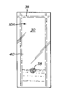
Referring initially to Figures 1, 2 and 3, there is
shown the~integument 10 of the present invention for
containing and culturing organic material, such as,
plant and animal tissue and cells and microorganisms
including bacteria, viruses, fungus, molds and single
cell algae. Integument 10 comprises membrane 12 which,
in the preferred embodiment, encloses plant tissue from a
parent plant or cultivar during the first three stages of
micropropagation. However, it should be understood that
the integument of the present invention may be used for
culturing any type of organic material. When sealed,
membrane 12 completely and entirely surrounds and en-
closes the culture from the ambient environment.
The integument 10 is made by folding membrane 12
over at 14 such that two sides 16, 18 are formed. Sides
16, 18 are heat sealed at 24, 26 along the entire length
thereof and adjacent to longitudinal edges 20, 22 of
membrane 12 so as to form an envelope. The envelope
shaped integument 10 includes a cellule 30 forming an
expandable chamber for containing the plant tissue and
growth medium. The cellule 30 has an approximate average
volume of 50 ml for most varieties of plants. As can be
appreciated, the size and volume of the chamber of
cellule 30 can be varied to host the particular tissue or
plantlet contained therein. Thus, cellule 30 may be of
various sizes. The cellule 30 has at least initially, an
open end 28 formed by the terminal edges 32, 34 of
membrane 12. End 28 serves as a port of entry of cellule
30 for receiving the plant tissue and media. As can also
be appreciated, rather than being made of a single folded
13~S5~33
membrane 12, integument 10 may be made of two individual
and separate pieces of material such as a base material
and a frontal material. In this embodiment, the bottom
of cellule 30 is formed by heat sealing the frontal
material to the base material near the lower terminal
edges th~reof as distinguished from the fold at 14 where
a single piece of material is used as described with
respect to Figures 1-3. Composite integuments may be
formed to take advantage of the strength of one material
and the permeability to oxygen and carbon dioxide of the
other, as an example.
The membrane 12 is a polyethylene material which is
pliable and collapsible such that it can be stored and
shipped in rolls. Further, the polyethylene is so
inexpensive as to be disposable upon completion of any
particular stage of the micropropagation process.
Preferably, the membrane 12 is made of high density
polyethylene. One preferred membrane 12 is made from
0.94 to 0.96 gm/cc density po~yethylene. The material
for membrane 12 should withstand sterilization in an
autoclave which may reach tempertaures of 2500F at 15
p.s.i., for example.
Referring now to Figures 4 to 6, the integument 10
is shown in each of the first three stages of micropro-
pagation. As is shown, after the plant tissue and media
have been received by cellule 30, the port of entry at
open end 28 is heat sealed at 36 along the entire length
thereof and adjacent to the terminal edges 32, 34 of
membrane 12 to close and seal cellule 30 containing the
plant tissue and media therein. At this time the plant
tissue is completely and entirely encapsulated from the
ambient environment and sealed from biological contami-
nants in the ambient environment. Figures 4 to 6 schema-
tically illustrate the integuments 10A, B and C investing
the plant tissue and media in each of the first three
stages of micropropagation. Figure 4 depicts the meri-
stematic tissue 38 from a parent plant or cultivar
, ~ .
~3~ 13
invested within integument 10A together with suitable
media 40 such as Murashige Minimal Organic Medium manu-
factured by Carolina Biological Supply Company. Figure 5
illustrates the use of another integument lOB during the
secGnd stage of tissue culturing. The initial tissue
culture 42 from Stage 1, or a portion of such tissue, is
transferred to cellule 30B containing a suitable Stage 2
medium 44 such as Murashige Shoot Multiplication Mediums
A, B and C manufactured by Carolina Biological Supply
Company. Figure 6 shows an individual plantlet 46 grown
in Stage 2 enclosed by another integument 10C and placed
in a medium 48 such as Murashige Pretransplant Medium
manufactured by Carolina Biological Supply Company to
stimulate cell differentiation and the growth of indivi-
dual plantlets such as 46, each plantlet 46 developing
roots 47 and foliage 49.
Although the integument 10 has been shown and
described as providing a single cellule 30 for enveloping
a~ individual culture, it is preferred that the
integument form a plurality of cellules. Referring now
to Figure 7, there is shown an integument pack 50.
Integument pack 50 is made of membrane 12 and is formed
similarly to~integument 10 of Figure 1. For most culture
investments, the integument pack 50 has dimensions of
approximately 12 inches wide and 6 inches high.
Integument pack 50 is formed by folding membrane 12 over
at 52 so as to form sides 54, 56. As distinguished from
integument 10, sides 54, 56 are heat sealed along the
entire longitudinal length thereof at 58, 60, 62, 64, 66,
68 and 70 to form six individual cellules 72. Individual
tissue samples 74 and media 76 are shown invested in each
of the cellules 72. The plant tissue and media may be
for any of the first three stages of micropropagation as
represented in Figures 4 to 6. The ports of entry at the
upper end 78 have been heat sealed at 80 along the entire
length of integument pack 50 to close cellules 72 after
the tissue 74 and media 76 are inserted into the cellules
.
~3~5~33
72. An upper flap or band 82 may be formed at the upper
ends 78 of membrane 12 for the purpose of suspending
integument pack 50 in the vertical position. Suitable
connection means such as apertures 84, 86 may be provided
through band 80 for attachment means such as drapery
hooks or S-hooks to suspend integument pack 50
vertically. Suspending the growing tissue and plants
vertically at different elevations markedly reduces the
amount of space required in the growing areas. The~
suspension of cultures above others is allowed because of
the translucency of integument packs 50. Further, the _
vertical suspension of integument packs 50 at different
elevations will also enhance air movement within the
growing area. Many conventional growing area layouts
concentrate the tissue cultures or plants at a given
elevation within the growing area, such as on countertops
or working surfaces, such that there is a limited move-
ment of air between the plants. Thus, by increasing
light transmission and the availability of air for gas
exchange by suspending integument packs 50, growth of the
tissue and plants is enhanced and the growing area
requirements are reduced.
The cellule, and thus the integument, is sized in
accordance with the culture to be grown. Referring now
to Figures 8 to 13, there is shown another embodiment of
the integument shown in Figures l and 7 that is adapted
and sized for the micropropagation of lettuce, spinach or
other leafy vegetables. The integument 90 for enclosing
the tissue for a leafy vegetable is made of a membrane
92, membrane 92 being like that of membrane 12 for
integument lO or integument pack 50 as shown in Figures l
and 7 respectively.
Integument 90 is made by membrane 92 being extruded
in tubular form having a circumference of approximately
24 inches. The tubular membrane 92 is folded into
quarter panels 94, 95, 96, 97 and one eighth panels 98,
lO0 and 102, 104, best shown in Figures 8, 11 and 13.
13~5~:33~
One eighth panels 98, 100 and 102, 104 are formed by
folding quarter panels 95 and 97 at 106 and 108,
respectively. Quarter panels 94, 95, 96 and 97 were
formed by folding tubular membrane 92 into quarter
lengths, at folds 110, 112, 114, 116. Folds 106, 108 are
directed inwardly as shown in Figure 13 and one end 118
of tubular membrane 92 is heat sealed at 120 in the
folded position as shown in Figure 12 to produce a
ceilule 122 to house the leafy vegetable tissue and
media. The cellule 122 has a volume of approximately
1000 ml which can be varied according to the particular
leafy vegetable plant tissue grown therein. The other
end 124 of tubular membrane 92 is initially left open as
a port of entry 126 to receive the leafy vegetable tissue
and media.
Cellule 122 preferably includes a foliage chamber
130 and a root chamber 132 with an open neck 134
therebetween, best shown in Figure 10. Chambers 130, 132
and neck 134 are formed by heat sealing portions of one
eighth panels 100 and 104 to quarter panel 96 at 136 and
138 and by heat sealing portions of one eighth panels 98
and 102 to quarter panel 94 at 140 and 142.
Additionally, upon expanding integument 90, heat seals
136, 138 and 140, 142 create creases at 106, 108 and 146
shown in Figures 8 and 9 to form root chamber 132.
The foliage chamber 130 and root chamber 132 of
cellule 122 permit a separation of the foliage from the
root system during growth and more particularly to
separate the foliage from the media. A plantlet is
positioned within cellule 122 such that the foliage grows
within foliage chamber 130 and the root system extends
from the foliage chamber 130 down through neck 134 and
into the root chamber 132 whe~e the media is disposed.
Given a cellule 122 with a volume of approximately 1000
ml, the root chamber 132 is sized to contain
approximately 50 ml of media. By maintaining the
integument 92 in the vertical position, all media will
13~5g33
12
flow downward into root chamber 132. This downward flow
is facilitated by the angular heat sealing at 136 and
138. Thus, the media is thereby kept separate from the
foliage. This permits the foliage to be kept clean of
media and to permit the leafy vegetable to grow in a
preferred and desirable symmetric shape. Without the
division of cellule 122 into a foliage and a root
chamber, the.leafy vegetable would grow in a haphazard
form losing its symmetry. Further, with the reduced neck
portion 134 separating cellule 122 into a root chamber
132 and foliage chamber 130, the media is retained in the
' root chamber 132 and its flow into the foliage chamber
130 is prevented or retarded when the integument 90 is
tipped or inverted since the media will tend to flow into
the upper angular portions 139 of root chamber 132
instead of flowing through neck position 132.
hdditionally, the reduced neck portion 134 tends to
secure a mature plant in position within cellule 122
since the plant's roots will grow into a mass having a
size larger than the cross sectional area of neck portion
134. This growth of root mass also acts to impede the
flow of media into the foliage chamber 130.
The material for membrane 12 of integuments 10, 50
and for membrane 92 of integument 90 is critical to
providing the desired environment for the tissue and
plantlet during the first three stages of
micropropagation and in particular enhancing growth by
permitting optimum gas exchange and light transmission.
Gas exchange, for example, is needed for the necessary
biochemical actions required for culture growth.
Understanding the role of the gases and gas exchange
requires an explanation of the utilization of each gas
individually.
Two functions of green plant growth are
photosynthesis and respiration. Photosynthesis is the
biochemical process where green plants convert carbon
dioxide and water into complex carbohydrates in the
,,.. , :
- 13C~55~33
. 13
presence of light of a given wave length and intensity
-for a given period of time. The process is affected by a
nùmber of environmental factors including quality of
light, availability of water, availability of carbon
dioxide, temperature, leaf age and chlorophyll content of L
the tissue. Photosynthesis is also referred to as a
carbon dioxide fixation. The exact chemistry of the
process is complex but in essence, chlorophyll in the
presence of~carbon dioxide, water and light converts the
carbon dioxide and water into complex carbohydrates that
are in turn converted into sugars and utilized by the
plant as a food source.
One of the by-products of this process is the
production of free oxygen. Fixation of carbon dioxide by
plants accounts for a large portion of their carbon
content and subsequent weight increase during growth.
The exact uptake of carbon dioxide by plants varies from
species to species. However, a range between eight and
eighty milligrams of carbon dioxide per hour for 100
cubic centimeters of tissue surface can be used as an
approximation of the carbon dioxide intake for most
plants exposed to good environmental conditions. This
intake can be directly related to the dry weight of plant
tissue. At an uptake rate of 25 milligrams of carbon
dioxide per hour for 100 cubic centimeters of tissue
surface, an increase of 5% of the original weight of the
tissue can be realized in a one hour period. From this
overview of photosynthesis and carbon dioxide fixation,
it is clear that among the critical factors affecting
piant growth is the availability of carbon dioxide.
The other function relating to the gases of interest
is respiration. This process is essentially an oxidation
reduction reaction where oxygen serves as the oxidizer to
the carbohydrates and sugars formed during the process of
photosynthesis. Again, the exact chemistry involved is
very complicated. However, the end result is a release
of chemical energy necessary for continued growth of the
.
13~S~33
plant. As in photosynthesis, or carbon dioxide fixation,
a number of environmental factors affect the uptake of
oxygen for the respiratory process. These include
temperature, light, tissue starvation, availability of
oxygen and tissue age. While respiration is believed to
take place at all times in plant tissue, there is a noted
increase in this activity in the absenee of light. This
is believed to be a result of the decreased creb cycle
activity in the absence of light.
Oxygen uptake for use in respiration varies from
species to species and while no generally accepted range
has been established for plants in ideal environmental
conditions, uptake of up to 350 microliters to 1,480
microliters per gram of fresh tissue has been recorded.
There has been no direct correlation of fresh weight to
oxygen uptake. There is also a difference in oxygen
uptake from tissue to tissue within a given plant. Woody
tissue and starch storage organs have the lowest uptake,
while root tips and other regions containing meristematic
cells have the,highest uptake rate. This can be directly
related to the activity of growth in a given area of the
plant where the most active areas require the greatest
energy production and consume the greatest amount of
oxygen. From this, it is clearly defined that the
presence of available carbon dioxide and oxygen is
essential to the continued growth of green plant tissue.
In prior art micropropagation procedures, the
exchange of oxygen and carbon dioxide between the plant
tissue disposed within a glass or plastic container for
protection from contamination has been severly limited in
that the gas exchange must take place through the cotton
packing disposed in the bore of the rubber stopper,
between the loose fit of the top and the container and a
plastic lid or top, or through the slits in the baffled
plastic top. This curtailment of gas exchange has
limited the growth of the plant tissue. The material of
membranes 12, 92 provides a marked enhancement of
,. .. . .
, .
13~5g33
permitted gas exchange as compared to the prior art glass
or plastic containers.
The membranes 12, 92 are made of a translucent and
semipermeable material. The preferred material is a high
density polyethylene, material no. 9650T, lot no. T011235
manufactured by the Chevron Chemical Company of Orange,
Texas. It has a permeability to water vapor of 0.32
grams per 100 square inches per 24 hours for a sheet
which is 1 mil in thickness. It is preferred that the
material of membrane 12, 92 have a thickness of 1.25
mils. Other matexials which have the desired light
translucency, gas permeability and contaminant
impermeability are also available for membranes 12, 92.
For example, certain translucent low density polyethylene
is suitable and even allows greater gas permeability than
the preferred high density polyethylene; however, such
low density polyethylene cannot withstand the high
temperatures of the autoclave and must be sterilized
through other means. Other polymeric materials may be
used which have greater permeability than the preferred
high density polyethylene; however, if the permeability
is too great, the media drys out as the water in the
media solution vaporizes and passes through membrane 12,
92 and out of the integument. The high density polyethy-
lene at a thickness of 1.25 mils forms a molecular
structure during the extrusion process which is espe-
cially useful as a membrane for integuments. The high
density polyethylene is made from linear crystalline
polymers of suitable molecular weight with high tensile
strength and extension modulus, a high degree of symme-
try, strong intermolecular forces and a controlled degree
of cross-linking between layers. The cross-links between
adjacent layers of polymers are introduced to prevent the
polymeric chains from slipping under applied stress. The
lightly cross-linked adjacent uniform layers of polymers
of the high density polyethylene for membranes 12, 92
form interstices therebetween which allow the preferred
i3~5~3
16
diffusion and osmosis therethrough for the desirable gas
exchange and light transmission between the ambient
envixonment and the plant tissue. These interstices are
smaller than .01 micrometers so as to preclude the
passage therethrough of even the smallest microorganisms,
such as viruses. It also provides rigidity to facilitate
the transfer and handling of the cultures. Upon sealing
off the cellule, the culture is completely enveloped and
-enclosed from the ambient atmosphere and environment, as~
distinguished from prior art containers, so as to prevent
any introduction of contaminants.
The necessary gas exchange between the culture and
the atmosphere of the ambient environment due to the
production of the by-product oxygen by the plant during
photosynthesis and the oxygen uptake of the plant during
respiration takes place by osmosis. The gases diffuse or
propagate through the semipermeable membrane 12, 92,
which separates the miscible gases in the ambient
atmosphere and within the cellule, in moving to equalize
their concentrations. The osmotic pressure or unbalanced
pressure between the ambient atmosphere and cellule gives
rise to the diffusion and osmosis causing an interaction
or interchangé of gases by mutual gas penetration through
the separating semipermeable membrane 12, 92. Thus, the
inventive membrane of the integument permits the tissue
to breathe by osmosis and air to diffuse through the
semipermeable membrane and yet prevent the passage of
biological contaminants.
The material of membranes 12, 92 is translucent and
allows the passage and diffusion therethrough of light
rays having at least the wavelengths of 400 to 750
nanometers. Individual wavelengths of light in the range
of 400 to 750 nanometers are required by individual
photosynthetic agents, such as the chlorophylls, in green
tissue plants to provide the reactions necessary for life
and growth. The reduced thickness of the material for
membranes 12, 92 and the uniformity of molecular
17
structure formed in part by the extrusion process for the
material for membranes 12, 92 permits greater light
transmission to the tissue sample enclosed by the
integuments than has previously been allowed by the glass
and plastic of prior art containers. The approximate
1.25 mil thickness of the material for membrane 12, 92 as
compared to the much thicker prior art glass or plastic
containers, substantially enhances the amount of light
and the various individual wavelengths of light which are
received by the tissue culture. It is important that
each wavelength of light necessary for each
photosynthetic agent to react pass through the
integument. The uniformity and light cross-linking of
the molecular structure of the material for membranes 12,
92 provides a pathway of lesser resistance for light.
The molecular structure of glass and plastic of the prior
art containers is more complicated and thus creates a
more complex pathway through the glass or plastic through
which the light must pass to ultimately reach the plant
tissue. Thus the thicker and more complex molecular
structure of the prior art glass and plastic containers
inhibits light passage and may filter out certain
wavelengths of light necessary for the photosynthetic
agents of green tissue plants.
The new and improved integument of the present
invention permits the utilization of a new and improved
process for micropropagation. This process includes four
stages as hereinafter described.
Staqe 1: Initial Tissue Culturing
The cultivars or parent plants to be micropropagated
are maintained under carefully controlled greenhouse
conditions in an attempt to yield plant tissue which
minimizes the growth of microorganisms and particularly
any biological contaminants. After selection of the
optimal parent, an area of the plant with meristematic
(undifferentiated~ tissue is identified, and a bulk
,
13~5933
18
sample, which includes the meristematic tissue, is
removed from the parent plant. This area is usually
where active growth takes place, such as at the tips of
stems or at lateral buds (between the leaf apex and the
connection to the stem).
To prevent contamination of the culture by
biological contaminants, the meristematic tissue is
excised from. the bulk sample and transferred to the
growing medium under a laminar flow hood which removes
airborne contaminants. Prior to the placement of the
meristematic tissue sample into cellules 72 of integument
pack 50, five ml of a suitable media (as distinguished
from 10 ml in the prior art tissue culturing process)
such as Murashige Minimal Organic Medium manufactured by
Carolina Biological Supply Company is inserted into
cellules 72. This medium is an agar-based substance
containing all the re~uired nutrients for tissue growth.
Integument pack 50, containing the media therein, is then
rolled and sterilized in an autoclave. This procedure
tends to close the open upper side 78 of cellules 72.
See Figure 7. Later, under the laminar flow hood, the
integument packs 50 are unrolled and its cellules 72
opened one at a time prior to tissue placement. A
meristematic tissue sample, typically a 0.2 to 1.0 mm
cube, is then placed into an individual cellule 72 of
integument pack 50, a single cellule being shown in
Figure 4.
After tissue placement, the ports of entry into
cellules 72 again tend to immediately close, reducing the
length of time that the samples are exposed to the
environment and that contaminants can enter. ~hereafter,
the upper ends 78 of cellules 72 are heat sealed at 80,
thereby forming a complete investment and envelope around
the plant tissue. In this state, the plant tissue is
completely impermeable to contaminants as distinguished
from the prior art containers.
.. . .
13~9~33
19
The integument packs 50 are then exposed to
approximately 300-500 foot-candles of light during this
first stage.
Using the present inventive process, precise
temperature and humidity conditions need not be
maintained in ~he culture room. In the prior art
process, as temperature changes occurred, atmosphere
would be drawn into and expelled around the tops of the
glass containers containing the tissue cultures, thereby
increasing -the risk of contaminations from airborne
contaminants which had not been removed by the prior art
air filtration system. Further, the 80% humidity level
was typically maintained in the prior art in order to
prevent the media from drying out through evaporation:
Such is not critical in the inventive process.
Furthermore, and importantly, the inventive process, as
distinguished from the prior art process, can be carried
out in an environment which does not require a sterile,
filtered air-flow since each cellule 72 of the integument
pack 50 is contaminant impermeable.
Once the tissue culture has been established, and it
is growing in the initial culture and has been certified
contaminant-free, it is ready for Stage 2.
Staqe 2: Tissue Culture Multiplication
During Stage 2, the initial tissue culture resulting
from Stage 1 is multiplied. Under the laminar flow hood,
the cellules 72 of the integument packs 50 of Stage 1 are
opened with a sharp sterilized knife and the tissue
samples, or portions thereof, are transferred to a second
set of unused integument packs 50, an individual integu-
ment pack being shown in Figure 7. Multiplication of the
tissue culture occurs by using a different media. The
media used for Stage 2 cultures differs from that used in
Stage 1 culturing and includes hormones to induce rapid
growth and multiplication of the tissue. Suitable Stage
2 media include Murashige Shoot Multiplication Nediums A,
- . ~
~3~5933
B, and- C, available from the Carolina Biological Supply
Company. Again, only 5 ml of media are required as
compared to the 10 ml in the conventional prior art
process. The integument packs 50 of Stage 2 are then
heat sealed and suitably disposed on a rack within a
culture room. About 300 to 5~0 foot-candles of light are
provided. During this period, Stage 2 growth yields
primarily non-differentiated tissue growth. The cells in
each tissue sample multiply rapidly during Stage 2 to
form a cluster of primarily undifferentiated tissue
cells, the size of which depends upon the plant variety.
The desired cell multiplication takes approximately 20 to
45 days, again depending upon the plant variety.
- After each Stage 2 cycle, the integument packs
containing the cultures are immersed in a solution of
sodium hypochloride, rinsed, returned to the laminar flow
hood, opened, and the tissue is removed. The tissue is
then subdivided by cutting into a number of small pieces,
'each of which will then be cultured. Each time the
tissue samples are divided, the individual smaller tissue
samples are inserted into cellules 72 of unused integu
ment packs 50. All of these steps are performed in the
laboratory un'der a laminar flow hood. ';
Each culture is grown and divided in a 20 to 45 day
cycle until a sufficient number of tissue samples have
been produced to meet production goals. As an example,
if each tissue culture emerging from Stage 1 produces a
cluster of tissue which in turn yields five tissue
samples capable of culturing, over 15,000 cultures will
have been produced at the end of seven months of Stage 2
multiplication. With the exception of a few naturally
occurring mutations or "sports," each of these resulting
cultures of Stage 2 can then be grown into an individual
plant'which will be genetically identical to the parent
plant. Thus, when the desired number of cultures have
been produced in Stage 2, the tissue cultures then are
ready for Stage 3 production.
~3a\5933
21
Stage 3: Differentiation and Plant Formation
During Stage 3, the cellules 72 of the integument
packs 50 of Stage 2 are opened and tissue samples therein
are divided and transferred to a third set of unused
integument packs 50 as shown in Figure 7. Although a
single plant tissue growing into a plantlet is shown
disposed within each individual cellule 72 in Figure 7,
during Stage 3, a plurality of plant tissues may be
disposed within an individual cellule if desired. This
may be done to save additional space. However, in the
inventive process, the plantlets may be grown separately
in the new integument packs 50, eliminating the need for
plant separation and the damage associated with
untangling roots and foliage of several individual
plants.
During Stage 3, the individual tissue samples grown
in Stage 2 are placed in a media which stimulates cell
differentiation and the growth of individual plantlets,
each plantlet developing roots and foliage. Suitable
Stage 3 media includes Murashige Pre-Transplant Mediums,
available from the Carolina Biological Supply Company.
The purpose of Stage 3 is to grow individual plantlets
and prepare them for greenhouse culture. As distin-
guished from the prior art process, during Stage 3, the
same size or a larger size integument pack 50 can be
used. Initially in Stage 3, the plants are still grown
in the culture room during this phase of development, but
they are placed under increased light conditions so as to
promote photosynthesis and growth. Approximately 2000
foot-candles of light are provided. The differentiation
and growth process of Stage 3 requires between 20 and 45
days depending upon the plant variety. Because the
integument packs 50 are contaminant impermeable, once
individual plantlets have formed, the plantlets can be
removed to the greenhouse to harden during the later
portions of Stage 3 and need not be housed in a culture
room for the entire Stage 3 period. This can signifi-
-
~3~S933
cantly reduce the time normally required for the hard-
ening process and reduce the size of the culture room.
Some commercial growers will purchase their plants
upon completion of Stage 3. Many, however, will wait
until the plants have completed Stage 4, the final
production stage. If purchased at the end of Stage 3,
the plants produced by the inventive process need not be
immediately planted. They may be maintained for up to
one month simply by keeping the plantlets in their'
integument packs under conditions of reasonable tempera-
ture and light. This is advantageous in commercial
production where the Stage 3 plants are sometime shipped
directly to the grower, who may lack the time to plant
them immediately. In the prior art, since the plantlets
have been removed from the sterile environment of the
culture room, the commercial grower must immediately
remove the plantlets from the shipping containers, rinse
them to remove the media in which the contaminants can
thrive, and then plant the plantlets immediately.
Because the plantlets purchased by growers at the end of
Stage 3 are shipped and maintained in the integument
packs 50, they are contaminant impermeable and, there-
fore, without the danger of contamination. The advantage
in the new process is that the grower does not have to
plant immediately. When the grower is ready to plant, he
can simply slit the Stage 3 integument open, rinse and
deposit the plantlet into the soil medium.
Staqe 4: Greenhouse Culture and Hardeninq
At Stage 4, the plantlets are removed from the
integument packs 50 of Stage 3 and are transf¢rred to a
greenhouse where they are individually planted in a soil
medium. The plant's tolerance to light must be increased
so that the plant can adapt to its natural environment.
This process is called "hardening" the plant. The
plant's tolerance to light is gradually increased in
Stages 3 and 4. During Stage 4, the plants are exposed
13~33
23
to up to 8,000 foot~candles in the greenhouse where
growth and hardening is to take place. The exposure of
the foliage of the plant directly to the atmosphere
permits the plantlet to later grow in its natural
environment without the protection of the integuments
used in Stages 1 to 3.
The soil used in Stage 4 is typically a
pre-sterilized peat moss mix. Depending upon the type of
plant, most commercial plants remain in the greenhouse 30
to 90 days before they are shipped to the grower.
Use of the inventive process permits all stages of
the micropropagation process to be less time consuming
than their prior art counterparts, because the new and
improved integuments are more easily and quickly handled.
Thus, more tissue culture samples can be processed per
day. Further, because the integuments consume less space
than prior art containers, the costs associated with the
culture room and the greenhouse are reduced.
TABLES 1 AND 2
Table 1 compares the contamination rate using the
inventive integuments and related process versus the
prior art process and containers, in this instance test
tubes, using different plants in an environment without
sterile filtered air. The tissue samples were cultured
for 28 days in each stage under identical conditions,
except that 10 ml of media was used with the prior art
containers, and 5 ml was used with each of the cellules
72 of the integument packs 50.
._
130S~33
TABLE 1: CONTA~lINATIONS PER 2 0 0 CULTURES PER STAGE
STAGE 1 STAGE 2 STAGE 3
Test Present Test Present Test Present
Tube Invention Tube Invention Tube Invention
Alocasia
Lindanii
(Alocasia) 66 25 55 0 51 0
California
(Boston Fern) 61 29 59 0 52 0
Hillii
(Boston Fern) 73 25 47 0 43 0
Nephrolepis
Biserrata
Furcens
~Fishtail Fern) 68 28 * * * *
Boston Curly
Frond
(Boston Fern) * * 52 12 * *
Boston
Roosevelt
Compacta
lBoston Fern) * * * * 44 o
* These Tables reflect the results of the limited
tests which had been conducted at the time of this
application. These tests were not conducted pur-
suant to a predetermined procedure whereby each
~ plant underwent every stage of the micropropagation
process. These tests were conducted using available
tissue samples from a variety of plants, the tissue
samples being in various stages of development. For
this reason, certain stages of the micropropagation
process were never conducted for certain plants.
1305933
- 25 - 74330-2
TABLE 2: TI5SUE GROWTH RATES
(AVERAGE WEIGHT PER SAMPLE)
STAGE 1 STAGE 2 STAGE 3
(Note 1) (Note 2) (Note 1)
Test Present Test Present Test Present
Tube Invention Tube Invention Tube Invention
Alocasia
Lindanii
(Alocasia) 0.38g 1.70g 1.03g 4.5g 1.52g 5.41g
Californi
(Boston Fern) * * 1.03g 4.s7g 1.31g 5.47g
Hillii
(Boston Fern) * * 0.38g 4.38g 1.03g 5.05g
Boston Curly
Frond
(Boston Fern) * * 0.39g 4.08g * *
Boston
Roosevelt
Compacta
(Boston Fern) * * * * 1.39g 4.68g
* These Tables reflect the results of the limited tests
which had been conducted at the time of this applica-
tion. These tests were not conducted pursuant to a
predetermined procedure whereby each plant underwent
every stage of the micropropagation process. These
tests were conducted using available tissue samples from
a variety of plants, the tissue samples being in various
stages of development. For this reason, certain stages
of the micropropagation process were never conducted for
certain plants.
Notes:
1) Plants were grown for 28 days in Stages 1 and 3 using
Murashige Minimal Organic in all cases.
; 2) Plants were grown for 28 days in Stage 2 using Murashige
Fern Multiplication in all cases except for the
.' ~' - . ~ .
13~5933
Alocasia Lindanii, where Murashige Shoot ~lultiplication
A was ~sed.
Tables 1 and 2 illustrate a reduction in contami-
nation and an increase in growth rate and in the number
of new tissue cultures and plantlets produced from an
individual meristematic tissue of a cultivar using the
inventive integuments and related process. For example,
the Alocasia Lindanii of Table 1 shows that the prior art
container and process had 172 contaminated tissue cu~--
tures per 600 cultures while the integument and process
of the present invention had only 25 contaminated cul-
tures. Thus, the present invention reduced contaminated
cultures by approximately 85~. Table 2 shows that the
growth of the Alocasia Lindanii culture using the integu-
ment and process of the present invention had an increase
in average weight of approximately 4.5 times over the
prior art process during Stage 1, an increase of approxi-
mately 4.4 times over the prior art process during Stage
2, and an increase of approximately 3.6 times over the
prior art process during Stage 3. Over the three stages,
the inventive integument and process produced a growth
rate approximately 4 times greater than that of the prior
art containers and process.
The following are further examples of the use of the
new and improved integument and culturing process.
EXAMPLE I: Tissue Culture of NePhrolepis Exaltata Whit-
manii
An experiment was conducted for the micropropagation
of the fern Nephrolepis Exaltata Whitmanii, wherein the
results of employing the integument and process of the
present invention were compared with those obtained using
the prior art containers and process. Stages 1 to 4
where utilizing the inventive integument and process are
described first, followed by a description of the prior
art containers and process.
131G 5933
Inventive Intequment and Process
In preparing the media for Stage 1, 4.4 grams of
pre-mixed Murashige Minimal Organic medium and 30 grams
of sucrose were added to 500 ml of distilled water. The
solution was stirred until the ingredients had dissolved.
Additional distilled water was then added to bring the
final volume of the solution to 1000 ml. The pH of the
solution was then adjusted to 5.5. 8 grams of agar were
then added and the mixture was heated until the agar
dissolved. 5 ml of the media was then transferred to
each of 200 cellules 72 of the integument packs 50. The
unsealed ports of entry of the cellules were then covered
with nona~sorbent paper towelling and the integument
packs were autoclaved for fifteen minutes at 15 psi. The
integuments were removed from the autoclave while still
warm and placed under a laminar flow hood to complete
cooling.
In preparing the meristematic tissue, 250 stolons of
the fern were removed from the preselected parent plant
and were wrapped in a sterile gauze. This gauze packet
containing the stolons was then soaked with 500 ml of
sterile distilled water to which two drops of wetting
agent, such as Palmolive Green manufactured by Procter &
Gamble of Cinncinati, Ohio, had been added.
This packet was sonicated for three minutes. The
packet was then placed in a sterile container and covered
with 500 ml of a 10~ sodium hypochloride solution to
which two drops of a wetting agent had been added. The
container was covered with a tight fitting lid and
vigorously shaken by hand for one minute. The container
was then placed in the ultrasonic cleaner and sonicated
for ten minutes, after which it was then removed and
sprayed with a 90% isopropyl alcohol solution and placed
in the laminar flow hood to air dry. The lid was removed
and the 10% sodium hypochloride solution was drained off.
The gauze packet containing the stolons was then
rinsed three times with sterile distilled water
- , ~
S~33
28
(approximately three minutes for each rinse). The packet
was removed from the container, laid on a sterile work
surface under the laminar flow hood, and the gauze packet
was opened. The clean stolons were separated and
approximately one inch of the active growing end was
removed from each stolon. One active end was placed in
each of the cellules 72 containing media.
The top of each cellule was heat sealed using a wire
sealer at 300F for ten seconds. The integument pack was~
then labeled and ~he process was repeated until all the
tissue had been so placed.
The integument packs were placed in the culture
room; which was maintained at 80F with sixteen hours of
light and eight hours of darkness per twenty-four hour
period. The cultures were examined every twenty-four
hours for contamination and growth.
During the first five days of Stage 1, twenty-six of
the 200 cultures contaminated. At the end of ten days,
some initial growth was observed in all of the remaining
cultures. Some frond development was noted in all
cultures by the end of the twentieth day, and the
cultures were ready for Stage 2 multiplication by the end
of the twenty-eighth day.
To prepare the media for Stage 2, 4.6 grams of
premixed Murashige Fern Multiplication Medium and 30
grams of sucrose were added to 500 ml of distilled water.
This was stirred until a solution was formed. Additional
distilled water was added to bring the volume to 1000 ml.
The pH was adjusted to 5.3. 8 grams of agar was then
added to the solution and the solution was heated until
the agar had dissolved. 5 ml of the solution was then
added to each of 200 unused cellules 72 of integument
packs 50. The open ports of entry of the cellules were
covered with nonabsorbant paper towels and the integument
packs were autoclaved for fifteen minutes at 15 psi.
While still warm, the integument packs were moved to the
laminar flow hood and allowed to cool.
:~3~5~33
29
To prepare the tissue cultures from Stage 1, the
integument packs containing active clean cultures from
Stage 1 were first completely immersed in a 10% sodium
hypochloride solution for three minutes, then removed and
rinsed with sterile water. The integument packs were
dried with a sterile paper towel and laid on a sterile
work surface under the laminar flow hood.
- One cellule was opened at a time, using a sterlized
No. 11 scapel, by making a lengthwise cut down the center
of the cellule. The tissue samples were removed with
sterilized instruments and placed on a sterile work
surface. If more than one active growing point was
present on the removed tissue sample, the sample was
divided into individual growing points. These individual
growing points were then planted in the prepared cellule
containing the Stage 2 multiplication media. After the
cellules were filled, they were sealed using the
above-descxibed wire heat sealer.
The Stage 2 integument packs were then labeled and
moved to the culture room, which was maintained at 80F
with sixteen hours of light and eight hours of darkness
per twenty-four hours. The cultures were checked every
24 hours for contamination and growth.
During the 28 day test period no contamination was
noted in any of the cultures. During the first ten days,
accelerated growth was noted in all cultures. At the end
of twenty-eight days, the cultures were ready for stage
3.
In preparing the media for Stage 3, 4.4 grams of
pre-mixed pretransplant medium was mixed with 30 grams of
sucrose and added to 500 ml of distilled water. This was
then stirred until the ingredients had dissolved. Addit-
ional distilled water was added to bring the volume to
1000 ml. The pH was then adjusted to 5.5. 8 grams of
agar was added, and the solution was heated until the
agar had dissolved. 5 ml of the media was placed in each
unused Stage 3 cellule 72 of integument pack 50. The
. .
-,
13~5~33
unsealed ports of entry of the Stage 3 cellules were then
covered with non-absorbant paper towelling and the
integument packs were autoclaved for fifteen minutes at
15 psi. While still warm, the integument packs were
removed from the autoclave and placed in the laminar flow
hood to finish cooling.
The tissue samples emerging from the Stage 2
cellules were.used as the Stage 3 source materials. The
Stage 2 integument packs were first immersed in 10%
sodium hypochloride solution for three minutes, and then
rinsed in distilled water. The integument packs weEe
dried with sterile paper towelling and placed on a
sterile work surface under the laminar flow hood. Each
cellule of the integument pack was opened by cutting
lengthwise down its center with a s~erile scalpel. The
tissue was removed, placed on a sterile work surfacet and
then rinsed with st~rile water and blotted dry with
sterile paper towelling. Each tissue sample was then
weighed. The average weight per sample was 4.5 grams.
The tissue emerging from Stage 2 was then subdivided
into as many pieces of active growing tissue as could
feasibly support good Stage 3 growth. Each division was
then placed in a cellule 72 of an unused integument pack
50 which was sealed using the wire sealer at 300F for
ten seconds.
The integument packs were then labeled and moved to
the culture room maintained at 80F with sLxteen hours of
light and eight hours of darkness per each twenty-four
hours. The cultures were checked every twenty-four hours
for contamination and growth.
During the twenty-eight day test of Stage 3, no
contamination was observed in any culture. Root develop-
ment was noted at the end of the first week and good
frond development appeared by the end of the second week.
After twenty-eight days, the resulting plantlets
were ready to enter Stage 4. Under the laminar flow
hood, the plantlets were removed from the integument
130Sg33
31
packs, rinsed with distilled water, blotted dry and
weighed. The average weight was 5.4 grams per tissue
sample.
The Prior Art Process
The media preparation for the prior art process was
the same as described abov~, except that twice as much
media was prepared, and, rather than being placed into
the integuments of the present invention, 10 ml of media
was placed into each of 200 25 x 150 mm sterilized test
tubes. The tubes were capped with conventional plastic
caps.
The tissue preparation for the Nephrolepis Exaltata
Whitmanii was also the same as described above. However,
the results obtained following Stage 1 were dramatically
different. Twenty cultures became contaminated during
the first 5 days, and an additional 26 were lost during
the 28 day Stage 1 period. It was not until the 15th day
that all tissue samples showed some growth, and by the
20th day only one half of the samples showed frond
development.
The Stage 2 media was the same as that described
above, except that, once again, twice as much was
prepared and placed into each Stage 2 test tube. The
Stage 1 test tubes could not be immersed in the sodium
hypochloride solution because there would be leakage
through the caps. Instead, under the laminar flow hood,
their outer surfaces were sterilized by spraying with a
90% isopropyl alcohol solution before the tissue samples
were removed from their Stage 1 test tubes and placed
into Stage 2 containers.
During the first 5 days of Stage 2 growth, 18
cultures became contaminated, and an additional 38
samples were lost to contamination between the 14th and
28th days. It was not until the 10th day that
accelerated growth in the samples was observed. The
average weight per sample at the completion of the 28 day
130S~33
32
Stage 2 was only 1.03 grams as compared to 4.5 grams
using the inventive integument and process.
- For Stage 3, once again twice as much media as that
used with the inventive process was prepared and placed
into each Stage 3 test tube. Again, rather than
immersing the Stage 2 test tubes in sodium hypochloride
solution, the outer surface was sprayed with the alcohol
solution while under the laminar flow hood.
During the first 5 days of Stage 3 growth, 18
cultures became contaminated, and an additional 35
samples were lost to contamination between the 14th and
28th days. Root development did not appear on the
majority of samples until the 14th day, and minimal frond
- development did not appear until the 24th day. The
average weight per sample at the completion of the 28 day
Stage 3 was only 1.3 grams as compared to 5.4 grams using
the inventive integument and process. At this point, the
majority of the samples were not ready for transfer to
Stage 4. It is estimated that such samples would have
required approximately 45 days of Stage 3 growth to
achieve the size and maturity necessary for transfer to
Stage 4.
EXAMPLE II: Lettuce Production From Tissue Culturing
Lettuce was produced in tissue culture using the
inventive integument and process, as described below.
The Stage 1 media preparation was the same as that
described above for the Nephrolepis Exaltata Whitmanii.
A non-heading lettuce variety known as butter leaf
was selected. This variety has a normal production time
from seed of 45 to 50 days. Tissue was first removed
from the apical dome of thirty greenhouse-raised plants.
The leaves were stripped, and the roots were removed
exposing the stem, which was rinsed in running water.
The apical dome was then removed.
The apical dome was placed in a clean container and
covered with 10% sodium hy~ochloride solution to which
two drops of a wetting agent had been added. This was
13C~S~3~3
sonicated for ten minutes and the tissue was rinsed three
times in sterile distilled water.
Under the laminar flow hood, final tissue samples,
which were still covered with leaf, were excised from the
primary apical dome of the plant and subdivided three to
four times to yield 100 tissue samples. Each individual
tissue sample was placed in the cellule 72 of an
integument pack 50, already each containing 5 ml of Stage
1 media. Each cellule was then sealed using a wire
sealer at 300F for ten seconds. The integument packs
were labelled and moved to the culture room which was
maintained at the same temperature and light conditions
as described with respect to Example I. The cultures
were examined every twenty-four hours for growth and
contamination.
During the first five days, thirty-five cultures
contaminated, but no further contamination occurred. On
the fifth day, good root development was noted in all the
remaining cultures, and by the end of the seventh day,
all cultures had developed leaves and were actively
growing. A tremendous increase in tissue mass was noted
by the end of the tenth day, at which time a majority of
the cultures~had developed one inch long leaves. By the
twenty-eighth day, the average leaf size was three
inches, and all cultures were ready for Sta~e 2.
In preparing the media for Stage 2, 4.8 grams of
Murashige Premixed Multiplication Medium A and 30 grams
of sucrose were added to 500 ml of water. This was
stirred until the ingredients had dissolved and distilled
water was added to make the final volume 1000 ml. The pH
was adjusted to 5.5. 8 grams of agar was then added to
the solution, and it was heated until the agar had
dissolved. 5 ml of media was put into each cellule 72 of
integument packs 50. The open ports of entry of the
cellules were covered with nonabsorbent paper towelling
and the integument packs were autoclaved for fifteen
minutes at 15 psi. While still warm, the integument
~38S~33
34
packs were placed in the laminar flow hood to complete
cooling.
The tissue samples emerging from Stage 1 were used
for Stage 2 cultures.
The Stage 1 integument packs were completely
immersed in a 10% sodium hypochloride solution for three
minutes to effect surface sterilization. They were then
rinsed in sterile water and dried with sterile paper
towelling. Under the laminar flow hood, the cellules`
were individually opened by cutting lengthwise down the
center, and the tissue was removed and placed in -a
sterile work surface under the laminar flow hood. All
roots-and leaves were removed from the tissue and, where
possible, the remaining tissue was subdivided. The
subdivided tissue samples were then placed into the
unused Stage 2 integument packs, one tissue sample per
cellule. After each cellule was filled, it was heat
sealed with the wire sealer.
A11 integument packs were labelled and placed in the
culture room, maintained at the light and temperature
conditions as described with respect to Example I. The
cultures were examined every twenty-four hours for growth
and contamination.
No Stage 2 cultures were lost to contamination. By
the end of the fifth day, there was a substantial in-
crease in the tissue mass. By the tenth day, there was
good root development along with primary leaf develop-
ment. By the end of the fifteenth day, clearly defined
plantlets were visible in locations which indicated that
lateral buds had developed. The lateral buds continued
to grow until the end of the twenty-eight day test
period. By this time, well-developed plantlets were
ready for additional subculture.
To prepare the media for Stage 3, 4.4 grams of
premixed transplant medium and 30 grams of sucrose were
added to 500 ml of distilled water. The solution was
stirred until the ingredients were dissolved. Additional
~3~55~
distilled water was added to make the final volume 1000
ml. The pH was adjusted to 5.5.
ml of this media was dispensed into the
alternative integume~t embodiment 90 specially desi~ned
to promote the growth of leafy vegetables. The integu-
ment employed was twelve inches long with a three inch
long root chamber at the base.
- The open ports of entry of the cellules 122 of
integument 90 were folded over and closed with paper
clips and the integuments 90 were then autoclaved for
fifteen minutes at 15 psi. While still warm, the inte-
guments were moved to the laminar flow hood to finish
cooling. -
The active tissue samples from Stage 2 were used as
source materials. The Stage ~ integuments were immersed
in a 10~ sodium hypochloride solution for three minutes
to effect surface sterilization, then rinsed in sterile
water, dried with sterile paper towels, and laid on a
sterile work surface under the laminar flow hood. The
cellules 122 of the integuments 90 were opened by cutting
lengthwise down their centers, after which the tissue was
removed and placed on the sterile work surface.
Individual plantlets were then removed from the
primary tissue mass, and were placed in the center of
each of the integuments 90 specially designed for leafy
vegetable growth~ The top of each integument 90 was then
sealed with the wire sealer at 300F for twenty seconds.
The integuments were labeled and moved to the culture
room which was maintained at the light and temperature
conditions as described with respect to Example I.
No contamination was noted in any of the cultures.
By the end of the fifth day, all cultures showed good
root development. Leaf development was noted on the
sixth day, and it progressed very rapidly. Leaves three
inches long were observed in all cultures by the
fifteenth day, and full leaf development was noted on the
thirtieth day. Comple~e lettuce plants were harvested on
13t;~S5~3;~
36
the thirty-fifth day. All had well-developed leaves
suitable for consumption. The plants averaged seven
inches in length, this measurement being taken from the
bottom of the lowest leaf to the top of the plant. The
plants also had well-developed interiors with densely
packed leaves. Normally, this type of lettuce is
nonheading and it takes ~5-50 days to produce a similar
sized plant from seed.
EXAMæLE III: Lettuce Production from Seed
An experiment was conducted to determine whether the
use of the integument and method of the present inventi~n
enhanced lettuce growth when lettuce was grown from seed.
The experiment was conducted as described below.
The media used was Murashige Minimal Organic, with
30 grams of sucrose and 8 grams of agar dissolved therein
by the techniques described above for Nephrolepis
Exaltata Whitmanii. The final pH was adjusted to 5.5. 5
ml of the media was then placed into the cellules 72 of
integument packs 50 depicted in Figure 7.
Black-seeded Simpson lettuce was used. Two hundred
commercially obtained seeds were wrapped in gauze and
surface sterilized by sonicating for ten minutes in a 10~
sodium hypochloride solution to which two drops of a
wetting agent had been added. The gauze packet was then
removed and rinsed three times in sterile distilled
water, with each rinse lasting for three minutes. The
gauze packet was then placed on a sterile work surface
under a laminar flow hood and the seeds were separated
into two equal groups of 100 each. One hundred of the
seeds were planted in the cellules 72 of integument packs
50 with one seed per cellule 72.
After each cellule 72 was filled, it was sealed,
labeled and placed in the culture room where it was
checked daily for growth and contamination.
The other 100 seeds were planted in a seed starting
mix consisting of peat moss, pearlite and vermiculite.
The seed was sown on the top of the pre-moistened mix,
13~S9~33
and pressed into the soil. The flat was labeled and
placed in the culture room under the same light and
temperature conditions as the seeds planted in the
integuments, the same conditions described in Example I.
In the first five days, three cultuxes in the
integuments were lost to contamination. Root development
was noted in all cellules by the end of the third day,
and primary leaf development was noted on the fourth day.
Well-developed seedlings were observed in the integuments
on the fifth day.
By the end of the seventh day, no growth was noted
in the planted seeds, and a problem was suspected. A
microscopic observation revealed evidence of fungal
attack on all the seeds. It is suspected that the
surface sterilization of the seed removed some natural
fungal defense mechanism. The experiment was terminated
at this point and repeated as described below.
200 seeds of the black seeded Simpson were obtained
as described above. This time, however, only the 100
seeds which were intended for planting in the integuments
were treated with the sodium hypochloride solution and
sonicated as described above. These 100 seeds were
planted in cellules 72 of integument packs 50 containing
the same media described above.
The other 100 untreated seeds were pressed into the
freshly prepared pre-moistened soil mix described above.
Both the integument packs 50 and the flats with the
untreated seeds were then placed into the culture room,
which was maintained an 80 F with 16 hours of light and
8 hours of darkness per day. Both the integument packs
and flats were checked daily for contamination and
germination. The soil in the flats was misted daily to
moi~ten the soil.
During the first five days, two cultures in the
integument packs were lost to contamination. Root
development was noted in the third day with primary leaf
development occurring on the fourth day. Well developed
13~S~33
38
seedlings were observed on the fifth day. By the end of
the tenth day, the seedlings in the integument packs had
grown to over one inch in length and had well-developed
root systems. At the end of the twenty day test period,
these seedlings had filled the cellules of the integument
packs with well-developed leaves. 98% of the seeds in
the integument packs germinated.
Only 30% of the seeds planted in the soil mix first
showed primary leaf development on the seventh day. By
the end of the eighth da~, only 68 of the 100 seeds had
germinated. Root development was not observable as the
roots were beneath the soil. The final resultant
seedlings averaged only one inch in height with one to
two secondary leaves. Eight additional seedlings were
lost. At the end of the test, only 60% of the starting
seeds originally planted in soil had produced seedlings.
While this experiment was conducted through the use
of integument packs 50 such as illustrated in Figure 7,
given the high germination rates of the seeds grown using
the inventive process, it is preferable to grow lettuce
from seed in an integument 90 such that the plantlets
produced would not have to be transferred from an
integument pack 50 to an integument 90. Due to the low
cost of the integument 90 and the seed itself, any
integument 90 containing a seed which fails to germinate
can easily be identified and disposed of. Further,
eliminating the steps of transferring plantlets from
integùment packs 50 to integuments 90 would eliminate the
significant labor costs otherwise incurred.
Exam~le IV: Fungal and Bacterial Production and Culture
Storage
Tests were conducted to determine if the integument and
method of the present invention would allow for the growth,
isolation and storage of bacterial and fungal cultures. The
tests were performed in the manner described below:
Fungal Cultures:
~3~5~3
39
A semisolid Minimal Organic Media with 30 grams of
sucrose and 8 grams agar was prepared by techniques de-
scribed above for Nephrolepis Exalata Whitmanii. The final
ph was adjusted to 5.5. 5 ml of the media was then placed
into cellules 72 of integument packs 50 depicted in Figure
7. The integument packs 50 were then autoclaved for 15
minutes at 2500F at 15 p.s.i. They were then placed in the t
laminer flowhood and allowed to cool. Several cellules 72
of integument packs 50 were innoculated with Rhizoctonia
solani. This was done by inserting a steril innoculation
loop into a pure culture of Rhizoctonia solani, removing it
and then inserting it into a cellule 72 of integument pack
50. The ihnoculant loop was placed in the center of the
cellule and moved down until contact was made with the
media. The loop was then resterilized and the process
repeated until all cellules 72 of integument pack 50 had
been innoculated. The integument packs 50 were heat sealed,
labeled and placed in the culture room. This process was
repeated using pure cultures of Rhizopus stolonifer and- _
Penicillium italicum on several integument packs 50.
To determine if cross contamination would occur between
cellules 72 of integument pack 50, several integument packs
were innoculated in the following manner. The first, third
and fifth cellule 72 of the integument pack 50 was innocu-
lated with Rhizoctonia solani, Rhizopus stolonifer and
Penicilliym italicum respectively, leaving cellule 2 and 4
containing only sterile media. The integument packs were
sealed and each cellule containing an innoculant was label-
ed. The integument packs were placed in the culture room.
After 5 days all cultures were checked by visual
observation for growth of fungus. All innoculated cultures
showed positive growth and no growth was observed on those
cellules that were not innoculated. The cultures were
checked several times during a six month period. While
fungal growth slowed as the media was consumed, the cultures
remained viable. Little or no drying of the media was
~3~tS5~33
noted. Further, th~ resulting fungal cultures appeared to
be pure, i.e., uncontaminated from outside microorganisms or
from the other fungal cultures.
Bacterial Culture:
Several integument packs 50 were innoculated with the
bacteria Serratia marcescens, obtained from a pure culture
in the method described above for fungal cultures.
The cultures were checked by visual observation after 5
days and several times after that during a 6 month peri~d.
All innoculated cellules of the integuments showed positive
growth of Serratina marcescens. It was observed for certain
bacterial cultures that the bacteria migrated towards the
inne~ surfaces of the integument membrane where the gas
exchange with the ambient atmosphere was greatest. There
was no bacterial growth or contamination of the cellules of
the integuments that were not innoculated, and those that
were innoculated appeared pure. At the end of a 6 month
period all cultures were viable with little or no loss of
media due to drying.
Certain microorganisms live and grow anaerobica~ly.
Although no such experiments have yet been conducted, the
integument for culturing these microorganisms can be made
from less permeable materials than the polyethylene of the
preferred embodiment so as to preclude a gaseous interchange
between the ambient environment and the organic material.
Similarly, some microorganisms prefer or require the absence
of light. When culturing such microorganisms, an opaque
material can be used for the membrane.
Examples I - III above demonstrate that the integument
and method of the present invention yields dramatic
improvements in plant micropropagation and tissue culturing.
These same improvements will follow irrespective of whether
the plants cultured are horticultural, agricultural or even
aquatic in variety. It is believed that the invention will
yield dramatic improvements in animal and human tissue
culturing as well. Example IV demonstrates the ability of
the integument and method described herein for culturing
13~55~3
41
microorganisms, such as viruses, single celled algea, fungus
and bacteria, and in preventing the cultured microorganisms
from escaping from the integument and contaminating or
infecting others.
The Examples and embodiments described are exemplary
only and not limiting. Many variations and modifications of
the processes and the integuments are possible, and are
within the scope of the invention. Accordingly, the scope
of protection is not limited by the above description but
only by the claims which follow, and that scope includes all
equivalents of the subject matter of the claims.
,. .