Note: Descriptions are shown in the official language in which they were submitted.
:L3~63L5~
10-175C~ Descr_ption
Partible Forceps Instrument for Endoscopy
Technical Field
_ _ .
This invention relates generally to flexible forceps
instruments for endoscopy and, more particularly, to a
partible forceps instrument.
Back~round Art
Flexible forceps instruments for endoscopic use,
such as gastroscopy, bronchoscopy and the like, typically
are constructed as an inseparable ;nstrument having a
cable with forceps pincers welded to one end and a spring
mounted operating handle welded to the other end. The
; cable is enclosed within a flexible, coiled sheath that
is permanently attached to the operating handle at one
end and to the base of the forceps pincers at the other
end. Pushing the cable forward relative to the sheath
by means of the handle causes the pincers to open.
~ome of the disadvantages of inseparable forceps
instruments are that they are difficult to clean after
use and that they are difficult to repair when damaged.
During use, the interior of the forceps, the cable
and the sheath become soiled. The delicate forceps
become clogged with material which is difficult to
dislodge. Even subjecting the instrument to ultrasonic
cleaning will not thoroughly clean the operating cable
and shea~h in as much as the ultrasound dislodges par-
ticulate matter but does not flush it out of the sheath.
; Thus, the dislodged matter may dry and block the cable
or the forceps.
The forceps instrument may be damaged in use when
the operator attempts to force the flexible cable and
sheath through an endoscope with the result that the
coiled sheath develops a kink which inhibits or impairs
the relative movement of the cable. The instrument may
i~3~6~S4
also be damaged when the operator applies too much ten-
sion on the cable in an attempt to close the forceps on
a specimen. Since the base of the forceps is mounted
to the sheath, too much tension on the cable can rupture
the weld between the cable and the forceps pincer mechanism.
Typically, repair involves returninq the instrument to
the manufacturer for rebuilding. The advantage of in-
separable forceps instruments is the stability and handling
afforded by unitary conskruction.
Disclosure of the Invention
:
; 10 The present invention provides a partible forceps
instrument for endoscopy which has the stability and
handling of conventional inseparable forceps and which
has separable parts and a cleaning fixture to provide
for complete cleaning and flushing of the instrument.
The separability allows for replacement or interchange-
ability of parts and permits for a smaller inventory of
forceps parts.
More particularly, the invention is a flexible
forceps instrument comprising a cable sheath, a forceps
having a scissor mechanism demountably attached to a
distal end of the sheath, a connector attached to a
proximal end of the sheath, a manual operating assembly
demountably attached to the connector and an operating
cable inside the sheath attached at a distal end to the
scissor mechanism of the forceps and demountably attached
at a proximal end to the manual operating assembly.
The present invention also provides for a flexible forceps
instrument system which includes a cleaning fixture
~ attachable to the sampling segment in place of the manual
;~ 30 operating assembly. The fixture is in the form of a
hollow ~ube having an inlet at one end for receiving
cleaning fluid under pressure and having a coupler at
the other or outlet end to attach the fixture to the
connector of the sampling segment.
~3~36~S4
The invention further provides for a sampling segment
having a connector whereby the sampling segment is useable
with the manual operating assembly for endoscopic use
or is placed in condition for cleaning after use by
attachment to the cleaning fixture.
One advantage of the construction of the present
invention is that the biopsy instrument may be disassembled
to facilitate cleaning the interior of the sheath and
the cable.
Another advantage of the present invention is that
a cleaning fixture is provided to facilitate ~he in~ro-
duction of cleaning fluid under pressure into the interior
of the sheath to flush out trapped particulate matter.
Yet another advantage of the present invention is
that a sampling segment is provided which allows various
lengths of sheath enclosed cable and various forceps
designs to be used interchangeably with the same manual
operating assembly.
A further advantage of the present invention is
that the sampling segment allows for replacement or
~0 repair of damaged sheaths or damaged forceps without
the need for replacing or repairing the entlre instrument~
Other advantages and a more complete understanding
of the invention will be had from the following detailed
description when taken in conjunction with the accompanying
drawings.
Brief Description of the Drawings
Figure 1 is a partial plan view of the flexible
biopsy instrument;
Figure lA is a cross-sectional view of Figure 1
taken along the line lA-lA of Figure 1;
Figure 2 is a partial longitudinal-sectional view
of Figure 1 taken along the line 2-2 of Figure l;
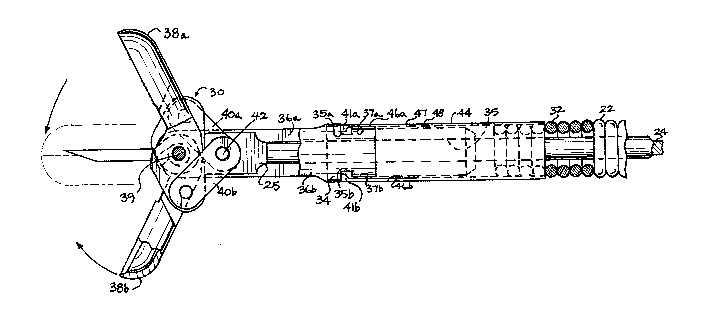
Figure 3 is a partial cross-sec-tional view of a
demountable forceps of the flexible biopsy instrument;
.: ... .. .. .
~L3~6~
Figure 4 is a partial cross-sectional view of a
unitary forceps and sheath of the flexible biopsy in-
strument;
Figure 5A is a side view of the connector of the
; biopsy instrument; and
Figure 5B is a side view partially in section of
the cleaning fixture.
Best Mode for Carrying Out the Invention
Referring to Figures 1-2r the fle~ible forceps
instrument, shown generally at 10, has a sampling segment
20 and a operating segment SO.
The sampling segment 20 is composed of a coiled
spring sheath 22, an operating cable 24 within the sheath
22/ a connector 26 attached to the proximal end 28 of
the sheath 22, and a sampling forceps 30 attached to
the distal end 32 of the sheath 22.
As shown in Figure 3, the sampling forceps 30 has
a pincer supporting body 34 with a passage 35 through
which the operating cable 24 passes. Two laterally
spaced prongs 36a, 36b extend distally from the body 34
between which pincers 38a, 38b are mounted on a fixed
pivot shaft 39. Pincer operating links 40a, 40b pivotally
connect the distal end 25 of the operating cable 24 to
`!
the pincers. The operating cable 24 is connected to
~`~ the pincer operating links through a pivot 42. A similar
forceps 30' is shown in Figure 4, in which like parts
are designated by the same reference numeral as in the
forceps 30, but with a prime designation.
The sampling forceps 30 as shown in Figures 3 and
4 are of the biopsy cup type with needle. It is to be
understood that many types of forceps such as, but not
limi~ed to, biopsy Eorceps, grasping forceps, or scissors
may be employed.
In the preferred embodiment shown in ~igure 3, the
sheath 22 is demountably mounted on ~he body 34 of the
:: - ,- ~ , ,.
~3~5~
sampler forceps 30. A sheath collar 44 is attached to
the dis~al end 32 of the shea~h 22. The sheath collar
44 has a pair of distally projecting fingers 46a, 46b
extending from diametrically opposite sides of the collar
44~ ~he width of each finger is approximately one-half
the diame~er of ~he collar 44~ Each finger has a base
portion, as at 47, formed by a pair of slits in the
collar, as at 48, extending proximally into the collar
44. This construction and the spring steel of which
the collar is formed impart spring-like resiliency to
the finger.s. Each of the fingers 4Ga, 46b is provided
with an inwardly facing tooth 41a, 41b. The teeth 41a,
41b snap into and out of a pair of corresponding op-
positely facing flat grooves 35a, 35b in a pair of op-
positely acing mounting portions 37a, 37b of the body
34 of the sampler Eorceps 30. The mountiny portions
37a, 37b are in the form of flat external surfaces
against which the fingers abut and which effectively
reduce the diameter of the body 34 to allow ~he collar
44 to be snapped onto the body 34 via the projections
46a, 46b and the teeth 41a, 41b without presenting an
increased diameter periphery. In another embodiment as
shown in Figure 4, ~he sheath 22' is non-demountably
attached to the body 34' of the sampler forceps 30' by
a friction closure 49 or the like.
As shown in Figures 1 and 5A, the connector 26 has
a reduced diameter distal portion 27 with a tapered end
29. The distal portion 27 has external threads 31 about
which the proximal end 28 of the sheath 22 is threaded
and then permanently attached by welding or the like.
The adapter 26 has an increased diameter proximal end
35 with external threads 37 and an internal conical
surface 33.
The operating segment 50, as best shown in Figures
1 and 2, has a central shaft 52, a cable slide 62 slidably
.~ ..
-` ~3q:~6'~
mounted on the shaft 52, a thumb ring 54 with a connecting
col]ar 56 crimpea to be freely rotatable on a proximal
end 57 of the shaft 52, and an internally threaded coupler
~` nut 58 freely rotatably about a reduced diameter distal
portion 60 of the shaft 52. As best seen in Figure 2,
the reduced diameter distal portion 60 of the shaft 52
has a tapered portion 64 and a snap-ring groove 66.
The snap-ring groove 66 receives a snap-ring 68 which
mounts the coupler nut 5~ on the reduced diameter distal
portion 60 of the shaft 52. The coupler nut 58 serves
to demountably attach the sampler segment 20 to ~he
operating segment 50. The tapered portion 64 of the
shaft 52 engages the internal conical surface 33 of the
increased diameter proximal portion 35 of the connector
26 and provides a stable connection that resists defor-
mation or flexion during manipulation of the instrument.
The cable slide 62 is in the form of a spool to
~' facilitate gripping between the index and third finger
of the operator's hand. The slide 62 has a central
portion 80 and increased diameter flanges 82a, 82b to
define the spool shape. The central shaft 52 has an
; elongated lateral passage 84 which forms a pair of tracks
70a? 70b, extending substantially the length of the
shaft 52, upon which the cable slide 62 is slidably
mounted. The elongated lateral passage 84 communicates
with a reduced diameter passage 89 extending longitudinally
through the distal portion 60 of the shaft 52 that receives
the operating cable 24.
As best seen in Figure lA, the cable slide 62 has
a throughbore 86 to accommodate the tracks 70a, 70b.
Rotation of the slide 62 about the central shaft 52 is
prevented by means of a core member 88 which is located
in longitudinal recesses 90a, 90b in throughbore 86. A
::
'~3~5~
pair of counter-sunk set screws 92a, 92b within the
flanges 82a, 82b retains the core member 38 within the
slide 62.
The operating cable 24 is demo~mtably attached to
the slide 62 by means of a pair of t:humb screws 106a,
106b which fasten the cable 24 in a bore 110 of the
core member 88 at longitudinally spaced locations.
A compression spring 112 is interposed between the
slide 62 and the distal end of the :Lateral passage 84
around the operating cable 24. The spring 112 biases
the slide 62 and the operating cable 24 to a position
in which the forceps 30 are closed. An operator moving
the slide 62 so as to compress the spring 112 causes
the operating cable 24 to move distally toward the fixed
pivot shaft 39 to cause the pincers 38a, 38b to open in
order to take a biopsy sample. When the operator is
satisfied the pincers are properly located to take a
sample, the slide 62 is released from the index and
third finger and the expansion of the spring 112 moves
the slide 62 and operating cable 24 proximally to cause
the pincers 38a, 38b to close.
After the instrument has been used to take a biopsy
specimen, the instrument is prepared for cleaning and
re-use by disassembly into the sampler segment 20 and
the operating segment 50. The thumb screws 92a, 92b in
the cable slide 62 are loosened to release the operating
cable 24 from the bore 110 of the core member 88. The
coupler nut 58 is rotated to free the operating segmen~
50 Erom the sampling segment 20. If the body 34 to
which the operating cabïe 24 is a~tached is demoun~ably
mounted on the sheath 22 as shown in Figure 3, the body
and cable are removed by unsnapping the teeth 41a, 41b
of he fingers 46a, 46b of the sheath collar 44 from
the flat grooves 35a, 35b in the body 34 of the sampler
forceps 30 and the cable pulled out of the sheath 22
:
~3~6~S~
from the distal end. If the operating cable 24 is non-
demountably attached to the sheath 22, as with the body
34' shown in Figure 4, no further disassembly prior to
cleaning is possible.
A cleaning fixture 120r as shown in Figure 5B,
facilital:es cleaning the operating segment 20 and com-
prises a hollow tube 122 having an internally threaded
- coupler 124 at one end and an inlet 126 at an other end
;~ to receive a source of cleaning fluid under pressure,
such as from a syringe. The inlet end 126 may be in
the form o a luer lock or the like. The coupler 124
is freely rotatable about a reduced diameter portion
128 of the tube 122. The reduced diameter portion 128
has tapered portion 130 and a snap-ring groove 132 which
receives a snap-ring 134. The snap-ring 134 retains
the coupler 124 on the reduced diameter portion 128 of
the tube 122. The coupler 124 of the cleaning fixture
120 is substantially identical to the coupler nut 58 of
the operating segment 50, so that the cleaning fixture
120 may be interchanged with the operating segment 50.
For cleaning the sampling segment 20, the coupler 124
of the cleaning fixture 120 is attached to the externally
threaded increased diameter portion 35 of the connector
26. The tapered portion 130 of the cleaning fixture
120 engages the internal conical surface 33 of the con-
nector 26 and provides a fluid tight connection for
introducing cleaning fluid into the interior of the
connector 26 and sheath 22. Cleaning fluid may be in-
troduced urlder pressure with a syringe having a luer-
lock couplable to the inlet 126. This fixture is
especially advantageous when the cable cannot be removed
from the sheath because it is otherwise difficult to
flow fluid through the sheath. The present invention
thus provides a system in which a separable or partible
~3[316:~5~
forceps and a cleaning fixture cooperate to provide the
advantages set forth.
Variations and modifications of the invention will
be apparent to those skilled in the art from the above
detailed description. Therefore~ it is to be understood
that, within the scope of the appended claims, the inven-
tion can be practiced otherwise than as specifically
shown and described.
';
