Note: Descriptions are shown in the official language in which they were submitted.
130'~4~30
BACKGROUND OF THE INVENTION
The present invention relates to chemiluminescent
processes. The present invention relates more particularly
to the detection of nucleic acid hybrids, antibodies,
antigens and en~ymes using chemiluminescence. Still
further, -the present invention concerns chemiluminescence
devices.
The present invention also concerns prolonged
enhanced chemiluminescence. More particularly, the present
invention relates to stabili~ation of the enzyme in enhanced
chemiluminescence reactions by the use of nitrogen-
containing compounds.
The present invention further relates to the
detection of a nucleic acid hybrid by chemiluminescent
reactions.
Luminescence is defined as the emission of light
without heat. In luminescence, energy is specifically
channeled to a molecule so that a specific light-emitting
state is produced without greatly increasing the temperature
of the molecule. The color is determined by the character
of the light-emitting state involved, and does not change
when the energy or method to produce it is changed.
Chemiluminescence is defined as luminescence
wherein a chemical reaction supplies the energy responsible
for the emission of light (ultraviolet, visible, or
infrared) in excess of that of a blac~body (thermal
radiation) at the same temperature and within the same
spectral range. Chemiluminescence thus involves the direct
conversion of chemical energy to light energy. Below 500C,
the emission of any light during a chemical reaction
involves chemiluminescence. The blue inner cone of a bunsen
burner or the Coleman gas lamp are examples.
Many chemical reactions generate energy. Usually
this exothermicity appears as heat, that is, translational,
rotational, and vibrational energy of the product molecules;
whereas, for a visible chemiluminescence to occur, one of
J - 2 -
130 7~80
the reaction products must be generated in an excited
electronic state (designated below by an asterisk') from
which it can undergo deactivation by emission of a pho-ton.
Hence a chemiluminescent reaction, as shown in reactions (a)
and (b) be]ow, can be regarded as the reverse of a
photochemical reaction.
A + B ~C* ~ D (a)
C* --~C + hv (b)
The energy of the light quantum hv(where h is
Planck's constant, and v is the light frequency) depends on
the separation between the ground and the first excited
electronic state of C; and the spectrum of the
chemiluminescence usually matches the fluorescence spectrum
of the emitter. Occasionally, the reaction involves an
additional step, the transfer of electronic energy from C*
to another molecule, not necessarily otherwise involved in
the reaction. Sometimes no discrete excited state can be
specified, in which case the chemiluminescence spectrum is a
structureless continuum associated with the formation of a
molecule, as in the so-called air afterglow: NO + O-~N02 +
hv (green light).
The efficiency of a chemiluminescence is expressed
as its quantum yield ~, that is, the number of photons
emitted per reacted molecule. Many reactions have quantum
yields much lower (10 8 hv per molecules) than the maximum
of unity, Einsteins of visible light (1 einstein = Nhv,
where N is Avogadro's number), with wavelengths from 400 to
700 nm, correspond to energies of about 70 to 40 kcal per
mole (300 to 170 kilojoules per mole). Thus only very
exothermic, or "exergonic," chemical processes can be
expected to be chemiluminescent. Partly for this reason,
most familiar examples of chemiluminescence involve oxygen
and oxidation processes; the most efficient examples of
these are the enzyme-mediated bioluminescences. The glow of
phosphorus in air is a historical]y important case, although
the mechanism of this complex reaction is not fully
. - 3 -
~3C~'7480
understood. The oxida-tion of many organic subs-tances, such
as aldehydes or alcohols, by oxygen, hydrogen peroxide,
ozone, and so on, is chemiluminescent. The reaction of
heated either vapor with air results in a bluish "eold"
flame, for example. The efficiency of some
chemiluminescences in solution, such as the oxidation of
luminol (I) (see formula below) and, especially, the
reaction of some oxalate esters (II) (see formula below)
with hydrogen peroxide, can be very high (0 = 30~).
NH2
~ ~ RO- C- C- OR
(I) (II)
It is believed that the requirements for
chemiluminescence are not only sufficient exothermicity and
the presence of a suitable emitter, but also that the
chemieal process by very fast and involve few geometrical
ehanges, in order to minimize energy dissipation through
vibrations. For example, the transfer of one eleetron from
a powerful oxidant to a reductant (often two radical ions of
opposite eharge generated eleetroehemieally) is a type of
proeess which can result, in some eases, in very effective
generation of eleetronie exeitation. An example, with 9,10-
diphenylanthraeene (DPA), is shown in reaetion (e).
DPA + DPA - ~DPA* + DPA (c)
The same is true of the decomposition of
four-membered cyclic peroxides (III) into carbonyl products,
shown in reaction (d), whieh may be the prototype of many
chemiluminescences.
. ~ .
~30~74~30
A special type of chemiluminescence is
bioluminescence.
Bioluminescence is definea as the emission of
light by living organisms, due to an energy-yielding
chemical reac-tion in which a specific biochemical substance,
called luciferin, undergoes oxidation, cataly~ed by a
specific enzyme called luciferase.
There are many specific luciferins and luciferases
which are chemically different, each involved in some
different living luminescent organism. The flash of the
firefly, the brillant "phosphcrescence" or "burning" of the
ocean, or the eerie glow of mushrooms deep in the forest at
night are but a few examples of these different
bioluminescent organisms.
Since bioluminescence is a type of
chemiluminescence, it is not necessary to have a live
organism to obtain light emission. The simple preservation
of the chemicals involved will suffice. This can be done in
some cases by rapidly drying the organism under mild
conditions.
Dried firefly tails (lanterns) emit light when
ground up with water. This light emission dies away within
a few minutes, but can be restored by the addition of
adenosinetriphosphate (ATP), a key coenzyme in the energy
metabolism of cells. In this case, ATP reacts with the
luciferin of fireflies to give the luciferyl adenylate
intermediate and pyrophosphate (PP).
Using lantern extracts from hundreds of thousands
of fireflies, scientists at Johns Hopkins University
determined the chemical structure of firefly luciferin to be
C13H12N2O3S2~ It can now be synthesized. The reaction of
luciferyl adenylate with oxygen is postulated to give a
four-membered-ring alpha-peroxylactone intermediate and to
release adenosinemonophosphate (AMP). This breaks down in
the energy-yielding step to give carbon dioxide and a
light-emitting excited molecule. This loses its energy as a
- 5 -
... .
1307480
photon (hv), in the yellow reyion of the spectrum in this
case.
Firefly luciferin and luciferase from preserved
light organs are used in a very sensitive biochemical test
to detect AIP.
A postulated pathway for firefly luciferin is as
follows:
0~ 5 ~ ~ O
Luci~er~e ¦ + ~A~TP,
W ' B
~N ~ ~ C--A M P
,1,+
~N N ~"0
S
~N N ~ 0
The luminescence of the firefly occurs as a brief
flash, coming from the inside of photogenic cells in the
lantern, under the control of the nervous system. Quite a
different situation occurs in the small marine crustacean
Cypidina, which is found in the waters off the coast of
Japan. It synthesizes its luciferin and luciferase in
separate glands. To emit light, it simply squirts luciferin
and luciferase into the water, where the reaction occurs,
separate from the animal. The light may function to divert
or trick predators.
- 6 -
1307~30
The chemist:ry of Cypridina luciferin has been
determined by a yroup of chemists in Japan. C22H270N7 is
postulatecl to react directly with oxygen at the position
indicated by the arrow (below), forming a type of alpha-
peroxylactone similar to the firefly molecule. In the final
step, carbon dioxide is also released, alony with the
excited molecule, which in this case emits in the blue.
A postulated pathway for Cypridina ]uciferin is as
follow~,:
o~ ~CH2--CH~
CC--CH
0 ~ ~ CH~
~ J ~ N( C H ~ `t H--C
,1 +
0--0
~C--C--R~
Rl ;~ Rl
¦- c R J .
IL R ~-- ~ A
~i
Il - 7 -
:13C~7~8~
Like fireflies, dried Cypridina emit light when
ground up with cool water; the preserved luciferin and
luciferase are released from the glands as they are crushed.
The light gradually fades as the luciferin is oxidized, but
5 the addition of more luciferin restores light in the
exhausted extract. Luciferin can be obtained either
synthetically, or in the natural form by grinding up dried
Cypridina in hot water. The heat destroys the luciferase,
which is a protein, but leaves the luciferin active. When
cooled and mixed with the exhausted extract, luminescence is
observed. This is the basis for the clcassical
luciferin-luciferase test.
Luminescent bacterial emit a continuous blue-green
light. Such bacteria can be isolated directly from sea
water or from the surface of a dead fish and will grow
rapidly on any medium containing 3% salt (equivalent to sea
water) and some fish or meat extract.
A postulated pathway for bacteria luciferin is as
follows:
CH2--(CHOH)3--Cff2--O--1 OH
H OH
c~ N~ C::~O
H O
Redvced ribon-vin ph~nph~
+2
\ + RCHO
¦I,vcif~nae ll~vin ccrnpl~
Ribofl~vin pho5ph-~e + hv (grern~
~0
+ CH3(CH~)loC + H20
OH
~3(~7~
Chemiluminescent detection is one of the most
sensitive ways of de-tectinq an analyte. The process,
although sensitive, suffers from several disadvantages. In
most cases the chemiluminescent reaction mediated emission
of light has a very short lifetime, i.e., light emission is
very quick, so that a sophisticated device has to be
developed to monitor the extent of light emission and also
to determine the extent of -the presence of an analyte. It
is also difficult ~o couple the interacting systems to the
analyte without destroying or changing the property of the
interacting partners.
Recently, it has been demonstrated that if a
substance, for example, an iodophenol or a benzothiazole
derivative is present during the chemiluminescent emission
mediated by horseradish peroxidase, the reaction rate is
retarded and simultaneously the quantum yield of the light
emission is enhanced (European Patent Application No. 0 116
454; European Patent Application No. 0 103 784; UK Patent
Application No. 820 62 63; Gary H.G. Thorpe, Robert Haggart,
Larry J. Kricka and Thomas P. Whitehead, "Enhanced
Luminescent Enzyme Irnmunoassays For Rubella Antibody,
Immunoglobulin And Digoxin", Biochemical and Biophysical
Research Communications, Vol. 119, No. 2, pp. 481-487, March
15, 1984; Thomas P. Whitehead, Gary H.G. Thorpe, Timothy
J.N. Carter, Carol Groucutt and Larry J. Kricka, "Enhanced
Luminescence Procedure For Sensitive Determination Of
Peroxidase-labelled Conjugates In Immunoassay", Na-ture, Vol.
305, pp. 158-159, September 8, 1983; Gary H.G. Thorpe, Larry
J. Kricka, Eileen Gillespie, Susan Mosely, Robert Amess,
Neil Baggett and Thomas P. Whitehead, "Enhancement Of The
Horseradish Peroxidase Catalysed Chemiluminescent Oxidation
Of Cyclic Diacyl Hydrazides By 6-Hydroxybenzothiazoles",
Anal. Biochem.). Although this method has been shown to be
useful in the detection of an analyte by conventional
immunoassay methods, it has never been demonstrated,
however, whether this method could be utilized to detect a
nucleic acid hybrid.
~3~7~80
Irwin ~ridovich, "The Stimulation Of E~orseradish
Peroxidase By Nitrogenous Ligands", The Journal of
Biological Chemistry, Vol. 238, No. 12, December 1963, pp
3921-3927, describes the stabilization of peroxidase in
solution with nitrogenous ligands.
It has been demonstrated heretofore that a
chemiluminescent reaction occurs where the emission is due
to an iron initiated activation of bleomycin. The self-
inactivation reaction is affected by the presence of DNA
In Photochemistry Photobiology, Vol. 40, pg
823-830, (1984), it was described that photoemission is
quenched by target molecules such as DNA and that the
presence of DNA does not prevent the iron-initiated activa-
tion of bleomycin, by the so-called self-inactivation
lS reaction associated with chemiluminescence. The article
went on to state that these findings seem to suggest that an
electronically excited intermediate of bleomycin can alter
bio-molecules though, in that case, the nature of the
excited state was not precise.
Swedish patent application 8200479 describes
chemiluminescent detection of nuclelc acid hybrids.
European patent application 0 070 687 concerns a
light-emitting polynucleotide hybri.dization diagnostic
method.
Heretofore chemiluminescence reactions proceeded
too quickly and thus resulted in light of only a short
duration. The use of enhancers have somewhat extended and
amplified the light from chemiluminescence reactions,
however, the duration and intensity of the emitted light is
still in many instances inadequate.
Immunoassay is one of the most widely used
analytical techniques in the clinical laboratory. At
present the majority of immunoassays employ a radioactive
isotope, especially iodine-125, as a label. However,
radioactive isotopes have a number of major disadvantages.
First, the method of labelling involves the use of highly
radioactive and hence potentially hazardous reagents.
-- 10 --
'~
4HV
Second, the shelf life of the radio-actively labelled
substance is often relatively short not only because by its
very nature the radioactive isotope is continuously decaying
but also because radioactively labelled proteins are often
unstable. ~hird, it is often difficul~ to label proteins
sufficiently to provide a sensitively and rapidly detectable
reagent. Fourth, the disposal of radioactively labelled
substances is inconvenient.
These disadvantages have stimulated a search for
viable alternatives to the radio label. To be suitable as a
label a substance should meet at least the following three
requirements:
a. it should be detectable both rapidly and in
very small quantities when attached to a ligand such as an
antigen or an antibody.
b. it should be possible to attach it, without
affecting its determination, to a ligand such as an antigen
or an antibody; and
c. once attached, it should not significantly
alter the properties of the ligand.
Some of the most promising alternative labels are
either substances which can themselves take part in a
reaction resulting in the emission of luminescent light or
substances which, on suitable treatment, produce compounds
capable of ta~ing part in a luminescent reaction.
Heretofore, the use of luminescence in immunoassays has
suffered since the measurement of luminescence is a rapid
process and may be completed in a matter of seconds rather,
than the several minutes generally required for the
measurement of radioactivity.
Luminescence has been employed in three major
luminescent or luminometric immunoassay systems:
a. Organoluminescent or organoluminometric
immunoassays wherein chemiluminescent or bioluminescent
compounds which participate directly in luminescent
reactions (i.e., which are converted to an excited state and
then return to a non-excited state with the emission of a
photon) have been used to label ligands such as proteins,
., -- 1 1 --
1307~80
hormones, haptens, steroids, nucleic acids, metabolites,
antigens and/or antibodies. Examples of suitable compounds
include luminol and isoluminol;
b. Luminescent catalysts or cofactor immunoassays
wherein catalysts or cofactors of luminescent reactions have
been used as labels. An example of a suitable catalyst is
the enzyme peroxidase; and
c. Enzyme linked immunoassays wherein
luminescent reactions have been used to determine the
products formed by the action of enzyme labels on suitable
substrates. An example of this type of immunoassay is the
determination of antibody linked glucose oxidase by reacting
the enzyme/antibody reagent with glucose to form a hydrogen
peroxide and then measuring the amount of hydrogen peroxide
produced by adding luminol under controlled conditions to
initiate a luminescent reaction.
The sensitivity of the above assays is de-termined
in part by the lower limit for detection of the label or the
product of the label. In the case of luminescent or
luminometric assays the sensitivity of the system will
depend partially on the light emitted in -the luminescent
reaction per unit of labelled material.
It is known that chemiluminescent detection is one
of the most sensitive ways of detecting an analyte. The
process, although sensitive, suffers from several
disadvantages. In most cases the chemiluminescent reaction
mediated emission of light has a very short lifetime, i.e.,
light emission is very ~uick, so that a sophisticated device
has to be developed to monitor the extent of light emission
and also to determine the extent of the presence of an
analyte. It is also difficult to couple the interacting
systems to the analyte without destroying or changing the
property of the interacting partners.
SUMMARY OF THE INVENTION
It is an ob~ect of the present invention to
provide chemiluminescence reactions emitting light of long
duration and high intensity.
- 12 -
13C~7~8~
It is also an objec-t of the present invention to
provide chemiluminescence devices capable of prolonged light
duration.
It is a further object of the invention to detect
nucleic acid hybrids.
It is still another object of the present
invention to detect antibodies and antigens using
chemiluminescence.
Another object of the present invention is the
detection of enzyme in a sample.
It is also an object of the present invention to
provide nucleic acids capable of participating in a
chemiluminescent reaction.
It is another object of the invention to provide
methods of detecting nucleic acids in unknown samples.
It is a further object of the the invention to detect
nucleic acid hybrids.
These and other objects are realized by the
present invention.
The present invention concerns a chemiluminescence
process comprising the contacting of a chemiluminescence
precursor, e.g., a 2-3-dihydro-1,4 phthalazinedione, an
oxidant, e.g., hydrogen peroxide, an enzyme, e.g., a
peroxidase enzyme, and a nitrogen compound selected from the
group consisting of ammonia and a water-soluble organic
amine.
The present invention also concerns a
chemiluminescence device comprising a vessel and a means for
combining a chemiluminescence precursor, an oxidant, an
enzyme and a nitrogen compound selected from the group
consisting of ammonia and a water~soluble organic amine.
The present invention also concerns a
chemiluminescence process comprising the contacting of a
chemiluminescence precursor, e.g., a
2-3-dihydro-1,4-phthalazinedione, an oxidant, e.g., hydrogen
peroxide, an enzyme, e.g., a peroxidase enzyme, and a
nitrogenous compound selected from the group consisting
" - 13 -
~3(~8()
of ammonia and water-soluble organic amines, and a
chemiluminescence enhancer, e.g., 4-iodophenol or
6-hydroxybenzo-thiazole.
The presen-t invention further concerns a nucleic
acid probe capable of participating in a chemiluminescent
reaction comprising
a. a defined nucleic acid sequence, and
b. a chemiluminescence precursor photochemically
linked to the nucleic acid sequence.
Another nucleic acid probe according to the
present invention comprises
a. a defined nucleic acid sequence, and
b. a chemiluminescence enhancer linked, for
example, covalently linked, to the nucleic acid sequence.
Such probe can be used as a participant in an enhanced
chemiluminescent reaction and also as a substrate for
luciferase type enzymesby a photochemical linker.
The present invention also concerns a further
nucleic acid probe capable of participating in an enhanced
chemiluminescent reaction comprising a defined nucleic acid
sequence, the sequence being linked to any one of
a. a chemiluminescence precursor,
b. a chemiluminescence enhancer, and
c. an enzyme,
the remaining two of (a), (b) and (c) not linked to said
sequence, being in a mixture with the linked sequence. The
nucleic acid probe can exist as a homogeneous mixture, e.g.,
solution, a heterogeneous phase or in a hybridized form.
The hybridized form can exist as a homogeneous mixture
e.g., solution, or as a heterogeneous phase.
The present invention also concerns a method for
determining a particular single stranded polynucleotide
sequence, e.g., by hybridization, in a test medium,
comprising the steps of:
(a) combining the test medium with a polynucleotide
probe having a base sequence substantially complementary to
the sequence to be determined under conditions favorable to
, . . .
- 14 -
~ 3G79~80
hybridization between the probe and the sequence to be
determined,
(b) labeling either the resulting hybrids or probe
which have not hybridized with the sequence to be determined
with one of the participants in an enhanced chemiluminescent
reaction involving a chemiluminescent precursor, an enzyme,
an oxidant, and a chemiluminescence enhancer,
(c) initiating such chemiluminent reaction wi.th the
labeled hybrids or probe, and
(d) detecting the resulting light emission.
The present invention concerns another method for
determinincJ a particular single stranded polynucleotide
sequence in a test medium, comprising the steps of:
(a) immobilizing single stranded nucleic acids in the
test medium,
(b) contacting the immobilized nucleic acids with a
polynucleotide probe having a base sequence substantially
complementary to the sequence to be determined under
conditions favorable to hybridization between the probe and
the sequence to be determined,
whe.rein the probe
(l) is labeled with a chemiluminescence label
selected from the participants in an enhanced
chemiluminescent reaction involving a
2,3-dihydro-1,4-phthalazinedione chemiluminescent precursor,
a peroxidase enzyme and a chemiluminescence enhancer, or
(2) comprises a binding site for a specific
binding partner,
(c) separating resulting immobilized hybrids from
probe which have not hybridized with the sequence to be
determined, and where the probe comprises the binding site
adding the binding partner which is labeled with the
chemiluminescence label,
(d) initiating the chemiluminescent reaction with the
separated, labeled, immobilized hybrids, and
(e) detecting the resulting light emission.
- 15 -
130~7~80
The present invention also relates to a further
method for determining a par-ticular single stranded
polynucleotide sequence in a test medium, comprising the
steps of:
ta) combining the test medium with a polynucleotide
probe having a base sequence substantially complementary to
the sequence to be determined to form hybrids having an
antigenic determinant which distinguish them from single
stranded nucleic acids,
wherein the probe is either in an immobilized form
or comprises a binding site whereby the probe is
immobilizable by contact with an immobilized form of a
binding partner for such binding site,
(b) when the probe is in the immobilizable form,
contacting the resulting Hybrids with the immobilized
binding partner,
(c) contacting the resulting immobilized hybrids with
an antibody reagent capable of binding to the distinguishing
antigenic determinant, which antibody reagent is labeled
with one of the participants in an enhanced chemiluminescent
reaction involving a 2,3-dihydro-1,4-phthalazinedione
chemiluminescent precursor, a peroxidase enzyme and a
chemiluminescence enhancer,
(d) separating into fractions the labeled antibody
reagent that becomes bound to immobilized hybrids from that
which does not bind,
(e) initiating the chemiluminescent reaction in one of
the separated fractions, and
(f) detecting the resulting light emission.
The present invention relates to an enhanced and
delayed chemiluminescent assay particularly useful for
clinical diagnosis of certain kinds of disease states which
can be monitored by immunological reactions or by nucleic
acid hybridization method. The invention can also be
utilized for straightforward sample analysis where one of
the reacting components for the assay is already present in
the test sample in an unknown amount. The diagnosis of
disease states by using immunoassay and also by nucleic acid
r~ ~ 1 6
~ 3C~'7480
hybridization assays require highly sensitive detection
systems. Since the amount of analyte present is usually
very little, -the assay condition should provide enough
amplified detection. For example, in the detection of an
infectious agent such as a microorganism in a blood sample,
it is possible to extract DNA from the blood sample which is
already infected by the microorganisms and use a nucleic acid
probe specific for ~hat microorganism. The detection can be
conducted by hybridization with the DNA extracted from the
test blood sample and the nucleic acid probe specific for
the microorganism which presumably has infected the blood
sample~
Nucleic acid hybridization technology can also be
used for the detection of genetic diseases which are not
manifested through an infectious agent, for example, a point
mutation on the beta-hemoglobin gene givesrise to a defect
known as sickle cell anemia. People who are affected with
some mutation and also who are carriers of such defects have
a specific sequence of nucleic acid in their genome which
can be detected by hybridization technology. For the
detection of a single gene point mutation it is essential
that a highly sensitive technique is available because of
the low concentration of the defective gene. Usually the
radioactively labeled isotopes are used for the detection
process. The present invention provides a highly æensitive
chemiluminescence assay which is mediated by a
peroxidase-like enzyme and a diacylhydrazide-like substrate
for light substrate for light emission in the presence of a
peroxide. Among the other assays where the present
invention is useful includes the assay of elastin or the
assay of glucose by using glucose oxidase peroxidase system.
The principle and the utility of these assays are known in
the art and have discussed hereinabove, wherein it was
demonstrated that a chemiluminescence type assays can be
used for the detection of elastin or glucose and that
chemiluminescent type assay can be used for immunoassay
- 17 -
13~74~30
purposes. The present invention is based on a surprising
observation that certain nitrogenous ma-terials alone or with
enhancers slow down the rate of emission of light and
prolong the activity of the enzyme for a long period of time
in a chemiluminescence reaction. From the combination of
these two effects it can be concluded that the nitrogeneous
materials enhance and delays the chemiluminescence emission
from diacylyhydrazides mediated by peroxidase and hydrogen
peroxide.
The present invention also concerns processes for
detecting a nucleic acid hybrid.
In one process according to the present invention
for detecting a nucleic acid hybrid an unknown DNA contain-
ing sample is contacted in a mixture, for example, a
solution, with a probe comprising contacting a defined
nucleic acid sequence linked, e.g., photochemically linked,
such as by the use of furocourmarin, to a chemiluminescence
precursor, the mixture containing an oxidant, an enzyme and
a nitrogen compound selected from the group consisting of
ammonia and a water-soluble organic amine, and then
determining the extent of light emission.
In another process according to the present
invention for detecting a nucleic acid hybrid, an unknown
DNA-containing sample is contacted in a mixture, for
example, a solution, with a probe comprising contacting a
defined nucleic acid sequence and an enzyme linked to the
nucleic acid sequence, the mixture containing a
chemiluminescence precursor, an oxidant and a nitrogen
compound selected from the group consisting of ammonia and a
water-soluble organic amine and then determining the extent
of light emission.
In one process according to the present invention
for detecting a nucleic acid hybrid an unknown DNA
containing sample is contacted in a mixture, for example, a
solution, with a probe comprising contacting a defined
nucleic acid sequence linked, e.g., photochemically linked,
- 18 -
13(~7~85)
such as by the use of furocourmarin, to a chemilumine~cence
precursor, the mix-ture containing an oxidant, an enzyme, an
enhancer, and a nitrogen compound selected from the group
consisting of am~onia and water--soluble organic amines and
then determining the extent of light emission.
In another process according to the present
invention for detecting a nucleic acid hybrid, an unknown
DNA-containing sample is contacted in a mixture, for
example, a solution, with a probe comprising contacting a
defined nucleic acid sequence and an en~yme linked to the
nucleic acid sequence, the mixture containing a
chemiluminscence enhancer and a nitrogen compound selected
from the group consisting of ammonia and water-soluble
organic amines and then determining the extent of light
emission.
In one process according to the present invention
for detecting a nucleic acid hybrid an unknown nucleic
acid-containing sample is contacted in a mixture, for
example, a solution, with a probe comprising a defined
nucleic acid sequence and a chemiluminescence precursor
linked to the nucleic acid sequence and thereafter adding a
chemiluminescence enhancer and an oxidant and then
determining the extent of light emission.
In another process according to the present
invention ~or detecting a nucleic acid hybrid, an unknown
nucleic acid-containing sample is contacted in a mixture,
for example, a solution, with a probe comprising a defined
nucleic acid sequence and a chemiluminescence enhancer
linked to the nucleic acid sequence and thereafter adding a
chemiluminescence precursor and an oxidant and then
determining the extent of light emission.
A further process for detecting a nucleic acid
hybrid according to the present invention involves
contacting in a mixture, for example, a solution, an unknown
~ 35 nucleic acid-containing sample with a probe, such probe
; comprising
a. a defined nucleic acid sequence,
-- 19 --
13~7~30
b. a pho-tochemical linker bound to the nucleic
acid sequence,
c. a light bound to the linker,
d. a binding protein bound to the ligand, and
e. an enzyme bound to the binding protein, and
thereafter adding a chemiluminescence substance, a
chemiluminescence enhancer and an oxidant, and then
determining the extent of light emission.
The present invention also concerns
chemiluminescence assays.
A chemiluminescence immunoassay for the detection
of an antigen in an unknown sample according to the present
invention comprises contacting the sample with an antigen
linked to a chemiluminescence precursor or an enzyme,
lS contacting the sample and the antigen with an oxidant, a
nitrogen compound selected from the group consisting of
ammonia and a water-soluble organic amine and an enzyme if
the antigen is linked to a chemiluminescence precursor, or a
chemiluminescence precursor if the antigen is linked to an
enzyme, and determining the extent of light emission.
A chemiluminescence immunoassay for the detection
of an antigen in an unknown sample according to the present
invention eomprises contaeting the sample with an antibody
to the antigen, the antibody linked to a ehemiluminescence
precursor or an enzyme, contacting the sample and said
antibody with an oxidant, a nitrogen compound selected from
the group consisting of ammonia and a water-soluble organic
amine and an enzyme if the antigen is linked to a
ehemilumineseence preeursor, or a ehemilumineseenee
preeusor if the antigen is linked to an enzyme, and
determining the extent of light emission.
Another chemiluminescence immunoassay for the
deteetion of an antibody in an unknown sample according to
the present invention comprises contacting the sample with
an antibody to the antigen, the antibody linked to a
chemiluminescence precursor or an enzyme, contacting the
- 20 -
.~
~3~748()
sample and said antibody with an oxidant, a
chemilumine~cene enhancer and a nitrogen compound selected
from the group consis~ing of ammonia and water ~oluble
organic amines and an enzyme if the antigen is linked to a
chemiluminescence precursor, or a chemiluminescence
precursor if the antigen is linked to an enzyme, and
determining the extent of light emission.
Another chemiluminescence immunoassay for the
detectlon of an antibody in an unknown sample according to
the present invention comprises contacting the sample with
an antibody to the antigen, the antibody linked to a
chemiluminescence precursor or an enzyme, contacting the
sample and said an-tihody with an oxidant, a
chemiluminescence enhancer and a nitrogen compound selected
from the group consisting of ammonia and water-soluble
organic amines and an enzyme if the antigen is linked to a
chemiluminescence precursor, or a chemiluminescene
precursor if the antigen is linked to an enzyme, and
determining the extent of light emission.
The present invention further concerns a
chemiluminescence assay for the detection of a peroxidase
enzyme comprising contacting an unknown sample with a
chemiluminescence precursor, an oxidant and a nitrogen
compound selected from the group consisting of ammonia and a
water-soluble organic amine and determining the extent of
light emission.
The present invention further concerns another
chemiluminescence assay for the detection of a peroxidase
enzyme comprising contacting an unknown sample with a
chemiluminescence precursor, an oxidant, a chemiluminescence
enhancer and a nitrogen compound selected from the group
consisting of ammoniaand water-soluble organic amines and
determining the extent of light emission.
Still further, the present invention involves a
test kit for conducting chemiluminescence assays comprising
a chemiluminescence precursor, an enzyme, an oxidant and a
13(~7480
nitrogen compound selected from the group consisting of
ammonia and a water-soluble organic amine.
Still fur-ther, -the present invention involves
another test kit for conducting chemiluminescence assays
comprising a chemiluminescence precursor, an enzyme, an
oxidant, a chemiluminescence enhancer and a nitrogen
compound selected from the group consisting of a~nonia and
water-soluble organic amines.
The present invention also relates to a
chemiluminescence device composed of a vessel containing a
nitrogen compound selected from the groupconsisting of
ammonia and a water-soluble organic amine and
chemiluminescene reactants, i.e., a chemiluminescence
precursor, an oxidant and an enzyme. In an embodiment of
lS such device, the vessel contains at least two compartments
with each of two compartments containing at least one, but
not all of the chemiluminescence reactants, and means for
allowing the controlled flow of the nitrogen compound and
reactants from one compartment to the other.
The present invention also relates to another
chemiluminescence device composed of a vessel containing a
nitrogen compound selected from the yroup consisting of
ammonia and water-soluble organic amines and
chemiluminescence reactants, i.e., a chemiluminescene
precursor, an oxidant, a chemiluminescent enhancer and an
enzyme. In an embodiment of such device, the vessel
contains at least two compartments with each of two
compartments containing at least one, but not all of the
chemiluminescence reactants, and means for allowing the
controlled flow of the nitrogen compound and reactants from
one compartment to the other~
The present invention describes a surprising
observation which increases the life and intensity of the
enhanced chemiluminescence method by the synergestic
combination of certain nitrogenous compounds and enhancers
such as hydroxy benzothiazole or luciferin. When they are
- 22 -
~3~374~3~
used together they produce intenser light and prolonged
light then when they are separately present. The -total
amount of light emission is greater by virtue of -the present
invention than the sum of the individual light emissions,
e.g., from ammonium-containing buffers and
luciferin-containing substances.
Subpicog~am amounts of nucleic acid hy~rids can be
detected by the present invention, whereas for immunoassays
using chemiluminescence techni~ues only nanogram quantities
of the analyte, i.e., antibody or antigen, can be reliably
detected.
The presen-t invention is based on the surprising
observation that under certain conditions nucleic acids do
not have an appreciable effect on the process, SQ that
enzyme, for example, horseradish peroxidase, mediated
chemiluminescent reactions can be utilized to detect the
presence of very small amounts of DNA, RNA or any other
nucleic acid after the nucleicacid has been hybridized to
the corresponding unknown test sample or to the complemen-
tary nucleic acid sequences.
BRIEF DESCRIPTION OF THE DRAWINGS
Figure 1 shows the effects of calf thymus DNA onrate of light emission.
Figure 2 shows the effects of biotinylated DNA on
the rate of light emission.
Figure 3 shows the effects of nick-translated
biotinylated DNA on the rate of light emission.
Figure 4 shows the effects of angelicin on the
rate of light emission.
Figure 5 shows the effects of biotin on the rate
of light emission.
Figure 6A shows the effects of luciferin with
biotinylated DNA on the rate of light emission.
Figure 6B shows the effects of luciferin with
unbiotinylated DNA on the rate of light emission.
- 23 -
~:,
13~7~J
Figure 7 depicts the detection ]imits of
unbiotinylated Adenovirus DNA vs. biotinylated Adenovirus
DNA.
Figure 8 depicts the detection of hybridi~ed
biotinylated Adenovirus DNA.
Figure 9 depicts the detection of hybridized
biotinylated PsR 322 DNA.
Figure 10 is a plot of light intensity versus time
for a buffered amine, for luciferin, without amine or
luciferin and for buffered amine plus luciferin (according
to the present invention).
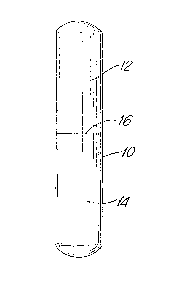
Figure 11 is an elevational view of a
chemiluminescence device according to the present invention.
DETAILED DESCRIPTION OF THE INVENTION
Non-limiting examples of nitrogen compounds for
use in the present invention include ammonia anditssalts,
heterocyclic aromatics and water-soluble amines, e.g.,
organic amines. Exemplary of salts of ammonia for use in
the present invention are, for example, acetate, chloride,
nitrate, sulfate, phosphate and borate salts, primary,
secondary, tertiary and quaternary ammonium salts where the
protons are exchanged with alkyl or aryl residues.
Non-limiting examples of heterocyclic nitrogen compounds for
use in the present invention include imidazoles and their
alkyl derivatives and pyridine and alkyl derivatives
thereof. Amines for use in the present invention include
alkyl amines, polyamines, aryl amines e.g. benzylamines.
Non-limiting examples of polyamines for employment in the
present invention include putrescine (butylene-diamine),
spermine, spermidine, and their alkyl salts. Thiazines can
also be used in the present invention as the nitrogen
compound. Exemplary thiazines are thionine and methylene
blue.
Alkylamines for use in the present invention are
- 24 -
13Q~7~80
exemp]ified by the formula
/ Xl
N - X2
x3
where Xl, X2 and X3 are the same or different and are
aliphatic saturated hydrocarbon radicals. Non-limiting
examples of aliphatic saturated hydrocarbon radicals for
use in the present invention include unsubstituted and
substituted alkanes having 1 to 8 carbon atoms, preferably
1 to 8 carbon atoms. Non-limiting examples of substituents
for such substituted alkanes include hydroxy, nitro, halo
10 (e.g., fluoro, chloro, bromo, iodo), carboxy, amide and
the like.
Chemiluminescence precursors for use in the
present invention include 2,3-dihydro-1,4-phthalazinediones
("DPD"). Preferably the 2,3-dihydro-1,4-phthalazinedione is
15 of the formula
Rl o
2 ~ NH
R3 ~ NH
R4
wherein Rl is amino and each of R2, R3 and R4 is hydrogen,
optionally substituted Cl-C6-alkyl or alkenyl, hydroxyl,
Cl-C6-alkoxy, carboxyl, amino or R2 is amino and each
20 of Rl, R3 and R4 is H, unsubstituted or substituted
Cl-C6-alkyl or alkenyl, hydroxyl, Cl-C6-alkoxy, carboxyl
or amino, or Rl and R2 are together and are an amino
or substituted amino derivative of a benzo-group, and each
of R3 and R4 is H, optionally substituted Cl-C6-alkyl
25 or alkenyl, hydroxyl, Cl-C6-alkoxy, carboxyl, or amino.
Particularly preferred chemiluminescence precursors are
5-amino-2,3-dihydro-1,4-phthalazinedione (luminol) and
6-amino-2,3-dihydro-1,4-phthalazinedione (isoluminol).
Substituted alkyl, alkenyl and amine radicals for
30 use in the present invention are well known in the art.
Non-limiting examples of substituents for such substituted
- 25 -
13C~80
radicals include halogen, e.g., chloro-, fluoro-, bromo- and
iodo-, hydroxy, carboxy, nitro, cyano and thiol.
Furthermore, amine radicals for use in the present invention
can be substituted by alkyl, preferably having 1 to 10
carbon atoms, and alkenyl, preferably having 2 to 10 carbon
atoms. Hydroxyl radicals for use in the present invention
can be substituted by halogen, alkyl, preferably having 1
to 10 carbon atoms, or alkenyl, preferably having 2 to 10
carbon atoms.
Generally any peroxidase enzyme can be used in the
present invention. Non-limiting examples of enzymes for use
in the present invention include horseradish peroxidase
(HRP), microperoxidase and lactoperoxidase.
Any oxidant which reacts with the chemiluminescence
precursor to cause excitation of the chemiluminescence
precursor so that it emits light in a luminescent reaction,
may be employed in the present invention. Particularly
preferred oxidants are hydrogen peroxide, perborate ion and
sodium peroxidate.
An example of a buffered amine for use in the
present invention is ammonia.
Non-limiting examples of chemiluminescence
enhancers include 4-chlorophenol, 4-bromophenol, 4-iodo-
phenol, 4-bromo-2-chlorophenol, 2,4-dichlorophenol, 3,4-di-
chlorophenol, 4-methylphenol, 4-tert. butylphenol, ethyl
3-(4-hydroxyphenyl)propionate, 4-benzylphenol,
4-(3'-methylcrotyl)phenol, 4-styrylphenol, 4-(2'4'-dinitro-
styryl)phenol, 4-hydroxylcinnamic acid, alpha-cyano-4-
hydroxycinnamic acid, 4-phenylphenol, 4-(4'-hydroxyphenyl)
phenol, 2-chloro-4-phenylphenol, 4-(4'-hydroxyphenyl)benzo-
phenone, 4-(phenylazo)phenol, 4~(2'-carboxylphenylazo)
phenol, 4-phenoxyphenol, 4-(4'-hydroxyphenoxy)phenol,
4-hydroxyphenol sulphide, 4-hydroxyphenyl disulphide,
naphth-2-ol, 1-bromonaphth-2-ol, 6-bromomaphth-2-ol and
1,6-dibromonaphth-2-ol. A particularly preferred enhancer
is 4-iodophenol.
--~r - 26 -
13C~ 30
O-ther non-limiting examples of chemiluminescence
enhancers for use in the present invention include
6-hydroxybenzothiazoles, such as 6-hydroxybenzo-thiazoles
of the formula
X
x3
wherein R is H~ CN or optionally substituted thiazole, and
each of Xl, X2 and X3 is H, optionally substi.tuted
Cl-C6-alkyl or alkenyl, hydroxyl, substituted hydroxyl,
C1-C6-alkoxyl, carboxyl, amino or substituted amino.
Particularly preferred chemiluminescence enhancers are
firefly luciferin (4,5-dihydro-2-(6-hydroxy-2-
benzothiazolyl)-thiazole-4-carboxoylic acid) and
dehydroluciferin.
Light emission from the chemiluminescent reaction
of the present invention, although depending primarily on
the choice of enzyme, oxidant, chemiluminescent precursor
and buffered amine or enhancer will also be determined hy
secondary factors such as temperature, pH, reagent
concentration, mixing speed and method of light measurement.
To maximize the sensitivity of the present system these
secondary factors should be adjusted to obtain the maximum
light emission, in a reproducible and easily measurable
manner, with the signal to background ratio as high as
possible.
~he conditions chosen generally involve a com-
promise involving the enzyme or catalytic activity of the
oxidant, the kinetics of the reaction, the apparatus
employed, the signal to background ratio and the sensitivity
required.
In order to achieve optimum results the present
chemiluminescent reactions should be conducted under
moderate conditions of temperature ranging from 10C to
- 27 -
13C~74~30
50C, and pH, in the range of 6 to 10, preferably hetween 7
and 9. The luminescence of the process of the present
invention is not limited to these temperature ranges and
temperature is not per se critical. Suitable buffering
substances that can be employed in the present invention are
phosphate, tris (hydroxmethyl) aminomethane,
2-amino-2-methyl-1,3-propanediol, acetate, carbonate and
borate.
The following reagent concentrations (when added
to a solution) are particularly suitable for use in the
present invention:
enzyme : .01 ng to 5000 mg/liter
oxidant : 10 ,u mol to 300 mmol/liter
chemiluminescent
substance : 0.5 u mol to 200 mmol/liter
nitrogen compound : 5 u mol to 500 mmol/liter
chemiluminescent
enhancer : 5 ~ mol to 100 mmol/liter
One aspect of the present invention involves the
detection of nucleic acid hybrids.
One nucleic acid probe for use in the process of
the present invention comprises a nucleic acid sequence
bound to a liyand, such ligand bound to a binding protein
and such binding protein bound to an enzyme. The nucleic
acid sequence can be bound to the ligand by an intercalator
compound such as a furocourmarin or a phenanthridine
compound, or by a non-intercalator compound such as
netropsin, distamycin and bis-benzimidazole. Particularly
preferred intercalator compounds are furocourmarins, for
example, angelicin (isopsoralen), psoralen and derivatives
thereof, e.g., 4-aminomethyl-4-5'-dimethyl angelicin,
4'-aminomethyltrioxsalon, 3-carboxy-5- or 8-amino- or
-hydroxy- psoralen, as well as mono- or bls-azido aminoalkyl
methidium or ethidium compounds.
Non-limiting examples of intercalating agents for
use in the present invention are exemplified in the
following Table:
-28 -
1307~80
T A B L E
Intercalator Classes and
Representative Compounds Literature References
A. Acridine dyes J. Lerman, Mol . Biol ., 3 , 18
(1961); sloomfield et al,
Physical Chemistry of Nucleic
Acids, Chapter 7, pp. 429-476,
Harper and Rowe, NY (1974);
proflavin, acridine Miller et al, siopolymer
orange, quinacrine, 19, 2091 (1980)
acriflavine
B~ Phenanthridines Bloomfield et al, supra
Miller et al, supra
ethidium
coralyne Wilson et al, J. Med. Chem.,
19, 1261 (1976)
ellipticine, Festy et al, FEBS Letters,
ellipticine cation and 17, 321 (1971); Kohn et al,
derivatives Cancer Res., 35, 71 (1976);
LePecq et al, PNAS (USA), 71,
5078 (1974); Pelaprat et al,
J. Med. Chem., 23, 1330 (1980)
C. Phenazines Bloomfield et al, supra
5-methylphenazine cation
D, Phenothiazines ibid
chlopromazine
E. Quinolines ibid
chloroquine
quinine
F. Aflatoxin ibid
G. Polycyclic hydrocarbons ibid
and their oxirane
derivatives
3,4-benzpyrene
benzopyrene diol Yang et al. Biochem. Biophys.
epoxide, l-pyrenyl- Res. Comm, 82, 929 (1978)
oxirane
- 29 -
'~7
1307~0
benzanthracene- Amea et al, Science, 176, 47
5,6-oxide (1972)
H. Actinomycins Bloomfleld e-t al, supra
actinomycin D
5 I. Anthracyclinones ibid
beta-rhodomycin A
daunamycin
J. Thiaxanthenones ibid
miracil D
10 K. Anthramycin ibid
L. Mi-tomycin Ogawa et al, Nucl. Acids
Res., Spec. Publ. 3, 79
(1977); Akhtar et al, Can. J.
Chem., 53, 2891 (1975)
15 M. Platinium Complexes Lippard, Accts. Chem. Res.,
11, 211 (1978)
N. Polyintercalators
echinomycin Waring et al, Nature, _52, 653
(1974); Wakelin, Biochem. J.,
157, 721 (1976)
quinomycin Lee et al. Biochem. J.,
triostin 173, 115 (1978j, Huang et al,
BBM928A B ochem. 19, 5537 (1980);
tandem Viswamitra et al, Nature,
2~ 817 (1981)
diacridines LePecq et al, PNAS (USA),
7~ 2915 (1975), Carrellakis
et al, Biochem. Biophys. Acta,
418, 277 (1976); Wakelin et
al, Biochem, 17, 5057 (1978);
Wakelin et al, FEBS Lett.,
104, 261 (1979); Capelle et
al, Biochem., 18, 3354 (1979);
Wright et al, Biochem., 19,
5825 (1980); Bernier et al,
Biochem. J., 199, 479 (1981);
King et al, Biochem., 21, 4982
(1982)
- 30 -
., .
~30~80
ethidium dimer Gaugain et al. Biochem,
17, 5078 (197~); Kuhlman et
al, Nucl. Acids Res. 5, 2629
(1978); Marlcovits et al,
Anal. Biochem., 94, 259
(1979); Dervan et al, JACS,
100, 1968 (1978); ibid 101,
3664 (1979)
ellipticene dimers Debarre et al, Compt. Rend.
and analogs Ser. D., 284, 81 (1977)-
Pelaprat et al, J. ~ied. Chem.,
23, 1336 (1980)
heterodimers Cain et al, J. Med. Chem.,
21, 658 (1978); Gaugain et al,
Biochem." 17, 5078 (1978)
trimers Hansen et al, JCS Chem. Comm.,
162 (1983); Atnell et al,
JACS, 105, 2913 (1983)
o. Norphillin A Loun et al, JACS, 104, 3213
(1982)
P. Fluorenes and Bloomfield et al, supra
fluorenones
fluorenodiamines Witkowski et al, Wiss.
Beitr.-Martin-Luther-Univ.
Halee Wittenberg, 11 (1981)
Q. Furocoumarins
angelicin Venema et al, MGG, Mol. Gen.
Genet., 179, 1 (1980)
4,5'-dimethylangelicin Vedaldi et al, Chem.-Biol.
Interact, 36, 275 (1981)
psoralen Marciani et al, Z. Naturforsch
B, 27(2), 196 (1972~
8-methoxypsoralen Belognzov et al, Mutat. Res.,
84, 11 (1981); Scott et al,
Photochem. Photobiol., 34, 63
~i981)
5-aminomethyl-8- Hansen et al, Tet. Lett.,
methoxypsoralen 22, 1847 (1981)
4,5,8-trimethyl- Ben-Hur et al, Biochem.
psoralen Biophys, Acta, 331, 181 (1973)
13~ 30
4'-a~inomethyl--1,5,8- Issacs et al, siochem~ 16,
trime-thylpsoralen 1058 (1977)
xanthotoxin Hradecma et al, Acta Virol.,
(Engl. Ed.) 26, 305 (1982)
khellin BeaumGnt et al, Biochem.
siophys. Acta, 608, 1829
(1980)
R. Benzodipyrones Murx et al, J. Het. Chem.,
12, 417 (1975); Horter et al,
Photochem. Photobiol., 20, 407
(1974)
S. Monstral Fast Blue Jurarranz et al, Acta
Histochem., 70, 130 (1982)
Particularly useful intercalating agents are the
azidointercalators. Their reactive nitrenes are readily
generated at long wavelength ultraviolet or visible light
and the nitrenes of arylazides prefer insertion reactions
over their rearrangement products (White et al, Methods ln
Enzymo_ , 47, 644 (1977)). Representative azido-
intercalators are 3-azidoacridine, 9-azidoacridine, ethidium
monoazide, ethidium diazide, ethidium dimer azide (Mitchell
et al, JACS, 104, 4265 (1982)), 4-azido-7-ch].oroquinoline,
and 2-azidofluorene. Other useful intercalators are the
furocoumarins which form C2+2~ cycloadducts with pyrimidine
residues. Alkylating agents can also be used such as bis-
; 25 chloroethylamines and epoxides or aziridines, e.g., a-
flatoxines, polycyclic hydrocarbon epoxides, mitomycin, and
norphillin A.
Suitable angelicin derivatives for use in the
present invention have the following formula
R,2 Rl
0~0
R3
R4
- 32 -
13~ 80
wherein Rl, R2~ R3 and R4 are as follows
Rl R2 R3 R4
H H H H
CH3 H CH3 H
CH3 CH3 CH3 CH2H
CH3 H CH3 CH20CH3
CH3 H CH3 CH2NH2
CH3 H CH3 CH2Cl
CH3 H CH 3 ~ \N- CH2
Many other compounds with different Rls can be
synthesized following published procedures.
Suitable psoralen derivatives for use in the
present invention have the formula
R,2 Rl
R3 ~ oR
in which
R, Rl and R3 each independently is hydrogen or
lower alkyl,
R4 is hydrogen, lower alkyl or
lower alkyl substituted by hydroxy,
lower alkoxy, amino, halo and~or
o
C~ N
, and
13~7~8~
R2 and R5 each independently is hydrogen, hydroxy,
carboxy, carbo-lower alkoxy or lower alkoxy.
Angelicin derivatives are superior to psoralen
compounds for monoadduct formation. If a single-stranded
probe is covalently attached to some extra double-stranded
DNA, use of phenanthridum and psoralen compounds is
desirable since these compounds interact preferentially to
double-stranded DNA in the dark.
Non-limiting examples of nucleic acid sequences
for use in the present invention can be singly or doubly
stranded DNA or RNA or fragments thereof, such as are
produced by restriction enzymes or even relatively short
oligomers.
In anembodiment of the present invention the probe
is immobilized on a solid support, for example,
nitrocellulose paper.
Non-limiting examples of ligands for use in the
present invention include haptens and biotin, e.g., biotin-
N-hydroxysuccinimide and biotin-P-nitrophenyl ester.
Non-limiting examples of binding proteins for use
in the present invention include antibodies, avidin and
streptavidin.
In one embodiment for carrying out the present
invention, the labelled probe immobilized by hybridization
on nitrocellulose paper, i.e., enclosed in a transparent
container, is placed on high speed photographic film such as
a "POLAROID*" film cartridge. The immobilized probe and film
cartridge, and suitable reagents in solution form (the
reagents employed depend upon the probe utilized, for
example, if the probe contains a chemiluminescence
substance, then the reagent solution will contain an
enhancer, an oxidant and an enzyme) would be injected into
the vessel to contact the immobilized probe. Light emitted
by virtue of a reaction between the reagents and the probe
would then be detected on the film. It should be noted that
the wavelength of light emitted would depend on the reagents
*Trade-mark
- 34 -
~l; `
~3074~30
employed. If hybridization occurs, iight will be emitted.
If hybridization does no-t occur, light will not be emitted.
Probes And Formats For Hybridization
There are different types of probes and formats
which can be used for hybridization assays and detection by
following the method of the present invention.
Essentially any nucleic acid hybridization format
can be followed for the purposes of the present invention in
which either the hybrids formed between the probe and the
sequence to be determined or the probe which has not
hybridized with the sequence of interest are labelable with
the selected chemiluminescence label. As is known in the
art, the labeling of such hybrids or unhybridized probe can
be accomplished before or after the actual hybridization
reaction. Normally, the probe is either labelled or
labelable through a specific bridying reaction or the formed
hybrids are subsequently labeled, usually through a specific
bridging reaction. A central novel feature of the present
invention is the advantageous application of the phenomenon
of enhance chemiluminescence to the detection of nucleic
acid hybridization.
The probe will comprise at least one single
stranded base sequence substantially complementary to or
homologous with the sequence to,be detected. However, such
base sequence need not be a single continuous polynucleotide
segment, but can be comprised of two or more individual
segments interrupted by nonhomologous sequences. These
nonhomologous sequences can be linear, or they can be
self-complementary and form hairpin loops. In addition, the
homologous region of the probe can be flanked at the 3'- 5'-
termini by nonhomologous sequences, such as those
comprising the DNA or RNA of a vector into which the
homologous sequence had been inserted for propagation. In
either instance, the probe as presented as an analytical
reagent will exhibit detectable hybridization at one or more
- 35 -
130~glV
points with sample nucleic acids of interest. Linear or
circular single stranded polynucleotides can be used as -the
probe element, with major or minor portions being duplexed
with a complementary polynucleotide strand or strands,
provided that the critical homologous segment or segments
are in single stranded form and available for hybridization
with sample DNA or RNA. Particularly preferred will be
linear or circular probes wherein the homologous probe
sequence is in essentially only single stranded form (see
particularly, Hu and Messing, Gene, 17, 271-277 (1982)).
The formats where a single polynucleotide sequence
is used as a probe is common in the prior art. The probe
can be labeled in such a way that will be able to
participate in the chemiluminescent reaction. This can be
achieved by labelling the probe with a ligand as, for
example, biotin which specifically binds to a protein and
that protein can be a carrier for the chemiluminescent
reaction component, as for example linked covalently to
luminol or horseradish peroxidase.
The probe can also be directly linked to the
chemiluminescent reaction partners. The probe can be
photochemically linked to luminol or horseradish peroxidase.
The probe can also be produced in such a fashion that after
the hybridization the hybrid will behave immunologically
distinct from the rest of the reaction components, for
example, if a DNA probe is used for the detection of RNA or
an RNA probe is used for the detection of DNA, the DNA/RNA
hybrid produces immunologically specific antibodies which
will recognize those hybrids and those specific recognition
can be utilized for the detection of the hybrid. If the RNA
probe is immobiliæed, the hybrid is likewise immobilized and
an antibody specific for the RNA/DNA hybrid is reacted with
the hybrid. If the antibody carries a label which can
participate in the chemiluminescent reaction, the hybrid can
be detected via the antibody, and the chemiluminescent
process. As, for example, if the RNA/DNA hybrid specific
~3V~7~
antibody is covalently linked to horseradish peroxidase
after the hybridization and interaction with the
peroxi~ase-linked antibody it should be possible to initiate
chemiluminescent reaction by adcling the precursor and an
oxidant.
There are several other ways a nucleic acid can be
made immunogenic and immunologically distinct from the other
nucleic acids. Antibodies which are selective for RNA/RNA
or DNA/DNA hybrids are also known and can be similarly used.
In addition, if a nucleic acid interacts with an
intercalator, the nucleic acid complex becomes
immunologically distinct from the unreacted nucleic acid.
In a hybridization format if a probe is prepared such that
the probe will provide such interaction sites after the
hybridization, an antibody assay can be conducted for the
detection of the hybrid.
Practice of the analytical methods of -the presen-t
invention is not limited to any particular hybridiæation
format. Any conventional hybridization technique can be
used. As improvements are made and as conceptually new
formats are developed, such can be readily applied to
carrying out the present method. Conventional hybridization
forms which are particularly useful include those wherein
the sample nucleotide acids or the polynucleotide probe is
immobilized on a solid support (solid-phase hybridization)
and those wherein the polynucleotide species are all in
solution (solution hybridization).
Solid-Phase Hybridization Formats
In solid-phase hybridization formats, one of the
polynucleotide species participating in hybrization is fixed
in an appropricate manner in its single stranded form to a
solid support. Useful solid supports are well known in the
art and include those which bind nucleic acids either
covalently or noncovalently. Noncovalent supports which are
.
.~..
13~1'7480
generally understood to involve hydrophobic bonding include
naturally occurring and synthetic polymeric materials, such
as nitrocellulose, derivatized nylon, and fluorinated
polyhydrocarbons, in a variety or forms such as filters or
solid sheets. Covalent binding supports are also useful and
comprise materials having chemically reactive groups or
groups, such as dichlorotriazine, diazobenzyloxymethyl, and
the like, which can be activated for binding to
polynucleotides.
A typical solid-hase hybridization technique
begins with immobilization of sample nucleic acids onto the
support in single stranded form. This initial step
essentially prevents reannealing of complementary strands
from the sample and can be used as a means for concentrating
sample material on the support for enhanced detectability.
The polynucleotide probe is then contacted wlth the support
and hybridization detected by the methods as described
herein.
Normally, -the probe is labeled directly or
indirectly through one or more specific binding pairs with
the selected chemi.luminescence label. As used herein,
indirect labeling, immobilization, or other modification
through one or more specific binding pairs intends the
coupling of one of a pair of mutually binding substances to
the material to be labeled, etc., e.g., probe, and the
labeling, immobilization, etc. of the other member of the
pair. Useful binding pairs include biotin/avidin (including
egg white aviden and streptavidin), haptens and antigens/
antibodies, carbohydrates/lectins, enzymes/inhibitors, and
the like as are known in the art. One can also use bridging
pairs, such as coupling biotin or a hapten to the material
to be labeled, etc., and also to the label, solid-phase,
etc., and using avidin or an anti-hapten, respectively, to
bridge the two.
- 38 -
,. . .
~3ai~80
When using labeled probe and immobilized sample
nucleic acids, the resulting hybrids are separated from the
unhybridized probe, and the chemiluminescence reaction is
initiated in one or the other of the separate fractions.
Alternatively, the hybrids and unhybridized probe do not
have to be separated if hybrids are detected by anti-hybrid
antibodies which distinguish the hybrids from the
unh~bridized single stranded probe. Such antibodies can be
selective for mixed DNA/RNA hybrids or selective on RNA/RNA
or DNA~DNA hybrids, or can be selective for intercalator
duplexes where an intercalatin~ agent has been introduced to
the hybrids. Such antibody reagents will be described in
more detail below~
An alternative method to those involving sample
nucleic acid immobilization uses immobilized probe and
detection of resulting immobilized hybrids with an
anti-hybrid antibody labeled directly or through specific
binding pairs with the selected chemiluminescence label as
described above. When presented to the hybridization
reaction in an immobilized form, the probe can be in any
appropriate form that enables the probe, and any components
of the reaction mixture that have become associated
therewith by hybridization and/or by binding of the
anti-hybrid reagent, to be subsequently isolated or
separated from the remaining mixture such as by
centrifugation, filtration, chromatography, or decanting. A
variety of compositions and configurations of an immobilized
probe will thus be evident and available to the worker in
the field. Essentially any form of the probe that is
insoluble in the reaction mixture can be used. For example,
the probe can be aggregated or otherwise precipitated,
attached to an insoluble material, polymer, or support, or
entrapped in a gel such as agarose or polyacrylamide (see
Meth. Enzymol., 12B:635 (1968) and PNAS, 67, 807 (1970)).
- 39 -
~3~7~80
It is particularly preferred to employ a solid suppor-t to
which the probe is attached or ~ixed by covalent or
noncovalent bonds, the latter including adsorption methods
that provide for a suitably stable and strong attachment.
The solid support can take on a variety of shapes and
compositions, including microparticles, beads, porous and
impermeable strips and membranes, the interior surface of
reaction vessels such as test tubes and microtiter plates,
and the like. Means for attaching a desired reaction
partner to a selec-ted solid support will be a matter of
routine skill to the worker in the field.
One method for adsorbing the probe onto
nitrocellulose membranes involves saturating a solution of
probe with sodium iodide and spotting or filtering aliquots
onto the membrane (Bresser et al, DNA, 2, 243 (1983)). The
sodium iodide facilitates denaturation of the probe and
enhances adsorption onto the membrane. Alternatively, the
probe can be treated with glyoxal. usually at concentrations
around 1 molar (M), and then adsorbed onto the membrane. The
probe is fixed by baking at around 80C under vacuum for a
period in the range of 2-4 hours. (P.S Thomas, M th. In.
Enzymol., 100, 255 (1983)).
Covalent immobilization of RNA or DNA probes can
also be accomplished. A wide variety of support materials
and coupling techniques can be employed. For example, the
probe can be coupled to phosphocellulose through phosphate
groups activated by carbodiimide or carbonyldiimidazole
(E.K.F. Bautz, and B.D. Hall, Proc. Nat'l. _cad. Sci. USA,
_, 400-408 (1962); T.Y. Shih and M.A. Martin, Biochem, 13,
3411-3418 (1974). Also, diazo groups on
m-diazobenzoyloxymethyl cellulose can react with guanine and
thymidine residues of the polynucleotide (B.E. Noyes and
G.R. Stark, Cell, 5, 301-310 (1975); J. Reiser et al,
Biochem. Biophys. Res. Commun., _, 1104-1112 (1978)).
Polysaccharide supports can also be used with coupling
through phosphodiester links formed between the terminal
-- ~0 --
~,..`i
~`
~3~P7~
phosphate of the polynucleotide and the support hydroxyls
ky water soluble carbodiimide activation (D. Richwood,
_iochim. Biophys. Acta, 269, 47-50 (1972); P.T. Gilham,
Biochem, 7, 2809-2813 (1968)), or by coupling nucleophilic
sites on the polynucleotide with a cyanogen br~mide
activated support (D.J. Arndt-Jovin et al, Eur. J. Biochem.,
54, 411-418 (1975), U. Linb~rg and S. Ericksson, Eur. J.
Biochem., 1~, 474~479 (1971)). Further, the 3'-hydroxyl
terminus of the probe can be oxidized by periodate and
coupled by Schiff base formation with supports bearing
amine or hydrazide groups tP.T. Gilham, Method. Enzymol.,
21, 191-197 (1971); H.D. Hansske et al, Method. Enzymol.,
59, 172-181 (1979)). Supports having nucleophilic sites
can be reacted with cyanuric chloride and then with the
polynucleotide (H.D. Hunger et al, Biochim. Biophys. Acta,
653, 344-349 (1981)).
In general, any method can be employed for
immobilizing the probe, provided that the complementary
single stranded sequence is available for hybridization to
sample nucleic acids. Particular methods or materials
are not critical to the present invention.
Another method of interest is the sandwich
hybridization technique wherein one of two mutually
exclusive fragments of the homologous sequence of the probe
is immobilized and the other is labeled. The presence of
the polynucleotide of interest results in dual hybridization
to the immobilized and labeled probe segments, again with
the same ultimate measurement results of support-associated
labeled hybrids. See Methods in Enzymology, 65, 468 (1980)
-
and Gene, 21, 77-85 (1983) for further details.
For purposes of better illustration, the following
solid-phase hybridization methods involving detection with
antibody to intercalated duplexes are particularly useful in
the present invention.
- 41 -
~,
13CJ 7~8(~
In a first me-thod, the single stranded nucleic
acids from the liquid -tes-t medium are first immobiliæed on a
solid support. A hybridiza-tion reaction mixture is then
formed by contacting the immobilized sample nucleic acids
with the probe which in this case comprises, in addition to
the complementary sinyle stranded portion, at least one
double stranded portion WhiCh iS chemically linked with the
intercalator in the form of lntercalation complexes. A
particularly useful form of the probe is -the circular form
described by Hu and Messing, _upra. The resulting
hybridization aggregate comprises the immobilized
polynucleotide of interest hybridized with the probe which
has a covalently linked, intercalated double stranded
region. The solid support carrying immobilized duplexes is
then preferentially separated from the remainder of the
reaction mixture. The antibody is added, preferably labeled
with the selected chemiluminescence label, and the resul-ting
immobilized antibody bound to intercalation complexes in the
aggregate is separated from the remainder of the reaction
mixture. The antibody bound to the support is then
determined to complete the assay. Alternatively, the
antibody in the separated solu-tion can be determined;
although this will generally be less perEormed since a large
excess of antibody is normally used.
A variation oE this method is to employ a probe
such as above, but not having covalently linked intercalator
bound to the double stranded region. Rather, the
intercalator is added -to the immobilized aggregate resulting
in the formation of intercalator complexes in both the
double stranded portion of the probe and the duplexed region
formed by hybridization.
A second method is based on a sandwich format
where a reaction mixture is formed among the test medium
containing the sequence of interest and the first and second
probes, each comprising respectively at least one base
- 42 -
1307~:80
sequence complemen~ary to a mutually exclusive portion of
the sequence of interest. The first probe is immobilized on
a solid support and the second probe is modified with
covalently linked, in-tercalation complexes as in the
previous method. The resulting hybridization aggregate
comprises the sequence of interest hybridized to both the
immobilized ~irst probe and the intercalation
complex~modified second probe. The antibody is added,
preferably in labeled form, and the resulting immobilized
antibody bound to intercalation complexes in the aggregate
is separated from the remainder of the reaction mixture.
The bound antibody is determined to then complete the assay.
There are several useful variations of this second
method. First, as in the case of the variation of the first
method, one can employ a probe which does not comprise
covalently linked intercalator, but rather can add free
intercalator to the immobilized aggregate, resulting in the
formation of intercalator complexes with all available
double stranded regions. Also, as an alternative to using a
second prohe with a double stranded portion, one can use a
probe of entirely single stranded nucleic acid with
intercalator chemically linked thereto so that upon
hybridization there are formed intercalation complexes, or
with intercalator being added so that intercalation occurs
between the duplexes formed between the two probes and the
sequence to be detected.
In a third method, the sample nucleic acids are
contacted with immobilized probe and preferably the
resulting immobilized duplexes are separated from the
remainder of the reaction mixture. In this format, the
probe is in single stranded form. The resulting
hybridization product comprises the immobilized probe
hybridized with the sequence of interest. Also, this format
allows significant reappealing between complementary regions
of sample nucleic acid which can take place on the
- 43 -
<, ~ .
1;3(3748(~
immobilized aggregate. Such reannealing works to the
advantage of the assay since i-t provides additional double
stranded nucleic acid for subsequent intercalation. The
next step in the assay is to add intercalator and the
antibody, again preferably in a labeled form. The assay is
completed by separation and antibody determination steps as
in the previous formats.
Finally, there is a fourth method wherein the
single stranded sample nucleic acids are contacted with
immobilized probe where, in this case, such probe is
chemically, e.~., covalently, to the intercalator such that
duplex formation in the region of the linked intercalator
results in formation of intercalation complexes. This is a
highly advantageous format in that the probe is both
immobilized and modified, requiring no immobilization or
modification step to be performed at the time of the assay.
The resulting aggregate comprises covalently llnked,
intercalation complexes in the region of hybridization
between sample and probe nucleic acids and in any reannealed
sample regions. Antibody is then added and the assay
completed as in the previous formats. This format provides
the advantage of eliminating the need for the analyst to
handle solutions of the free intercalator which in some
cases can be potentially hazardous. A simple variation of
this technique is to immobilize sample nucleic acids rather
than the labeled probe and proceed in the normal fashion.
This is somewhat less advantageous but is a practical assay
approach.
Solution-Phase Hybridization Formats
In addition to the above described solid-phase
formats, a variety of solution-phase hybridization formats
can also be applied to the present invention. Such formats
are characterized by the feature that the hybridization step
involves soluble forms of both the sample nucleic acids and
- 44 -
13~7~8(1
the probe. This can result in significantly faster
hybrizaLions since the kine-tics are much faster when both
strands are in solution compared to when one is immobilized.
Normally, subsequent to the hybridization step, -the
resulting hybrids are rendered immobile for purposes of
detec-tion. Such immobilization can be accomplished in a
variety of ways. Conventionally i-t is known to selectivel~
immobilize complexes by exposure to adsorbents such as
hydroxyapatite and nitrocellulose membranes.
A particularly useful approach to immobilizing
hybrids formed from a solution-phase hybridization involves
the use of a probe which comprises a reactive site capable
of forming a stable covalent or noncovalent bond with a
reaction partner and obtaining immobilization by exposure to
an immobilized form of such reaction partner. Preferably,
such reactive site in the probe is a binding site such as a
biotin or hapten moiety which is capable of specific
noncovalent binding with a binding substance such as avidin
or an antibody which serves as the reaction partner. After
the hybridization step then, one c~n add an immobilized form
of the reaction partner, e.g., binding substance, which will
effectively bind and immobilize the hybrids through the
reactive site on the probe.
Essentially any pair of substances can comprise
the reactive site/reactive partner pair which exhibit an
appropriate affinity for interacting to form a stable bond,
that is a linking or coupling between the two which remains
substantially intact during the subsequent assay steps,
principally the separation and detection steps. The bond
formed may be a covalent bond or a noncovalent interaction,
the latter being preferred especially when characterized by
a degree of selectivity or specificity. In the case of such
preferred bond formation, the reactive site on the probe
will be referred to as a binding site and the reaction
partner as a binding substance with which it forms a
noncovalent, commonly specific, bond or linkage. Such
- 45 -
13~7q~80
bindin~ site can be present in a single stranded
hybridizable portion of the probe or can be present as a
result of a chemical modification of the probe. Examples of
binding sites existing in the nucleotide sequence are wher~o
the probe comprises a promoter sequence (e.~., lac-p~omoter,
trp-promoter) which is bindable by a promoter protein (e.g.,
bacteriophage promoters, RNA polymermase), or comprises an
operator sequence (e.g., lac operator) which is bindable by
a repressor protein (e.g., lac repressor), or comprises
rare, antigenic nucleotides or sequences (e.g., 5-bromo or
5-iododeoxyuridine, Z-DNA) which are bindable by specific
antibodies (see British Patent Specification 2,125,964).
Binding sites introduced by chemical modification of the
probe are particularly useful and normally involve linking
one member of a specific binding pair to the probe nucleic
acid. Useful binding pairs from which to choose include
biotin/avidin, haptens and antigens/antibodies,
carbohydrates/lectins, enzymes/inhibitors, and the like.
Where the binding pair consists of a proteinaceous member
and a nonproteinaceous member, it will be preferred to link
the nonproteinaceous member to the probe since the
proteinaceous member may be unstable under the denaturing
conditions of hybridization of the probe. Preferable
systems involve linking the probe with biotin or a hapten
and employing immobilized avidin or anti-hapten antibody,
respectively. Preparation of useful ligand-labelbd probes
is known in the literature (Langer et al, Proc. Natl. Acad.
Sci., 78, 6633 (1981); Broker, Nucl. Acids Res., 5, 363
(1978); Sodja et al, Nucl. Acids Res., _, 385 (1978); Tchen
et al, Proc. Hatl. Acad. Sci., 81, 3466 (1984)).
Immobilization of the binding substance can follow
conventional techniques.
A large variety of methods are known for
immobilizing proteins on solid supports and these methods
are applicable to the immobilization of the binding
substance (see Methods in Enzymology, Vol. 44 (1976)).
-- 46 --
~3(~480
Antibodies, for e~ample, are immobilized either by covalent
coupling or by noncovalent adsorption. Noncovalent methods
frequently employed are adsorption to pol~styrene beads or
microparticlex and to polyvinylchloride surface. Marly
covalent methods are used for immobilizing proteins and a
few include cyanogen bromide activated agaroses and
dextrans; glutaraldehyde activated nylons and
polyacryla~ides; and epoxides on acrylic and other supports.
When the probe is presented for hybridization with
the sequence of interest in an immobilizable form, the
subsequent steps of immobilization of the formed duplexes
through a property of the probe and addition of the
anti-hybrid rea~ent can proceed in any desired order.
Immobilization and anti-hybrid addition can be accomplished
by simultaneous addition of the involved reagents and
materials, or one can precede the other, with or without
intervening wash or separation steps, in either order.
Where ordered additions are followed, of course one will
take into account the concentrations of the added reagents
so as not to oversaturate the formed hybrids and inhibit
interaction therewith of the second added materials.
Although immobilized probes or immobilizable
probes which become bound to solid supports by specific
bindin~ processes described above are preferred,
immobilizable probes can be bound to supports by processes
with relatively low specificity. In this case the support
~ould bind the hybridized probe but not the unhybridized
form. Then the amount of hybrid would be measured with the
antibody reagent. An example of a support of this type is
hydroxyapitite which binds DNA-RNA and RNA-RNA duplexes but
not the single stranded species (Brenner and Falkow, Adv. in
Genet., _6, 81 (1973)).
Also, a chemically active or activatable group can
be introduced into the probe and allowed to react with the
solid support following the hybridization. This system
would give a covalently immobilized probe and the amount of
- 47 -
13CI'7~?~0
hybrid coupled to the support can be determined with the
antibody.
n addition to -the above methods, solution-phase
hybridization formats can be performed wherein the hybrids
a~e immobilized by binding of immobilized or immobilizable
anti-hybrids antibody reagents. Such antibody reagents can
be specific for in~ercalated duplexes or for DNA/RNA,
: RNA/RNA or DNA/DNA hybrids as described herein. Researching
immobilized duplexes are detected by using directly or
indirectly labeled probe, labeled second anti-hybrid
antibody, or a labeled second probe.
Anti-Hybrid Antibody Reagent and Detection Schemes
The antibody reagent used in the preferred
` embodiments of the present invention is principally
characterized by its ability to bind the hybrids formed
between the probe and complementary sample nucleic acids to
the significant exclusion of single stranded
polynucleotides. The antibody reagent can consist of whole
antibodies, antibody fragments, polyfunctional antibody
aggregates, or in general any substance comprising one or
more specific binding sites from an anti-hybrid antibody.
When in the form of whole antibody, it can belong to any of
the classes and subclasses of known immunoglobulins, e.g.,
IgG, IgM, and so forth. Any fragment of any such antibody
which retains specific binding affinity for the hybridized
probe can also be employed, for instance, the fragments of
IgG conventionally known as Fab, E`(ab'), and F(ab')2. In
addition, aggregates, polymers, derivatives and conjugates
of immunoglobulins or their fragments can be used where
appropriate.
The immunoglobulin source for the antibody reagent
can be obtained in any available manner such as conventional
antiserum and monoclonal techniques. Antiserum can be
obtained by well-established techniques involving
immunization of an animal, such as a mouse, rabbit, guinea
- 48 -
,`t'~
13(~7~3Q
pig or goat, with an app~opriate immunogen. The
immunoglobulins can also be obtained by somatic cell
hybridization -techniques, such resulting in what are
commonly referred to as mcnoclonal antibodies, also
involving the use of an appropriate immunogen.
Useful anti-hybrid antibodies include those
selective for intercalated nucleic acid duplexes as well as
those which bind DNA/RNA, RNA/RNA, or DNA/RNA hybrids
specifically.
Antibodies to i-ntercalated duplexes are raised
against an immunogen which usually comprise an ionic complex
between a anionic protein or protein derivative (e.g.,
` methylated bovine serum albumin) and the anionic
intercalated duplexes. Preferably, the intercalation is
covalently linked to the duplex. Alternatively, the
intercalator duplex complex can be covalently coupled to a
carrier protein.
The preparation of antibodies to DNA/DNA is
described in European Patent Publication No. 135,139.
Immunogens for stimulating antibodies specific for
DNA-RNA hybrids can comprise homopolymeric or
heteropolymeric polynuc].eotide duplexes. Among the possible
homopolymer duplexes, particularly preferred is poly(rA)-
poly(dT) (Kitagawa and Stollar, Mol. Immunol., 19, 413
(1982)). However, in general, heteropolymer duplexes will
be preferably used and can be prepared in a variety of ways,
including transcription of ~X174 virion DNA with RNA
polymerase (Nakazato, Biochem, 19, 2835 (1980)). The
selected RNA DNA duplexes are adsorbed to a methylated
protein, or otherwise linked to a conventional immunogenic
carrier material, such as bovin serum albumin, and injected
into the desired host animal (see Stollar, Meth. Enzymol.,
70, 70 (1980)).
Antibodies to RNA DNA duplexes can be raised
against double stranded RNAs from viruses such as reovirus
or Fiji disease virus which infects sugar cane, among
- 49 -
`.'~
1307~8~
others. Also, homopolymer duplexes such as poly(rI)
poly(rC) or poly(rA) poly(rU), among others, can be used for
immunization as above.
When the antibody reagent is used to detect
hybrids, it will usually be labeled with the
chemiluminescence label by suitable synthetic means.
Alternatively, the antibody reagent can be
detected based on a native property such as its own
an-tigenicity. A chemiluminescence labeled anti-(antibody)
antibody or protein A will bind to the primary antibody
reagent where the label for the second antibody or protein A
is a conventional label as above. Further, antibody can be
detected by complement fixation or the use of labeled
protein A, as well as other techniques known in the art for
detecting antibodies.
Where the antibody reagent is labeled, as is
preferred, the labeling moiety and the antibody reagent are
associated or linked to one another by direct chemical
linkage such as involving covalent bonds, or by indirect
linkage such as by incorporation of the label in a
microcapsule or liposome which is in turn linked to the
antibody. Labeling techniques are well-known in the art and
any convenient method can be used in the present invention.
Emitted light can be detected by convention means,
such as by a photomultiplier tube, the signal from which can
be fed to and displayed or recorded on a recorder,
oscilliscope or scalar. The light could also be quantified
on a luminometer.
Depending on the type of label employed, the assay
may be either heterogeneous or homogeneous. In the former
case complex fluids such as serum may be analyzed, however,
in the latter case, a preliminary extraction or purification
step may be necessary.
Typical heterogeneous and homogeneous luminescent
- 50 -
~307~0
or luminometric immunoassays are outlined below:
1. Heterogeneous Luminescent of Luminometric
Immunoassay
In this type o~ immunoassay the substance to be
assayed is reacted with an antibody thereto. The free
antibody is then separated from the hound antibody. The
reaction is quantified by labelling either the antibody, the
substance to be assayed or another molecule which can react
with the free or bound moieties after separation.
2. Competitive Heterogeneous Luminescent Immunoassay
In this case an unknown amount of the substance to
be assayed is mixed wi~h a known amount of said substance
coupled with a label and a known, but limited, amount of an
antibody thereto. A competitive reaction between the
labelled and unlabelled substance for the antibody ensues.
The complexes between antibody and unlabelled substance and
between antibody and labelled substance are separated from
the free labelled and unlabelled substance.
The amount of labelled substance bound to antibody
is related to the amount of unlabelled substance in the
solution beiny assayed. These quantities may be determined
either by measuring the amount of label bound to antibody or
by measuring the amount of free labelled substance
remaining. Examples of this type of assay wherein
peroxidase is the label and the antibody is bound to a solid
phase, via the walls of a glass test tube, are given in
UK 2,044,927A.
3. Two-Site Heterogeneous Luminometric Immunoassay
In this type of immunoassay the substance to be
assayed is first bound to an unlabelled antibody thereto
which in turn is bound to a solid phase support, such as
plastic. The complex (between antibody and substance) is
then treated with a labelled antibody.
Analysis for the labelled antibody in the solid
complex obtained may then be affected by separating the
solid complex from the solution, and then determininy either
- 51 -
~3~7480
the amount of label present in the separated solid complex
or the amount of label present in the residual labelled
antibody dissolved in the solution.
In alternative embodiments of this type of
S immunoassay the substance to be assayed may either be bound
consecutively to the labelled antibody and to the
unlabelled, solid supported antibody or be bound to both the
labelled and unlabelled antibody in one binding step.
4. Homogeneous Luminescent or Luminometric
Immunoassay
This is applicable to immunoassays wherein the
label is an amino or a substituted amino derivative of a
2,3-dihydro-1,4-phthalazinedione. It depends upon the
light emitted from the free labelled substance of interest
(or antibody thereto) being of a different intensity or
wavelength to the light emitted from the bound labelled
substance of interest (or antibody thereto).
In one example it was found that the intensity of
light emitted from the reaction of a (progesterone-
isoluminol deriv) conjugate was added, followed by a knownamount of haem and hydrogen peroxide. The light emitted was
measured and the amount of progesterone present in the
unknown sample thereby determined from the standard curve.
(The more progesterone present in the unknown sample, the
less free IgG is left at equilibrium and the lower is the
light yield of the luminescent reaction).
In this way the determination of progesterone may
be achieved without the requirement of a separation step.
In all of the above immunoassays the quantifying,
detecting or locating step may be the luminescent reaction
of the present invention.
The antibodies employed in the above immunoassays
may be purchased commercially or prepared by known
immunological techniques. The antibodies may be in the form
of a complex mixture of antibodies or they may be one or
more monoclonal antibodies. Only a small volume of antibody
- 52 -
13Q7480
is generally required and it is maintained at the conditions
of pH, ionic strength and temperature appropriate for its
activity.
Antibodies to -the following non-exhaustive list of
substances may be usefully employed in immunoassays
utilizing the present luminescent reaction: proteins such as
insulin, alphafetoprotein and fe~ritin, hormones such as
growth hormone, parathyroid hormone, follicle stimulating
hormone, luteinising hormone, tyroid stimulatin~ hormone,
adrenocorticotrophic hormone, glucagon, prolactin and
calcitonin, haptens/steroids such as estriol, progesterone
and cortisol, drugs such as digoxiin, antigens such as cell
surface antigens and carcino embryonic antigen and
antibodies such as mumps virus antibody, human
immunoglobulin G (IgG), rabbit IgG, sheep IgG, guinea pig
IgG, donkey IgG and human immunoglobulins E and M.
Figure 11 depicts a chemiluminescence device in
accordance with the present invention. Device 10 has two
compartments 12 and 14 separated by a valve means or
membrane 16. Compartments 12 and 14 each contain one, but
not all of the chemiluminescence reactants, i.e.,
chemiluminescence precursor, oxidant and enzyme. A buffered
amine is contained in one or both of compartments 12 and 14.
A typical vessel would, for example, contain a
chemiluminescence precursor in compartment 12 and an
oxidant, enzyme and buffered amine in compartment 14. A
valve means 16 would control the gravity flow of
chemiluminescence precursor from compartment 12 into
compartment 14. Light is emitted when the flow begins. To
stop the emittance of light, the valve means 16 would be
closed to stop the flow of chemiluminescence precursor.
The invention will now be described with reference
to the following non-limiting examples.
- 53 -
?74~0
Examples
Example 1: reparationofLigand-Bound Probe DNA:
Although the method below is illustrated with a
specific nucleic acid it can be used for any DNA probe.
There are other various methods of labeling (nick
translation, for example) nucleic acid probes known in the
literature. A general method for labelling nucleic acids,
hence a test sample is described below:
Covalent
¦Nucleic Acid ¦-t ¦photoreactive ¦- Light ~ Com plex
lintercalator
chemical reaction with ~
reactive ligand ,
Ligand labeled nucleic
acid.
In the above, the Eollowing were employed:
a) an ADENOVIRUS DNA or pBR322 probe (commercially
available plasmid DNA probe from ENZO Biochem,
New York and BRL - Bethesda Research Laboratory.
Maryland).
b) the photoreactive intercalator was an aminomethyl
angelicin
c) the reactive ligand was N-hydroxysuccinimido
biotin.
The probe was first photochemically reacted with
an intercalator. The intercalator was then reacted with a
reactive residue of biotin. The order can be changed so
that biotin residues are reacted first with a photoreactive
intercalator then the product can be photochemically reacted
with the probe.
- 54 -
1;3C)t7~0
A 50 ~cJ DNA probe was dissolved in 0.500ml borate buffer
(lOmn pH 8.2) and to the solution 5~1 (5~g) aminomethyl
angelicin (lmg/ml in H20) WdS added. The solution was
irradiated at 346 nm for 30 minutes. The reacted nucleic
acid was purified by precipitation with ethanol. The -NH2
residue of the bound angelicin was reactive and could be
modified with N-hydroxysuccinimide derivative of biotin (NHS
biotin). This was done by dissolving aminomethyl-angelicin
coupled nucleic acids (lmg/ml) in borate buffer (lOmM pH
8.2) and adding 10 times molar excess of NHS biotin
tdissolved in DMF lOmg/ml). The mixture was shaken for 8
hours at room temperature. The resulting biotinylated DNA
was purified by dialysis against phosphate buffer (lOmM
NaH2P04, lOmM Na2 HP04, lmM EDTA pH 7.5). The resulting
biotinylated probe was ready for hybridization.
Example 2: Dot-blot Assay for DNA
100 ng to lpg photochemically biotinylated DNA were
spotted on BioRad (Richmond, California, U.S.A.)
nitrocellulose paper, baked in an oven at 80C for 2 hours;
saturated with BSA (bovine serum albumin) by immersing the
paper in 3% BSA at 42C for 20 minutes. Excess BSA was
removed by taking the paper out of the container and
blotting it between two pieces of filler papers. The paper
was then incubated in a solution containing Streptavidin
(0.25mg/ml, 3.0ml total volume), for 20 minutes at room
temperature. It was then washed three times with a buffer
containing Tris O.lM, pH 7.5 O.lM NaCl, 2mm MgC12 and 0.05~
Triton XTM. It was incubated with biotinylated horseradish
peroxidase (O.lOmg/ml) for 15 minutes at room temperature.
This was followed by three washings with Tris (O.lM, pH
7.5), O.lM NaCl, 2mM MgC12 and 0.05~ Triton X-100 . The
spots were punched out and the discs containing the DNA were
placed in microtiter plate wells which were painted black on
the sides. After the punched paper circles were placed in
the microtiter plate wells 0.8ml buffer containing 40mM tris
ji7
.
1307~
and 40mM ammonium acetate (p~l ~.1) was added to each well.
Then 10,ul of 1:1 (v/v) mixture ~f 39mM luminol in DM~ and
30mM H2O2 in water was added and a photograph of emitted
light was taken. After the light decayed more H2O2 +
luminol mixture was added. The reaction was continued for
three days with approximately 50~ loss of enzyme activity.
Example 3: Hybridization of Biotinylated Probe and
Detection by Chemiluminescent Reaction
Solutions:
A. Tris-HCL buffer (lM; pH 7.5)
B. 0.5M NaOH solution
C. Tris-HCl (0.5M; pH 7.5)
D. 3 Molar NaCl
E. SSC X 20: 175 g NaCl
88 y Na-Citrate
water to make 1 liter
pH adjusted to 7.0 with HCl
This was diluted with water to produce
different SSC concentrations
F. Prehybridization Solution:
45~ formamide
50mm Na-phosphate buffer pH 6.5
5xSSC
5x Denhardts solution
2001ug/ml single-stranded DNA in water
G~ Hybridization Solution:
45~ formamide
20mm Na-phosphate buffer pH 6.5
5xSSC
5x Denhardts solution
100,ug/ml single-stranded DNA in water.
Method:
l~g to lpg of test sample DNA and control DNA (should
not hybridize with the probe) were spotted onto
_ 56 _
: ,.
~.3~'74~0
ni-trocellulose paper. The DNA samples were denatured ~y
contacting -the paper with a 3MM Whatman cellulose paper
(which was soaked in and saturated with 0.5M NaOH) for 7
minutes. Then the nitrocellulose paper was brought in
contact with another wet 3MM paper (which was soaked in
Solution A for neutralization). The paper was dried after
minutes. The neutralization and drying under vaccum was
repeated three times.
The nitrocellulose paper containing the immobilized
denatured DNA was then con~acted with a 3MM paper soaked in
and saturated with solutions C and D for 5 minutes. The
paper was then baked at 80C under vacuum for two hours.
The filter was then placed in a plastic bag containing 10mls
of Solution F. The bag was incubated at 42C for 2 hours in
a water bath. After prehybridization, the paper was taken
out and placed in another bag containing 10mls of solution G
and lug labeled denatured probe (product of Example 1).
Hybridization was conducted at 42C for 16 hours.
The nitrocellulose paper was then washed sequentially
as follows:
a. with 250ml lxSSC -~ 0.1% SDS: 2 washes, 3 minutes
at room temperature.
b. with 250ml 0.2SSC -~ 0.1% SDS: 2 washes,
3 minutes at room temperature.
c. with 250ml 0.16xSSC + 0.1% SDS: 2 washes, 15
minutes at 50C
d. with 50ml 2xSSC + 0.1 SDS: 1 wash, 1 minute at
room temperature.
The hybrids were then detected by a chemilumunescent
reaction as follows : The filters with the hybrids were
saturated with BSA (bovine serum albumin) by immersing the
paper in 3~ BSA at 42 C for 20 minutes. Excess BSA was
removed by taking the paper out of the container and
blotting it between two pieces of filter paper. The paper
was incubated in a solution containing Streptavidin
(0.25mg/ml, 3.Oml total volume), for 20 minutes at room
- 57 -
,~ 7.
1307~80
-t~mperatu~e. It was then washed three times with a buffer
containing Tris O.lM, pH 7.5 O.lM NaCl, 2mm MgC12 and 0.05~
Triton x . Next the filter was incubated with biotinylated
horseradish peroxidase (O.lOmg/ml) for 15 minutes at room
temperature. This was followecl by three washings with Tris
(O.lM, pH 7.5), O.lM NaCl, 2mM MgC12 and 0.05% Triton X-100
and one washing with lOmM Tris (pH 8.0) buffer. Spots were
punched out and the discs containing the DNA were placed in
a microtiter plate with wells which were painted black on
the sides. After the punched paper circles were placed in
a microtiter plate wells, 0.8 ml buffer containing 40 mM
Tris and 40mM ammonium acetate (pH 8.1) was added to each
well. Then lO,ul of a 1:1 mixture of 39mM Luminol (in DMF)
and 30mM H202 (in water) was added. Light emission was
recorded on "P~LAROID"* instant film by exposing it directly
in the film holder.
In a dark room the light emitting papers were brought
in contact with each other. The wet paper was wrapped with
transparent plastic paper, e.g., "SARAN WRAP*", and put
directly on the open film (cover pulled using a film
holder). After they were exposed, the cover was replaced
and the film was developed and processed by pulling it out.
Example 4: Preparation of Enzyme-Eabeled Probe and
Chemiluminescent Detection of Nucleic
Acid Hvbrid
As has been described by Renz et al Nucleic Acids
Res., 12, 3435 (1984), a nucleic acid probe is chemically
linked to horseradish peroxidase and is hybridized to the
immobilized test sample (Example 3). The method and
conditions of hybridization are identical to the published
procedure in NAR., 12, 3435 (1984). After the hybridization
the paper is washed with Tris buffer (lOmM, pH. 8), spots
are punched out and are detected as described in Example 3.
No post-hybridization BSA blocking is necessary when an
enzyme-labeled probe is used.
*Trade Mark
- 5~ -
13~7480
Example 5: Preparation of Photoreactive Isoluminol
Derivati.ve and Hybridization
Scheme:
CH3 CH3
~+ [~cC\/ ,~
H3C ~ ~ (2) ~ O
2 NH- C -(CH2)2- COOH
(1) (3)
O
dicyclohexyl / //
carbodiimide / ~N- OH
~ O
~ - 59 -
130'7~0
CH~
O ~o ~"0
H~CJ~ ~
NH--C--(CH~)2--COO--N
(5)
~ 1l
H2N--(CH2)~--N~ NH
(6) a
6~N-(6 ~minohc~.yl)-N-cthyl~
unino 2, 3, dibydrophthnll-2ir~
1, ~dbae
\ (AHEn
CH~
0~0
H~CJ~NH--C--(CHI)2--C--NH--(CH2)~--N~ NH
--~ C ~ NH
(7) a
Ang AHEI
-- 60 --
~;
,. . .
13~4~30
100m~ of ~'aminomethyl-4,5'-dimethylangelicin (1)
and 0.4gm of succinic anhydride are shaken together in
anhydrous pyridine (5ml) for 24 hours. Pyridine is
evaporated, the residue is treated with methanol and the
product is evaporated to a gummy mass. The solid is placed
in 10ml dimethyl-formamide (DMF) and 0.2gm of dicyclohexyl
carbodiimide and 0.4gm of N-hydroxysuccinimide are added.
The reaction is carried out for 24 hours. The reaction
mixture is cooled to -20C to precipitate dicyclohexylurea,
which is removed by centrifugation. The resulting product
is reacted with three times molar excess of AHEI (6) in DMF.
The reaction is conducted by incubating the mixture for 12
hours at room temperature. DMF is then evaporated under
reduced pressure. The resulting solid can be used without
purification. The solid is dissolved in 10ml D~IA and 1.0ul.
of this solution is added to lml probe (50~ug) to be labeled
and photoirradiation conducted as in Example 1, then is
hybridized as in Example 3.
After hybridization, the spots are separately placed
in microtiter plate wells. l,ul (0.lmg/ml) horseradish
peroxidase, lml Tris-ammonium buffer (40mM Tris + 40mM
ammonium) and 0.5ml, 5mM H2O2 are added. Light emiSSiOn is
recorded by exposuring "POLAROID" film.
Example 6 ~ onucleotide Detection After Hybridization
This example is divided into four parts.
6a. Synthesis of an amine containing oligonucleotide.
6b. Reaction of 6a product with N-hydroxysuccinimido
biotin
6c~ Purification of 6b product.
6d. Hybridization and detection of oligonucleotide
by chemiluminescence.
- 6I -
`~r'
~h
13~)7~80
Example 6a: Synthesis of a Reactive Amine-containing
_, . .
Oligonucleotide
~ ~ ~ H7~-CF3 H ~ N-C-CF3
K2PdC14 HO ~
OH 5 8
-l
~3CO ~ Cl
$~
OCH3
pyridine
- 62 -
?.~
13()7480
o o
~ ~ H Cl - P~
H3CO ~ C-O o~N ~ OCH3
[~ ~ CH2C12
OCH3
H3C ~ (-O ~ ~ ~ ~ H_CI._CF3
OCH3 P -N
H3CO
Scheme fox the synthesis of 1 to be added to l9A' at the 5'
end.
- 63 -
13C~7~30
The syn~ s utlined in the above scheme
5~Chloromercuri-2'-deoxyuridine, (5) prepared
according to the method of D.E. serstrom and J.L. Ruth, J.
Carbohydrates, Nucleosi s, and Nucleotides, 4, 257 (1977),
was -treated with 3-trifluroacetamido-1-propene (7) tM.
Paileand, W.J. Hubsch, Monatshe tefurChemie, 97, 99
(1966)) and K2PdC14 in methanol to give
5-trifluoroacetamidoallyl-2'deoxyuridine (8) in 2~ yield
after two chromatographies and a crystallization from
methanol. Reaction of 8 with 4,4'-dimethoxytrityl chloride
in pyridine produced 9 in 85~ yield after flash
chromatograph (W.C. Still, M. Kahn, A. Mitra, J. Org Chem.,
43, 2923 (1978), which was subsequently treated with
N,N-diisopropylaminomethoxy chlorophosphine (L.J. McBride
and M.E~. Caruthers, Tet. Letters, 24, 245 (1983)) (10) to
give 1 as a white solid after precipitation from pentane
l9-unit oligonucleotides HB19A':
3'-GA-GGA-CXC-CTC-TTC-AGA-CG-5'
was prepared using a DNA synthesizer. Three separate l~umole
batches of each oligonucleotide were made and each was
attached to a solid support and fully protected by a
dimethoxytrityl radical. The dimethoxytrityl protecting
group was removed from the 5'-terminus and 1 was attached to
the l9-unit chain without the DNA synthesizer, but using the
same reagents and conditions the machine (synthesizer)
typically employs.
The product of this process, after removal from the
support, is an oligonucleotide with a
S'-(-aminoallyl-5'-(4,4'-dimethoxytrityl)-2'-deoxy-uridine
unit at the C-5' end, viz.,
o
~ HN ~ - NH2
H3CO ~ C-O
H3CO / \
HO O-o]igonucleotide
, 64
~3Q74~30
The product polynucleotides were 1astly de-tri~ylated with
brief exposure to a 3~ trichloroacetic acid then purifled by
polyacrylamide gel electrophoresis.
The polynucleotide HB19A' is a unit polynucleotide
corresponding to a portion of human DNA which codes for the
polypeptide beta hemoglobin, specifically that region of the
DNA wherein lies the mutation which manifests itself in the
formation of sickle~cell hemoglobin and the genetic disorder
known as sickle cell anemia.
Infrared (IR) spectra were obtained as solutions in
CHC13 unless otherwise noted. The 1602 cm 1 band of
polystyrene film was used as an external calihration
standard.
Proton magnetic resonance ( H NMR) spectra were
obtained in CDC13 solution unless othexwise noted. Chemical
shifts are reported in parts per million downfield from the
internal standard tetramethylsilane, unless otherwise noted.
Carbon-13 magnetic resonance (1 C NMR) spectra were
obtained in CDC13 solution unless otherwise noted. Carbon
shifts are reported in parts per million downfield from the
internal standard tetramethylsilane, unless otherwise noted.
Phosphorous-31 magnetic resonance ( PNMR) spectra
were obtained in CDC13 solution unless otherwise noted.
Phosphorous shifts are reported in parts per million
downfield from an external aqueous 15% H3PO4 standard.
Thin layer chromatograph (TLC) was performed using
silica gel 60F-254 plates from E. Merck. Column
chromatography was performed using E. Merck Silica Gel 60
(70-230 mesh).
5-Trifluoroacetamidoallyl-2'deoxyuridine (8)
A suspension of 5-chloromercuri-2'-deoxyuridine (5)
(Bergstrom and Ruth, supra) (5.56 g; 12 nmol) in HPLC (high
perEormance liquid chromatography) grade methanol (120 ml)
was maintained under an inert gas atmosphere at ambient
temperature and treated with 3-trifluoro-acetamido-1-propene
(7) (Pailer and Hubsch, supra) (7.33 g; 48 mmol; 4
~3(~4~30
equivalents), a~d K2PdC14 (~.28 g; 1.1 equivalents). The
reaction gradually became black and was stirred for 22
hours. The mix-ture was treated with H2S gas ~or several
minutes, then filtered through CeliteTM rinsed with methanol
and evaporated to dryness under reduced pressure from a 80C
bath to give a crude semi-solid residue (7.0 g). The
residue was chromatogr~phed on a silica gel column developed
with CH2C12: MeOH (5:1). The band which stained a blue
color with mo~lified p-anisaldehyde reagent (Egon Stahl, Thin
Layer Chromato~raph, 2nd Edition, Springer-Verlong, N.Y.,
857 (1969)) and had an Rf=0.51 (CH3CN: MeOH 3-1) was
collected and evaporated to dryness in vacuo to give a
colorless form. The product was crystallized from a minimum
of methanol, filtered, washed with cold CHC13:MeOH (3:1) and
vacuum dried. The mother liquor was worked for a second
crop-total yield 1.01 g (22~). A recrystallization from
MeOH produced the title compound (8) as analytically pure
tiny white needles with mp = 183-4 after drying in vacuo
(<1.0 torr) a~ 64C overnight. IR (KBr) cm 3420, 3260,
20 1718, 1683 (br), 1560, 1478, 1283, 1190, 1102, 1061, 980;
788, 763, 737; 'HNMR (DMSO-d6) (Ref. DMSO-d6) 2.13 (d of d,
J = 6 Hz, 2H), 3.59 (br s, 2H), 3.70-3.97 (m, 3H), 4.25 (br
s, lH), 5.06 (br m, lH), 5.20 (br m, lH), 6.05-6.65 (m6,
4H), 8.01 (s, lH), 9.60 (br s, lH); NMR (DMSO-d6) (Ref.
25 DMSO-d) ppm 162.05, 155.29, 149.50, 138.05, 124.33, 124.14,
109.96, 87.53, 84.47, 70.23, 61.12, 39-93; (~)D = +8.01
(C = 0.87, MeOH).
Anal. Calculated for C14lll6N3O6F3: C, 44.33; H, 4.25;
N, 11.08
Found: C, 44.19; H, 4.10; N, 10.93
5-Trifluoroacetamidoallyl-5'-0-(4,4'-dimethoxytrityl)-
2'deoxyuridine (9)
A solution of 8 (0.60 g; 1.58 mmol) in anhydrous
pyridine (8 ml) was maintained under an inert gas atmosphere
and treated at ambient temperature with 4,4'-dimethoxytrityl
- 66 -
13()~4~CI
chloride (0.67 g; 1.25 equivalents). After stirring for 18
hours, the reaction was poured into ice wa-ter (70 ml) with
vigorous shaking. On standing one-third of an hour at 0C,
a gummy solid was separated out, leaving a nearly clear
solution which was decanted. The solid was washed once with
H2O (5 ml), then taken up in CH3C12 (10 ml), washed once
with brine (5 ml), then the CH2C12 solution was dried over
K2CO3, filtered and evaporated to dryness in vacuo to give a
brownish foam. The crude product was purified by flash
chromatography (Still et al, supra) on a column of silica
gel (Merck, Grade 60, 230-400 mesh, 60A) (75 g) developed
with 4.0~ MeOH in C~IC13 solvent (1.0 liter). Fractions of
ca. 20 ml each were collected in tubes containing pyridine
(10 ,ul) to inhibit deprotection of the 5'hydro~yl.
Fractions containing the major product band (Rf = 0.29;
MeOH: CHC13 7.93) were combined, filtered and evaporated to
dryness in vacuo to give 9 (0.91 g; 85%) as a slightly
yellowish foam. A fraction from the center of the elution
band was freed of solvent, taken up in ethylacetate (EtoAc),
treated with "NORIT 211"* (sold by General Norit Co.),
filtered through "CELITE"* (an analytical filter aid-sold by
Chem Alert) and evaporated to dryness under high vaeuum (<
1.0 torr) at 64C overnight to afford the analytical sample
as a colorless foam with mp = 105~110C (dee.). IR
(CHC13)em 3370, 2920, 1715, 1695, 1618, 1515, 1470, 1260,
1182, 1045, 842; H NMR (CDC13) ~ 2.38 (br m, 2H), 3.25-3.75
(m, 5H), 3.75 (s, 6H), 4.10 (br m, lH), 4.60 (br s, lH),
5.39 (d, J = 16 Hz, lH), 6.10-6.55 (m, 2H), 6.70-6.95 (m,
5H, 7.15-7.45 (m, 10H), 7.84 (s, lH); C NMR (CDC13) (Ref.
CDC13) pp~l 162.31, 158.74, 157.70, 156.01, 149.70, 144.04,
137.88, 135.65, 135.52, 130.12, 128.11, 127.26, 125.05,
113.48, 111.33, 86.94, 86.68, 85.25, 72.18, 63.60, 55.34,
42.66, 41.42.
Anal. Calculated for C35H34N3O8F3 : C, 61.67; H, 5.03;
N, 6.16
Found: C~ 61.47; H, 5.19; N, 5O95
*Txade Mark
- 67 -
13C~7~0
5-Trifluoroacetamidoallyl-5'0-(4,4'-dimethoxytrityl)-
2'deoxyuridine-3'-0-(N,N-diisopropylaminomethoxy)
ph _p ne (1) _ _ _
A solution of 9 (0.34 g; 0.5 mmol) in anhydrous
S CH2C12 (1.5ml) maintained under an argon atmosphere at
ambient temperature was treated first with anhydrous,
diisopropylethylamine (0.35ml; 0.259 g; 2mmol; 4
equivalents) then dropwise, over 1 minute with N,
N-diisopropylaminomethoxy-chlorophosphine (see McBride et
al, supra) (10) (0.19ml; ca 0.2 g; 2.2 equivalents). The
resultant colorless solution was stirred for 20 minutes then
transferred with EtOAc (20ml).~EtOAC) was previously washed
with saturated aqueous NaHCO3, then brine to a separatory
funnel, washed four times with brine (35ml each), dried over
Na2SO4, filtered and evaporated to dryness in vacuo to give
a colorless glass (0.51 g.). This crude product was ta~en up
in anhydrous benzene (2ml) and precipitated into rapidly
stirred anhydrous pentane (60ml) at -78C under an Argon
atmosphere. The resulting suspension was filtered, washed
with -78C pentane and vacuum dried at<l torr over KOH
overnight to obtain the title compound (1) (0.38 g; 93%) as
a white amorphous powder. IR (CHC13) cm 1 2965, 1722, 1698,
1618, 1518, 1470, 1262, 1185, 1045, 988, 842; 'H NMR
(CD2C12) ~0.95-1.30 (m, 12H), 2.20-2.60 (m, 2H), 3.24 and
3.37 (d of d, J= 13 Hz, 3H~ (P-O-CH3), 3.20-3.80 (m, 6H),
3.75 (s, 6H), 4.17 (br m, lH), 4.68 (v br m, lH), 5.42 (d,
J = 16 Hz, lH), 6.15-6.55 (m,3H), 6.75-6.95 (m, 4H),
7.20-7.50 (m, 10H) 7.79 (s, lH); C NMR (CD2C12) (Ref.
CD2C12) ppm 162.40, 159.21, 157.78, 149.78, 144.71, 138.34,
136.00, 130.53, 128.71, 128.45, 127.54, 125.66, 125.27,
113.82, 111.48, 87.23, 86.31, 85.60, 55.75, 43.78, 43.20,
42.94, 24.99, 24.60; PNMR (CD2C12) ppm 149.30, 148.87
14.11 (approximately 12% impurity), 8.18 (approximately 4%
impurity).
- 68 -
13Cl~
Attachment of 1 to Oligonucleotides
_ _ _ _ _ _ _
~ he l9-unit oligonucleotides were synthesized using an
Applied Bio-systems Model 380A DNA Synthesizer on control
pore glass solid support. Immediately prior to attaching 1
to the 5' end of ~he oligomer, the
5'-0-(4,4'-dimethoxytrityl) protecting group was cleaved on
the machine with 3% CC13CO2H in CH2C12 for 90 seconds. The
support-bound 5'deprotec-ted oligomer was washed with CH3CH
and dried in an argon stream. Subsequent steps were
performed without the machine (synthesizer), but using the
same chemistry;
1. The support-bound oligomer was removed from the
container (column) used for automated synthesis and
transferred to dry septum-cap vial under an argon
atmosphere.
2. The bound oligomer was treated with a 20-30 fold
excess of 0.5 MlH-tetrazole in anhydrous CH3CN.
It was incubated for 30 minutes with gentle
agitation.
3. Reagents were pipetted-off and the bound oligomer
was washed with three portions of CH3CN.
4. The solid support containing the bound oligomer
was treated with an excess of I2H2O-Lutidine-THF
(0.1 M : 1:10:40) and agitated for 15 minutes.
5. Reagents were pipetted and the bound oligomer was
washed with four portions of CH3CN.
6. The solid support containing the bound oligomer was
washed with an excess of
thiophenol-triethylamine-dioxane for 60 minutes.
7. Reagents were pipetted-off and the bound oligomer
was washed with four portions of MeOH.
8. The solid support containing the bound oligomer was
treated with concentrated aqueous NH40H for 2 hours
at ambient temperature (this removes protected
oligonucleotide from the support).
69 -
$30~4~30
9. To remove all pro-tecting groups, the oligonucleotide
was treated with concentrated aqueous NH40H and
heated at 50 C overnight (this removes all
protecting groups, except the dimethoxytrityl).
10. The support was filtered-off and the filtrate was
evaporated to dryness to obtain crude oligonucleotide.
The above ten steps were repeated for all batches of
support-bound oligonucleotide. Treatment of a portion of
each on a silica gel TLC plate with 3% CC13CO2H in CH2C12
produced the orange-red color of dimethoxytrityl cation
indicating the successful incorporation of 1 into the
oligonucleotides.
One bath of the modified HB19A' oligonucleotide was
detritylated with 3% CC13CO2H in CH2C12, and purified by
electrophoresis on polyacrylamide gel.
Example 6b: The reaction of a specific oligonucleotide
~ ~ with N-hydroxsuccinimido si_tin tNHs-biotin)
Two micrograms of l9A' amine or 19S'-amine from
Examples 6a were dissolved in 20 microliters of 10mM borate
buffer pH 8.16. To this, 5 microliters of a freshly
prepared DMF solution of N-hydroxysuccinimido biotin
(lOmg/ml) purchased from Pierce were added. The reaction
was allowed to proceed at room temperature for 16 hours.
After the reaction, the solvent was evaporated under reduced
pressure.
Example 6c: The separation of NHS biotin reacted l9A'
from the reaction mixture
HPLC separation is conducted on a Brownlee RP300*
guard column coupled to a Synchropack* RPP 4.1 x 10cm (Synchrom;
Linden, Indiana) column at ambient temperature with a
gradient of .lM triethylammonium acetate pH 7 to 0.lM
*Trade Mark
_ 70 -
.L ` .......................................... ,
130~ 80
triethylammonium acetate pH 7, 50~ acetonitrile is run over
a period of 10 - 120 minutes depending on the sample. The
detector is set at 254 nm and full scale is 0.15 absorbance
unit. In order to de~ermine the location of the derivatized
oligonucleotide product, a blank run is carried out with the
reaction mixture without the oligonucleotide. A new peak
appears after adding the oligonucleotide and corresponds to
the reaction product. After the product is separated and
collected in a fraction collector, the product is analyzed
by gel electrophoresis and from the next run an analytical
determination of the proper peak is found to be not
necessary. The oligonucleotide is then evaporated to
dryness under reduced pressure.
Example 6d: Hybridization and Detection of a Hybridized
Oligonucleotide
__
For demonstration purposes purified blood DNA is
immobilized on nitrocellulose paper, prehybridized as in
Example 3, hybridized with biotinylated oligonucleotide
product of Example 6c under conditions as described in
20 Conner et al, Proc. Natl. Acad. Sci., U.S., 80, 278 (1983)
and detected by chemiluminescent method as in Example 2.
Example 7:
Examples 2, 3, 5 and 6 were repeated using other
buffers instead of tris + ammonium. Such other buffers were
as follows:
(i) 40 mM tris + 40 mM imidazole pH 8.1
(ii) 40 mM tris + 10 mM pyridine pH 8.1
(iii) 40 mM tris + I0 mM sperimine pH 8.1
Better results were obtained with ammonium. All these
nitrogenous compounds in buffers (i), (ii) and (iii) were,
however, effective in delaying the chemiluminescence
emission and kept the enzyme in active form for a long
period of time, thus enhancing the emission.
I ~
.~.
~3C~;48U
Example 8 Assay for antirubella IgG-
As has been described by Thorpe et al, (Biochem.
iophys. Res. Cem., 119, 481 (1984)), a Rubazyme Kit (Abbot
Diagnostic) is used for this assay. Polystyrene beads are
coated wlth Rubella virus. The test sample is then
incubated with the virus coated beads in (10 mM tris (pH
7.5) buffer. The unreacted test components are removed by
separating the beads and washing them.
The beads are then reacted with antihuman IgG
(goat)/horseradish peroxidase (IgG-HRP) conjugate. After
removing any unreacted IgG-HRP the beads are placed in
microtiter plate wells. Enough buffer (approximately 1 ml)
40mM tris + 40mM ammonium acetate (pH = 8.1) is added to
submerge the beads. 40,ul 1:1 (v/v) mixture of 3mm luminol
15 (in DMF) and 30mM ll2O2 (in H2O) were added. Light emission
is monitored by exposing a "POLAROID"T instand film as in
Example 3.
Example 9: Synergistic effect of ammonium and luciferin
in a horseradish peroxidase mediated
_hemiluminescence reaction
I.ight emission was monitored in SLM4800
spectrofluorometer. Intensity was plotted against time.
A typical measurement is shown in Fig. 10.
A : Buffer is 40 mM tris + 40 mM (pH 8.1)
ammonium acetate without any enhancers.
L : Buffer is tris (pH 8.5)
Enhancer is luciferin (40 um)
A+L : Buffer is as in A
Enhancer is luciferin (40 um)
---- : Tris only - no ammonia; no enhancer
13~48()
The reaction was initiated by adding 1 ,ul (1 ,ug/H2Om
horseradish peroxidase followed to 2 ml (A), (L) or (A+L)
followed by 40 ul 1:1 (v/v) mixture of luminol 39 mM (in
DMF) and 30 mM H2O2 (in H2O) Eig. 1 clearly demonstrates
the synergistic effect. In other words, the sum of the
intensity of emission from A and L separately are less than
A+L.
Example 9A: Synergistic effects of ammonium and enhancers
on a peroxidase mediated chemiluminescence
reaction
A mixture containing
2 ml buffer;
100 ~ul 1 ng/ml (in tris) biotinylated horseradish
peroxidase (Sigma Chem. Co. ! St. Louis,
Missouri, U.S.A.); and
1-4 ,ul enhancer to produce a final concentration of
60 ,uM was taken in a fluorimeter cuvette. The
cuvette was placed in the measurement chamber of
a spectroflurimeter SLM 4800. All the devices
except the light source were turned on. 40 ,ul
H2O2-Luminol mixture (examples) was added.
Emission was measured up to 12 minutes. A typical
emission is shown in Fig. 1.
- 73-
~L3~)~4~3C)
Inteyrated values of the light emission are
presented in the table below:
Total _ Tris Enhancer Intensity
Time of Integrated
Plus No Intensity Arbitrary
Integration Ammonium Ammonium Compound Ratio units
180 seconds - X 4-iodophenol 56 11,201
1.80 X - 4-iodophenol 90 18 ,120
180 X - None 2 400
lO 180 - X None 1 200
180 - X I.uciferin 21 3436
180 X - Luciferin 37 5889
180 X - None 2 300
180 - X None 1 160
15 The buffer is always tris (pH 8.5).
From the above table it is clear that the
simultaneous presence of ammonium and an enhancer produces a
synergistic effects in emission - as is determined in a
fluorometer.
Example 10: Couplinc~ of Enhancer to a DNA probe
Firefly D-luciferin or -
~ N ~ N ~ COOH
HO
\
. . .
131~74~0
is activated with N-hydroxysuccinimide as in Example 5 and
then reacted with 4' aminomethyl-4,5'-dimethylangelicin
reacted probe as in Example 1. Hybridization is done in an
identical method to Example 3. The detection required the
additional of 1 lul (O.lmg/ml) horseradish peroxidase, O.lml
1:1 mixture of 0.5mM Luminol (in DMF) and 5mM H2O2. This
method reduces the background emission, since delayed
enhancer chemiluminescence is produce~ only from the
hybridized probe.
Examples 11, 12 and 13:
Examples 11, 12 and 13 were conducted in the same
manner as Example 2, but without luciferin. Instead of
luciferin an identical concentration of:
4-iodophenol (Example 11)
6-hydroxybenzothiazole (Example 12) and
4-phenylphenol (Example 13)
were used. The stock solutions of these enhancers were
prepared in ethanol in 10 times higher concentration (2 mM)
and only 10 ul is used instead of 100 ,ul (used for
luciferin).
Comparative results indicate that iodophenol shows
the greatest enhancement but the light emission decays faster
than luciferin or 6-hydroxy benzothiazole.
6-hydroxybenzothiazole is better than luciferin in delaying
and enhancing light emission. Phenylphenol shows similar
behavior to iodophenol. These conclusions are drawn from
the visual analysis of exposed films.
Example 14: The effect of DNA and DNA modifying
agents on the chemiluminescent reaction
delayed by luciferin
Fig. 1 shows the effect of DNA on light emission
rate measured in a SLM 4800 Spectroflurometer. Experiments
were carried out by adding 200 microliters(mcl) of 0.2mM
luminol plus 5mM hydrogen peroxide in 1:1 mixture to a
_ 75 -
13()74~0
solution containing lOOmcl of nucleic acid. The total
amoun-t of nucleic acid present in the mixture was
respectively l,ug, 5yg and lO,ug. The horseradish peroxidase
concentration was lOOng per ml. The specific activity was
250 units per mg purchased from Sigma Chemical Co., St.
Louis, Missouri, U.S.A. The horseradish peroxidase was
biotinylated so that when the nucleic acid hybridization was
studied the same enzyme was used. The total final volume of
the reaction mixture was adjusted to 2.4 ml by adding 10
millimolar tris buffer (pH of 8.1). Fig. 1 clearly
indicates that the nucleic acid has virtually no effect on
the process. Fig. 2 and Fig. 3 show that under identical
conditions biotinylated DNA might have some irradiating
effect on emission. The photochemically biotinylated
nucleic acid which had been biotinylated by reacting DNA
with biotin angelicin adduct and irradiated at 346 Nm did
not show the kind of effect which is shown by
nick-translated commercially available products. The effect
of the nick-translated nucleic acid is not clearly under-
stood, but at this point if photochemical biotinylationmethod is conducted the effect of the nucleic acid on the
chemiluminescent reactions can further be reduced by using
avadin. Fig. 4, Fig. 5 and Fig. 6 show the results of the
effect of angelicin, biotin and luciferin, respectively. In
order to utilize the horseradish peroxidase mediated process
for the detection of nucleic acid hybrids, four different
types of formats were utilized so that ultimately horse-
radish peroxidase of a similar enzyme assay could be used to
monitor the final reaction results.
Chemilumlnescence in the above Examples was
determined by photoradiography means on a "POLAROID" film
holder.
The film was exposed when the light emitter probe
and the film were in the casette separated only by a thin
transparent piece of solid material, e.g., "SARAN WRAP"
- 76 -
~'
8~)
transparent fiber, or a flat side of a microtiter plate.
o 37: The experimental protocol for Examples
15 to 37 was the same as for Example lA. The relative
intensities were measured by cutting out the recorded curves
and weighing in an analytical balance. Weights have been
tabulated as relative intensities in arbitrary units. The
results of Examples 15 to 37 are presented below.
Example 15: l-H-tetra~ole as the amine in the buffer
ENHANCER CONCENTRATION pH BUFFER Relative to Intensity(micromoles) __ (arbitrary units)
Iodophenol 60 8.5 1-H-Tetrazole(5mM) 2.5137
Iodophenol 60 8.5l-H-Tetrazole(lOmM) 2.4660
Iodophenol 60 8.51-H-Tetrazole(20mM) 2.0045
Iodophenol 60 8.51-H-Tetrazole(50mM) .7926
15 Iodophenol 60 8.5l-H-Tetrazole(lOOmM) .8276
Example 16- l-methylimidazole as the amine Ln the buffer
Iodophenol 60 8.51-Methylimidazo]e(5n~l) 3.4877
Iodophenol 6d 8.5l-Metllylimidazole(10ll~) 3.5858
Iodophenol 60 8.51-MethylimLdazole(20mM) 3.3958
20 Iodol~henol 60 8.51-Methylimidazole(50mM) 3.2640
Iodophenol 60 8.5l-Methylimidazole(lOOmM) 3.3324
Example 17: 2-methylimidazole as amine in the buffer
Iodophenol 60 8.52-Methylimidazole(5mM) 3.4739
Iodophenol 60 8.52-Methylimidazole(lOmM) 3.4430
Iodophenol 60 8.52-Methylimidazole(20niM) 3.4273
..~,,1
_L~
13~480
EN~ANCER CONCl~NTRA']ION pH BUFFER Relative to Intensity
_ _ __ _ _ _(m cromo es) __ ____ _ __ _ __ _ _ _ arbitrary units)
Iodophenol 60 8.5 2-Methylimidazole(50mM) 3.2461
Iodophenol 60 8.5 2-Methylimidazole(lOOmM) 3.0539
Example 18: 4-methylimidazole as amine in the buffer
Iodophenol 60 8.54-Methylimidazole(5mM) 3.4543
Iodophenol 60 8.54-Met'nylimidazole(lOmM) 3.4160
Iodophenol 60 8.54-Methylimidazole(20mM) 3.3388
Iodophenol 60 8.54-Methylimidazole(50mM) 2.7639
10 Iodophenol 60 8.54--Methylimidazole(lOOmM) 1.0850
Example 19: Imidazole as amine in the buffer
Iodophenol 1 8.5 Imidazole (50mM) .5826
Iodophenol 2 8.5 Imidazole (50mM) 1.3055
Iodophenol 3 8.5 Imidazole (SOmM) 1.7864
lS Iodophenol 4.3 8.5 Imidazole (50mM) 2.5260
Iodophenol 30 8.5 Imidazole (50mM) 2.8777
Iodophenol 60 8.5 Imidazole (50mM) 2.9960
Iodophenol 3 8.5 Imidazole (SmM) 1.5385
Iodophenol 3 8.5 Imidazole (lOmM) l.7156
20 Iodophenol 3 8.5 Imldazole (50mM) 1.7864
Iodophenol 3 8.5 Imidazole (lOOmM) .3399
Example 20: Iodophenol (varying concentrations) in
Tris Imidazole (lOmM/40mM)
pH values of 8.0, 8.5 and 9.0
25 Iodophenol 0.00 8.0 Tris(lOmM)/Imidazole(40mM) .0043
Iodophenol 25 8.0Tris(lOmM)/Imidazole(40mM) .0142
Iodophenol 50 8.0Tris(lOmM)/Imidazole(40mM) .1913
Iodophenol 1.0 8.0 Tris(lOmM)/Imidazole(40mM) .4269
Iodophenol 2.0 8.0 Tris(lOmM)/Imidazole(40mM) .9810
30 Iodophenol 3.0 8.0 Tris(lOmM)/Imidazole(40mM) 1.528
- 7~ -
. .
~L3C~7~
ENHANCER CONCENTRATION pH ~UFFER Relative to Intensity
_ _ (micromole_) _ _____ _ _ r_itrary units)
Iodophenol 4.3 8.0 Tris(lOmM)/Imidazole(40mM) 2.7118/3.5847
Iodophenol 30.0 8.0 Tris(lOmM)/Imidazole(40mM) 2.9330/3.5910
Iodophenol .25 8.5 Tris(lOn~)/Imidazole(40mM) .0436
Iodophenol .50 8.5 Tris(lOn~)/Imidazole(40mM) .2730
Iodophenol 1.0 8.5 Tris(lOmM)/Imidazole(40mM) .8126
Iodophenol 2.0 8.5 Tris(lOmM)/Imidazole(40~1) 3.1143
Iodophenol 3.0 8.5 Tris(lOmM)/Imidazole(40mM) 3.4117
Iodophenol 4.3 8.5 Tris(lOmM)/Imidazo]e(40mM) 3.4700
Iodophenol 30.0 8.5 Tris(lOmM)/Imidazole(40mM) 3.6142
Iodophenol .25 9.0 Tris(lOmM)/Imidazole(40mM) .0257
Iodophenol .50 9.0 Tris(lOmM)/Imidazole(40mM) .0831
Iodophenol 1.0 9.0 Tris(lOmM)/Imidazole(40mM) .6977
Iodophenol 2.0 9.0 Tris(lOmM)/Imidazole(40mM) 2.6106
Iodophenol 3.0 9.0 Tris(lOmM)/Imidazole(40mM) 3.5931
Iodophenol 4.3 9.0 Tris(lOmM)/Imidazole(40mM) 3.5622
Iodophenol 30.0 9.0 Tris(lOmM)/Imidazole(40mM) 3.6459
Example 21: Variation oE Buffer and pll and Enhancer Concentration
Iodophenol 60 8.0 Tris* (lOmM) .7190
Iodophenol 60 8.5 Tris (lOmM) .7686
Iodophenol 60 9.0 Tris (lOmM) 1.9568
Iodophenol 60 8.0Tris (40mM)/AA** (40mM) 1.3377
Iodophenol 60 8.5Tris (40mM)/AA (40mM) 1.6950
Iodophenol 60 8.5Tris (40n~) /AA (40mM) 1.6830
Iodophenol 60 9.0Tris (40mM) /AA (40mM) 1.9794
Iodophenol 60 9.0Tris (40mM) /AA (40mM) 1.9144
Iodophenol 60 9.5Tris (40mM) /M (40mM) 1.0224
* Tris: Tris(hydroxymethyl)amino methane
** AA: ammonium acetate
- 79 -
13~17~8~
ENHANCER CONCENTRATION pH B~FFER Relative to Intensity
(micromoles) _ (arbitrary units)
_ _ __ _ _ ___ _ . _ _
_A IATION OF J_D_ H~NOL CONCEN R_TION
Iodophenol. 20 8.5 Tris(40mM)/AA (40mM) l.4985
Iodophenol 20 8.5 Tris(40mM)/AA (40mM) l.4550
Iodophenol 60 8.5 Tris(40mM)/AA (40mM) 2.0555
Iodophenol 60 8.5 Tris(40mM)/AA (40mM) l.99l9
Iodophenol 80 8.5 Tri.s(40mM)/AA (40mM) 1.9082
Iodophenol. 80 8.5 Tris(40mM)/AA (40TnM) l.8261
Iodophenol lOO 8.5 Tris(40mM)/AA (40mM) l.7503
Iodophenol 100 8.5 Tris(40mM)/AA (40mM) 1.6098
Iodophenol 200 8.5 Tris(40mM)/AA (40mM) .9292
Iodophenol 200 8.5 Tris(40mM)/AA (40mM) l.O043
ARIATION_OF BUFFER pH
Iodophenol 60 8.1 Tris(40mM)/AA (40mM) .8919
Iodophenol 60 8.5 Tris(40mM)/AA (40mM) 2.0555
Iodophenol 60 9.0 Tris(40mM)/AA (40mM) 1.8480
Iodophenol 60 9.5 Tris(40mM)/AA (40mM) 1.8234
Examples 22 and 23: Variation oE Ammoni~lm Acetate and tris
concentration
Iodophenol 60 8.5 Tris (40mM)/AA (20mM) 1.6730
Iodophenol 60 8.5 Tris(40TnM)/ M (40mM) 2.055
Iodophenol 60 8.5 Tris (40mM)/AA (40mM) 1.9970
Iodophenol 60 8.5 Tris (40TIIM) /AA (60mM) l.4727
Iodophenol 60 8.5 Tris (40mM)/AA (60mM) l.4968
Iodophenol 60 8.5 Tris (40mM)/AA (80mM) 1.8140
Iodophenol 60 8.5 Tris (40mM)/AA (lOOmM) 2.0014
Iodophenol 60 9.0 Tris (40mM)/AA (20mM) l.4977
Iodophenol 60 9.0 Tris(40mM)/AA (60mM) 1.2867
- 80 -
~,
~3~7~
ENHANCER CONCENTRATION pH BUFFER Relative to Intensity
_ _ (micromole~s)_ __ _ _ _(arbitrary u its)
VARIATION OF TRIS CONCEN~I~RATION
Iodophenol 60 8.5Tris(40mM)/M (40mM) 2.0555
Iodophenol 60 8.5Tris(40mM)/ M (40mM) l.9970
Iodophenol 60 8.5Tris(60mM)/AA (40mY) 1.7368
Iodophenol 60 8.5Tris(80mM)/M (40mM) 1.7405
Iodophenol 60 8.5Tris(100l~l)/ M (40mM) 1.6450
Examples 24 to 27: Variation of BuEfer and pH with
6-hydroxybenzotlliazole as enhancer
Example 24:
Hydroxy- 60 8.0 Tris(lOmM) .5995
benzothiazole
Hydroxy- 60 8.0 Tris(40mM) .1039
benzothiazole
Hydroxy- 60 8.5 Tris(40mM) .1703
benzothiazole
Hydroxy- 60 8.5 Tris(40mM) .1611
benzothiazole
Hydroxy- 60 9.O Tris(40mM) .2935
benzothiazole
Hydroxy- 60 9.0 Tris(40mM) .3308
benzothiazole
Hydroxy- 60 9.5 Tris(40mM) .5280
benzothiazole
Example 25:
VARIATION OF pH
Hydroxy- 60 8.1 Tris(40mM)/AA (40mM) .7192
benzothiazole
Hydroxy- 60 8.5 Tris(40mM)/M (40mM) 1.7185
benzothiazole
Hydroxy- 60 9.0 Tris(40mM)/AA (40mM) .2559
benzothiazole
Hydroxy 60 9.5 Tris(40mM)/AA (40mM) .3097
benzothiazoie
- 81 -
13(~ 0
ENHANCER CONCENTRATION pH BUFFER Relative to Intensity
_ _ _(mic_omole~s? _ _ _ _ __ _ _ _ _ (arbitrary units)
Example 26:
VARIATION OF ~DROXYBENZOATE CONCENTRATION
5 Hydroxy- 20 8.5 Tris(40n~)/AA (40n~) 1.6634
benzothiazole
Hydroxy- 60 8.5 Tris(40mM)/AA (40mM) 1.7185
benzothiazole
Hydroxy-100 8.5 Tris(40mM)/AA (40mM) 1.9954
benzothiazole
Hydroxy-200 8.5 Tris(40n~)/AA (40mM) 1.4658
benzothiazole
Example 27:
VARIATION OF AMMONIU~ ACETATE CONCENTRATION
Hydroxy- 60 8.5 Tris(40mM)/AA (20mM) .200
benzothiazole
Hydroxy- 60 8.5 Tris(40mM)/ M (40mM) 1.7185
benzothiazole
Hydroxy- 60 8.5 Tris(40mM)/AA (60mM) .]703
benzothiazole
Hydroxy- 60 8.5 Tris(40mM)/AA (60mM) .1796
benzothiazole
Hydroxy- 60 8.5 Tris(40mM)/AA (80mM) .0982
benzothiazole
Hydroxy- 60 8.5Tris(40mM)/AA (lOOmM) .1278
benzothiazole
Example 28:
Hydroxy- 60 8.5 Tris(40n~)/A~ (40mM) 1.7185
benzothiazole
Hydroxy- 60 8.5 Tris(60mM)/AA (40n~) .4437
benzothiazole
Hydroxy- 60 8.5 Tris(80mM)jAA (40mM) .5342
benzothiazole
- 82 -
13~7'~30
ENHANCER CONCENTRATION pH BUFFER Relative to Intensity
_ (micromoles) _ (arbitrary units)
Hydroxy- 60 8.5 Tris(lOOmM)/AA (40n~) .8377
benzothiazole
E mples 29 to 37: Effect of nucleic acid bases and
a pyrrole derivative
Example 29:
Thymidine 60 8.0 Tris(lOmM) .0123
Thymine 60 8.0 Tris(lOmM) .0100
Adenine 60 8.0 Tris(lOmM) .0076
Adenosine 60 8.0 Tris(lOmM) .0085
Cytosine 60 8.0 Tris(lOmM) .0097
Guanine 60 8.0 Tris(lOmM) .0056
Guanosine 60 8.0 Tris(lOmM) .0134
Pyrrolidine- 60 8.0 Tris(lOmM) .0056
carbodithiotic Acid
Example 30:
Thymidine 60 8.5 Tris(40mM)/AA (40mM) .1822
Thymidine 60 8.5 Tris(40mM)/AA (40mM) .2996
Thymine 60 8.5 Tris(40n~)/AA (40mM) .2364
Adenine 60 8.5 Tris(40mM)/AA (40mM) .1743
Adenine 60 8.5 Tris(40mM)/AA (40mM) .1746
Adenosine 60 8.5 Tris(40mM)/AA (40mM) .1226
Adenosine 60 8.5 Tris(40mM)/AA (40m~) .1239
Guanine 60 8.5 Tris(40mM)/AA (40mM) .0661
Guanosine 60 8.5 Tris(40mM)/AA (40mM) .1059
Cytosine 60 8.5 Tris(40mM)/AA (40mM) .0724
Pyrrolidine- 60 8.5 Tris(lOmM) .0068
carbodithiotic Acid
Example 31:
*H-ATP 20 8.5 Tris(40mM)/AA (40mM) 0.2418
H-ATP 40 8.5 Tris(40mM)/AA (40mM) 0.2357
*8-(6-aminohexyl)aminoadenosine t' triphosphate
is abbreviated as "H-ATP"
- 83 -
~L3~)'74~30
ENHANCER CONCENTI~TION pil BUFFER Relative to Intensity
(micromoles) (arbitrary units)
____ __ __ _ _ _ _ _ _ ___
H-ATP 60 8.5 Tris(40mM)/ M (40mM) 0.3143
H-ATP 80 8.5 Tris(40mM)/AA (40mM) 0.2982
5 H-ATP 100 8.5 Tris(40mM)/ M (40mM) 0.2875
H-ATP 200 8.5 Tris(40mM) /M ~40mM) 0.1407
H-ATP 20 8 Tris(lOmM) 0.0127
H-ATP 40 8 Tris(lOmM) 0.0231
H-ATP 60 8 Tris(lOmM) 0.0450
10H-ATP 80 8 Tris(lOmM) 0.1175
H-ATP 80 8 Tris(lOmM) 0.1047
H-ATP 100 8 Tris(lOmM) 0.0616
H-ATP 200 8 Tris(lOmM) 0.0782
_xample 32:
Thymidine 20 8.5 Tris(401nM) /M (40mM) .0290
Thymidine40 8.5 Tris(40mM)/AA (40mM) .0344
Thymidine60 8.5 Tris(40mM) /M (40mM) .1821
Thymidine60 8.5 Tris(40mM)/AA (40mM) .2996
Thymidine80 8.5 Tris(40mM)/AA (40mM) .0992
Thymidine 100 8.5 Tris(40mM)/ M (40n~) .0525
Thymidine200 8.5 Tris(40mM)/AA (40mM) .0519
Example 33:
Thymidine20 8 Tris(lOmM) .0226
Thymidine40 8 Tris(lOmM) .0154
Thymidine 60 8 Tris(lOmM) .0164
Thymidine80 8 Tris(lOmM) .0176
Thymidine100 8 Tris(lOmM) .0178
Thymidine200 8 Tris(lOmM) .0194
Example 34:
Adenosine 20 8.5 Tris(40mM)/M (40mM) .0559
Adenosine40 8.5 Tris(40mM)/AA (40mM) .04240
Adenosine60 8.5 Tris(40mM) /M (40mM) .1226/.1239
~ - 84 -
1~,.`'
1307480
ENHANCER CONCENTRA'I'ION pH BUFFEI~ Relative to Intensity
(micr_mole.s) _ _ _ (arbitrary units)
Adenosine 80 8.5 Tris(40mM)/AA (40mM) .0477
Adenosine 100 8.5 Tris(40mM)/AA (40mM) .0503
Adenosine 200 8.5 Tris(40mM)/AA (40mM) .0658
Example 35:
Adenosine 20 8 Tris(lOmM) .0095
Adenosine 40 8 Tris(lOmM) .0109
Adenosine 60 8 Tris(lOmM) .0118/.0084
Adenosine 80 8 Tris(lOmM) .0152
Adenosine 100 8 Tris(lOmM) .0128
Adenosine 200 8 Tris(lOmM) .0104
_xample 36:
H-ATP/thymidille 60 60 8.5 Tris(40mM)/M (40mM) .1682
H-ATP/adenosine 60 60 8.5 Tris(40mM)/ M (40mM) .1901
H-ATP/Hydroxy- 60 60 8.5 Tris(40mM)/AA (40mM) 2.004
benzothiazole
H-ATP/Iodophenol 60 60 8.5 Tris(40mM)/AA (40mM) 2.4900
H-ATP/luciferin 60 60 8.5 Trist40mM)/ M (40mM) 2.5796
Thymidine/ 60 60 8.5 Tris(40mM)/ M (40mM) 2.7440
luciferin
Thymidine/ 60 60 8.5 Tris(40n~)/AA (40mM) 2.6921
Iodophenol
Thymidine/ 60 60 8.5 Tris(40mM)/AA (40mM) .6677
hydroxy-
benzothiazole
Adenosine/ 60 60 8.5 Tris(40mM)/ M (40mM) .5436
hydroxy
benzothiazole
Adenosine/ 60 60 8.5 Tris(40mM)/ M (40mM) 2.6155
Iodophenol
Adenosine/ 60 60 8.5 Tris(40mM)/ M (40mM) 1.0764
luciferin
` - 85 -
13C)~7~0
ENHANCER CONCENTRATION pHB~FFERRel.ative to Intensity
_ _ _ (rnicromole~s) _ (arbitrarY units)
_ ample 37:
H-ATP/thymidine 60 60 8 Tris(~LOmM) .1897
H-ATP/adenosine 60 60 8 Tris(:LOmM) .1907
H-ATP/hydroxy- 60 60 8 Tris(:LOmM) .8809
benzothiazole
H-ATP/Iodophenol 60 60 8 Tris(:LOmM) 3.0000
H-ATP/luciferin 60 60 8 Tris(:LOmM) 1.5788
Thymidine/luciferin 60 60 8 Tris(lOmM) 1.4650
Thymidine/ 60 60 8 Tris(lOmM) 3.0000
Iodophenol
Thymidine/hydroxy- 60 60 8 Tris(lOmM) 1.2265
benzothiazole
Adenosine/hydroxy- 60 60 8 Tris(lOmM) 1.6143
benzothiazole
Adenosine/ 60 60 8 Tris(lOmM) 3.0000
Iodophenol
Adenosine/luciferin 60 60 8 Tris(lOmM) 1.3586
It will be appreciated that the instant
specification and claims are set forth by way of
illustration and non limitation, and that various
modifications and changes may be made without departure from
the spirit and scope of the present invention.
,-~
- 86 -