Note: Descriptions are shown in the official language in which they were submitted.
1 308988
2B410/713
AZB/dmc
6890B
MICRODILATATION PROBE AND SYSTEM FOR PERFORMING
ANGIOPLASTY IN HIGHLY STENOSED BLOOD VESSELS
FIELD OF THE INVENTION
This invention relates to new and improved
catheters and systems for performing balloon
angioplasty procedures on stenosed blood vessels.
BACKGROUND OF THE INVENTION
Balloon angioplasty procedures have been used
in recent years with increasing success in the
treatment of obstructed arteries, such as the
coronary arteries. The procedure involves advancing
a catheter having a special balloon at its distal
end to the location of the stenosis. The balloon
portion of the catheter is placed, in its deflated
condition, in the stenosis and then is inflated
under high pressure to compress radially and
outwardly the biological material such as plaque
which forms the stenosis. A balloon dilatation
system of this type is illustrated in ~runtzig U.S.
Patent 4,195,637. In those situations in which
1308988
2B410/713
AZB/dmc
6890B
-- 2 --
balloon angioplasty can be used, its successful use
avoids the greater risk of complex and expensive
bypass surgery.
Not all arterial stenoses are treatable by
balloon angioplasty. Among the types of vascular
obstructions which have not been treatable with the
angioplasty technology are those in which the
passage through the stenosis is so narrow that the
balloon angioplasty catheter cannot be inserted into
the stenosis, even when the balloon is in its
collapsed, deflated condition. Thus, where the
opening in a stenosis was only enough to permit
passage of a guide wire, but not enough to permit
passage of a deflated angioplasty balloon, the
procedure could not be performed. Until the present
invention, such conditions disqualified the patient
from receiving the potential benefits of the
angioplasty technique. Instead, such conditions
required bypass surgery.
Also among the difficulties encountered in the
angioplasty technique has been in the advancement
and placement of the dilatation balloon catheter in
the intended branch of the arterial tree so that is
can be advanced into the stenosis to be treated.
Difficulties often are encountered in guiding the
catheter to the obstructed branch or portion of the
arterial tree.
1308988
-- 3 --
It is among the primary objects of the
invention to provide a dilatation catheter system
including a microdilatation probe which enables such
very narrow stenoses to be treated with the balloon
angioplasty technique, and in a manner in which the
catheter can be guided accurately.
SUMMARY OF THE INVENTION
A construction in accordance with the
present invention includes a balloon angioplasty probe
10 having a proximal end and a distal end, and comprising
an elongate proximal segment comprising a solid wall
tube, and an elongate distal segment which is shorter
and more flexible than the proximal segment, the distal
segment including a support wire having a smaller
diameter than the outer diameter of the proximal
segment. A balloon is mounted on the distal segment
and is supported on the support wire with the support
wire extending through the balloon. Means are provided
for communicating the lumen of the proximal tube with
20 the interior of the balloon to inflate and deflate the
balloon. The proximal segment is sufficiently
torsionally rigid and the length and diameter of the
supporting wire are such that the probe is capable of
transmitting controllably, from its proximal end to its
distal end, rotation applied at the proximal end.
More specifically, the probe is very small
in diameter and has a small diameter, thin-wall balloon
at its distal portion. The balloon is expandable to a
predetermined maximum diameter which is just slightly
30 greater than the collapsed diameter of the balloon
portion of the dilatation catheter.
In another aspect of the invention, the
probe is constructed and arranged to be advanceable
through the patient's vascular system and can be
controlled and manipulated from its proximal end so
that it can be steered selectively at forks in the
vascular system. The steering capability coupled with
~ 1
1308988
- 3a -
the very small diameter of the probe enables it also to
be used as a guide wire over which the angioplasty
balloon catheter can be advanced.
More specifically, the balloon angioplasty
probe comprises a small diameter, elongate flexible
member, at least a major portion of its length
comprising solid wall tubing, a balloon mounted to the
distal region of the member, and means for
communicating the lumen of the tubing with the interior
10 of the balloon to enable inflation and deflation of the
balloon. A leader segment extends distally of the
balloon and is formable at its distal tip to a preset
curve. The probe is sufficiently torsionally rigid
along its length as to be capable of substantially and
controllably transmitting torque from the proximal end
of the probe to and through the leader segment to the
distal tip of the leader segment.
~, '
- 1308988
2B410/713
AZB/dmc
6890B
In a further aspect of the invention the probe
and catheter are constructed to permit fluid
communication from the distal end of the catheter to
the proximal end for distal pressure monitoring as
well as for infusion of liquids, such as radiopaque
dyes.
The main body of the probe has a flexible,
elongate, hollow main shaft adapted to transmit
torque without whipping. A smaller diameter balloon
support wire is attached to and extends from the
distal end of the flexible hollow shaft. A helical
spring is mounted to the distal portion of the
support wire. The microdilatation probe balloon is
attached at its proximal end to the distal portion
lS of the main shaft. An inflation/deflation port is
formed in the hollow main shaft, distally of the
proximal balloon connection, to communicate with the
interior of the balloon for inflating and deflating
the balloon. The distal end of the balloon is
attached to the proximal end of the helical spring.
A distal segment of the probe which projects beyond
the microdilatation balloon, includes the helical
spring and portion of the support wire. The support
wire is tapered within the helical spring to provide
progressively increasing flexibility in a distal
direction. The distal end of the probe is adapted
1308988
2B410/713
AZB/dmc
6890B
to be bent to a curve and enables the probe to be
selectively directed and steered by rotating the
probe from its proximal end.
The microdilatation balloon is very thin. The
diameter of the collapQed, folded balloon portion of
the probe is small enough to fit through the main
lumen of the angioplasty catheter. In its inflated
condition, the microdilatation balloon defines an
outer diameter which is slightly greater than the
diameter of the collapsed balloon portion of the
angioplasty catheter. Additionally, the outer
diametral dimensions of the probe and the inner
diameter of the main lumen in the angioplasty
catheter are formed to define a clearance to provide
a continuous fluid passage to provide fluid
communication from the proximal to the distal end of
the combined probe and catheter, without requiring
removal of the probe.
The invention may be used in various
protocols. Where it can be determined in advance
that the angioplasty catheter will not itself be
able to cross the lesion, the angioplasty catheter
and microdilatation probe may be preassembled and
advanced, as a unit. In those instances where a
guide wire was used preliminarily to serve as a
guide for the angioplasty balloon and it becomes
apparent that the stenosis cannot be crossed by the
1308988
2B410/713
AZB/dmc
6890B
-- 6 --
angioplasty catheter, the guide wire can be removed
and exchanged for the microdilatation probe. The
probe then is advanced through the angioplasty
catheter until its distal end is projected beyond
the end of the catheter. The probe balloon
extension beyond the distal end of the angioplasty
catheter is confirmable by a radiopaque marker
arrangement on the microdilatation probe and
angioplasty catheter. Once the balloon of the
microdilatation probe is in the stenosis, the probe
balloon is expanded to enlarge the passage through
the stenosis. The balloon then is collapsed and the
angioplasty catheter can be advanced over the
microdilatation probe into the enlarged stenosis.
The angioplasty balloon then is expanded and the
dilatation procedure is completed.
It is among the objects of the invention to
provide a system by which an angioplasty procedure
can be performed on a stenosed blood vessel in
which the lumen through the stenosis is too small to
permit entry of the angioplasty catheter.
Another object of the invention is to provide
an angioplasty system which can be used to dilatate
a stenosis in which the opening is as small as about
.020 inches diameter.
130898~
2B410/713
AZB/dmc
6890B
Another object of the invention is to provide a
dilatation probe having a microdilatation balloon
for performing a preliminary dilatation to open the
stenosed balloon vessel to a degree large enough to
receive the main angioplasty catheter.
A further object of the invention is to provide
a dilatation system which utilizes a plurality of
telescoping tubular members telescoped within each
other, each of which has a balloon at its distal
end, in which the balloon on the inner member is
expandable to a diameter which is between the
unexpanded and expanded diameters of the balloon on
the next surrounding tube.
Another object of the invention is to provide a
dilatation catheter and probe system in which a
probe has a balloon which is expandable to a
diameter just slightly greater than that of the
collapsed balloon portion of the dilatation catheter.
Another object of the invention is to provide a
microdilatation probe having an outer diameter
approximately the same as the diameter of a guide
wire so that the probe may be exchangeable for the
guide wire without requiring catheter changes and
while the angioplasty catheter remains in place.
A further object of the invention is to provide
a system of the type described which allows the
angioplasty procedure to be performed in cases
2B410/713 1308988
AZB/dmc
6890B
which, before the invention, could not have been
performed and would have required bypass surgery.
Another object of the invention is to provide a
microdilatation probe and angioplasty catheter which
maintains fluid communication from the proximal end
of the catheter to the distal end while the probe is
in place in the angioplasty catheter so as to permit
pressure measurements and liquid infusion.
Still another object of the invention is to
provide a microdilatation probe which can be
manipulated from the proximal end and can be steered
with control adequate to be selectively guided
through a patient's arterial tree to a precise
intended location.
DESCRIPTION OF THE DRAWINGS
The foregoing and other objects and advantages
of the invention will be appreciated more fully from
the following further description thereof, with
reference to the accompanying
drawings wherein:
FIG. 1 iS an illustration of the balloon
dilatation catheter and microdilatation probe
extending through the catheter and illustrating the
probe balloon and dilatation balloon in their
respective deflated and inflated configurations;
2B410/713 1308988
AZB/dmc
6890B
FIG. 2 is a cross section taken through the
balloon catheter and probe as seen along the line
2-2 of FIG. l;
FIG. 3 is a longitudinal, fragmented
illustration of the microdilatation probe;
FIG. 4 is a fragmented, longitudinal
illustration, partly broken away and partly in
section of the dilatation catheter;
FIGS. 4A-4C are sectional illustrations of the
dilatation catheter as seen along the lines 4A-4A,
4B-4B and 4C-4C of FIG. 4, respectively;
FIG. 4C-l is a sectional illustration of the
dilatation catheter as seen along the line 4C-4C of
FIG. 4, but with the probe positioned in the
catheter and illustrating the configuration of the
sleeve extension when inflated;
FIG. 4C-2 is an illustration similar to FIG.
4C-l but with the sleeve extension in an evacuated,
collapsed configuration;
FIG. 5 is an enlarged longitudinal section of
the portion of the microdilatation probe which
includes the transition region from the proximal
segment to the distal segment:
FIG. 5A is a sectional illustration of the
transition tube as seen along the line 5A-5A of FIG.
5;
2B410/713 1308988
6890B
-- 10 --
FIG. 6 is an enlarged longitudinal sectional
illustration of the balloon portion and distal
segment of the microdilatation probe
FIG. 6A is a sectional illustration of the
probe balloon as seen along the lines 6A-6A of FIG.
6:
FIG. 6A-l is an illustration of the probe
balloon of FIG. 6A in an evacuated, collapsed
configuration:
FIGS. 6A-2 and 6A-3 are illustrations of the
collapsed probe balloon with its wings wrapped about
the support wire in an S-shaped configuration and a
C-shaped configuration, respectively:
FIG. 6B is a sectional illustration of the
sleeve extension of the probe when the probe is in
an inflated condition
FIG. 6B-l is an illustration of the sleeve of
FIG. 6B when in an evacuated, collapsed
configuration
FIG. 7 is an enlarged sectional illustration of
the juncture of the balloon and the balloon
extension sleeve
FIG. 8 is a diagrammatic illustration of the
aortic arch and the position of a guide catheter and
dilatation catheter in the aortic arch in readiness
to perform an angioplasty procedure:
, . .
2B410/713 1308988
AZB/dmc
6890B
-- 11 --
FIG. 9 is a diagrammatic illustration of a
stenosed artery with a dilatation catheter and guide
wire in the artery and illustrating a situation in
which the dilatation catheter cannot pass through
S the stenosis;
FIG. 10 is a diagrammatic illustration of the
microdilatation probe which has been advanced into
the stenosis of FIG. 9 in readiness to perform a
preliminary, partial dilatation;
FIG. 11 is a diagrammatic illustration of the
microdilatation probe balloon in an inflated
condition within the stenosis;
FIG. 12 is a diagrammatic illustration of the
dilatation catheter being advanced over the
microdilatation probe to locate the dilatation
balloon within the partially enlarged stenosis; and
FIG. 13 is a diagrammatic illustration of the
positioned dilatation catheter with its balloon
inflated.
DESCRIPTION OF THE PREFERRED EMBODIMENT
FIG. 1 illustrates a balloon dilatation
catheter 10 together with the microdilatation probe
12 extending through and protruding distally beyond
the catheter. The dilatation catheter 10,
2B410/713 1308988
AZB/dmc
6890B
-- 12 --
particularly when it is intended for use in a narrow
artery, such as in a coronary artery, is slender
and, for example, may have an outer diameter of the
order of .050 inches. As shown in FIG. 2 and FIGS.
4A-4C the dilatation catheter 10 has a main body 14
through which two lumens are formed, including a
main lumen 16 and a balloon inflation lumen 18. The
dilatation catheter preferably is formed extruded
plastic and may be formed with an internal web 15
lû which separates and defines the lumens 16, 18. In
the illustrative embodiment, both of the lumens 16,
18 are generally D-shaped in cross section. The
balloon inflation lumen 18 communicates with the
interior of a dilatation balloon 20 mounted at the
distal end of the catheter 10. The main lumen 16
extends fully along the length of the main body 14
of the catheter, from the proximal end of the
catheter to the distal tip where it opens at an
outlet opening 22. The proximal end of the
dilatation catheter is provided with a Y-fitting 24
through which communication may be had with each of
the main and inflation lumens 16, 18. For that
purpose, separate tubes, 17, 19 branch proximally
from the fitting 24. The tubes 17, 19 communicate
respectively with the main lumen 16 and inflation
lumen 18. Fittings, 21, 23 are provided at the
2B410/713 1308988
AZB/dmc
6890B
- 13 -
proximal ends of the tubes 17, 19 for connection
with syringes, pressure measuring devices and the
like.
By way of dimensional example, in a dilatation
catheter having an outer diameter of the order of
.050 inches, the main lumen may be of the order of
.022 inches in width at its smallest cross-sectional
dimension. The inflation lumen 18 is of even
smaller cross-sectional size, as will be described.
In performing an angioplasty procedure, the
dilatation catheter 10 is advanced through the
patient's arterial system to locate the dilatation
balloon in the narrowed lumen of the arterial
obstruction. The dilatation balloon 20 then is
inflated under substantial pressure to enlarge the
diameter of the lumen and to cause radial outward
compression of the plaque which caused the
obstruction. The dilatation catheter 10 may be
advanced to the arterial site to be treated through
a guide catheter. A guide wire also may be used to
advance and guide the catheter. The guide wire is
receivable in the main lumen 16 of the catheter 10
and is extended beyond the distal end of the
catheter 10. The use of a guide wire enables the
dilatation catheter 10 to be advanced over the guide
wire to narrower, more distal portions of the
arterial tree than can be achieved with the use of a
guiding catheter alone.
2B410/713
AZB/dmc 1308988
Among the difficulties which may arise in
angioplasty procedures is that although the
dilatation catheter may be advanced to the location
of the stenosis, the passageway through the stenosis
is too small to permit the collapsed balloon portion
of the dilatation catheter 10 to be inserted into
the stenosis. Thus, although the passageway through
the stenosis may have been large enough to permit
passage of a guide wire, the dilatation catheter
could not be positioned to perform the angioplasty
procedure. Under those circumstances, the patient
typically was required to undergo an immediate and
extensive surgical procedure, such as a coronary
bypass operation. The present invention provides a
system and technique by which such the angioplasty
procedure can be performed under such circumstances,
thereby avoiding the necessity of bypass surgery.
As shown in FIG. 1 the microdilatation probe 12
is of very slender construction and can be passed
through the main lumen 16 of the dilatation catheter
10 so that the distal end of the probe 12 can
protrude through the outlet opening 22, and extend
distally beyond the dilatation catheter 10. The
probe is illustrated in phantom in FIG. 2 to show
its relative size and shape with respect to the main
lumen 16. When the probe, having a circular cross
section, is disposed in the main lumen 16 there will
2B410/713 1308988
6890B
be substantial voids through the main lumen 16, on
opposite sides of the probe, through which fluids
may be administered to the patient and through which
blood pressure measurements may be taken. As will
described in further detail, the fluids may be
administered and the pressure measurements may be
taken without removing the probe 12 at all, thereby
enabling the angioplasty procedure to proceed
quickly. By way of example, the cross section taken
up by the outer diameter of the probe 12 preferably
is of the order of no more than about 50 or 60
percent of the cross sectional area of the main
lumen 16.
The microdilatation probe 12 has a balloon 26
which, when collapsed, defines a small enough
cross-sectional configuration that it can be
advanced through the main lumen 16 of the dilatation
catheter. The diameter of the probe balloon 26,
when fully inflated is just slightly greater than
the outer diameter of the dilatation catheter 10
when the catheter balloon 20 is deflated. In its
collapsed configuration the probe balloon 26 as well
as the remaining portions of the probe 12 define an
outer diameter corresponding to that of the guide
wire. As will be described in further detail, when
the dilatation catheter 10 cannot be advanced into
the lumen of the stenosis, the microdilatation probe
2B410/713 ~3~8988
AZB/dmc
6890B
12 can be passed through the main lumen 16 of the
dilatation catheter 10 to locate the collapsed probe
balloon 26 within the stenosis. The probe balloon
26 then is inflated to enlarge the passageway
through the stenosis to a size which will be able to
receive the dilatation catheter 10. The probe
balloon 26 then is deflated and the balloon
dilatation catheter 10 then is advanced into the
stenosis to complete the angioplasty procedure.
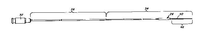
The microdilatation probe 12, illustrated in
FIG. 3, is longer than the dilatation catheter 10.
For example, with a dilatation catheter having a
length of approximately 150 centimeters the overall
length of the probe 12 preferably is of the order of
about 180 centimeters. The respective lengths of
the probe 12 and catheter 10 should be such that the
probe can be manipulated from its proximal end so
that the probe balloon 26 is extended distally and
completely out of the outlet opening 22 of the
dilatation catheter 10.
The probe 12 has a relatively long proximal
segment 28 which is formed from narrow, solid wall
tubing, such as hypodermic tubing. In the
illustrative embodiment, the proximal segment 28 may
be of the order of 150 centimeters long, about as
long as the dilatation catheter 10. The proximal
segment 28 is rigid torsionally so that it can
2B410/713 1308988
AZB/dmc
6890B
transmit substantially fully to its distal end
rotational motion imparted to the proximal end. As
will be described, the distal tip of the probe can
be bent to a preset curve. Rotation applied to the
probe can be controlled to selectively direct and
steer the curved distal end of the probe as it is
advanced. The proximal segment 28 also is flexible
and can bend longitudinally to follow the curvature
of the patient's arterial system. Preferably the
proximal segment 28 of the probe 12 is sufficiently
flexible that it can bend to follow the curve of a
patient's aortic arch which has a radius of the
order of between 2.5 to 3.5 inches in an adult.
As shown more clearly in enlarged FIG. 5, in
the preferred embodiment of the invention the hollow
tubular segment 28 has an outer diameter of .018
inches, a wall thickness of about .002 inches and an
internal diameter passage 30 of .014 inches. A
conventional fitting 32 is secured to the proximal
end of segment 28 to facilitate connection with an
inflation/deflation device, such as a syringe (not
shown).
The probe 12 includes a distal segment 34 which
extends from the distal end of the proximal segment
28 to the distal end of the probe 12. The distal
segment 34 includes a narrow diameter elongate
support wire 44 which is connected to and extends
2B410/713 1308988
AZB/dmc
6890B
- 18 -
distally of the proximal segment 28. The support
wire 44 is connected to the proximal tubing 28 by a
short transition tube 36. The transition tube 36 is
about one-half inch long and also is formed from
slender, flexible hypodermic tubing with a smaller
diameter than the proximal tube 28. In the
illustrative embodiment, the transition tube 36 is
formed from hypodermic tubing having an outer
diameter of .014 inches, a wall thickness of .003
inches and an inner diameter of .008 inches. The
- proximal end of the tubing 36 is received within the
distal end of the internal passage 30 of the
proximal segment 28 and is secured thereto as by
soldering or brazing. The solid support wire 44 is
attached to the distal end of the transition tube
36. The wire 44, which in the illustrative
embodiment is very slender, preferably .008 inches
diameter, is received in the distal end of the
passage 38 of the tubing 36 and is secured by
soldering or brazing. The support wire 44 plugs the
distal end of the tubing 36. In order to permit the
balloon 26 to be inflated and deflated, the
transition tube 36 is provided with apertures 46 on
opposite sides of the tube wall to provide
communication with the internal passages 38, 30 of
the probe. The apertures 46 may be defined by
forming a pair of longitudinal slots in the wall of
2B430/713 1308988
6890B
-- 19 --
the tubing 36. The support wire 44 provides support
for the probe balloon 26 and also extends distally
beyond the balloon 26, to form the core of a leader
segment 48. The leader segment includes a helically
wound radiopaque coil spring 50 which is attached to
the distal end of the core wire 44 in a manner
described below.
The probe balloon 26 is formed by molding high
strength polymeric material in a manner which
provides a thin balloon wall not greater than about
.001 inches thickness and, preferably, having a
thickness of the order of .0005 inches. The balloon
may be manufactured as described in U.S. patent
4,490,421 issued December 25, 1984 and reference is
lS made thereto for further details concerning the
manufacture of the balloon.
As shown in enlarged detail in FIG. 6, the
balloon includes a main cylindrical portion 52
which, in its fully inflated configuration defines
an outer diameter just slightly greater than the
outer diameter of the dilatation catheter 10 with
its balloon 20 collapsed. In the illustrative
embodiment, the probe balloon 26 preferably has an
outer diameter of 1.3 millimeters. As mentioned
above, the balloon is formed from a high strength
material which will not tend to stretch when
inflated. The length of the balloon 26 may be of
2B410/713
AZB/dmc
6890B 1308988
- 20 -
the order of 15 millimeters. The balloon is formed
to include tapering portions 54, 56 at the proximal
and distal ends respectively. The distal tapering
portion 56 merges into a narrowed neck 58 which fits
snugly about and against the proximal end of the
coil spring 50. The distal neck 58 of the probe
balloon 26 is adhesively attached to the coil spring
S0. As will be described in further detail, the
proximal end of the coil spring is soldered securely
to the core wire at the region where the distal neck
58 of the probe balloon 26 is joined. The proximal
tapering portion merges into a narrowed proximal
neck 60.
In order to communicate the interior of the
probe balloon 26 with the inflation/deflation
passages 30, 38 of the tubing, an extension sleeve
62 is adhesively attached to the proximal neck 60.
The extension sleeve 62 extends proximally over the
support wire 44. The proximal end of the extension
sleeve 62 preferably is formed from the same
material as the balloon 26 and is securely and
adhesively attached to the outer surface of the
transition tube 36, where it joins the main tube
28. The extension sleeve 62 defines an annular
passage 64 about the support wire 44~ The annular
passage 64 provides communication between the
apertures 46 and the interior of the balloon 26 for
inflation and deflation of the balloon.
2B410/713
AZB/dmc 1308988
As shown in FIG. 6 the leader segment 48 which
extends distally of the balloon 26 is of increasing
flexibility in a distal direction to provide a
relatively soft, flexible leading tip which reduces
the chance of trauma or injury to the blood vessel.
In the illustrative embodiment the leader segment is
about 3 centimeters long. The coil spring 50 is
soldered, at its proximal end to the support wire
44, as indicated at 66. The distal end of the
support wire 44 also is soldered to the coil spring
50 as indicated at 68. Soldered joint 68 and the
distal tip of the support wire 44 terminate short of
the distal tip 69 of the coil spring 50. The distal
tip 70 of the coil spring 50 may extend about five
millimeters beyond the soldered joint 68 and defines
a highly flexible bumper tip. A rounded weld bead
67 is formed at the distal tip of the spring 50.
The leader segment 48 is of increasing flexibility
in a distal direction. The support wire 44 is taper
ground and, for example, may be ground smoothly to a
.002 inch diameter at its distal tip 69.
The distal tip 70 of the coil spring 50
includes a flexible and bendable stainless steel
shaping ribbon 71 which is secured to the distal tip
69 of the support wire at one end, and to the distal
weld bead 67 at its other end. The shaping ribbon
is of slender, rectangular cross section, of the
2B410/713
AZB/dmc 130898fi
6890B
order of .001 inches by .002 inches. The shaping
ribbon is adapted to be bent to a desired curve and
to retain that curve when relaxed. The preset curve
enables the probe 12 to be steered by rotation of
the probe from its proximal end. The probe can be
rotated to direct the prebent distal tip 70 in
selective directions as desired within the patient's
blood vessels.
The probe also is provided with a radiopaque
marker band 72 which preferably is formed from
platinum. The marker band 72 is located proximally
of the main portion of the balloon 26. In the
illustrative embodiment it is securely attached to
the support wire 44. The marker band 72 provides a
means by which the physician can verify,
fluoroscopically, that the probe balloon 26 has been
extended beyond the outlet opening 22 of the
dilatation catheter 10, as a precaution before the
probe balloon 26 is inflated.
The microdilatation probe 12 is constructed so
that it can pass through the main lumen 16 of the
dilatation catheter but without blocking off fluid
communication along the main lumen 16. That enables
the advantages of the microdilatation probe to be
used without compromising the fluid infusion and
pressure monitoring capabilities of the dilatation
catheter 10. As shown in FIGS. 4, 4A, 4B and 4C,
2B410/713 1308988
6890B
the main lumen 16 of the dilatation catheter varies
in cross sectional dimensions and shape,
particularly through the distal region of the
catheter which contains the balloon 20. For
example, the D-shaped main lumen 16 as seen at the
section indicated at FIG. 4A is dimensioned to be
.028 inches high and .036 inches wide. The main
lumen 16 narrows at the section indicated at FIG. 4B
to .024 inches high by .032 inches wide. The main
lumen 16 then makes a transition to a circular shape
and, as indicated at the section at FIG. 4C, the
diameter may be .022 inches. At the distal outlet
opening 22 of the dilatation catheter the diameter
of the main lumen 16 is still further reduced, to
about .020 inches.
Among the difficulties presented when
attempting to pass a member through the very narrow
main lumen 16 of the catheter 10 is that the member
will tend to restrict fluid flow communication
through the main lumen, from the proximal to the
distal end of the catheter. The microdilatation
probe, having a main body diameter of .018 inches
provides sufficient clearance within the main lumen
16, particularly at the sides of the D-shaped cross
section of the lumen to permit quite adequate fluid
communication. In the more distal portions of the
catheter 10, such as at the portion indicated by the
AZB/dmc ~308988
6890B
- 24 -
cross section of FIG. 4C, the narrowed lumen 16
provides less clearance for fluid communication. In
order to maximize fluid communication through the
lumen 16 when the probe 12 is in place, particularly
through the more narrowed portions of the lumen 16,
the distal segment 34, and particularly the portion
of the distal segment 34 which is proximal of the
balloon 26, embodies a construction which assures
that a sufficiently large flow area will be
maintained throughout the main lumen 16 of the
catheter 10. To that end, the support wire 44 and
sleeve extension 62 are constructed so that when the
probe is deflated, the sleeve extension 62 will
collapse to a very small cross sectional area which
will not adversely obstruct the main lumen 16 of the
catheter 10, even in the more narrowed regions of
the main lumen 16. Additionally, the length of the
distal segment 34, proximally of the balloon 26 is
sufficiently long, about twenty centimeters, so that
the proximal segment 28 need not be inserted into
the more narrowed portions of the catheter lumen
16. Even when the probe 12 is advanced through the
catheter 10 to extend to its maximum distance beyond
the outlet 22 of the catheter 10, the distal end of
the proximal tubing 28 will remain proximal of the
balloon 26.
2B410/713 130898~
AZB/dmc
6890B
- 25 -
When the microdilatation probe is in use and
its balloon 26 is extended distally beyond the
outlet 22 of the catheter 10 the narrowed portion of
the main lumen, in the region of the dilatation
balloon 20 will be occupied by the narrow support
wire 44 and surrounding extension sleeve 62. When
the probe balloon 26 is inflated, the sleeve 62 will
be expanded to its full diameter, of the order of
.017 inches. As illustrated in FIG. 4C-l, when the
extension sleeve 62 is inflated to its diameter of
about .017 inches, only a relatively small annular
portion of the main lumen 16 is available for fluid
flow communication. Thus, during the interval when
the probe balloon is inflated, the ability to infuse
liquids and to take pressure measurements is
somewhat reduced. However, when the probe balloon
is deflated, by applying suction to the probe, the
sleeve extension 62 collapses about the slender
support wire 44, as suggested in FIG. 4C-2. The
sleeve 62 collapses in a manner which tends to form
flattened wings 62W which may curl against the inner
wall of the lumen 16, as suggested in FIG. 4C-2.
When in the collapsed configuration illustrated in
FIG. 4C-2, there is a very substantial open flow
area through the lumen 16 which permits full and
free liquid infusion and pressure measurement, as
desired. Because the probe balloon 26 is inflated
2B410/713
AZB/dmc 130898~
6890B
- 26 -
only very briefly during the entire procedure, and
is deflated, as shown in FIG. 4C-2 for most of the
time, the system displays the desired capability of
liquid infusion as well as pressure measurement.
In order that the probe may be passed through
the main lumen 16 of the dilatation catheter, the
probe balloon 26 also must be collapsible to a shape
and size which can be passed through the main lumen
16. The invention accomplishes these objectives by
using the slender, small diameter support wire 44
extending through the balloon and by using a balloon
with a very thin but high strength wall. When the
microdilatation probe 12 is to be inserted through
the catheter, the balloon 26 first is collapsed by
applying suction, such as by a syringe, to the
fitting 32. The balloon 26 and the extension sleeve
62 collapse, tending to form radially projecting
wings as illustrated in FIGS. 6A-1 and 6B-l,
respectively. The wings 62W and 26W wrap about the
support wire 44 when the probe is advanced through
the main lumen 16 of the dilatation catheter 10.
The wings 26W may wrap about the core wire 44 either
in an S-shaped configuration suggested in FIG.~6A-2
or in a C-shaped configuration shown in FIG. 6A-3.
In either configuration the overall diameter through
the collapsed and folded balloon portion of the
probe 12 includes six layers of the balloon material
2B410/713 130898~
AZB/dmc
6890B
- 27 -
in addition to the diameter of the support wire 44.
In accordance with the present invention, the
balloon is formed from a high strength thin material
having a wall thickness preferably not more than
about .001~. Thus, the aggregate diameter of six
balloon layers plus the support wire is about .014
inches. The probe balloon thus is collapsible to a
diameter which is about one fourth of its inflated
diameter and which can pass easily through the main
lumen 16 of the dilatation catheter 10 even in the
more restricted portions which may have a diameter
of the order of .022~.
The manner in which the system is used is
illustrated in FIGS. 8-14. As suggested
diagrammatically, a guide catheter 80 is inserted
initially in the patient's arterial system, usually
through the femoral artery and is advanced through
the aortic arch 82 to locate the distal tip 81 of
the guide catheter at the coronary ostium 84 leading
to the coronary artery 86 to be treated. The guide
catheter 80 typically is too large to be inserted
into the coronary artery 86 and serves only to
provide a path which leads the dilatation catheter
10 to the coronary artery 86. After the guide
catheter has been positioned the dilatation catheter
10 is advanced through the guide catheter 80 with
its dilatation balloon 20 collapsed. When the
2B410/713 13~8988
6890B
- 28 -
dilatation catheter projects out of the tip 81 of
the guide catheter it can be advanced into the
coronary artery 86. Under fortuitous conditions the
dilatation catheter 10 may be advanceable in that
manner to locate the inflation balloon 20 within the
stenosis. The balloon 20 then may be expanded and
the dilatation procedure completed, after which the
dilatation catheter 10 and guide catheter 80 can be
removed.
It may be preferable in some procedures to
introduce the dilatation catheter together with a
guide wire indicated diagrammatically and in phantom
at 88. In that protocol the guide wire 88 is
inserted into the dilatation catheter 10 and the two
are advanced, as a unit, through the guide catheter
80. When the coronary ostium is 84 reached, the
guide wire 88 may be advanced into the coronary
artery 86 and may be manipulated in an effort to
advance the guide wire into the branch of the
arterial tree in which the stenosis is located.
once the guide wire has been advanced through the
stenosis, the dilatation catheter is advanced over
the guide wire which guides it directly to the
stenosis.
FIG. 9 is a diagrammatic illustration of a
dilatation catheter 10 which has been advanced over
a guide wire 88 through the artery 86 to the
2B410/713
AZB/dmc 1308988
- 29 -
stenosis. In the embodiment illustrated in FIG~ 9
the opening through the stenosis 90 is large enough
to permit the guide wire 88 to pass but is not large
enough to permit entry of the distal end of the
dilatation catheter 10. As described above, before
the present invention, this situation was not
treatable by angioplasty and typically was treated
immediately with bypass surgery.
In accordance with the present invention,
however, the surgeon can withdraw the guide wire 88
while maintaining the dilatation catheter 10 in
place. The microdilatation probe 12 then is
substituted for the guide wire 88 and is advanced
through the main lumen 16 of the dilatation catheter
10. The microdilatation probe 12 is advanced with
its balloon 26 in a collapsed configuration
illustrated in either of FIGS. 6A-2 or 6A-3. The
diameter of the microdilatation probe 12 is about
the same as the guide wire 88. The probe 12 thus
can be advanced out of the distal opening 22 of the
catheter 10 and the balloon 26, in its collapsed
configuration, can be inserted into and through the
stenosis 90 as suggested in FIG. 10. Once it has
been verified that the probe balloon 26 is within
the stenosis 90 and is fully out of the main lumen
16 the probe balloon 26 can be inflated under
pressure to expand forcefully the probe balloon 26
2B410/713
AZB/dmc
6890B ~vo~8~
- 30 -
to its maximum diameter thereby making a preliminary
enlargement of the passageway through the stenosis.
FIG. 11 is an illustration of the dilatation probe
in its expanded configuration within the arterial
stenosis 90. As can be seen, the balloon 26 has
been inflated to enlarge the passage through the
stenosis to a diameter just large enough so that it
will be able to receive the distal end of the
dilatation catheter 10.
It is important that the probe balloon 26 is
not inflated until after it has been extended
distally beyond the end of the dilatation catheter
10. A marker band 72 on the probe provides a means
by which it can be verified that the probe balloon
has been extended out of the outlet opening. As
shown in ~IG. 4 the dilatation catheter 10 has a
pair of marker bands 74, 76 located adjacent the
proximal and distal ends, respectively, of the
dilatation balloon 20. The position of the probe
can be verified fluoroscopically. When the marker
band 72 on the probe is located sufficiently
distally of both marker bands 74, 76 on the
catheter, that indicates proper extension of the
probe 12 and readiness to inflate the probe balloon
26.
When the probe balloon 26 has been inflated to
enlarge the opening through the stenosis 90 the
probe balloon 26 is collapsed by aspirating the
2B410/713
AZB/dmc 13~8988
- 31 -
probe. With the balloon 26 evacuated the dilatation
catheter can be advanced over the microdilatation
probe 12 which then serves the function of a guide
wire to guide the dilatation catheter (FIG. 12).
The dilatation catheter then can be advanced over
the probe to locate the dilatation balloon 20 within
the partially dilatated stenosis. The dilatation
balloon 20 then is inflated as suggested in FIG. 13
to complete the angioplasty by compressing the
stenotic material radially outwardly. With the
coronary lumen thus enlarged the dilatation balloon
20 is deflated. The dilatation catheter 10 and
probe 12 then are removed to leave the artery with
an enlarged flow area where it had been previously
stenosed.
Modifications may be made to the procedure with
respect to the relative positioning of the probe and
catheter after the preliminary dilatation has been
performed. In some instances the surgeon may prefer
to advance the probe and catheter in unison without
any relative movement between the two, when
advancing the dilatation balloon 20 into the
preliminarily dilatated stenosis. In other
instances there may be special considerations
resulting in a decision not to advance the probe
while advancing the dilatation catheter into the
stenosis. That protocol, too, is available with the
2B410/713
AZB/dmc 1308988
- 32 -
present invention, by collapsing the probe balloon
which then will wrap to a compacted configuration as
the dilatation catheter is advanced over that
portion of the probe.
As described above, one of the features of the
probe 12 is the increased flexibility of the distal
segment 34 of the probe. The proximal segment 28,
as described, is sufficiently flexible so that it
can bend relatively easily through the aortic arch
(see FIG. 8). The bend from the aorta, into the
coronary ostium 84 and thereafter through the
coronary arteries are sharper and shorter radiused.
The length of the more flexible distal segment 34 is
sufficient so that the probe balloon can reach
deeply into the arterial tree without requiring the
stiffer proximal tubing 28 to pass through
relatively sharp bends, such as the bend from the
guide catheter to the coronary ostium. The distal
segment 34, which consists substantially of the
thin, flexible support wire 44 is able to make the
relatively sharp bends with ease. ThUs, the only
portion of the probe 12 which actually enters the
coronary artery is that which includes the slender
support wire 44. This support wire is very flexible
and is more easily bent to be able to negotiate
shorter radius bends encountered in the coronary
arterial tree.
2B410/713
AZB/dmc
6890B 13~8988
In some instances it may have already been
determined, by angiography that the stenosis to be
treated is so narrow that it is unlikely that the
dilatation catheter 10 will be able to pass through
the stenosis. Under those circumstances it may be
desirable to forego the use of a separate guide wire
and, instead, insert the dilatation catheter with
the microdilatation probe already in place within
the catheter, so that the probe 12 can serve as a
guide wire. When used in that manner it should be
understood that the probe is far more steerable than
conventional guide wires which have been used in the
past. The steerable characteristic of the probe is
due in large measure to the solid wall of the tubing
in the elongate proximal segment 28 of the probe.
The tubing is substantially torsionally rigid and
tends to transmit substantially all of its rotation
applied at the proximal end to the distal end.
Although the intermediate segment of the probe,
which includes the slender .008 inch diameter wire
is too small a diameter to effectively transmit
torque over relatively long distances, the distal
segment 34 is relatively short, preferably about
twenty-five centimeters and, therefore, does not
have too great of an adverse effect on the torque
transmission from the proximal end of the probe to
the distal end. The distal segment preferably is no
~308988
2B410/713
AZB/dmc
6890B
- 34 -
longer than about 25 centimeters, as compared to the
solid wall tubular proximal segment which is
approximately 150 centimeters long. Thus, by
forming a bend in the distal tip 70 of the leading
segment, the direction of the probe 12 can be
controlled by rotating the probe from the proximal
end.
From the foregoing it will be appreciated that
the invention provides a system and method by which
the angioplasty technique for treating arterial
stenoses can be extended to certain stenoses which
previously required bypass surgery. The system
enables a microdilatation probe to be advanced
through the dilatation catheter while maintaining
fluid communication from the proximal to the distal
end of the dilatation catheter even while the probe
is in place. Moreover, the invention provides these
advantages with a probe which can be steered to
selectively guide through the branches of a
patient's arterial tree and in which the probe can
be substituted for a guide wire.
It should be understood, however, that the
foregoing description of the invention is intended
merely to be illustrative thereof and that other
modifications and embodiments of the invention will
be apparent to those skilled in the art without
departing from its spirit.
Having thus described the invention what I
desire to claim and secure by letters patents is: