Note: Descriptions are shown in the official language in which they were submitted.
-1- 1 30q327
REAGENT AND METHOD FOR CLASSIFYING
LEUKOCYTES BY FLOW CYTOMETRY
The present invention relates to a reagent and a
5 method for classifying leu~ocytes in the practice of
clinical tes~ing. More particularly, the present inven~ion
relates to a reagent and a method for classifying leukocytes
with a flow cytometer by means of optical measurements on
fluorochrome-stained blood cells.
Leukocytes in the peripheral blood of normal subjects
can be classified as being of five types consisting of
lymphocytes, monocytes, neutrophils, eosinophils, and
basophils. Different leukocyte types have different
functions and counting of leukocytes in the blood according
15 to their type will provide valuable information for
diagnostic purposes. For instance, an increase in the
number of neutorphils is associated with such diseases as
inflammations, myocardial infarction and leukemia, and a
decrease in their number is associated with viral diseases,
20 hypoplastic anemia, agranulocytosis, etc. On the other
hand, an increase in the number of eosinophils is found in
such diseases as parasitosis, Hodgkin's disease and
allergosis. An increased number of monocytes occurs either
during the convalescence period of patients suffering from
25 infectious diseases or in such diseases as monocytic
leukemla.
Classification and counting of leukocytes have been
made most commonly by the differential counting method which
ls also referred to as the visual counting method or simply
30 as the manual method. In this method, a blood sample is
spread on a glass slide and the blood corpuscles in the
smear are fixed and stained for examination by miscroscop~
Tha technician identifies the type of individual leukocytes
according to their morphological features (e.g., their size,
35 the morphology of their nucleus and cytoplasm, and the
presence or absence of granules) or the degree of dye uptake
and performs classification and counting of them. At
.,~
~ 3~q3~7
--2--
ordinary laboratories, 100 - 200 leukocytes are usually
counted for each sample and the percentage of the total
leukocyte count occupied by each type of corpuscle is
recorded as a measured value.
The differential cour,ting method has several
disadvantages. First, microscopic obs~rva~ion must be
preceded by cumbersome procedures for preparing a specimen
that involve such steps as smearing a blood sample on a
glass slide, fixing the corpuscles and staining them.
10 Secondly, it is a gre-at burden for the technician to
identify subtle differences between corpuscles by micro-
scopic classification and counting. Thirdly, it is
difficult even for a skilled technician to yield consistent
counts by the manual method since aside from the small
15 number of leukocytes computed, the smeared sample often has
an uneven distribution of blood corpuscles.
Various methods have been proposed for eliminating
these disadvantages of the manual method of leukocyte
classification by achieving automation and such automated
20 techniques may be roughly divided into two types. The first
method consists of recording the images of corpuscles with a
video camera or some other suitable imaging device and
classifying the leukocytes by means of image processing on a
computer. The operating principle of this method is similar
25 to that of the conventional visual counting method but
primarily due to the existence of many corpuscles that defy
classification by processing with a computer, this method
has not yet become an ideal alternative to the manual
method. Furthermore, this method is not economically
30 feasible since it requires sophisticated equipment which is
large and costly.
The other approach toward automatic classification
and counting of leukocytes is based on a flow system. In
this method, a blood sample having corpuscles suspended in a
35 diluent is permitted to flow in such a way that the
corpuscles will individually (one by one) pass through a
narrowed detecting area and leukocyte classification is ma~e by
_3_ 1 3~9327
analyzing the signal generated by the detector. This second
method of leukocyte counting which makes use of a flow
system is further subdivided into two categories.
In a method of the first category, an electrolyte in
which all red cells that were present have been destroyed
with a lysing agent so ~Aat only leukocytes will be
suspended is permitted to flow through an orifice and the
change in electrical impedance that occurs at the orifice
when each corpuscle passes through it is detected, with the
10 magnitude of the detected signal being used as a basis for
classification of leukocytes. .
A method of the second category is characterized by
the use of a flow cytometer that comprises a light source,
a flow cell that permits the blood cells in a sample to flow
15 one by one through a constricted channel, a photometric unit
that detects light issuing from each blood cell, and an
analyzer for analyzing the detected signals. In this method,
the corpuscles in the sample which are stained are
illuminated under light and the fluorescence emitted from
20 the irradiated corpuscles is detected, optionally together
with scattered light, with leukocyte classification being
made in accordance with the intensity of the detected
signals.
Techniques that fall within the category of this flow
25 cytometric method are described, for example, in Japanese
Patent Publication No. 853/1984 and L. A. Kamentsky, Blood
Cells, 6, 121 - 140 (1980). According to these techniques,
a blood sample is stained with 10 volumes of an acridine
orange solution, incubated for 1 minute, and irradiated
30 under a light source such as an argon ion laser. The green
fluorescence and red fluorescence that are emitted from the
individual corpuscles are measured and classification and
eounting of leukocytes are subsequently made based on a two-
dimensional plot of the florescence measurements.
Other examples of teehniques that are classified as
being within the flow cytometrie approaeh are shown in
Unexamined Published Japanese Patent Application No.
,.~
1 30q327
--4--
20820/1975; H. M. Shapiro et al., J. ~istochem. Cytochem.,
_, (1), 39~ - 411, (1976); and idem, ibid, 25, (8), 976 -
989, (1977). According to these methods, a blood sample is
stained with 4 volumes of a Dye Solution I, incubated for 3
5 minutes, further mixed with 20% formaldehyde in a volume
equal to the blood, fixed for 5 minutes, and diluted with a
diluting Dye Solution II to obtain a concentration 15 - 20
times as low as the initial value. The so prepared specimen
is subjected to measurement with a flow cytometer.
The flow cytometer employed in these methods used
either three mercury lamps each of which produces a separate
wavelength of light, or three lasers, so as to excite the
three~ fluorescent stains in the dye solutions. The
parameters measured are three kinds of fluorescence, forward
15 scattered light, 90 scattered light and absorbed light.
Based on these six parameters, two-dimensional plots are
constructed in four stages and analyzed to make leukocyte
classification and counting.
Applicant's Canadian Patent Application Serial No.
20 546,199, filed September 4, 1987 discloses a one-step staining
process consisting of staining a blood sample with a dye
solution comprised of a buffer solution, inorgar~ic salts and
fluorescent stains. But this method has the problem that
unlyzed erythrocytes may adversely affect measurement data
25 to produce unreliable results.
In the first version of the method that uses a flow
system for leukocyte classification and counting, the
disruption of erythrocytes is a perequisite but depending on
a blood sample, it is impossible to effect complete lysis of
30 erythrocytes and the accuracy of measurements may be
impaired in such a case.
The examples of the flow cytometric approach that
are described in Japanese Patent Publication No. 853/1984
and Blood Cells, 6, 121-140 (1980) are characterized by the
35 performing measurements before dye absorption by the cells
reaches an equilibrium, or at the time when the difference
between the intensities of fluorescence from individual
1 30q327
leukocytes attains a maximum during the staining process.
However, the time required for attaining an appropriate
level of fluorescence intensity in a sample whose leukocyte
count is at either one of two extremes will be different
5 from the time for a normal sample and an appropriate
staining time must be selected for each samples. As a
further problem, this method relies solely on the
differential intensities of fluorescences for leukocyte
classification and does not necessarily ensure precise
10 separation between different leukocyte types such as
lymphocytes and monocytes.
The other examples of the cytometric approach that
are described in Unexamined Published Japanese Patent
Application No. 20820/1975, J. Histochem. Cytochem., 24 (1)
15 396 - 411 (1976) and supra, 25 (8) 976 - 989 (1977) have the
disadvantage that they involve many steps of operation, take
a prolonged staining time and require the use of reagents in
a complex system. Furthermore, practice of these methods
re~uires a very sophisticated and costly apparatus that
20 includes three light source and which is capable of
measuring six parameters. In addition, analysis of such a
large number of parameters is inevitably complicated and
requires an analyzer of large capacity.
The method described in aforementioned Canadian Patent
25 Application Serial No. 546,199 has the following problem. Erythrocytes
in the blood sample emit only fluorescence of very low
intensity, so if all that is needed is to measure the
intensity of fluorescence, erythrocytes will not interfere
with the counting of leukocytes even if coincidence of
30 erythrocytes and leukocytes occurs (i.e., erythrocytes and
leukocytes pass through the detecting portion
simultaneously). However, if one wants to measure the
scattered light, erythrocytes which produce scattered light
having an intensity comparable to that of the scattered
35 light emitted from leukocytes will interfere with the
counting of leukocytes. In this case, one may measure
fluorescence and scattered light simultaneously and regard
-6- 1 309327
as leukocytes only the corpuscles that emit fluorescence
having an intensity greater than a certain level. However,
if coincidence of leukocytes and erythrocytes occurs, the
scattered light from erythrocytes is superposed on the
5 scattered light from leukocytes, thereby making it difficult
to accomplish accurate measurement of scattered light from
the leukocytes.
In the invention described in aforementioned Canadian Patent
Application No. 546,l99, a blood sample is diluted by,
10 for example, 20 folds so that the probability of coincidence
of erythrocytes and leukocytes is reduced but the potential
interference by erythrocytes cannot be completely prevented.
Therefor, if eosir.ophils and basophils are excluded by
measurement of the intensity of fluorescence and if the
15 intensities of right-angle scattered light from the
remaining three types of leukocytes, i.e., lymphocytes,
monocytes and neutrophils, are potted, the populations of
the three leukocyte types cannot be completely separated
from one another as shown in Fig. 2b.
If the sample is further diluted so that the
probability of coincidence of erythrocytes and leukocytes is
reduced to such a level that the potential interference by
erythrocytes can be completely disregarded, the populations
of lymphocytes, monocytes and neutrophils can be completely
25 separated from one another as shown in Fig. 2c, which is a
plot of the intensities of right-angle scattered light from
these three types of leukocytes. However, in order to
ensure the desired precision of measurement, at least about
10,000 leukocytes must be counted. Therefore, the practical
30 value of diluting the blood sample is limited by the
prolonged time required for completion of measurement.
The aforementioned problems associated with the
interference by erythrocytes can be solved if they are
eliminated ~rom the blood sample by a suitable technique
35 such as lysing but this idea has not been put to practice
in the present art because of the absence of any erythrocyte
eliminating method such as lysing that matches the
_7_ l ',~n9 3~7
conditions of staininy. There i5 no prior art technique
that performs lysing of erythrocytes into five types by
staining with fluorochromes. Also, there if no
technique available that successfully lyses only
5 erythrocytes within one minute and which yet does not
deteriorate the right-angle scattered light (morpholosical
information) from leukocytes.
Blood samples for leukocyte counting that are free
from erythrocytes are commonloy prepared by the following
10 methods.
i) lysing of erythrocytes
a) treatment with a surfactant;
b) treatment with an ammonium salt (e.g. NH4Cl);
c) hypotonic treatment (at physiological pH)
15 ii) separation
d) centrifugation;
e) sedimentation;
f) others.
The methods (a) to (e) are briefly described below.
20 (a) Treatment with a surfactant:
This method inhibits subsequent staining and in
addition to lysing of erythrocytes, it causes morphological
changes in leukocytes such as loss of cytop asm and membrane,swell
and shrinking, thereby making it difficult to achieve 3-part
25 differentiation of leukocytes by signals of scattered llght.
Furthermore, leukocytes in the sample treated with a
surfactant will experience morphological changes with time.
(1) Treatment with an ammonium salt
This method inhibits subsequent staining. In
30 addition, the ammonium salt does not have a great ability to
lyse erythrocytes and a thick sample that is a 20-fold
dilution of the whole blood is difficult to prepare by this
method. Furthermore, it takes as many as 3 - 5 minutes to
achieve complete lysis of erythrocytes by method (b).
35 (c) Hypotonic treatment
This method leaves leukocytes intact while lysing
erythorocytes by making use of the fact that leukocytes are
, . .
-8- 1 309327
more resistant than erythrocytes in hypotonic solutions.
However, at a physiological pH and under condltions that
cause complete lysis of erythorocytes, part of the
leukocytes will be destroyed.
5 (d) Centrifugation, (e) Sedimentation
Both methods have such disadvantages as cumbersome
and lengthy procedures, and high incidence of leukocyte loss
and fluctuations in each leukocyte's count and ratio.
The present invention has been accomplished in order
10 to solve the aforementioned problems of the prior art
techniques for leukocyte classification and counting and it
provides a reagent and a method that enable accurate
classification and counting of leukocytes by simple
procedures.
In one aspect, the present invention provides a
reagent system of the following composition for use in
clasifying leukocytes into five types by flow cytometry:
(1) a dye that specifically stains eosinophils, such as
20 Neutral Red;
(2) a dye that specifically stains basophils, such as
Astrazon Orange G or Auramine O (with the former being
particularly advantageous);
(3) a buffer such as phosphate, citrate, borate, Tris
ttris-(hydroxy-methyl)-aminomethane], Hepes, glycine,
carbonate, collidine, or taurine; and
(4) an osmolarity compensating agent (i.e., an alkaline
metal salt including an alkali metal salt and an alkaline
earth metal salt).
In order to achieve a better resolution of monocyte
fractions, the following constituent (5) may be added:
(5) a dye that specifically stains monocytes and which is
at least one member selected from the group consisting of
DiOCl(3), DiOC2(3), DiOC3, DiOC5(3), DiOC6(3),-~A2 and 2-
tY-(l'-ethyl-4',5'-benzothlazolylidene)propenyl]-1-
ethyl,4,5-benzoxazolium iodide tDiOC3(3) being particularly
advantageous].
9 1 3')9~27
The dyes used as constituents (1), (2) and ~5)
respectively have the following chemical structural
formulae:
Neutral Red (C.I.No. 50 040 or C.I. Basic Red 5)
~ N ~ Me
Me2N N NH2
Astrazon Orange G (C.I. No. 48,035 or C.I.Basic Orange 21)
Me
CH=CH ~ Cl
Me H
Auramine O (C.I.No. 41,000 or C.I.Basic Yellow 2)
NH
2N C ~ 2
DiOCn~3) (l,l'-dialkyloxacarbocYanine); n=1,2,3,5 or 6
CH=CH-CH ~ N ~ .
(CH2)nH ( 2)nH
2-~y-(l~-ethyl-4'~5'-benzothiazolylidene)propeny~ -eth
4,5-benzoxazolium iodide
~ ~CH=CH-CH~
1~ 1
Et Et
, ~
1 309327
--10--
If the reagent system of the present invention is
used, no complicated preliminary treatments are necessary
and selective classification and countins of leukocytes can
be accomplished with a flow cytometer by simply performing a
5 one-step staining operation on the blood sample.
During the course of experimentation conducted on a
trial-and-error basis that finally led to the accomplishment
of the present invention, the present inventors found that
there were 17 dyes with which leukocytes could be stained
10 for classification into at least 4 different types based on
two-dimensional plots of two of the parameters for
measurement that consist of right-angle scattered light and
several fluorescence emissions, with an argon ion laser that
operates at 488 nm being employed as the sole light source.
15 For the names, color index numbers and fluorescence
characteristics of the individual dyes, see Table A below.
-11- 1 309327
. ,
U~
. C
~ E u~ ~ ~r ~ ~ o o~ o~ co u~ o c~ ul u~ o~
Ll U~ ~ ) ~1 O~ a~ o o~ a~ ~1 o ~1 o ~ ~ N _~ ~
~J E X-- u~ ~ ~ Lr) ~ ~r Lr) u~ ~ m Ln u~ ~D In Ir.
U r~
t~ ~ E
~0 ~ E~ o o~ ~ o ~ ~ ~ cn N ~ C~ C~ O ~
u~ U~ C~ ~ CO CO CO a~ _I ~D 1~
~A X-- ul Ln ~ ~ r ~ ~ ~ ~ er ~r ~ ~ ll~ ~ ~r
-
Z o~lIn u~o o o ~
. O N t'7 I I I I I O O O OO O O O O tU al
H u~ ~ ~D O --i C~
U ~ ~ u~ ~ ~r
~: _ _ ~ a~ R R O
R
.Q . OQ~ ;0 ~ O~XXI~
O 0 .c ~ ~ rl t: ~a 0 h R I I
b e ~ ~ O O O O O c C.c ~ a O a : j c I
_
a~ . ~ 0 a~ ..... _ ... ,
" ~ ~; = a a ~ a
1 3(~9327
-12-
Leukocytes can be classified into five or more types
if acridine dyes such as Acridine Orange and Rhoduline
Orange are used.
Fig. 1 is a schematic diagram of the optics of a flow
5 cytometer that may be employed in implementing the method of
the present invention,
Fig. 2a is a graph showing the relative intensities
of right-angle scattered light from five different types of
leukocytes;
Fig. 2b is a frequency distribution curve for the
intensities of right-angle scattered light from lymphocytes,
monocytes and neutrophils as influenced by the coincidence
of erythrocytes and leukocytes;
Fig. 2c is a frequency distribution curve for the
15 intensities or right-angle scattered light from lymphocytes,
monocytes and neutrophils in the absence of any coincidence
of erythrocytes and leukocytes;
Figs. 3 to 11 and 14 are two-dimensional plots of two
signals as used to classify leukocytes;
Fig. 12 is a graph showing the excitation and
emission spectra of fluorescence of neutral Red;
Fig. 13 is a graph showing the excitation and
emission spectra of fluorescence of Astrazon Orange G;
Fig. 15 is a graph showing the resolution between
25 eosinophils and neutrophils and that between basophils and
neutrophils as a function of the concentration of Neutral
Red;
Fig. 16 is a two-dimensional plot of the intensities
of red and green fluorescence as used to classify
30 leukocytes; and
Fig. 17 is a graph showing the resolution between
eosinophils and neutrophils and that between basophils and
neutrophils as a function of the pH of dye solution.
Fig. 18 is a graph showing the relation between the
35intensities of fluorescence of classified leukocytes and
wave lengths.
Figs. 3 to 5 are two-dimensional plots of the
intensities of right-angle scattered light and fluorescence
1 3~q327
-13-
1 as measured with a flow cytometer from leukocytes that were
stained with one of the 17 dyes listed in Table A such that
they were clearly distinguishable from erythrocytes and
platelets. The numerals and symbols used in these figures
have the following definitions: 1, lymphocytes; 2,
monocytes; 3, neutrophils; 4, eosinophils; 5, basophils;
Side Sc., the relative intensity of right-angle scattered
light; and FL., the relative intensity of fluorescence.
The separation pattern shown in Fig. 3 is typical
of staining the Xanthene dyes, oxacarbocyanine dyes or
acridine dyes. Similar patterns are obtained by
constructing two-dimensional plots of the intensities of
green and red fluorescence from leukocytes stained with
acridine dyes.
The separation pattern shown in Fig. 4 is typical
of staining with Neutral Red.
The separation pattern shown in Fig. 5 is typical
of staining with Astrazon orange G or Auramine 0.
The present inventors also found that there were
about 20 dyes with which luekocytes could be stained for
classification into three types and the separation pattern
that is typical of staining with these dyes is shown in Fig.
6.
If one of the dyes that produce a separation
pattern of the type shown in Fig. 4 is mixed with an
appropriate amount of one of the dyes that produce a
separation pattern of the type shown in Fig. 5, and if the
fluorescence of each dye is received, a pattern of the type
shown in Fig. 7 is produced by measurement of the
intensities of fluorescence and right-angle scattered
light. In this case, if Neutral Red of Azine dyes and
Astrazon Orange G of Methine dyes are used, alternative two
step analysis with three measurement parameters (i.e. right-
angle scattered light and red fluorescence and green
fluorescence of appropriate wavelength) is possible.
Eosinophils and basophils can be separated from other
,,~ ~&.
-13A- 1 309327
1 leukocyte types as shown in Fig. 8 (eosinophils 4 and
basophils 5). ~he remaining components of leukocytes (i.e.,
lymphocytes, monocytes and neutrophils) can be separated
from one another by the intensities of fluorescence and
right-angle scattered light as shown in Fig. 9.
-14- 1 3n9327
If a dye that produces a pattern cf the type shown in
Fig. 6 is added to dyes that produce the pattern of Fig.
7, a better resolution of lymphocytes, monocytes and
neutrophils is achieved to produce a pattern of the type
5 shown in Fig 10 (in which the respective leukocyte
populations are design2ted by 1, 2 and 3). In this case,
too, a two-stage analysis can be effected by first employing
green and red fluorescence (Fig. 8), then employing
fluorescence and right-angle scattered light (Fig. 11).
10 Dye Characterization
a. Neutral Red
This is a fluorochrome dye that selectively stains
leyukocytes. It stains eosinophils to a greater extent than
other leukocytes. A two-dimensional plot of the intensities
15 of right-angle scattered light and red fluorescence from
leukocytes stained with Neutral Red is shown in Fig. 4.
Fig. 12 shows the excitation and emission spectra of
fluorescence of Neutral Red. Neutral Red produces a
specific fluorescence of eosinophils in the ban~ of 580 - 640 nm
20 (orange to red).
A two-dimensional plot of the pattern shown in Fig. 4
is producad by using a dye solution having a pH of 5 - il
and a dye concentration of 3 - 300 yg/ml. Even if the dye
concentration is less than 3 yg/ml, a specific pattern of
25 the distribution of eosinophils is produced but the other
leukocytes are too noisy to be accurately measured. If one
needs to obtain only the signal of eosinophils, the dye
concentration may be at least about 0.1 yg/ml.
b. Astrazon Orange G
This is also a fluorochrome dye that selectively
stains leukocytes. It stains basophils to a greater extent
than other leukocytes. A two-dimensional plot of the
intensities of right-angle scattered light and green
fluorescence from leukocytes stained with Astrazon Orange G
35 is shown in Fig. 5.
Fig. 13 shows the excitation and emission spectra of
fluorescencs of Astrazon Orange G. Astrazon Orange G
-15- l 309~27
produces a specific fluorescene of basophils in the yellow-
green band having a central wa-~elength of about 54C nm.
A two-dimensional plot of the pattern shown in Fig. 5
is produced by using a dye solution having a pH of 5 - ll
5 and a dye concentration of 1 - 300 ~g/ml. A similar
separation pattern is obtained with Auramine O.
c. Other dyes
Other fluorochromes that stain
leukocytes can also be used. They stain monocytes to a
10 greater extent than other leukocytes. They are capable of
differentiating leukocytes into at least three types in
terms of right-angle scattered light and fluorescence as
shown in Fig. 6.
d. Combination of Neutral Red and Astrazon Orange G
Neutral Red produces a specific staining of
eosinophils while Astrazon orange G specifically stains
basophils, thereby producing a two-dimensional plot of the
intensities of right-angle scattered light and yellow to red
fluorescence as shown in Fig. 7. This plot is obtained by
20 using a dye solution having a pH of 5 - ll, a Neutral Red
concentration of O.l - 30 ~g/ml, and an Astrazon Orange G
concentration of l - 300 ~g/ml.
e. Combination of Neutral Red, Astrazon Orange G and other
dyes
By employing appropriate combinations of dyes of
groups d. and c., leukocytes can be stained in such a way
that a better resolution of monocytes (less contamination by
lymphocytes and neutrophils) can be attained as compared
with the case of using dyes of group d. alone. A two-
30 dimensional plot of the intensities of right-angle scattered
light and yellow to red fluorescence form leukocytes stained
with combinations of neutral Red, Astrazon Orange and other
appropriate dyes is shown in Fig. lO.
Illustrative dyes that fall under category c. and
35 which can be used to produce a separation pattern of the
type shown in Fig. lO include oxacarbocyanine dyes such as
DiOCl(3), DiOC2(3), DiOC3(3), DiOC5(3) and DiOC6(3), TA-2 (a
,
-16- l 30~327
styryl dye produced by Nippon Kankoh-Shikiso Kenkyusho Co.,
Ltd., Okayama, Japan), and cyanine dyes such as 2-[Y-(l'-
ethyl-4'-5'-benzothiazolylidene)-propenyl]-l-ethyl-4,5-
benzoxazolium iodide.
As shown in Fig. 14, oxacarbocyanine dyes used alone
will allow leukocytes to be classified into 4 types, with
eosinophils 4 stained to a smaller extent than neutrophils
3. If such dyes are mixed with Neutral Red which has a
strong specificity for staining of eosinophils, a plot of
10 the pattern shown in Fig. 10 is obtained, in which
eosinophils 4 are distributed above neutrophils 3.
There are many other dyes that belong to group c. but
because of several limiting factors such as dyeing
conditions, degree of dye uptake and the wavelength of
15 fluorescence emissions, those which are specifically
mentioned above and analogs thereof are the sole examples
that can be advantageously used in the present invention.
Other Components of_the Dy~e Solution
a. Buffer
The buffer is used to maintain the pH of the dye
solution at an optimum level. It is important that the pH
of the dye solution be maintained at an optimum level since
a dye's adsorption mass and this specificity to cytoplasmic
proteins vary with pH. Blood itself has a buffering action
25 to maintain a pH near 7.4, so the buffer must be added in an
amount sufficient to cancel this action and provide a
desired pH.
For this purpose, buffers such as phosphate~ citrate,
borate, Tris, Hepes, glycine, carbonate, collidine and
30 taurine are used in amounts ranging from 5 to 200 ppm.
b. Osmolarity compensating agent
The osmolarity compensating agent is used to prevent
leukocytes from experiencing such defects as extreme
deformation and lysis. For this purpose, alkaline metal
35 salts are used in amounts of 60 - 380 mM so as to provide an
osmolarity that is within the range of 40 - 250% of the
physiological osmolarity of human blood (280 mOsm/kg).
-17- l 30~327
In using the reagent sytem of the present invention,
the following precautions must be taken:
(a) If two or more dyes are mixed together, dyeing
conditions that permit the individual dyes to exhibit
5 intended specificities must be located since optimum
concentrations and pHs for achieving specific staining
usually vary from dye to dye.
(b) The ~nt of each of the dyes to be added must be
adjusted in such a way that a desired separation pattern is
10 produced since different dyes have different intensities of
fluorescence (fluorescence intensity is generally determined
by multiplying the ~uantity of illuminating light, Io, by
the molecular extinction coefficient, , ~uantum yield, 0,
dye concentration, c, and the compensation factor, a, which
15 is determined by the specific optics used), and
(c) The wavelength of light to be received must be
selected in such a way that a two-dimensional plot having a
desired specificity can be obtained.
According to a second aspect of the present
20 invention, it provides a method for classifying leukocytes
by the following steps:
(a) lysing the erythrocytes in a fresh sample of anti-
coagulated blood by adding it to a hypotonic first fluid
composed of Neutral Red that selectively stains eosinophils,
25 Astrazon Orange that selectively stains basophils, and a
buffer for maintaining an acidic pH range;
(b) stalning the leukocytes in the so-treated blood
sample by adding to it a second fluid that is composed of a
buffer for neturalizing the acid in the buffer in the first
30fluid and maintaining the pH of the resulting dye solution
at a staining pH, and an osmolarity compensating agent for
ad~usting the osmolarity of the dye solution to a value at
which the leukocytes remain unchanged in shape:
(c) permitting the stained sample to flow through a flow
3scytometer, differentiating leukocytes from all other
corpuscles and ghosts by intensity of fluorescence, and
-18- 1 3 n9 3 27
1 measuring the signals of fluorescence and right-angle
(rectangular) scattered light from leukocytes; and
(d) identifying the type of each of the leukocytes
based on said multiple signals emitted therefrom, counting
S the number of detected leukocytes according to their type,
and calculating the proportions of individual leukocyte
types.
Natural Red and Astrazon Orange G used in the
method of the present invention have the following chemical
formulae: ~ N ~ Me HCl
Me2N N NH2
Neutral Red (C.I.No.50,040 or C.I. Basic Red 5)
Me Me
~ CH=CH ~ . Cl~
Me H
Astrazon Orange G (C.I.No.48,035 or C.I.Basic Orange 21)
Of the multiple signals emitted fro leukocytes in
the method described above, the right-angle scattered light
signal reflects the structural information of an individual
white cell. The larger the nucleus of a white blood cell
and the more granules that are present in it, the greater
light reflection will occur in the cell to produce more
intense right-angle scattered light. A lymphocyte contains
very few or no granules, so the scattered light produced
from the lymphocyte is the weakest of all leukocytes. On
the other hand, a neutrophil contains many granules and has
a large nucleus, so that it produces the intense scattered
light. The intensity of scattered light which eosinophils
produce is substantially equal to that of scattered light
which neutrophils produce and basophils produce scattered
light the intensity of which is intermediate between the
intensities of scattered light from
, ~.
-19- 1 ~nq.~27
lymphocytes and neutrophils. For these reasons, the
relative intensites of right-angle scattered light from
individual leukocyte types are plotted as shown in Fig. 2a.
The fluorescence signal reflects the cytochemical
5 characters of leukocytes and depending on the interaction
between stains and individual leukocyte types, signals of
different intensities are produced form the leukocytes.
Therefore, leukocytes can be classified into five
types by first performing selective staining of eosinophils
10 and basophils so that the clusteres of these two types of
leukocytes can be separated from each other by the intensities of
two fluorescences, and subsequently differentiating the
remaining leukocytes ti.e.~ lymphocytes, monocytes and
neutrophils) by means of the intensity of right-angle
15 scattered light.
As will be understood from the foregoing explanation,
the method of the present invention has the advantage that
no cumbersome operations involving a complicated preliminary
treatment are required and that the leukocytes in blood
20 alone can be c]assified and counted with a flow cytometer
after a simple two-stage staining operation has been
completed.
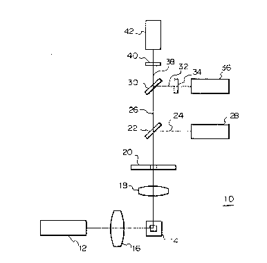
A specific example of the optics of a flow cytometer
employed in the present invention is hereunder described
25 with reference to Fig. 1. The optics shown in Fig. 1 is
used in a flow cytometer designed for measuring right-angle
scattered light, red fluorescence and green fluorescence.
The optics generally indicated by 10 uses an argon ion laser
12 as a light source and it operates at a wavelength of
30 488 nm, producing an output of 10 mW. Light emitted from
the laser 12 is converged by a cylindrical lens 16 and
illuminates a blood sample flowing through a flow cell 14.
When the stained leukocytes in the sample are
irradiated by the laser light, they produce scattered light
35 and fluorescence. The right-angle scattered light and the
fluorescence are converged with a condenser lens 18 and pass
through an aperture 20 to fall upon a dichroic mirror 22.
-20- 1 3nq327
The dichroic mirror 22 reflects the right-angle scattered
light 24 and transmits the flurescence 26. The rlght angle
scattered light 24 reflected from the dichroic mirror 22 is
detected in a photomultiplier tube 28. Of the
5 fluorescence 26 that passes through the dichroic mirror 22,
red fluorescence 32 is reflected by a dichroic mirror 30 and
green fluorescence 38 is transmitted through that mirror.
The reflected red fluorescence 32 passes through a color
filter 34 and is detected in a photomultiplier tube 36. The
10 transmitted green fluorescence 38 passes through a color
filter 40 and is detected in a photomultiplier tube 42.
In the method of the present invention, erythrocytes
in a blood sa~ple are disrupted by an acidic and hypotonic
treatment such as to reduce the disturbance that occurs in
15 the intensity distribution of right-angle scattered light on
account of coincidence of red and white blood cells.
As already mentioned, if a hypotonic treatment is
performed in the physiological pH range, not only the
erythrocytes but also some leukocytes will be destroyed. On
20 the other hand, if a hypotonic treatment is performed in an
acidic pH range, for example, at a pH between 2.0 and 5.0,
leukocytes will remain intact and only erythrocytes will be
disrupted. In this case, no morphological changes such as
loss of cytoplasm and membrane, swelling and shrinkage will
25 occur in leukocytes.
The mechanism by which erythrocytes are selectively
lysed is not clear but as erythrocytes are progressively
lysed by hypotonic treatment, embrittlement of their
membranes and acidic fixation of leukocytes will probabaly
30 proceed under acidic pH conditions, with the result that
only leukocyt~s which are more resistant than erythrocytes
remain intact.
As a result of this hypotonic treatment under acidic
conditions,most of the erythrocytes become "ghosts"
and "fragments". As a consequence,
the intensity of right-angle scattered light signals from
erythrocytes is reduced to no more than a half to a third of
-21- l 30~327
the intensity of right~angle scattered light slgnals from
lymphocytes, and the coincidence of red and white blood
cells can be disre~arded for practical purposes.
Since not all of the erythrocytes are reduced to
"fragments" by the hypotonic treatment under acidic
conditions, it is difficult to discriminate erythrocytes
from leukocytes solely on the basis of the intensity of
scattered light signals. Therefore, as already mentioned,
it is desirable to discriminate erythrocytes from leukocytes
10 by the intensity of a fluorescence signal.
The functions of Astrazon Orange G and eutral Red
used as fluorochromes in the present invention are
described below.
A sample of anti-coagulated blood is first mixed with
15 the first fluid so that the erythrocytes in the blood are
reduced to ghosts and fragments. Subsequently, the second
fluid is added so as to stain the leukocytes and platelets
in the blood.
It is speculated that the stains in the dye solution
20 (i.e,, first fluid) combine with the cellular constituents
(granules, in particular) in the leukocytes by ionic
adsorption. Astrazon Orange G would bind strongly to acidic
substances such as heparin and histamine in basophilic
granules and, as a consequence, the wavelength of
25 fluorescence emitted from Astrazon Orange G shifts from 520
- 540 nm to 560 - 580 nm (this phenomenon is generally
referred to as metachromasia). Astrazon Orange G also binds
to the granules in the other leukocytes (i.e., eosinophils,
lymphocytes, monocytes and neutrophils) but unlike in the
30 case of its binding to basophils, no detectable
metachromasia occurs. Astrazon Orange G binds weakly to the
surfaces of nuclei and cells and emits fluorescence in the
wavelength range of 520 - 540 nm.
Neutral Red also principally stains granules and
35 emits fluorescence of 620 nm. This dye binds to
eosinophilic granules to a greater extent than the granules
in other leukocytes, thereby emitting a stronger
'i~.
j,
-22- t 30q327
fluorescence radiation than that emitted from any other
leukocytes.
A two-dimensional plot constructed from the
measurement with a flow cytometer of a blood sample to which
5 both the first and second fluids have been added is shown in
Fig. 16, in which Red FL. signifies the relative intensity
of red fluorescence and Green FL. denotes the relative
intensity of green fluorescence. The numerals used in Fig.
16 have the following meanings: 1, lymphocytes, 2,
10 monocytes; 3, neutrophils; 4, eosinophils; 5, basophils; and
6, non-leukocytes, namely, platelets and erythrocytic ghosts
and fragments (the same symbols and numerals used
hereinafter have the same definitions).
In Fig. 16, the leuykocytes are clearly distinguished
15 from platelets and erythrocytic ghosts and fragments denoted
by 6 since the latter emit a lower intensity of green
fluorescence. Eosinophils 4 and basophils 5 are oompletely separated
from others in the two-dimensional plot of Fig. 16.
However, the other leukocytes (i.e., lymphocytes 1,
20 monocytes 2 and neutrophils 3) which do not emit any
specific fluorescence cannot be separated from one another
on the two-dimensional plot of the intensi'ies of green and
red fluorescences and can be classified as shown in Fig. 2C
based on the intensities of right-angle
25 scattered light.
The compositions, pHs and osmolarities of the first
and second fluids used in the method of the present
invention are described below in detail.
(1) Dye concnetration
30 a. Concentration of Astrazon Orange G
Astrazon Orange G produces the best separation of
basoph~ls and neutrophils when its final concenration is
15~/m~ with the staining pH being at 9.O. If the final
concentration of Astrazon orange G is less than 15-~g/m~, a
35 lower resolution results because of the decrease in the
intensity of green fluorescence from basophils. The same
result also occurs if the final concentration of Astrazon
-23- 1 309327
Orange G is more than 15 ppm and this is because of the
combined effect of the decrease in the intensity of green
fluorescence from basophils and the increase in the
intensity of green fluorescence from neutrophils. The
5 concentration of Astrazon Orange G that provides an optimum
resolution varies with pH. The adsorption mass of Astrazon
Orange G decreases with decreasing pH.
b. Concentration of Neutral Red
A good resolution between eosinophils and neutrophils
10 can be attained at the higher end of the concentration range
of neutral Red from 1 to lO,ug/mQ~ Eosinophils have better
staining characteristics at lower pHs.
c. Interaction between Astrazon Orange G and Neutral Red
Neutral Red also stains the granules in basophils
15 (i.e., the intensity of fluorescence it emits has no
specificity to basophils), so it inhibits selective staining
of basophils by Astrazon orange G. It is therefore
necessary to determine a concentration of neutral Red that
provides for good resolution between neutrophils and each of
20 basophils and eosinophils.
Fig. 15 shows the profiles of resolution between
eosinophils and neutrophils and between basophils and
neutrophils as a function of the concentration of neutral
Red with the concentration of Astrazon Orange G and pH fixed
25 at l~g/me and 9.0, respectively. In Fig. 15, the term
"green fluorescence ratio of basophils/neutrophils" means
the ratio of the intensity of green fluorescence from
basophils to that from neutrophils, and the term "red
fluorescence ratio of eosinophils/neutrophils" means the
30 ratio of the intensity of red fluorescence from eosinophlls
to that from neutrophils (the same expressions used
hereinafter have the same meanings). The higher the points
in the figure, the better separation that can be achieved
between neutrophils and basophils or eosinophils.
In Fig. 15, the separation between basophils and
neutrophils coincides with that between eosinophils and
neutrophils but in practice, there usually are fewer
-
, .. ~.
1 3n9327
-24-
basophils in leukocytes than eosinophils, so in order to
improve the resolution of basophils from neutrophils, it is
desirable to set the concentration of neutral Red at a
comparatively low level, say 2~g.mQ.
If the volume ratio of the first to second fluid is
set at 9:1 as in Example 7 to be described later in this
specification, the concentrations of Astrazon Orange G and
neutral red in the first fluid may be adjusted to 16.5ug/m
and 2.2~g/~,respecitvely, in order that their final
10 conc~ntrations will be at l~g/~ and 2~g/~ respectively.
(~) pH
a. Final pH to be attained as a result of mixing the first
and second fluids
Fig. 17 shows the profile of resolution between
15 neutrophils and basophils or eosinophils as a function of
pH, with the concentrations of Astrazon Orange G and Neutral
Red being fixed at 15.0 ppm and 3.0 ppm, respectively.
Obviously, the resolution of eosinophils from neutrophils
decreases with increasing pH. On the ~ther hand, the
20 resolution of basophils from neutrophils increases with the
increase in pH up to about 9.0 - 9.5 and decreases
thereafter.
As pH increases, the rate of basophils staining
increases(i.e., the time required for the intensity of
25 fluorescence to reach a maximum decrease), but once a
maximum fluorescence intensity has been reached, the
subsequent decrease in fluorescence intensity is rapid at high pH.
The staining rate of eosinophils does not vary greatly with pH.
Therefore, with the resolution of neutrophils from
30 each of eosinophils and basophils and the decreas~ in the
intensity of fluorescence from basophils being taken into
consideration, it is desirable to adjust the final pH to a
value in the neighborhood of 8.6 - 8.7. In the present
lnvention, the value of the final pH attained is referred to
35 as the "staining pH".
b. pH of the first fluid
-25- 1 3 n~ 327
The pH of the first fluid influences the lysing
efficiency of erythrocytes. Erythrocytes lyse rapidly at
pHs of 5.0 and below, and the lower the pH, the faster the
rate of lysis. However, at pHS below 2.0, proteins such as
5 hemoglobin begin to denature as the lysing of erythrocytes
progresses, and the rate of protein denaturation increases
as pH decreases. A denatured protein will clog at the time
when the final "staining" pH has been attained. In
consideration of these facts, it is desirable to adjust the
10 pH of the first fluid to be at a value between 2.0 and 5Ø
(3) Buffer
a. Buffer in the first fluid
The buffer in the first fluid is used to maintain the
pH of the first fluid at a level suitable for lysing
15 erythrocytes, and any buffer that has a pKa value of 3.5 +
1.5 may be employed for this purpose. Illustrative examples
include maleic acid, malonic acid, phthalic acid, diglycolic
acid, saliyclic acid, fumaric acid, tartaric acid, citric
acid and malic acid. In order to reduce the osmolarity of
20 the first fluid, the concentration of the buffer is
desirably held as low as possible. For the purposes of the
present invention, the concentration of the buffer in the
first fluid is preferably at 50 mM and below, more
preferably at 5 - 30 mM.
25 b. ~3uffer in the second fluid
The buffer in the second fluid is used to neutralize
the acid in the buffer in the first fluid and to maintain
the pH of the resulting dye solution at the staining pH.
Any buffer that has pKa value of 8.0 - 9.5 may be employed
30 for this purpose. Illustrative examples include Tris,
tricin, bicine, 2-amino-2-methyl-1,3-propanediol, taurine,
boric acid and serLne. These buffers are preferably used at
concentrations of at least 10 mM in terms o~ the final
concentration which is attained as a result of mixing of the
35 first and second fluids. For the purposes of the present
invention, the buffer in the second fluid advantageously has
a final concentration of 3Q - 100 mM.
1 3nq327
-26-
(4) Osmolarity
a. Osmolarity of the first fluid
The lower the osmolarity of the first fluid, the more
rapid the lysing of erythrocytes. For the purposes of the
5 present invention, the osmolarity of the first fluid is
preferably adjusted to a value in the range of O - 100
mOsm/kg, more preferably in the range of O - 50 mOsm/kg.
b. Osmolarity of the second fluid
The osmoraltiy of the second fluid determines the
10 fianl osmorarity which is to be attained as a result of
mixing the first and second fluids. The final osmorality
influences the ability of leukocytes to retain their own
shape and is preferably within the range of 150 - 600
mOsm/kg, more preferably in the range of 150 - 300 mOsm/kg.
The present invention is hereinafter described in
greater detail with reference to the following Examples 1 to
7, which are given here for illustrative purposes only and
are by no means intended to limit the present invention.
Example 1
20 Concentration of neutral Red and Astrazon Orange G:
To a 10 mM borate buffer solution (pH, 9.0)
containing 75 mM of NaCl, Astrazon Orange G and Neutral Red
were added in the amounts shown in Table 1, so as to prepare
dye solutions. Two milliliters each of these dye solutions
25 was mixed with 80~1 of a fresh sample of EDTA anti-
coagulated blood and the mixture were incubated for 1
minute. The so prepared specimens were permitted to flow
through a flow cytometer having the optical arrangement of
the composition shown in Fig. 1. The results of leukocyte
30 classification based on the measurement of the intensities
of green fluorescence, red fluorescence and right-angle
scattered light are shown in Table 1.
-27-
1 309327
Table 1
Concentration of
Astrazon
C t orange G
ration of \ gtml) 3 10 30 100
Neutral Red (yg/ml)
_ _
0.3 _*2) 5*3) 5*3) _*2)
1 3*5) 5*1) 5*3) 3*5)
3 4*4) 5*3) 4*4) 4*4)
4*4) 4*4) 4*4) 4*4)
1 *1) 5-part differentiation by red fluorescence *6) and
right-angle scattered light
*2) unclassifiable
*3) 5-part differentiation in which eosinophils and
basophils were first separated from others by red
fluorescence/green fluorescene *7), followed by 3-
part differential by right-angle scattered light
*4) 4-part differentiation by red fluorescence/right-
angle scattered light
*5) leukocytes were classified into 3 types by red
fluorescence/green fluorescence; provided that;
*6) red fluorescence 580 nm; and
*7) green fluorescence = 520 - 580 nm.
Example 2
p~:
A dye solution having a pH of 8.0 was prepared by
adding 10 ug/ml of Astrazon Orange G and 1 ug/ml of Neutral
Red to a 10 mM boarte buffer solution containing 75 mM of
NaCl. Two additional dye solutions were prepared in the
same manner as described above except that their pHs were
adjusted to 9.0 and 10.0, respectively. Using these dye
solutions, flow cytometry was conducted as in Example 1.
With the dye solution having a pH of 10.0, 5-part
differentiation of leukocytes could not be successfully
achieved by measurement of the intensities of red
fluorescence and right-angle scattered light. But the
-28- 1 30q 327
intended results could be attained by first differentiating
basophils 5 and eosinophils 4 from others in te ~ of green fluorescence
and red fluorescence and then distinguishing between the
remaining three types of leukocytes based on green
5 fluorescence and right-angle scattered light. With the dye
solution having a pH of 9.0, 5-part differentiation of
leukocytes could be accomplished based on red fluorescence
and right-angle scattered light. With the dye solution
having a pH of 8.0, 4-part differentiation was possible on
10 the basis of the red fluorescence and right-angle scattered
light.
Example 3
Concentration of NaCl:
Four dye solutions were prepared by adding 50, 75,
15 150 and 300 mM of NaCl to a 10 mM borate buffer solution
(pH, 9.0) containing 10 yg/ml of Astrazon Orange G and
1 ~g/ml of Neutral Red. Using these dye solutions, flow
cytometry was conducted as in Example 1. No significant
changes in separation pattern were observed within the
20 tested range of NaCl concentrations and 5-part
differentiation of leukocytes could successfully be achieved
with each of the dye solutions.
Example 4
Concentration of buffer:
A dye solution was prepared by adding 75 mM NaCl,
10 ~g/ml of Astrazon Orange G and 1 ~g/ml of Neutral Red to
a borate buffer soloution (pH, 9.0) wherein the buffer was
incorporated in an amount of 3 mM. Two additional dye
solutions were prepared in the same manner as described
30 above except that the buffer concentration was ad~usted to
10 mM and 30 mM, respectively. Using these dye solutions,
flow cytometry was conducted as in Example 1. No
significant changes in separation pattern were observed
within the tested range of buffer concentrations and 5-part
differentiation of leukocytes could successfully be achieved
with each of the dye solutions.
Example 5
:'k
-29- l ~ nq 327
Wavelength of fluorescence
Flow cytometry was conducted as in Example l using a
dye solution that was composed of a lO mM borate buffer
solution (pH,9.0) containing 75 mM NaCl, lO ~g/ml of
5 Astrazon orange G and l ~g/ml of Neutral Red. The analysis
was based on the measurement of the intensities of right-
angle scattered li~ht and six fluorescence emissions not
shorter in wavelength than 520 nm, 540 nm, 560 nm, 580 nm,
600 nm and 620 nm, respectively, that were collected with a
lO photomultiplier tube 36 in the optics shown in Fig. l. A
total reflection mirror was used instead of a dichroic mirror 30,
and a long-pass filter as a color filter 34.
As the wavelength of fluorescence collected was
increased, the resolution between basophils and lymphocytes
15 decreased whereas the resolution between eosinophils and
neutrophils increased. The efficiency of 5-part
differentiation of leukocytes was particularly high when
fluorescence emissions having wavelengths not shorter than
560 nm and 580 mn were collected.
20 Example 6
Wavelengths of red and green fluorescence
Flow cytometry was conducted as in Example 5, with
the wavelengths of red and green fluorescence collected
being varied as shown in Table 2 below.
Table 2
Green fluorescence Red fluorescence
(nm) (nm)
a. 540 - 600 >560
b. 540 - 600 >580
c. 540 - 580 >560
d. 540 - 580 >580
e. 500 - 540 >560
When fluorescence emissions having the wavelengths c.
or e. were collected, basophils and eosinophils were
35 selectively stained to permit good resolution from the other
leukocytes.
-30- 1 3~9327
The foregoing examples show that the reagent system
of the present invention will produce good results when it
is used under the following conditions.
Astrazon Orange G : 3 - 100 ~g/ml
Neutral Red : 0.3 - 10 yg/ml
pH : 8.0 - 11.0
Fluorescence wavelength
Green Fl. : 500 - 580 nm
Red Fl. :>560 nm
Example 7
This is an example of the method of the present
invention as it was carried out with the composition of the
reagent system described above being adjusted to an optimum
15 range.
Reagents:
1) First fluid
Astrazon Orange G 16.5 ppm
(selective dye for
basophils)
~0
Neutral Red 2.2 ppm
(selective dye
for eosinophils)
Citric acid/sodium hydroxide 10 mM
(buffer)
pH, 3.0; osmolarity, 10 mOsm/kg
2) Second fluid
Taurine/sodium hydroxide 500 mM
(buffer)
Sodium chloride (osmolarity 300 mM
compensating agent)
pH, 9.7 - 9.8; osmolarity, 2,600 mOsm/kg
Staining Procedure
Eighteen parts by volume of the first fluid was added
35 to one part by volume of EDTA 2K anti-coagulated blood.
After agitation, the mixture was incubated at 25C for 20
seconds. Thereafter, 2 parts by volume of the second fluid
-31- 1 3~327
was added and, after agitation, the mixture was incuvated at
25C for 40 seconds. The finally attained staining
conditions were a pH of 8.7 and an osmolarity of
260 mOsm/kg.
Emission Characteristics of Fluorescene:
The fluorescence emission intensity vs. wavelength
characteristics of the individual leukocyte types as stained
with the reagent system described above are shown in Fig.
18.
10 Selection of Filtration and Dichroic Mirrors-
Based on the emission characteristics shown in Fig.
18, the following filters and dichroic mirros were selected
as optimum devices:
Dichroic mirror 22 530 nm
(reflect blue light)
Dichroic mirror 30 600 nm
(reflect red light)
Color filter 34 600 nm
(long-pass filter trans
mitting wavelengths
not shorter than 600 nm)
Color filter 40 540 nm
(long-pass filter trans-
mitting wavelengths
not shorter than 540 nm)
25 Results of Analysis
A two-dimensional plot of the intensities of red and
green fluorescences as measured with a flow cytometer under
the conditions described above is shown in Fig. 16.
Population 6 (consisting of platelets, red cell ghosts and
30 fragments) was successfully separated from leukocytes, and
it was possible for both an eosinophil cluster 4 and a
basophil cluster 5 to be separated from all other leukocytes
with high resolution. The remaining leukocytes will also
successfully separated from one another with good
35 resolution, as indicated in Fig. 2c which is a frequency
distribution curve for lymphocytes 1, monocytes 2 and
neutrophils 3. In Fig. 2c, Side Sc. signifies the relative
-32- 1 309327
intensity of right-angle scatterd light and Freq. stands for
frequency.
In Examples 1 to 7, all measurements are initiated
after the necessary procedurs of staining have been
5 completed (namely, after staining has reached an
equilibrium). There~ore, the sample will not experience any
time-dependent change during measurements, and an
appropriate level of the intensity of staining or reaction
can be attained within a certain period of time no matter
10 how large or small the number of leukocytes in the sample
is. This allows for consistent results in measurement and a
fluorescence signal of an adequate intensity can be attained
even if a light source of a comparatively low output is
used. In Examples 1 - 7 described above, an argon ion laser
15 of 10 mW was employed as a light source in the flow
cytometer.
However, the light source in the flow cytometer used
in the present invention is not limited to the afore-
mentioned argon ion laser of low output and any of the other
20 light sources can be emplyed, such as a mercury arc lamp,
xenon arc lamp, a He-Cd laser, a He-Ne laser and a krypton
ion laer, as well as an argon ion laser of high output. If
these light sources are used, the conditions of staining,
reaction and measurement may be selected as appropriate.
The reagent system and the method of the present
invention as applied to classify and count leukocytes in
blood by flow cytometry have the following advantages.
(1) A sample of measurement can be prepared by simple
preliminary treatments that consist of merely adding anti-
30 coagulated blood to a dye solution.
(2) The sample can be prepared in approximately one
minute and this provides a rapid access time for
measurement.
(3) Since measurements are conducted after the neceesary
35 procedures of staining have been completed, the sample will
not experiene any time-dependent change during measurements
and an appropriate intensity of staining or reaction can
~33- 1 3n9 327
always be attained within a certain period of time
irrespective of the nature of the sample (whether it is
normal or contains an extremely large or small number of
leukocytes). This eliminates the need to change the
5 staining time from sample to sample.
(4) Since measurements are conducted after staining has
been completed to provide a high staining intensity, a light
source of low output may be employed. In addition, only one
light source need to be used and two or three parameters appro-
10 priately selected from among two channels of fluorescenceand one cahnnel of right-angle scattered light may be
measured. Because the number of parameters to be measured
and analyzed in this few, the reagent system of the present
invention can be used to accomplish flow cytometry of blood
15 with a simple and inexpensive apparatus.
(5) The reagent system of the present invention has a
very good ability to stain blood cells in a differential
manner and therefore enable leukocytes to be classified with
good resolution.
20 ~6) The method of the present invention effects
measurement not only of fluorescence but also of right-angle
scattered light and this contributes to better classifi-
cation of leukocytes including separation between
lymphocytes and monocytes.
25 (7) In accordance with the method of the present
lnvention, erythrocytes are selectively lysed by an isotonic
treatment under acidic conditions. Since the coincidence of
erythrocytes and leukocytes is eliminated by this treatment,
a very efficient separation between lymphocytes, monocytes
30 and neutrophils can be achieved by means of a rlght-angle
scattered light signal.
(8) Leukocytes can be classified into five types with a
very high resolution by first separating eosinophils from
basophils on the basis of a fluorescence signal, and then
35 separating the remaining leukocytes (i.e., lumphocytes,
monocytes and neutrophils) based on right-angle scattered
light.
1 3n~3~7
-34-
(9) In the method of the present invention, separation of
leukocytes from other corpuscles including their ghosts and
fragmetns is achieved on the basis of fluorescence
intensity, so correct measurements are ensured even if not
5 all erythrocytes have been reduced to fragments.
Acco-ding to the method of the present invention,
accurate and reproducible measurements are ensured by
counting no less than 10,000 leukocytes for each sample.