Note: Descriptions are shown in the official language in which they were submitted.
1311~66
NOVEL ENDOTRACHEAL TUBE AND MASS SPECTROMETER
This invention relates to a device which measures
the quantity and composition of inhaled and expired
gases from a conscious or unconscious human subject
and then computes the pulmonary function and cardiac
output of the subject from this data information.
It is desirable, and often necessary, to deter-
mine the cardio-respiratory function in human subjects.
The function of the cardio-vascular and respiratory
system is to supply oxygenated blood to the body tis-
sues and to remove the CO2 produced by the tissues forexcretion by ventilation of the lungs. The amount of
blood pumped or vented and the amount of 2 and CO2
in the blood, as well as the volume of lung ventila-
tion, are critical reflections of the adequacy of the
circulatory and respiratory function.
During exercise, during disease states, or during
surgery, these physiological parameters are adaptively
altered and may be impaired. In order to diagnose and
131~5`~6
treat cardio-respiratory dysfunction, it is necessary to
measure and evaluate these parameters. This is particularly
true during surgical anesthesia, where the anesthetist must
maintain cardio-respiratory homeostasis that may become
impaired from the anesthetic agent or from complications
during surgery. It is also necessary to assess these
parameters in critically ill non-surgical patients while
being managed in critical care units. Moreover, assessment
of cardiac output and other cardio-respiratory functions,
which should be a key part of exercise testing, is not
evaluated routinely because there are presently no effective
non-invasive methods which are acceptable.
Invasive systems are available, but cannot be routinely
used because the insertion procedure (via catheter or the
like) is time-consuming and involves risk. Non-invasive
devices, such as the ultrasonic Doppler device, have been
developed, but cannot be used routinely and are unable to
continuously accurately determine the cardio-respiratory
function.
The present invention provides a novel non-invasive
device, which continuously measures the quantity and
composition of the inhaled and expired gases from a human
subject, and then calculates the pulmonary function and
cardiac output from this data.
More specifically, this invention provides a non-
invasive system for determining the cardio-respiratory
function comprised of a specially designed endotracheal tube
having a miniature mass spectrometer mounted thereon, which
is operable to continuously measure 2~ C02, total volume and
temperature of respired air, as welI as tissue P02 and PC02
and other gases exchanged from the tracheal tissue
- ~311a~6
compartment. It will be appreciated that such measurements
provide the data to permit rapid calculation of the cardiac
output, as well as a determination of the adequacy of tissue
perfusion.
Therefore, this invention provides a novel endotracheal
tube and a novel miniature mass spectrometer which cooperate
with each other and continuously and rapidly measure the
cardio-respiratory function of a human subject.
In carrying out this invention, a specially designed
endotracheal tube is provided, which has several auxiliary
passages along its length. The ventilation function of the
endotracheal tube is not altered, and the sample gases are
circulated through the auxiliary passages to the mass
spectrometer for quantitative analysis. In the preferred
embodiment of the invention, the endotracheal tube is
preferably a disposable item and may be readily detached from
the mass spectrometer motor pump module.
In one aspect the invention provides an apparatus for
sampling and analyzing the lung gases from a human subject,
comprising: a gas sampling device for insertion into the
mouth of a human subject including a double wall tube having
upper and lower ends and including inner and outer tube
elements, the interior of said inner tube defining a central
passage therethrough, means dividing the space between the
inner and outer tube elements into a plurality of elongate
passages extending throughout the length of the double wall
tube, a plurality of openings in the lower end portion of
said inner tube element, each opening communicating with one
of said passages, a capillary restriction member positioned
within said inner tube having a plurality of capillary
openings therein for restricting the flow of air through said
131i~6~ `
- 3a -
central passage, a miniature motor pump mass spectrometer
module, including a mass spectrometer, means connecting the
mass spectrometer to a vacuum source, means connecting the
motor pump mass spectrometer module with the upper end of
said gas sampling device for intercommunicating a pair of
said passages with said motor pump mass spectrometer module
whereby lung gases are directed to said mass spectrometer, a
differential pressure transducer with a pair of said passages
whereby the gas pressure located below and above said
capillary restriction member are sensed, and electronic
circuitry cooperating with said mass spectrometer and said
pressure transducer for analyzing the lung gases and lung
capacity of the human subject.
In a further aspect the invention provides a disposable
gas sampling device for use in obtaining respiratory gases
for analysis from the human subject, comprising: an elongate
flexible double wall tube for insertion into the mouth of a
human subject, said double wall tube defining a central
passage therethrough and having upper and lower ends and
including inner and outer tubular elements, a plurality of
axially extending, peripherally arranged auxiliary passages
located between the inner and outer tubular elements of the
double wall tube, including a gas sample passage, a return
passage, and a pair of pressure sensing passages, a plurality
of openings in the lower end portion of the inner tubular
element, including a gas sample opening, a return opening,
and a pair of pressure sensing openings, said gas sample
opening communicating with said gas sample passage to permit
lung gases to pass in an upward direction, said return
opening communicating with said return passage through which
lung gases are returned, each of said pressure sensing
openings communicating with one of said pressure sensing
passages, a capillary restriction member positioned within
1311~66
- 3b -
the inner tube element adjacent the lower end portion thereof
and having a plurality of axially extending capillary
passages therein for restricting the flow of gases through
the central passage of the double wall tube, one of said
pressure sensing openings being located below the capillary
restriction member and the other pressure sensing opening
being located adjacent but above said capillary restriction
member, and means on the upper end of said double wall tube
to permit the upper end of the sampling device to be
connected in communicating relation to a differential
pressure transducer and to a mass spectrometer, whereby the
lung gases can be analyzed and the lung capacity determined.
Figure 1 is a perspective view of the endotracheal
~31~5~
tube and mass spectrometer unit as it is applied to a
patient;
Figure 2 is an enlarged side elevational view of
the endotracheal tube and mass spectrometer and motor
pump module, with certain parts thereof broken away
for clarity;
Figure 3 is a cross-sectional view taken approxi-
mately along the line 3-3 of Figure 2 and looking in
the direction of the arrows;
Figure 4 is a cross-sectional view taken approxi-
mately along the line 4-4 of Figure 2 and looking in
the direction of the arrows;
Figure 5 is a perspective view illustrating a
mouthpiece as an alternative form to the endotracheal
tube;
Figure 6 is a cross-sectional view taken approxi-
mately along the line 6-6 of Figure 2 and looking in
the direction of the arrows;
Figure 7 is a cross-sectional view taken approxi-
mately along the line 7-7 of Figure 2 and looking in
the direction of the arrows;
Figure 8 is a cross-sectional view taken approxi-
mately along the line 8-8 of Figure 2 and looking in
the direction of the arrows;
Figure 9 is a diagrammatic cross-sectional view
illustrating the location of certain passages in the
valve in the manifold block;
1311~
Figure 10 is a cross-sectional view taken approxi-
mate].y along the line 10-10 of Figure 2 and looking in
the direction of the arrows;
Figure 11 is a cross-sectional view taken approxi-
mately along the line 11-11 of Figure 2 and looking in
the direction of the arrows;
Figure 12 is a cross-sectional view taken approxi-
mately along the line 12-12 of Figure 2 and looking in
the direction of the arrows;
Figure 13 is a cross-sectional view taken approxi-
mately along the line 13-13 of Figure 12 and looking in
the direction of the arrows;
Figure 14 is a cross-sectional view taken approxi-
mately along the line 14-14 of Figure 9 and looking in
the direction of the arrows, but rotated through an arc
of 90 degrees;
Figure 15 is a cross-sectional view taken approxi-
mately along the line 15-15 of Figure 14 and looking in
the direction of the arrows, but rotated through an arc
of 90 degrees;
Figure 16 is a sectional view of the motor pump
unit partially exploded to illustrate details of con-
struction thereof, but rotated through an arc of 90
degrees;
Figure 17 is a diagrammatic view of the inven-
tion, showing the major components thereof; and
Figure 18 is a circuit diagram of the circuitry
~311~6~
--6--
employed to operate the novel system.
Referring now to the drawings and, more specifi-
cally, to Figures 1 through 4, it will be seen that one
embodiment of the novel endotracheal tube and mass
S spectrometer apparatus, designated generally by the
reference numeral 10, is thereshown. The apparatus
10 is comprised of a flexible, preferably disposable,
endotracheal tube 11 formed of suitable insert flex-
ible plastic material, which is detachably connected
to a motor pump and mass spectrometer module 12. The
endotracheal tube 11 is comprised of an inner tube 13
and an outer tube 14. The double wall endotracheal
tube 11 may be formed in any conventional manufactur-
ing operation, such as a single-piece extrusion, or
the endotracheal tube structure may be assembled from
two tubes. The inner tube 13 defines a central pass-
age 15 throughout its length and the central passage
serves to ventilate the lungs in the manner of a con-
ventional endotracheal tube.
The inner tube 13 and outer tube 14 are inter-
connected together by a plurality of elongate inter-
connecting wall elements 16, which coopexate with
the inner and outer tubes to divide the inter-tubular
space into a plurality of circumferentially arranged
passages. The inner and outer tubes are joined
together at their respective lower ends, as at 17,
while the upper ends of the inner and outer tubes
--7--
are provided with and connected to an outturned rigid
annular member 18, as best seen in Figure 15. The
interconnecting wall elements 16 divide the inner
tubular space into circumferentially arranged pass-
ages 19 through 26, respectively. These passagesextend throughout the length of the endotracheal tube.
A capillary restriction member 27 is positioned
within the inner tube 13 adjacent the lower end 17 of
the endotracheal tube and is provided with a plurality
of capillaries or passages 28 therethrough. Referring
now to Figures 2 and 4, it will be seen that the capil-
laries or passages 28 extend axially through the capil-
lary restriction member 27 so that gases flowing
through the passage 15 must pass through the capil-
laries 28. It will be appreciated that a pressuredifferential exists on opposite sides or ends of the
capillary restriction member 27.
Referring again to Figure 2, it will be seen that
the lower end portion of the endotracheal tube 11 is
provided with an opening 29 in the inner tube 13 there-
of, which communicates with the passage 19. A second
opening 30, located adjacent the opening 29, com-
municates with the passage 20. Inhaled and expired
gases from the human subject pass through the opening
30 into the passage 20 and flow in an upward direc-
tion so that passage 20 defines a sample passage. Con-
versely, a portion of these sample gases is returned
`` 1 3 ~
--8--
through the passage 19 and is discharged through the
opening 29 into the lower tracheal area. Therefore,
the passage 19 constitutes a return passage, and the
gases flow in a downward or return direction.
It will also be noted that the inner tube 13 has
an opening 31 therein, located above the capillary
restriction member 27, which communicates with the
passage 21. An opening 32 in the inner tube 13,
located below the capillary restriction member 27,
communicates with the passage 23. The passages 21
and 23 are connected to a differential pressure trans-
ducer for sensing and analyzing the gaseous pressure
located below and above the capillary restriction mem-
ber to thereby determine the lung volume or capacity.
The outer tube 14 has a pair of flexible sleeve-
like members 33 secured thereto adjacent the lower end
portion of the endotracheal tube. These flexible
sleeve-like members 33 are longitudinally spaced
apart, and each has its upper annular edge portion
34 and its lower annular edge sealingly secured to
the outer wall. The volumetric space located between
each sleeve-like member 33 and the outer tube defines
a chamber 36. Thus, each sleeve-like member 33 cooper-
ates with the outer tube 14 to define a pair of inflat-
able balloons that may be selectively inflated anddeflated by the operator.
In this regard, the outer tube 14 has a pair of
1311~
_9_
lonqitudinally spaced apart openings 37 therein, each
communicating with one of the chambers 36. Each open-
ing also communicates with the passage 22, through
which air may be passed to inflate each of the respec-
tive balloons 33 or to allow the balloons to be de-
flated. The balloons provide a dual function, one
of which is to engage the tracheal wall of the human
subject and function as a retaining means. The inflat-
able balloons 33 also cooperate with the tracheal wall
of the human subject to define a tracheal wall sampling
cell for measuring tracheal tissue 2 and CO2, which
closely reflect arterial PO2 and provide an approxi-
mation of arterial PCO2 because the metabolic rate
of the trachea is very low.
Referring again to Figure 2, it will be noted
that the sleeve-like members or balloons 33 are
illustrated in an inflated condition for engaging
the trachea wall 38. These balloons 33 cooperate
with the tracheal wall 38 to define a tracheal sampl-
ing cell 39 defined by the volumetric space located
between the inflated balloons 33, the outer tube 14,
and the tracheal wall 38.
It will be seen that the outer tube 14 has an
opening 40 therein, which communicates with the pass-
age 24. The outer tube 14 also has an opening 41
therein, which communicates with the passage 25.
When the balloons 33 are in the inflated condition,
1 3 ~
--10-- ,
the opening 40 intercommunicates the passage 24 with
the tracheal sampling cell 39, while the opening 41
intercommunicates the passage 25 with the tracheal
sampling cell. Sample gases from the tracheal cell
flow upwardly through the sample passage 24 for analy-
sis by the mass spectrometer, while tracheal sample
gases are returned through the return passage 25 to
the tracheal sampling cell.
The upper end of the endotracheal tube is detach-
ably connected to the motor pump and mass spectrometer
module 12 by means of a manifold unit 42, which con-
stitutes a component of the pump and mass spectrometer
moduleO The manifold unit 42 includes a manifold body
43 having a reduced portion 43a, which projects into
the inner tube 13 of the endotracheal tube 11. The
manifold body has an external threaded portion 44,
which is threadedly engaged by an internally threaded
nut 45 having an inturned annular lip 45a. The in-
turned annular lip 45a engages the rigid annular
member 18 secured to the upper end of the endotracheal
tube and releasably secures the endotracheal tube to
the manifold unit. The rigid annular member 18 has
openings therein which are in registry with the respec-
tive passages of the endotracheal tube.
The manifold body 43 has an L-shaped passage 46
therethrough, which extends through the reduced por-
tion 43a and communicates with the large ventilating
131~
passage 15 of the inner tube 13. The manifold body is
provided with a fitting 47 having a flexible hose 48
connected thereto, which is connected to a source of
oxygen and anesthesia gas for ventilating the lungs
of the human subject in a conventional manner, as best
seen in Figure 17. It will, therefore, be seen that
a mixture of oxygen and anesthesia gas is circulated
through the passage 46 and into the ventilation pass-
age lS of the endotracheal tube for circulation to
the respiratory system of the human subject when the
human subject is anesthetized.
Referring now to Figures 7 through 9 and 14 and
15, it will be seen that the manifold body 43 is pro-
vided with a passage 49, which communicates with the
sample passage 20 of the endotracheal tube 11. The
manifold body is also provided with a passage 50, a
passage 51, and a passage 52 therein. Passage 50
communicates with the return passage 19 in the endo-
tracheal tube, and passage 51 communicates with the
tracheal sample passage 24 of the endotracheal tube.
Passage 52 in the manifold body communicates with the
passage 25 of the endotracheal tube and returns tra-
cheal tissue sample gases to the tracheal sampling
cell 39.
The manifold body 43 is also provided with pass-
ages 53, 59, and 56 therein, as best seen in Figure
7. Passage 53 communicates with passage 21 of the
1311 ~
12-
endotracheal tube and passage 56 communicates with
passage 23 therein. Passage 59 communicates with
passage 22 in the endotracheal tube, through which
air under pressure passes to inflate or deflate the
balloons 33.
It will be seen that the gas pressure from the
zone located below and above the capillary restric-
tion member 27, respectively, passes through the
passages 53 and 56 to a differential pressure trans-
ducer where the lung capacity or volume is determined.In this regard, the passage 53 is provided with a
fitting 54 having a hose 55 attached thereto which,
in turn, is connected to the differential pressure
transducer. Similarly, the passage 56 is provided
with a fitting 57 having a hose 58 connected thereto,
which is also connected in communicating relation
with the pressure transducer. Finally, the passage
59 is provided with a fitting 60 having a hose 61
connected thereto, which is connected to a suitable
small pump or similar pressure producing device,
which is operable for inflating and deflating the
balloons 33.
The manifold unit 43 is also provided with a
cylindrical recess 62 therethrough, which accommo-
dates a rotatable valve 63. The rotatable valve 63includes a generally cylindrical valve body 64 hav-
ing a pair of spaced apart valve ports 65 and 66
-13-
therethrough, as best seen in Figures 8, 9, 14, and
15. The valve body 64 is provided with a small handle
67 at one end thereof to facilitate the rotation of
the valve body in the manifold body. The valve body
is also provided with axially spaced apart seals 68
of well-known construction, as best seen in Figure 8.
A photoelectric position sensor unit 69 is secured
to the valve body 64 and is provided with suitable
electrical conductors for sensing the position of the
valve body during operation of the gas-sensing appara-
tus 10. In this regard, the valve body is rotatable
through an arc of 90 degrees to selectively inter-
communicate the passages 49 and 50 with the mass
spectrometer or to intercommunicate the passages 51
and 52 with the mass spectrometer device. This arrange-
ment permits lung gases to be sampled and measured or,
alternatively, tracheal tissue gases to be sampled and
measured. The photoelectric sensor unit 69 will pro-
duce a visual signal indicating which sampling pro-
cedure is being monitored.
Referring now to Figures 10, 11, 13, and 16, itwill be seen that the manifold unit is connected to
an air driven gear motor pump unit 71, which is com-
prised of a generally cylindrically shaped motor pump
body 72. Any suitable means, such as locking pins
or the like, may be used to detachably secure the gear
motor pump unit 71 to the manifold unit 62. The motor
131~
-14-
pump body 72 has a hollow interior provided with a
divider plate 73 that divides the interior of the
pump body into a motor chamber 74 and a pump chamber
75. The divider plate 73 engages an annular shoulder
72a in the pump body 72 for properly positioning the
divider plate. The divider plate 73 has spaced apart
axle pins 7~ and 77 projecting from one surface
thereof. The axle pins 76 and 77 project to the
motor chamber 74, and each defines the center of a
pair of cylindrical sub-chambers of the motor chamberO
The divider plate 73 also has spaced apart axle pins
78 and 79 extending from the other surface thereof
and projecting into the pump chamber 75. It will
also be noted that the axle pins 78, 79 each define
the center of a pair of sub-chambers of the pump
chamber. It will further be noted that the axle
pin 76 is disposed in coaxial relation with the
axle pin 78, while the axle pin 77 is disposed in
coaxial relation with axle pin 79.
Referring again to Figures 9, 10, 11, and 14,
it will be seen that the motor pump body 72 is pro-
vided with a pair of laterally spaced apart axially
extending passages 80 and 81 therein. The motor pump
body 72 is also provided with an axially extending
passage 82 therein and a radially extending passage
83 therein. The passages 80 and 81 communicate with
the pump chamber 75, while the passages 82 and 83
1311 ~
-15-
communicate with the motor chamber 74. The passage
80 defines a sample passage through which sample gases
from either the lung or tracheal sampling cell are
directed, while the passage 81 defines a return
passage through which lung gas samples or tracheal
tissue gas samples are returned. The passage 82 de-
fines an air inlet passage, which provides the motive
power for driving the motor pump unit. The air pass-
age 83 defines an outlet passage through which the air
under pressure for driving the motor pump unit is dis-
charged. In this regard, the pump body is provided
with a fitting 84 having a hose 85 connected thereto
through which air is discharged from the air outlet
passage 83.
Referring again to Figure 10, it will be seen
that an upper drive gear 86 has a central opening 87
therein and is journaled on the axle pin 76 for
rotation relative thereto. Drive gear 86 is provided
with a plurality of gear lobes 88 symmetrically
arranged and disposed in meshing relation with a
lower driven gear 89. The lower driven gear 89 is
provided with a central opening 90 and is journaled
on axle pin 77. The lower driven gear 89 is also
provided with gear lobes 91, each having a magnetic
element 92 embedded therein. The drive gears 86 and
89 are shaped to be positioned within the motor cham-
ber 74 so that the outer peripheries of each gear lobe
~ 3 1 ~
-16-
are disposed closely adjacent the inner surfaces of
the motor chamber. Again, it will be noted that the
axis rotation of the drive gear 86 and the driven gear
89 each define the center of the sub-chambers of the
motor chamber 74.
Referring now to Figure ll, it will be seen that
the pump chamber 75 is provided with a lower driven
gear 93 having a central opening 94 therein and is
journaled on axle pin 79. The lower driven gear 93
has a plurality of symmetrically arranged gear lobes
95, each having one of a plurality of soft iron core
elements 96 embedded therein.
The driven lower gear 93 in the pump chamber 75
is disposed in meshing relation with a driven upper
gear 97 having a central opening 98 therein and jour-
naled on axle pin 78. The driven gear 97 is also
provided with gear lobes 99. It will be noted that
the outer peripheral surfaces of the gear lobes of
the driven gear 93 and the driven gear 97 are dis-
posed closely adjacent the inner surfaces of thepump chamber 75. It w.ill also be noted that, when
the drive gear 86 of the air driven motor pump is
driven by a stream of air under pressure introduced
through the passage 82, gear 86 will drive the gear
89 and this rotating motive force will be transmitted
by the interacting magnetic elements and soft iron
core elements 92 and 96, respectively, to drive the
1 3 1 ~
lower gear 93 and, ultimately, the gear 97. The air
stream for driving the motor unit is constantly being
exhausted through the air passage 83 during operation
of the motor unit and the pump unit.
The motor pump body 22 is provided with a closure
plate 110 having a central outlet opening 111 therein.
Gas samples from either the lungs or the tracheal tis-
sue sample cell are exhausted from the pump chamber
through the outlet opening 111 into the mass spectro-
meter device 100.
Referring now to Figure 13, it will be seen that
the motor pump unit 71 is detachably connected to a
miniature mass spectrometer device 100, which includes
a housing or body 101 of generally cylindrical configu-
ration formed of stainless steel or the like. Althoughnot shown in the drawings, it is pointed out that the
mass spectrometer housing 101 will be connected to the
motor pump body 72 by any suitable releasable connect-
ing means, such as coupling pins or the like, to permit
easy and ready connection and disconnection of these
units.
The mass spectrometer housing 101 is of double
wall construction and includes an outer cylindrical
wall 102 and an inner cylindrical wall 103 spaced
from the outer wall to define a generally cylindrical
cooling chamber 104 therebetween. The housing 101
also includes a front end wall 105 integral with the
1 3 ~
-18-
cylindrical inner and outer walls. A generally cir-
cular ceramic header 106 defines the rear wall of the
housing 101 and engages the double cylindrical walls
thereof in sealing relation therewith.
The front end wall 105 is provided with an axial
opening 107 therein, which is closed by a closure
plate 108, having laser formed inlet ports 109. In
the embodiment shown, three inlet ports are provided
through which the sample gases to be measured pass.
The inlet ports 109 are closely grouped together and
each is of approximately 2.5 microns diameter. The
use of three separate ports to the mass spectrometer
allows for redundancy with regard to blockage by
foreign matter. Since the spectrometer vacuum system
is operated in a conductance limited regime, obstruc-
tion of one or more ports will be immediately recog-
nized by a change in the operating pressure. Such
blockage will be flagged to the operator, but will
not impede the continuing use of the spectrometer.
Sample gases to be measured are exhausted through
the outlet passage 111 of the motor pump unit and into
the inlet ports 109 of the mass spectrometer device.
However, it will further be noted that a volumetric
accumulator space 112 is defined between the closure
plate 108 of the mass spectrometer device and the
closure plate 110 for the pump chamber 75.
The sample gases to be measured are directed
1311~
--19--
through the inlet ports 109 into the interior 113 of
the mass spectrometer housing 101. It will be noted
that the housing 101 is provided with an inlet passage
fitting 114 which communicates with the cooling cham-
ber 104. The inlet fitting 114 is provided with a
hose 115, which is connected to a source of cooling
air under pressure for controlling the temperature
of the interior 113 of the mass spectrometer device.
It will further be noted that a passage 116 inter-
communicates the cooling chamber 114 with the air
inlet passage 82 in the motor pump body 72. Thus,
it will be seen that air under pressure, which is
used to cool the mass spectrometer, is also used to
drive the motor pump gear drive unit 71.
Referring again to Figure 13, it will be seen
that a circular entrance plate or electrode 117, hav-
ing a central opening 118 therein, is spaced from,
but pcsitioned adjacent the closure plate 108 with
the opening 118 generally in alignment with the inlet
20 ports 109. The entrance plate 117 is connected to a
suitable electrical conductor 119 which extends
through and is fused in the header 106. A grid
helix or cage 120 is welded or otherwise secured to
the entrance plate 117 and projects therefrom. A
pair of small wire brackets 121 is secured to the
entrance plate 117 and to the coils of the grid helix
120.
1311~
-20-
It will be noted that the axis of the grid helix
120 is disposed in coaxial relation with the opening
118 in the entrance plate. A circular end plate or
extractor electrode 122, having a central opening 123,
is connected to an electrical conductor 124, which
projects through and is fused to the ceramic header
106. The opening 123 in the end plate 122 is disposed
in coaxial relation with the opening 118 and the axis
of the grid helix 120. The volumetric space defined
within the grid helix 120 defines an ionization zone
and with the entrance and end plates constitutes com-
ponents of an ion generator.
The ion generator also includes a pair of elec-
tron emission filaments, which are disposed at an
angle of 90 degrees relative to each other, and the
filaments are made of palladium and coated with con-
ventional barium-strontium emission compound. In
normal operation, one of these filaments is heated
until it is emitting electrons, while the other fila-
ment is maintained in a warm condition in order tomaintain it free of contaminants. Should the fila-
ment in use break, or otherwise fail, then the second
filament will be heated to take over the supply of
electrons. The energized filament is maintained at
a potential of 100 volts negative to the wire grid
helix 120 and electrons are, therefore, accelerated
to an energy of 100 e.v. in the gap between the
1 3 1 ~
-21-
energized filament 125 and the grid helix 120.
On arrival at the grid helix 120, some of the
electrons land on the wire, causing a flow of cur-
rent, which is normally called the emission current.
This current is sensed by the electronic circuitry,
in a manner to be more fully described hereinbelow,
and is stabilized to a constant value by altering
the power supply to the filament.
Other electrons pass between the turns of the
grid helix and into the ionization zone. It is
pointed out that the ratio of electrons hitting the
grid, and thus providing the stabilization informa-
tion, to electrons passing into the ionization zone
is determined solely by the transparency of the grid.
The transparency of the grid is the ratio of wire
diameter to the turn spacing.
Inside the grid, some of the electrons collide
with the gas molecules, thereby producing ionization
and fragmentation, while other electrons pass through
the grid without interacting with the molecules or
without striking the grid coils. These non-interacting
electrons pass into a generally U-shaped trap elec-
trode 127, which is maintained at the same potential
as the grid helix 120. In this regard, each filament
25 125 has a U-shaped trap electrode 127 disposed in dia-
metrically opposed relation therewith. Each U-shaped
trap electrode is provided with a single central ion
~ r~
-22-
collector wire 128 which monitors the total number of
ions generated per unit time and, hence, measures the
gas pressure inside the spectrometer. The ion collec-
tor wire 128 for each U-shaped trap electrode 127 pro-
jects through and is fused in the header 106.
The number of ions generated with the grid helix,
therefore, depends on the number of ions of a given
species within the grid helix, the ionization cross-
section of a given species, when bombarded with 100
e.v. electrons, and the density of electrons within
the grid helix. As the electron density is maintained
constant by the emission stabilizer and the ionization
cross-section does not vary with time, the number of
ions of a given species generated per unit time depends
only on the partial pressure of that species within
the ionization zone. The ions migrate to the end of
the grid helix 120 remote from the entrance aperture
118 in the entrance plate 117. A fraction of these
ions passes through the opening 123 in the end plate
122 and into the linear accelerator mechanism 129 of
the spectrometer.
It will be appreciated that the components of the
spectrometer, within the inte~ior 113 thereof, includ-
ing the linear accelerator mechanism, are all built
2S or attached to the multi-pinned glass or ceramic feed-
through header 106, which is of general standard con-
struction. The linear accelerator mechanism 129 is
-23-
comprised of a plurality of substantially identical
axially spaced apart triangular plates or acceleration
electrodes 130, which are stacked or mounted on mount-
ing rods 131 provided with annular separator or spacer
elements 132 formed of mica. An elongate upper bus
rail 133 and a lower bus rail 133a provides energy
for the plates 130, and these bus rails project through
and are fused to the header 106. In the embodiment
shown, the plates 130 are connected alternately to
each rail 133 and rail 133a. For example, plates 1,
3, 5, 7, and 9 . . . are connected to the uppermost
rail 133 illustrated in Figure 13, while plates 2, 4,
6, 8 . . . are connected to the lowermost rail 133a.
The plates 130 each have a central opening 134 there-
in, and these openings are dipsosed in axial align-
ment with each other and with the opening 123 of the
extraction electrode 122.
The linear accelerator is also provided with an
ion collector element or plate 135 positioned closely
adjacent the header 106 and in alignment with the
axes of the openings 134 of the accelerator electrodes
130. The ion collector plate 135 is provided with a
suitable electrical conductor 136, which projects
through and is fused to the header 106. A shield
box 137 is positioned around the ion collector ele-
ment 135, but is provided with an aperture 138
therein, which is also disposed in axial alignment
13~1 ~36~
-24-
with the openings 134 of the accelerator electrodes.
The shield box 137, which is secured to an electrical
conductor 139 mounted in the header 106, serves to
minimize the radio frequency transferred to the low
level ion collector plate 135.
It will be noted that the mass spectrometer hous- -
ing 101 has an opening 140 therein, which communicates
with the interior 113 thereof. A fitting 141 is
secured to the housing in the opening 140 and is pro-
vided with thin-walled steel hydroformed bellows 142
having an effective diameter of 1.5 centimeters and
connected to a vacuum source, such as the vacuum pump
VP, as best seen in Figure 17. It is pointed out
that since the mass spectrometer device is of minia-
ture construction and, in the embodiment shown, hasa path length of the order of one centimeter, the
spectrometer may be operated at a vacuum of lE-3 torr.
Vacua of this magnitude may be obtained by remote
pumping, and it is these parameters which permit
the motor pump and mass spectrometer module to be
mounted directly on the end of the endotracheal tube.
Although the spectrometer shown has a path length of
approximately one centimeter, it is pointed out that
miniature spectrometers having a path length within
the range of approximately one-half to two centimeters
would also be effective.
In order to understand the operation of the mass
1311 3~ ~
-25-
spectrometer, the potentials existing therein must be
defined. As pointed out, the electron emission fila-
ment 125 is operated at a constant 100 volt potential
negative to the grid helix 120 and the entrance plate
117. The grid helix 120 and the entrance plate 117
are operated a few volts negative to ground potentials
and ions formed within the grid helix or cage 120
exist with epithermal energies at a potential a few
volts below ground potentialO The exact pGtential
of the initial ions can be adjusted during initial
setup of the spectrometer in order to set the spec-
trometer mass resolution to the desired value.
The ion collector wire 128 of each U-shaped trap
electrode 127 is operated at a potential 20 volts
negative to the associated electron emission fila-
ment 125, thereby attracting positive ions, while
preventing electrons from the filament from landing
on the collector wire. It is pointed out that each
U-shaped trap electrode and its associated ion collec-
tor wire is substantially identical to that of theconventional Bayard Alpert ionization gauge. In
normal Bayard Alpert gauge practice, the ion collec-
tor wire is operated at zero potential in order to
minimize leakage current to and from the collector
wire to ground or other electrodes. ~owever, in the
present embodiment, the ion current is relatively
large due to the high operating pressure of the
1311i~
-26-
spectrometer and so there is negligible loss of sensi-
tivity or stability associated with this method of
operation.
The end plate 122 of the ionization zone is oper-
ated at a potential of 5 volts more negative than the
grid helix 120 and the entrance plate 117. Ions in
this zone are, therefore, attracted to the end plate
122 and pass through the opening 123 therein into the
linear accelerator mechanism 129.
As soon as the ions pass through the end plate
122 of the ion generator, they are accelerated by a
potential of -100 volts supplied to the first elec-
trode 130 of the linear accelerator 129. This accelera-
tion achieves two desirable effects, namely, it reduces
the velocity spread of the ionized particles to the
order of 2.5%, and the end plate 122 and the first
accelerator electrode 130 of the linear accelerator
electrically cooperate with each other to form a gap
lens. The particles coming from the ionization zone
are diverging, and this gap lens reconverges the par-
ticles so they will focus at the upstream end of the
linear accelerator 129. As the linear accelerator
129 is itself focusing, the strength of this first
lens is adjusted (by adjusting the spacing between
the end plate 122 and the accelerator electrode 130)
so that the ions of interest are focused on the ion
collector plate 135 at the downstream end of the
-27-
linear accelerator 129.
The accelerator plates 130 are energized by the
bus rails 133 and 133a, and these bus rails are both
maintained at the same potential as the initial
accelerator electrode downstream of the end plate
122. However, each of the bus rails 133 and 133a have
a superimposed symmetrical radio frequency voltage
whose peak value is 5 volts. Therefore, at a given
instant of time, if the uppermost rail 133 is at +3
volts relative to the acceleration voltage, then the
lowermost rail 133a is at -3 volts. Consider now an
ion whose arrival time at the gap between plates 1
and 2 of the linear accelerator 129 is when plate 1
is at +5 volts, and plate 2 is at -5 volts. This
ion will be accelerated by a 10-volt potential and
will enter plate 2 with an energy 10 electron volts
greater than before.
Suppose the velocity of this ion is such that,
during its passage through the thickness of plate 2,
the time taken was equal to one-half cycle of the
radio frequency energy. When the ion emerges from
the plate 2 and crosses the gap into plate 3, the
lowermost bus rail 133a and plate 2 is now at a
potential of +5 volts, while the uppermost rail 133
and plate 3 is at a potential of -5 volts. This ion,
therefore, gains a further 10 electron volts of energy
on entering the third plate. If the thickness of the
131 1-3~ ~
-28-
plates or electrodes is chosen so that this ion stays
in phase, always taking a time equal to a half cycle
of the radio frequency to traverse the plate, then
it will receive a 10 electron volt energy gain at
each gap, finally emerging from a 5-gap, 6-plate stack,
with 150 electron volts of energy.
Consider now a continuous flux of ions, all with
the correct initial velocity to traverse the thick-
ness of a plate in one-half cycle of the radio fre-
~uency, but arriving at all possible times with respectto the potentials of the radio frequency cycle. Some
of the ions will arrive in gaps, as described above,
at the optimum time; some will arrive when there is
no radio frequency potential difference, and will
pass through the structure without acceleration,
while still others will arrive when the radio fre-
quency potential is in the retarding direction and,
thus, will exit at the stack with less than the ini-
tial 100 e.v. of energy.
Now consider an ion which arrives at the accelera-
tor stack with the correct velocity. No matter what
its arrival time in the first gap with respect to the
instantaneous potentials due to the radio frequency
energy, due to the different time this ion will take
to traverse the thickness of the plates, its instant
of traversing the inner plate gaps can never exactly
coincide with the maximum accelerating potential
1311~6~
-29-
difference at each gap. The phase of this ion is
constantly changing relative to the phase of the
radio frequency potential difference across each
gap. At best, such an incorrect velocity ion will
S receive only a part o the maximum acceleration and
will exit the gap structure with less than the maxi-
mum possible energy.
The velocity, therefore, is proportional to the
inverse square root of the mass. Therefore, for a
given frequency of the radio frequency energy, there
will only be one mass of ion which has the correct
velocity to receive maximum acceleration when crossing
all of the gaps. Altering the frequency of the radio
frequency energy will, therefore, alter the optimum
dwell time in each plate. Changing the radio frequency
is, therefore, the method of tuning the spectrometer
to optimally accelerate ions of different velocities
and, therefore, different masses.
In order to perform the desired measuring function,
one must identify the ions, which have been optimally
accelerated by all gaps of the accelerating section.
The ions are collected by the ground potential elec-
trode plate 135 facing the opening 134 in the last
accelerator plate 130. Since the ions were generated
in the ionization zone at a potential negative rela-
tive to zero, unaccelerated ions cannot land on the
collector plate 135. By changing this potential to
~311~
-30-
more negative values, one can require more and more
acceleration to have been obtained by an ion before
it can land on the collector plate surface and, thus,
produce an ion current signal as a measure of the
ion flux. For example, if the grid helix 120 is
operated at a potential of -30 volts, then ions need
to gain 30 electron volts (e.v.) in the linear accel-
erator in order to land on the collector plate. Thus,
by requiring an energy gain of a predetermined magni-
tude, only ions, for example, of one mass can land onthe collector plate 135. This selectivity can be
varied at will by altering the required energy gain,
naturally at the expense of rejecting more of the
ions of the correct mass.
In order to examine the quantitative form of the
ion flux curves, an effective method is to perform a
monte carlo calculation, starting with particles with
different velocities, and different projection angles
at the entrance to the linear accelerator 129 at random
times relative to the radio frequency cycle. Follo~-
ing these particles through the accelerator stack then
enables a flux distribution for any combination of
particle velocity (and, therefore, mass) and radio
frequency can be obtained.
While the mass spectrometer device 100 has the
inefficiency that many of the ions of the selected
mass do not land on the ion collector plate 135
131~
-31-
because these ions arrive at the first gap of the
linear accelerator 129 at the wrong phase of the
radio frequency cycle, this inefficiency is of no
moment in the present application. It will be appre
ciated that, since the acceleration of the ions is
produced by an axial electrical field, differentia-
tion between ion species can be obtained in an ex-
tremely compact structure. As pointed out above,
the mean free path needed for the majority of the
ions to be able to reach the collector plate 135
without large angle collisions is very small, which
implies that the mass spectrometer device 100, of
the embodiment disclosed will work effectively at
pressures around lOE-3 and up to lOE-2 torr. Fur-
ther, since the present mass spectrometer devicemeasures respiratory gases from a human subject,
these sample gases are available in liter quantities
at atmospheric pressure on the atmospheric side of
the inlet ports or orifices 109 of the mass spectro-
meter.
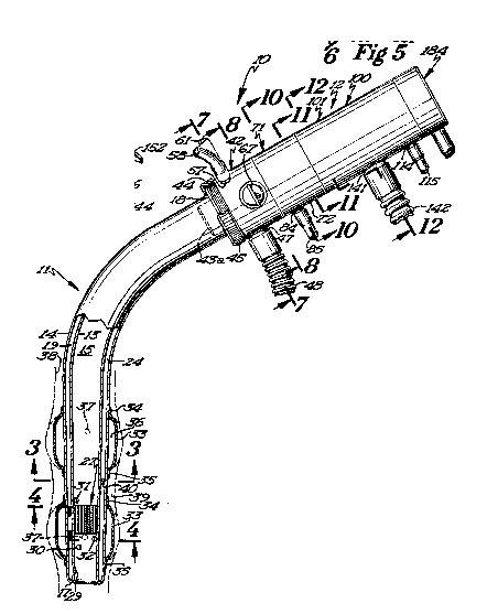
Referring now to Figures 5 and 6, it will beseen that a disposable mouthpiece device 143 is
thereshown and constitutes an alternate form for
the endotracheal tube as a device for continuously
circulating the gases to be measured through the motor
pump and mass spectrometer module 12. The disposable
mouthpiece device 143 is formed of a suitable flexible
131~fi~
-32-
plastic material and includes a double-walled mouth-
piece tube 144 comprised of an inner tube 145 and an
outer tube 146. Although not shown in the drawings,
suitable interconnecting wall elements interconnect
the inner and outer tubes and define four auxiliary
passages, rather than the seven passages embodied
in the endotracheal tube 11. The inner tube 145 de-
fines a large central passage in the manner of the
endotracheal tube. Referring again to Figure 6, it
will be seen that the mouthpiece tube 144 is provided
with an elongate passage 148 and an elongate passage
149, which extends throughout the length of the mouth-
piece tube. The lower end of the mouthpiece tube is
provided with an opening 150, which communicates with
passage 148, and an opening 151, which communicates
with passage 149. The sample gases from the lungs
will be directed through the opening 50 and upwardly
through the passage 148, while a portion of these
gases will be returned through the passage 149 and
discharged through the opening 151. Thus, the pass-
age 148 constitutes a sample gas passage, while pass-
age 149 defines a return passage.
A face engaging flange element 152 is secured
to the outer tube 146 and is adapted to engage the
exterior surface of the face of the human subject
adjacent the subject's lips. A capillary restric-
tion member 154 is positioned adjacent the lower end
131~
-33-
portion of this mouthpiece tube, and the capillary
restriction member is provided with a plurality of
capillaries or passages 155 which extend therethrough,
as best seen in Figure 6~ In this regard, the con-
struction of the capillary restriction member is sub-
stantially identical to that shown in the endotracheal
tube.
The inner tube 145 is provided with an opening
157 therein located below the capillary restriction
member 154 and is also provided with an opening 158
therein adjacent, but spaced above the capillary
restriction member. Although not shown in the draw-
ings, opening 157 communicates with a passage that
directs lung gases via the manifold unit to the
differential pressure transducer. Similarly, open-
ing 158 also communicates with a passage that directs
the lung gases through the manifold unit to the
differential pressure transducer. The disposable
mouthpiece device 143, therefore, permits measure-
ment of the lung gases by the mass spectrometer andalso permits measurement of the lung capacity by means
of the differential pressure transducer.
The disposable mouthpiece device is intended pri-
marily for conscious non-anesthetized patients for
use in assessing cardiac output and other cardio-
respiratory functions during office visits, exercise
testing, such as a treadmill procedure or measuring
6 ~
-34-
response to drugs. It will be appreciated that the
mouthpiece device will not have the degree of effi-
ciency of the endotracheal tube, since the lung gases
are being obtained at the rear portion of the human
subject's mouth. The dead space defined between the
rear or lower end portion of the mouthpiece device
and the lower tracheal or bronchial tree area naturally
renders the mouthpiece device less efficient than the
endotracheal tube. However, the mouthpiece device
can be readily used with conscious patients with little
or no discomfort.
Referring now to Figures 17 and 18, it will be
seen that the spectrometer electronics in cooperation
with the mass spectrometer 129 are thereshown in dia-
grammatic form. The electronic circuitry is designatedgenerally by the reference numeral 160 and is comprised
of a base unit 161 and a head unit 162. The head unit
is of light portable construction, containing the com-
putation read-out and operator input and may be mounted
on the anesthetist's trolley, as illustrated in Figure
17. The base unit does not require the operator or
anesthetist to manipulate or otherwise interact with
the components thereof and therefore may be positioned
at floor level with other components, such as the
vacuum module.
Since the spectrometer requires potentials of
approximately 100 volts, and is in close proximity
131~
-35-
to the patient, all the spectrometer electrical supply
requirements are constructed as a ground isolated
system, using isolating transformers and construction
approved for this application. The purpose of such a
ground isolated system is to insure that a leakage
to ground via the patient, or otherwise, causes an
unmeasurable flow of current, thus, protecting the
patient from harm. In addition, active ground current
monitoring from the isolated electronics will produce
an immediate shut-down and notify the operator of
fault conditions.
Transmission between the head and base unit is
by two fibre optic lines which not only provide ground
isolation, but also eliminate the transmission of
electro-magnetic interference to the head unit. In
the embodiment shown, one of the fibre optic lines
165 transmits information to change the frequency of
the radio frequency energy applied to the spectrometer
in order to tune ions of different mass. The outgoing
fibre optic line 166 provides digitized information
of the ion current at the spectrometer collector plate
135, the ion current collected by the ion collector
wire 128, as well as monitoring the potential supplied
to the spectrometer system in absence of fault condi-
tions.
The base unit 161 includes a D.C. power source163 which is provided with a filament electrical
~311~
-36-
current supply 164, which is controlled by the elec-
tron emission current received on the grid helix 120
of the ionization zone, and which stabilizes this
emission current at a given value. The filament cur-
rent supply 164 also senses for a voltage applied --
no filament current flow situation, and if found
switches over to the second filament. The head unit
is notified of the switchover and flags the operator
by a visual or audible signal. During start-up of
the apparatus lO, the head unit also senses and deter-
mines that current is flowing through both filaments
and start-up is prevented if only one filament is
operable. The current to the inactive filament will
be supplied through a resistor until the additional
filament is needed.
The D.C. power unit 163 is also provided with a
lO0 volt electron acceleration current supply 168,
which forms the feedback signal Eor the filament sup-
ply 167. A 120-volt supply current 169 serves to bias
the pressure sensing ion collector wire 128. This
current is isolated from the rest of the spectrometer
and supplies zero volt bus so that the ion current
flow can be sensed at bus potential and transmitted
to the head unit 162.
A lO0-volt current supply 17~ supplies the current
to operate the linear accelerator 129. This current
supply is modulated at audio frequency by a sinusoid
131~
-37-
of 1-2 volts amplitude. The final ion current in the
ion collector plate is phase detected with the audio
signal as a reference. The resultant output is used
to vary the exact value of the 100-volt potential
in order to maximize the ion current, that is, to have
a minimum first harmonic component in the phase de~
tected ion signal. This correction maintains the
spectrometer 100 in tune, as well as compensating for
dimensional tolerances in the spectrometer head and
possible slump of the structure due to mistreatment.
This seeking function is responsible for the main-
tenance of the spectrometer 100 in calibration without
day-to-day adjustment by an operator.
A programmable D.C. supply unit 171 has a 30-volt
programmable current supply or conductor 172, which is
controlled by input from the head unit 162. The cur-
rent supply 172 sets the potential at which the ions
are generated and, therefore, the acceleration that
they must obtain in order to land on the grounded ion
collector plate 135. The potential of this supply is
also sensed by the head unit 162.
The base unit 161 also includes a programmable
radio frequency generator 173 whose radio frequency
signal is transmitted to the spectrometer by two
coaxial cables 174, each terminated by its iterative
impedance. The radio frequency generator consists
of a group of quartz crystal controlled oscillators,
~3~1~66
-38-
together with harmonic multipliers, and a wide band
power amplifier capable of delivering a 5-volt pea~
amplitude radio frequency signal to the spectrometer.
The output level is rectified and fed back to the
power amplifier in order to keep the amplitude of
the radio frequency amplitude constant. Switching
between oscillators is performed on command by con-
trol means of the head unit 162 in order to select
different ion species for analysis. In general, the
switching rate between ion species will be determined
by the settling time of the ion collection amplifier.
The base unit 161 is also provided with an auxili-
ary thermistor and pressure transducer power unit 175
having a current supply conductor 176 and a current
supply conductor 177 electrically connected with a
pair of auxiliary thermistors, the function of which
will be set forth more clearly hereinbelow. A pair
of current supply conductors 178 also electrically
connects the power unit 175 with a differential pres-
sure transducer 179. Output signals from the differ-
ential pressure transducer 179 are transmitted to a
multiplexor unit 180 via con~uctors 181. The multi-
plexor unit 180 is a component of a data acquisition
system, which also includes a 10-bit analogical digi-
tal converter 182 and control electronics for trans-
mitting the digitized information to the head unit
162.
131~
-39-
The multiplexor unit 180, as well as transmitting
spectrometer ion current data, spectrometer pressure
ion current data, and parameters of the spectrometer,
also transmits data obtained from zener reference
sources in order to check the continual functionality
of the multiplexor-analog digital converter system.
An ion current amplifier 183 is mounted as a head
amplifier in the connection socket 184 of the mass
spectrometer header 106 and is followed by a second
amplifier (not shown) at the module end of the con-
necting cable for the connection socket. The use of
a head amplifier eliminates the problems associated
with a large cable capacitance which would otherwise
be associated with a remote amplifier.
The head unit 162 constitutes a system controller,
which communicates with the spectrometer system through
the fibre optic cables 165 and 166, as described herein-
above. The system controller actually constitutes a
micro-computer 185 to serial input-output ports and
can be fabricated from any of the commercial central
processing units of desired capacity. In the embodi-
ment shown, it will be seen that the micro-computer
or CPU has been duplicated for maximum reliability
and for continuation of monitoring, should a run-
wild or halt occur in the system. It is also pointedout that each CPU is provided with a watch dog set to
detect malfunction.
13115~6
-40-
The system also includes a display and user inter-
face 186, which may be a conventional screen-keyboard,
a custom LCD panel, or a touch-sensitive input tablet.
The head unit includes a digital analog converter 187,
which can be operated from the incoming signal line
from the head unit 162. The digital analog converter
187 is connected through one of the multiplexor unit
channels to the analog digital converter 182. Start-
up testing, therefore, programs a staircase wave form
on the digital analog computer, with the analog digi-
tal converter 182 converting each step. This enables
the monotonacity in the absence of missing codes to be
checked by both the digital analog converter 187 and
the analog digital converter 182. Under operating
conditions, the digital analog converter 187 sets the
levels of the power supply, which generates the start-
ing potential of the ions.
Although not shown in the drawings, provision is
made for an output line to a central recording area
where the spectrometer data can be continuously
recorded in a tamper-resistant environment in order
to provide information on the progress of the patient
during surgery, and to have documentation available
for future use, such as possible future litigation.
During operation of the apparatus, the rotatable
valve 63 in the manifold unit will be manipulated to
a position for measuring the sample gases from the
1311566
-41-
lower trachea or from the tracheal sampling cell 39.
If the lung gases are being sampled, then the head
unit of the apparatus will indicate that this function
is being performed and that the tracheal sampling cell
S 39 is not in use.
In a normal man, during the steady state, lung
profusion and cardiac output are essentially equal.
All CO2 recovered and all 2 consumed from the respira-
tory air reflects right ventricular blood flow. Since
the output of the right and left ventricles are vir-
tually identical, this is the cardiac output. The
general equation expressing this relationship is the
well-known form of the Fick equation:
Q = Vx/(Cax-Cvx)
where
Q = cardiac output
Ca = arterial concentra-tion of x
CVx = mixed venous concentration of x
Vx = total amount of x.
This expression is positive for uptake conditions
(X = 2) and negative for output conditions (X = CO2).
The cardiac output may be measured by a single
breath method procedure or by a CO2 rebreathing method.
The single breath method uses the respiratory exchange
ratio taken during a prolonged expiration (10 seconds).
A detailed discussion of the single breath method and
the computations used therein are expressed in the
` 1311~66
-42-
article entitled, "Estimation of True Venous and
Arterial PCO2 by Gas Analysis of a Single Breath",
by T. S. Kim, et al., appearing in the Journal of
Applied Physiology, 21(4): 1338-1344; 1966.
The respiratory exchange ratio taken during a
prolonged expiration (10 seconds) is expressed as
follows:
(R) = ---____ CV CO2 - CACO2
V 2 Ca 2 ~ CV2
In the operation of the subject apparatus, the
mass spectrometer R produces data which is continuously
calculated during a prolonged expiration, giving rise
to a curve. The slope (S) of the tangents at various
intervals is used to derive a family of instantaneous
R values from the following form of the alveolar air
equation:
S = R - (I-R) FACA2
I = (I-R) FAO2
Solving for R:
R = ----------------________
I - (FAO2 x S) - FAO2
The various instantaneous values of R are then
plotted against their corresponding measured PC02
(arterial-alveolar). This allows the estimation of
the appropriate arterial and mixed venous CO2 tensions
that must have existed. The arterial PCO2 is obtained
from the R value averaged from several normal expired
~3~156~
-43-
breaths preceding a greater than normal inspiration
followed by a prolonged expiration (10 seconds).
In man, the time mixed venous PCO2 is obtained
from the R value intercept at R = 0.32. At this value
of R = 0.32, arterial and venous PCO2 are equal (no
pressure gradient), yet CO2 is still exchanging at
one-third the usual rate because of the Haldane effect.
At R = 0.32 every unit volume of 2 taken up by hemo-
globin from the venous blood displaces exactly 0.32
volumes of CO2 without a change in PCO2. Thus, by
determining precisely the alveolar-arterial PCO2 when
the instantaneous exchange ratio (R) has fallen to
0.32, a value is obtained which must be equal to the
mixed venous PCO2.
The key to this single breath method is the
ability to determine PO2 and PCO2 at the same instant
in time which is made possible by the miniature mass
spectrometer attached to the upper end of the endo-
tracheal tube or mouthpiece device, thereby eliminat-
ing a maximum amount of dead space. The correlation
of this data with heart rate is critical in the single
breath method.
When the cardiac output is determined by the CO2
rebreathing method, this method is also simple and is
carried out in less than 30 seconds. Specifically,
this method depends on assessing the time it takes
for rebreathed CO2 to reach a plateau. The precise
1311566
-44-
detail of this method is described in an article
entitled, "Cardiac Output Determination by Simple
One-Step Rebreathing Technique" by L. E. Farhi
et al., published in Respiration Physiology (1976),
28, 141-159. However, the present apparatus obviously
increases the accuracy and shortens the time needed to
reach a plateau because the dead space is reduced.
Heart beat correlation with the CO2 rebreathing
method is important, but not as cirtical as with the
single breath method. It is pointed out that, during
the single breath method and the CO2 rebreathing
method, the lung gases are circulated through the
sample gas passage 20 in the endotracheal tube and
are returned through the return passage 19. If the
mouthpiece device 143 is used, the sample gases are
directed upwardly through the sample gas passage 148
and are returned through the return passage 149.
The lung capacity or volume is determined by the
differential pressure transducer and receives gas
samples from below and above the capillary restriction
member 27 in the endotracheal tube or the capillary
restriction member 154 in the disposable mouthpiece
device. The location of the restriction capillary
member 27 in the endotracheal tube or the capillary
restriction member 154 in the mouthpiece device is
chosen so that the temperature of the capillaries
will be close to body temperature, thus eliminating
S
-45-
condensation of moisture within the capillaries. Flow
of gases through these capillaries produces a pressure
differential which is communicated through their asso-
ciated passages to the differential pressure transducer
for measuring respiratory volume.
Although both of the balloons 33 are inflated
and serve as retention members during sampling of
lung gases by means of the endotracheal tube, these
balloons also define the tracheal sampling cell for
use in making an analysis of the tissue cell gases.
Sampling from this tracheal sampling space is under-
taken when it equilibrates with the tracheal tissue
and will measure tracheal tissue 2 (and CO2), which
will closely reflect arterial PO2. This sample gas
will be representative of tissue PO2 in other parts
of the body and, therefore, indicate the accuracy
of oxygenation. This particular procedure should
reduce or entirely eliminate the need for arterial
samples, thereby makiDg effective arterial PO2
determination a non-invasive procedure of choice.
Sampling from the tracheal sampling cells 39 is
done by flushing argon gas through the openings or
ports which communicate with the tracheal sampling
cell and circulating the argon gas through the mass
spectrometer for analysis until the argon gas reaches
an equilibrium condition in the tracheal sampling
cell.
131~
-46-
It is also pointed out that, by introducing a
tracer amount of gas, such as acetylene, into the
expired mixture, the lung to trachea circulation
time can be measured. This is a useful physiological
measurement to assess the efficiency of blood circu-
lation. Moreover, the uptake kinetics Gf acetylene
can be used to assess local blood flow in tracheal
tissues.
In addition to measuring the physiological para-
meters and characteristics set forth hereinabove, thepresent apparatus can measure other aspects of respira-
tory functions. In this regard, the apparatus can
measure the anatomic respiratory dead space, the
physiological respiratory dead space, the pulmonary
diffusing capacity for 2 and CO2, and the anatomic
and physiological shunt flow.
It will be appreciated that, regardless of the
source of the gas samples being measured, these gas
samples will be directed into the mass spectrometer
via the motor pump unit 71. The use of an air driven
gear pump insures that a steady flow of gases passes
through the sampling orifices of the mass spectrometer.
Even though a ripple in the flow rate occurs as a
result of the meshing frequency of the gear teeth,
this ripple in the flow rate is unimportant relative
to the frequency of breathing.
In order to monitor the air flow, two thermistors
1311~6~
-47-
are placed in the sampling gas stream. One of the
thermistors is placed upstream of the circulator
gears of the pump, and one is placed downstream of
the circulator gears of the pump. The upstream
thermistor will be used to correct the respired air
volumes to standard temperature, while the downstream
thermistor is supplied with sufficient electrical
energy to self-heat it to a few degrees above the
ambient temperature. Periodically, the flow of elec-
trical energy to this downstream thermistor is inter-
rupted and its cooling rate measured. The cooling
rate will indicate that there is a continuous flow
of yases around the measuring loop. If the sample
gas passage or the return passage is blocked, or if,
for any reason, the circulator gears do not rotate,
the abnormal cooling rate of the downstream thermistor
will make the instrument operator aware of this mal-
function. The positioning of thermistors on opposite
sides of the pump gears provides thermal isolation
between them.
It will be appreciated that a major portion of
the sample gases that are directed to the motor pump
unit will be returned via the particular return pass-
age and that only a small portion of the sample gas
will be discharged into the mass spectrometer. The
return of the sample gas for complete mixing of the
alveolar gas does not alter the measured lung ventila-
1311~6~
-48-
tion. This return and remixing of the sample gas
reduces the delay time for the gas to reach the mass
spectrometer analysis to a value negligible compared
with inhalation and exhalation times.
From the foregoing, it will be seen that the
novel gas sampling apparatus provides a non-invasive
means for accurately and routinely monitoring cardiac
output and the cardio-respiratory function in conscious,
as well as anesthetized or unconscious, human subjects.
Since the endotracheal tube and mouthpiece are dispos-
able, the motor pump and mass spectrometer module may
be readily disconnected therefrom and reused after
centrifuging. The gas sampling efficiency of the
apparatus, especially the endotracheal tube, is greatly
enhanced by the elimination of dead space. The provi-
sion of a miniature motor pump - mass spectrometer
module allows this efficiency to be obtained. Minia-
ture mass spectrometers having a free mean path of
the order of one-half to two centimenters in length
are simply unknown in medical and related fields.
Thus, it will be seen that we have provided a
non-invasive apparatus for assessing the cardiac
output and other respiratory functions which func-
tions in a more efficient manner than any heretofore
known comparable apparatus.