Note: Descriptions are shown in the official language in which they were submitted.
t3~25t2
RIGID VIDEO ENDOSCOPE WITH HE~T STERILI2ABL~ SNEP.~H
Technical Field
This invention relates to a video endoscope and
more particularly to such an endoscope having a moisture
05 tight inner cylindrical hody containing electronics and
optics which can be disinfect~d by soaking and an outer
rigid sterilizable sheath for containing and covering
the inner body during an operation, the outer sheath
being sterilized by heat and having a sterilizable
sleeve for extending over the trailing cable containing
the electronic and optic fibers.
sack~round Art
Over the years many developments have been made in
lS the endoscope art. Particularly, these developments
have been attempts to provide endoscopes which will
serve a variety of functions and which are maintained in
a sterile condition during use.
Erse~ et al. UOS. Patent Nos. 3,794,091 and
3,809,072 each disclose a flexible sheath which is
sterile at the time of manufacture and can be rolled up
onto an endoscope to provide sterility. However, there
is no seal at the proximate end of the sheath and
therefore bacteria can enter between the endoscope and
sheath and there is no provision for maintaining the
distal end of the endoscope in a sterile or protected
condition.
Russel U.S. Patent No. 3,866,601 discloses a
speculum in which a penetrating tube slidably receives a
guide tube and is surrounded by a flexible sheath.
Ibe U.S. Patent No. 4,132,227 discloses an
; endoscope surrounded by a hollow cylindrical sheath
~ extending toward but not to the distal end of the
1312512
--2--
endoscope in order to create a fluid channel in the
space between the sheath and the endoscope.
Smith U.S. Patent No. 4,201,199 discl~ses an
endoscope surrounded by a rigid glass or plastic tube
05 having an enlarged bulb at its dista:L end to space
tissue away from the viewing window of the endoscope.
The window is formed at an angle to provide viewing of a
site offset from the axis of the endoscope.
Yoon U.S. Patent No. 4,254,762 discloses an
endoscope surrounded by a sheath havin~ a transparent
lens at its distal end. The sheath may be at least
partially open at its distal end for use with endoscopes
having biopsy channels.
~-lampson U.S. Patent No. 4,327,735 discloses a
catheter surrounded by a transparent, collapsible sleeve
throu~h which the catheter projects at its distal end.
Silverstein et al. U.S. Patent No. 4,646,722
discloses another endoscope having a sterile flexible
sheath which can be rolled up along the endoscope. A
channel is provided between the endoscope and sheath
through which biopsies can be ta~en. The sheath is not
sealed at the upper end and will not maintain the
sterility which is required within an operating room.
D'Amelio V.S. Patent No. 4,721,0~7 discloses
another flexible sheath for use on an endoscope which
has no seal at the upper end and does not provide the
sterility required in an operating room.
Sidall et al. U.S. Patent No. 4,741,326 discloses a
further flexible sheath which is rolled up along the
endoscope and does not provide sterility or protection
of the entire endoscopic device.
Brown British Patent No. 1,405,025 discloses a
proctoscope surrounded by a concentric tube ~or
providing a fluid channel.
" 3 ~312512
Disclosure of the Invention
In accordance with the present invention, a rigid
video endoscope is provided which comprises an inner
cylindrical body member having a distal end and a
05 proximate encl. A li~ht transmitting element is sealed
to the distal end of the body member. An image sensor
is mounted against the light transmitting element within
the body member. An electronic cable within the body
member has a distal end connected to the image sensor
and a proximate end extending beyond the proximate end
of the body member and connectable to a video control
unit. At least one fiber optic bundle for light
transmission is provided within the body member and has
a distal end adjacent the light transmitting element and
1~ a proximate end extending beyond the proximate end of
the body member and connec-table to a xenon, halogen or
incandescent light source. A strain relief fixture is
sealingly attached to the proximate end of the body
member with the electronic cable and jacketed fiber
optic bundle extending therethrough and sealed to the
fixture. An outer rigid cylindrical heat sterilizable
sheath, having a distal end and a proximate end, is
provided for receiving the inner body member and is of
substantially the same len~th as the body member. A
window is sealed to the distal end of the sheathO An
accordion-folded, heat sterilizable, cylindrical sleeve
is mounted adjacent the proximate end of the sheath and
is extendable along the electronic cable and the optical
bundle for a substantial dis~ance to maintain sterility
of the vi~eo endoscope within the sterile field of the
operating room. Means is provided for releasably
locking the body member within the sheath.
A tab can be provided on the sleeve for extendiny
it along the electronic cable and fiber optic bundle. A
4 1312512
releasable locking means can include a bayonet slot at
the proxi~ate end of the sheath and a pin at the
proximate end of the body member which is releasably
en~ageable with the bayonet slot. If desired, the
05 window can be a prism for viewing at an angle to the
lon~itudinal axis of the endoscope.
In one embodiment the body member is concentrically
ali~ned with the sheath. In another embodiment the body
member is eccentrically mounted within the sheath. In
this later arrangement the window has apertures therein
and channels within the sheath corresponding in number
to the apertures to provide access to the site under
investi~ation. These channels can be used for providing
gas or a steerable device can be inserted through one of
them to carry out a procedure, such as taking a biopsy
at the site. It also could provide a channel for a
laser fiber. The body member may be backed filled with
nitrogen under pressure to minimize the possibility of
any liquids entering into that device.
With this arrangement, the inner cylindrical body
member contains all of the optics and electronics and
can be disinfected by soaking it in a disinfecting
solution. However, disinfecting is not sterilization
and therefore is generally not acceptable for use within
the operating room. It particularly is not acceptable
in open surgical procedures and in orthopedic sur~ery
and neurosurgery. Thus, the rigid sterilizable sheath
on the exterior can be properly sterilized by heat
treatment and then slipped over the inner cylindrical
body. The accordion-folded sleeve on the sheath can be
extended along the optical bundle and electronic cable
for a sufficient distance to provide a sterile barrier
between them and the operating site. This outer sheath
can be made of disposable material or it can be
_5_ 131251 2
resterilized for subsec~uent ~Isage. Thus, all portions
of the device which come in contact with the patient can
be sterili~ed even though the associated electronics and
optics cannot be sterilized. After use, any
05 contaminates from the patient's body will he removed
with the outer sheath and not contact the inner body
member. Thus, the contamination cannot to transmitted
to the next patient since the body member will be
inserted in another sterile sheath.
The term "light" transmitting element" as used
herein is intended to include any type of light
transmitting device which may have any one or several
optical qualities. For example, it may simply be a
transparent panel made of glass, plastic or sapphire.
On the other hand, it may comprise one or more lenses
for magnification or to increase the field of view. It
can include a series of adjustable lenses to provide
variable magnification and serve as a microscope.
Additional advantages of this invention will become
apparent from the description follows, taken in
conjunction with the accompanying drawings.
srief Description of the Drawings
Figure 1 is a perspective view of the inner
cylindrical body member which forms a part of the video
endoscope of this invention;
Figure 2 is a perspective view of the outer
cylindrical heat sterilizable sheath which forms the
other part of the video endoscope of this invention;
Figure 3 is a condensed, longitudinal section,
taken along line 3-3 of Figure 1, showing the internal
details of the cylindrical body member;
Figure 4 is an enlarged vertical section, taken
-6- 13~25~
along line 4-~ of Figure 3, showing the arrangement of
the image sensor and optical fiber bundles;
Figure 5 is an enlarged vertical section, taken
along line 5-5 of Figure 3, showing t:he position of the
05 electronic cable and the fiber optic bundles within the
body member;
Figure 6 is a condensed longitudinal section, taken
along line 6-6 of the Fi~ure 2, showing further details
of the heat sterilizable sheath;
Figure 7 is a condensed longit~dinal section,
showin~ the cylindrical body mem~er positioned within
the heat sterilizable sheath;
Figure 8 is a fragmentary perspective view of an
alternative video endoscope;
Figure 9 is an enlarged vertical section, ta~en
alon~ line 9-~ of Figure 8, showing the arrangement of
the body member within the sheath and the positioning of
the gas channels;
Figure 10 is a fragmentary perspective ~iew,
similar to Figure 8, but showing a passageway for use
with a steerable device; and
Figure 11 is a fragmentary section of an
alternative construction wherein the window of the
sheath is in the form of a prism.
~est Mode For carrying Out the Invention
In accordance with this invention a video endoscope
is provided which has an inner body member B, shown in
Figures 1 and 3, for containing the optics and
electroni~cs for the endoscope and an outer rigid, heat
sterilizable sheath S for receiving the body member and
providing a sterile outer casing for coming in contact
with the patient and for extending over the connecting
~7~ 1312512
cables to provide a sterile environment for the
operative procedure on th0 patient.
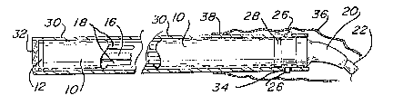
q`he inner body member M has a cylindrical housing
lO provided at its distal end with a light transmittiny
05 device 12 which is sealably attached thereto to minimize
the possibility of any fluids entering at this location.
An image sensor 14, such as a CCD is mounted on the
inside of light transmitting device 12 ~or receiving
light transmitted through the window at the
investiga~ive site, such as within a body caviky. The
image sensor also may be a CCD, CID, CPD or M~S device,
depending upon application. This CCD may be on the
order of l.0 mm x l.0 mm. A typical laproscope is 0.8
cm to 1.2 cm in diameter and would utilize a
correspondingly larger CCD device, a typical arthroscope
is 4.0 mm in diameter and would utilize a much smaller
CCD sensor. The window may include optics to focus an
image on the CCD and provide a focal length of 0.5 cm to
15 cm, depending on the intended use of the endoscope.
Field of view may be altered by the use of different
lenses and may range from 15 to 140 or more. An
electronic cable 16 has a distal end connected to the
image sensor 14 and runs longitudinally through housing
10, as shown in Figure 3.
Conveniently, optical fiber bundles 18 have distal
ends positioned adjacent light transmitting element 12
and spaced around image sensor 14. The exact number of
op~ical ~iber bun~les and t~pe of fiber will depend upon
the particular usage of the endoscope. Four such
optical fiber bundles have been shown in the drawings,
one being positioned on each of the four sides of image
sensor 1~. However, a greater or smaller number could
be provided, as required. The number of optical fiber
bundles may be optimized to allow transmission of
~ .
-8- 1312~12
various light frequencies, including laser light. These
optical fiber bundles 1~ also run through housing 10 and
pass throu~h a strain relief fixture 20 which seals the
proximate end of housing 10. This fixture is also
05 sealed around these cables and extend beyond the fixture
throuyh the center of a connecting cable 22 whose
opposite ~nd is connected to a video processing unit 24
having a suitable viewing screen (not shown) and light
sources (not shown) as required and as is apparent to
one skilled in the art. The cylindrical housing is
completely sealed against the entry of moisture by
window 12 and fixture 20. It also may be backfilled
with nitrogen gas under pressure to help keep moisture
out. After each use, it can be soaked in a
disinfectant, such as gluleraldehyde or Chlorox.
Conveniently, the distal end of housing 10 is
provided with oppositely extending pins 26 for
connection to a bayonet slot on sheath 5 as described
below. Also, an 0-ring 28 is provided for forming a
~0 seal with outer sheath S, as described below. The seal
could also be a threaded seal, with a threaded collar on
the outer unit fitting into a threaded collar in the
inner unit.
Sheath S comprises a cylindrical housing 30 which
has a window 32 at its distal end and a bayonet slot 34
at its proximate end for cooperating with pins 26 to
lock inner body member B within outer sheath S.
Advantageously, an accordion-folded sleeve 36 is
provided adjacent the proximate end of housing 30 and
has a fla-nge 38 attached to housing 30, as by adhesive
and includes a pull tab 40 for extending the sleeve over
strain relief fixture 20 and connecting cable 22 as best
seen Figure 7. Th~se parts can be made of Teflon or
other materials which can withstand high sterilization
_9_ 1312512
temperatures. The window can be made of ~lass or
sapphire or polycarbonate ~hich can stand the
steriliæation heat or other materials which are clear
and withs~and hi~h temperatures of heat s~erilization.
05 Referrint~ to Figure 7, it can be seen that when the
endoscope is ready for use, the inner body member is
locked within outer sheath S and light transmitting
element 1~ thereof is adjacent window ~2 of the sheath.
Thus, the inner body member is completely encased in the
sterile outer sheath. Furthermore, the sterile sleeve
36 extends over the strain relief fixture and connecting
cable to provide a completely sterile endoscope in the
operating room and particularly at the site at which the
operation Gr medical procedure is being conducted.
~fter use, the inner body member can be removed
from the outer sheath S and sleeve 36 by twisting it
slightly to release pins 26 from bayonet slot 34
whereupon it can be withdrawn. The outer sheath and
sleeve 36 can be thrown away or it can ~e heated to a
sufficient temperature and pressure for sterilization.
Also, it will be apparent that any contamination
from the body of the patient which may repose on the
outer sheath or the sleeve will be stripped away from
the inner body member along with the outer sheath and
therefore not be transmitted to the body member. The
inner body member can then be soa~ed in a disinfectant
and reinserted in another sterile outer sheath and
sleeve for use on a subsequent operation. Obviously, a
supply of the relatively inexpensive outer sheath and
sleeve assemblies can be maintained so that the inner
body member can be quickly made ready for a subsequent
operation. The video endoscope of this invention can be
used as a laparoscope, cystoscope, arthroscope, and for
-lo- 13t2512
pelviscopy. Wlth suitable optics, it could be used as a
sterile operating microscope.
An alternative embodiment is shown in Figures 8-11
wherein a larger outer sheath S' is provided around
05 inner body member B . This sheath S' has a cylindrical
housing 30' with a lens 32' at the distal end and a
sleeve 36' at the proximate end attached by means of a
flange 38'. The additional space is occupied by one or
more tubes or channels, such as tubes ~2 and 44 for
supplying gas or fluids to the site under investigation.
Conveniently, these tubes have control valves 46 and 48
respectively for controlling the flow of the gases and
to prevent gas leakage during a procedure. The gas can
be carbon dioxide which may be provided for the purpose
of clearing and distending the site under investigation
for better viewing. Also, a vacuum could be applied
through one of the channels for aspiration of unwanted
material from the viewing site. Conveniently, the
distal ends of these tubes are connected to apertures 50
and 52, respectively in window 32'. These channels or
tubes can be used for insertion of laser fibers, biopsy
devices, grasping devices, etc.
In Figure 10 the same device is shown for use with
a steerable device~ In this case, a channel or tube 54
is provided which enters through housing wall 30' and
extends longitudinally therealong and through aperture
52. Within this tube 54 is a steerable device, such as
a kiopsy sampling device 56 which is operated by a joy
stick 58, as shown.
Finally, a further alternative embodiment is shown
in Figure 11 wherein the outer sheath S is provided with
a prism 60 in place of the front window 32 so that the
device can be used to view at any suitable angle to the
longitudinal axis of the endoscope. The image sensor
1 3 1 25 1 2
may be placed a-t various angles to the longitudinal axis
of the tubular housing, for instance at 30, 45, or 90.
In this variation, the window in the heat sterilizable
sheath would ~e placed at a corresponding angle to match
05 the orientation of the sensor.
From the foregoing, the advantages of this
invention are readily apparent. ~n endoscope has been
provided which is formed in two parts, an inner body
member and an outer sheath. The inner body member
contains all of the optics and electronics and is sealed
against moisture so that it can be soaked in a
disinfectant between usages. However, in most
situations such disinfecting is not sufficient for safe
subsequent use in the operating room. Therefore, an
outer sheath is provided which can be heat sterilized
prior to use and can be slipped over the inner body
member and releasably locked thereto to provide an outer
sterile covering. The outer sheath includes an
accordion shaped sleeve at the proximate end which can
be extended over the trailing cables of the inner member
which contain the optical fibers and elPctronic cables
to provide a sterile covering so that the device can be
used in the operating room at the operating site. After
`~ use, the inner body member can be removed from the outer
sheath and the sheath can either be thrown away or
resterilized. Also, any contamination from the body o~
the patient will be removed with the outer sheath and
will not come in contact with the inner body member,
thereby minimizing any transmittal of disease from one
patient to the next.
This invention has been described in detail with
reference to particular embodiments thereof, but it will
be understood that various other modifications can be
effected within the spirit and scope of this invention.