Note: Descriptions are shown in the official language in which they were submitted.
2 6 ~
This invention relates to a simple procedure for
measuring the hemoglobin concentration in a sample
of whole blood, and more particularly to a
procedure which can be quickly performed by a
relatively unskilled technician.
Two measurements commonly performed on the whole
blood are the hematocrit and hemoglobin. The
hematocrit is the percentage volume that packed
red blood cells occupy in a centrifuged sample of
whole blood, and the hemoglobin content is the
weight of the hemoglobin per unit volume of whole
blood. The numeric ratio of hemoglobin to
hematocrit is reEerred to as the mean corpuscular
hemoglobin concentration (MCHC), and in normal
individuals it is close to 33.9%. When an
individual is suffering from certain diseases,
however, the ratio may vary from about 38% down to
26%. Thus, the determination of both the
hematocrit and hemoglobin are important for the
discovery and diagnosis of anemia or other blood
disorders.
In a large laboratory the measurements of
hematocrit and hemoglobin are usually made
concurrently in an automated analyzer, but in a
small clinic or in a physician's office, they must
be made separately, using two different
techniques. The hematocrit may be presently
performed by filling a small bore glass tube with
anticoagulated whole blood, sealing one end of the
tube, and centrifuging the tube to pack the red
blood cells. After packing, which takes about
three to five minutes in a small centrifuge, the
length of the packed red blood cell column and the
total filled length are measured, and the
hematocrit, expressed as a percentage, is
13192~9
calculated. It can be appreciated that little
skill is required to prepare the tube or take the
measurements. U.S. Patent Nos. 4,027,660 issued
June 7, 1977 to S.C. Wardlaw et al; 4,181,609
issued January 1, 1980 to S.C. Wardlaw et al;
4,156,570 issued May 1978 to S.C. Wardlaw; and
4,558,947 issued December 17, 1985 to S.C.
Wardlaw; and others describe a procedure which
involves drawing a sample of anticoagulated whole
blood into a capillary tube, placing a float in
the tube with the blood sample, and centrifuging
the blood sample to cause the float to settle into
the red cell layer to elongate the buffy coat in
the blood sample. This prior art technique can be
used to measure hematocrit as well, by merely
taking into account the expansion of a portion of
the blood sample by the float when calculating the
total length of the blood components, the observed
total length being scaled down by the measuring
instrument to compensate for the presence of the
float.
On the other hand, the measurement of the
hemoglobin concentration is considerably more
complicated. To perform this test, in a small
clinic or physician's office, the blood sample
must be more accurately diluted to a ratio of
either 1:250 or 1:500, depending on the equipment
used. The dilution is made by accurately taking a
tiny sample of the blood into a pipette and
delivering it into a container containing an agent
which dissolves the red blood cells, and cyanide,
which converts the hemoglobin to a more easily
measurable form. This mixture, after standing for
three to ten minutes, is then placed in a
photometer where the light attenuation at 560nm
(green) is compared to that of standard solutions.
1319269
From these comparisons, the concentration of
hemoglobin can be calculated. There are many
published variations of this method, but all
acceptable means to date require the accurate
measurement of the light attenuation in an
instrument designed for this purpose. Further,
the need for accurately handling small quantities
of the sample requires a higher level of skill
than does the performance of the hematocrit, and
is therefore also a source for analytical errors.
We have discovered a procedure for measuring
hemoglobin in a blood sample using basically the
same paraphenalia and instruments which are
presently used to measure hematocrit. Our
procedure is based on our discovery that the
hemoglobin concentration of the packed red blood
cells is inversely proportional to the depth to
which the float of the prior art sinks into the
red cell layer. The microprocessor in the
measuring instrument will be programmed to convert
additional depth measurements into the hemoglobin
concentration. The process steps used to perform
the procedure of this invention are as follows.
The whole blood sample is drawn into the
centrifuge tube, preferably a capillary tube,
anticoagulated, and the float is positioned in the
tube. After the bottom of the tube is plugged,
the sample is centrifuged so as to layer out the
blood onto red cell, buffy coat, and plasma
layers. During centrifugation, the float settles
into the red cell layer. The hematocrit will then
be measured generally as per the prior art.
The hemoglobin is measured as follows. As stated
above, the MCHC of the red blood cells is the
concentration of hemoglobin within them, normally
13l~26~
about 340 g/l. Therefore, about 1/3 of each cell
is hemoglobin, the rest is water and a small
concentration of salts and minor proteins of
relatively constant concentration. It follows
from this that virtually the sole contributor to
density differences between different patients'
red blood cells is their hemoglobin concentration.
Therefore, the packed red blood cell density is
proportional to the MCHC, and if this value (MCHC)
can be accurately ohtained, the hemoglobin content
can be calculated as:
Hemoglobin = Hematocrit x MCHC.
The apparatus of the invention comprises a
transparent tube of constant bore diameter, into
which is placed a resinous float. Anticoagulated
blood is drawn into the tube, either by capillary
action or by slight suction. The exact quantity
is not critical, as long as there is sufficient
blood to buoy the float. One end of the tube is
sealed, and the tube is then centrifuged at
approximately 10,000G's for approximately five
minutes. This is the same regimen that is
currently used to perform the standard hematocrlt
determination. When the centrifugation is
complete, the float, which has a specific gravity
between that of plasma and that of the packed red
blood cells, will be partially buoyed up by the
packed red blood cells. The hematocrit is
measured by taking the ratio between the length of
the blood sample in the tube (the total length of
the packed red blood cells, buffy coat, and
plasma) and the length of the packed red blood
cell coll~n only. The volume of the float must,
of course, be accounted for in making this
calculation. Because the depth of the float is
inversely proportional to the density of the
~319269
packed red blood cells, the red blood cell density
may be calculated and converted to hemoglobin
content as follows.
The sum of the buoyant forces on a floating object
that has reached equilibrium, such as the float
used in this invention, are zero. The buoyant
forces in a three phase system such as ours which
consists of a cylindrical plastic float; packed
red blood cells; and plasma can be expressed as
follows:
(Dr - Df) x Lr + (Dp - Df) x Lp = 0
wherein Dr is the density of the packed red blood
cells; Df is the density of the plastic float; Lr
is the length of the float which is submerged in
the packed red blood cells; Dp is the density of
the plasma; and Lf is the length of the float
which is submerged in the plasma.
In the aforesaid equation, the density of the
packed red blood cells is not known. The density
and length of the plastic float are known, and are
inputted into the microprocessor memory.
Likewise, the density of the plasma is known and
is inputted into the microprocessor memory. The
length of the float which is submerged in the red
blood cells is measured and is thus inputted into
the microprocessor. Finally, the length of the
float which is submerged in the plasma is
calculated by the microprocessor by subtracting
the length of the float submerged in the red blood
cells from the total length of the float. The
microprocessor can thus solve the equation for the
density of the red blood cells (Dr)~
~3i~269
Once Dr has been calculated, MCHC can be
calculated by the mlcroprocessor by solving the
following equation:
MCHC = (Dr x Ks) + Ko
The MCHC constants, Ko and Ks, may be determined
empirically by taking red blood cell density
measurements for a number of diverse samples and
correlating the density with the MCHC as
determined by conventional reference measurements.
The slope constant (Ks) and the offset constant
(Ko) of the best-fit correlation equation are then
used to calculate the MCHC from the red blood cell
density. Once calculated, Ks and Ko will not be
changed unless the critical parameters of the
paraphenalia, i.e. float density, etc. are
changed.
From the MCHC, the hemoglobin concentration may be
determined as previously described, i.e.,
Hemoglobin = Hematocrit x MCHC.
It is therefore an object of this invention to
provide an improved procedure for measuring the
hemoglobin content in a sample of whole blood.
It is an additional object of this invention to
provide a procedure of the character described
wherein the hemoglobin content is measured as a
function of the extent to which a float sinks into
the packed red cell layer of a centrifuged blood
sample contained in a tube.
It is a further object of this invention to
provide a procedure of the character described
wherein the hemoglobin and hematocrit measurements
1319269
can be made quickly and easily with, or without,
the use of an automatic computing device.
These and other objects and advantages of the
invention will become more readily apparent from
the following detailed description thereof when
taken in conjunction with the accompanying
drawings, in which:
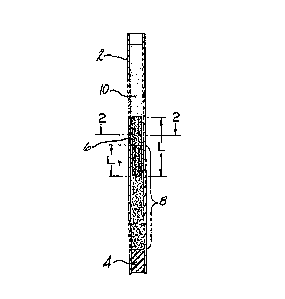
FIGURE 1 is a side elevational view of a glass
tube containing a centrifuged blood sample and a
float which has settled into the red blood cell
layer of the centrifuged blood sample; and
FIGURE 2 is a cross sectional view of the tube
taken along line 2-2 of FIGURE 1.
Referring to the drawings, the tube 2 is
preferably a glass capillary tube which may have
an anticoagulant coated on its inside bore well.
The bottom of the tube 2 is closed off with a clay
plug 4 or with a plastic cap which can be snapped
over the end of the tube 2 after the blood sample
is drawn into the tube 2. The float 6 is placed
in the tube 2, and when the blood sample is
centrifuged in the tube 2, the float 6 settles
into the red call layer, which is designated by
the numeral 8. Above the float 6 is the plasma
layer 10. The float 6 will have a preset known
axial length L, and the technician taking the
measurements will measure the distance Lr~ whlch
is the length of the float 6 which has sunk into
the red cell layer. The float shown in the
drawings has a fluted cross-sectional
configuration. This configuration imparts a
smaller cross-sectional area to the float 6 so
that the observed axial length of the centrifuged
131926~
blood sample, and particu]arly the buffy coat,
will not be significantly elongated. The flutes 7
on the float 6 will serve to maintain the coaxial
relationship with the tube 2. As previously noted
a fluted cross-sectionally-reduced float is not
essential to performing the hematocrit and
hemaglobin measurements. In this embodiment, the
cross sectional area of the float should
preferably be no more than about 2/3 of the cross
sectional area of the tube bore.
The blood used for the test must be anticoagulated
so that the red blood cells and plasma will
separate. This may be accomplished by drawing the
blood into an anticoagulant-containing vessel
prior to loading the blood into the tube, or by
incorporating an anticoagulant, such as heparin,
or the like, into the transparent tube itself.
This would allow the filling of the tube directly
from a finger puncture.
It can be appreciated that this procedure takes no
more time and requires no more skill than the
measurement of the hematocrit alone. It can also
be appreciated that an optical scanner, such as
described in U.S. Patent No. 4,156,570 issued May,
1978 to S.C. Wardlaw, or U.S Patent No. 4,558,947,
issued December 17, 1985 to S.C. Wardlaw, could be
used to read the lengths and automatically compute
the results. Because this procedure relies upon
two primary measurements (length and density), the
test does not require standardization.
There are two general embodiments of paraphenalia
used to perform the procedure of the invention.
The first is as shown in the drawings and
described above, and the second is identical to
2 6 9
the device described in U.S. Paten-t No. 4,077,396
issued March 7, 1978 to S.C. Wardlaw et al, in
that a buffy coat-expanding float ls used. In the
latter case, the buoyant effects of the expanded
buffy coat layers must be taken into account,
however, the readings can be computed by a
microprocessor which has been appropriately
preprogrammed as set forth hereinafter.
When the float is large enough to perform the
buffy COât measurements, as described in the
aforesaid U.S. patents issued to Wardlaw alone and
with others, the buoyant effect that the expanded
buffy coat exerts on the float can be compensated
for as follows. When such a float is used, the
three cellular components of the buffy coat will
add to the buoyant forces exerted on the plastic
float and must, therefore, be taken into account
when calculating the red blood cell density.
Therefore the following equation will be used.
(Dr - Df) x Lr +(Dg - Df) x Lg + (Dlm - Df) x
Llm + Dpl ~ Df) x Lpl + (Dp - Df) x Lp = 0
wherein: Dp, Dr/ Df, Lp, Lr/ and Lf are as
identified above;
Ll is the observed length of the float
disosed in the platelet layer of -the
blood sample;
Lml is the observed length of the float
disposed in the monocyte/lymphocyte cell
layer of the blood sample;
L~ is the observed length of the float
disposed in the granulocyte cell layer
of the blood sample;
Dg is the density of the granulocyte
cell layer;
-;;. _ g
.~ ~
13~269
Dlm is the density of the
lymphocyte/monocyte cell layer; and
Dpl is the density of the platelet
layer.
The instrument which is used is adapted for
measuring the white cell component counts, as
described in the aforesaid prior art. Thus the
microprocessor will have inputted information as
described above, and will also have the
granulocyte, lymphocyte/monocyte, and platelet
densities inputted. During the measurement
procedure, the lengths of the float disposed in
the granulocyte, lymphocyte/monocyte, and platelet
layers will be measured, and thus inputted into
the microprocessor. The value of Lf will be
calculated by the microprocessor as the difference
between the total float length minus the
cumulative lengths of the float which are
submerged in the red cells, granulocytes,
lymphocyte/monocytes, and platelets. Dr can then
be calculated by the microprocessor. Once Dr is
calculated, the hemoglobin value is determined as
set forth in the first example.
This technique was tested by determining the
hemoglobin and hematocrit of 100 patients. The
results obtained by the invention were virtually
identical to those obtained in the hospital
laboratory using automated analyzers (relative
standard error of 2.7~).
It will be readily appreciated that the procedure
of this invention will quickly and easily render
the hematocrit and hemoglobin measurements in an
anticoagulated whole blood sample. The procedure
can be conducted by a relatively unskilled person
-- 10 --
1319269
and can be performed with a single blood sample.
The procedure is particularly adapted for use in
small clinics and in the physician's office, but
can also be used in larger laboratories and
hospitals.
Since many changes and variations in the disclosed
embodiments of the invention may be made without
departing from the inventive concept, it is not
intended to limit the invention otherwise than as
required by the appended claims.
-- 11 --