Note: Descriptions are shown in the official language in which they were submitted.
~32~702
--1
METHOD AND DEVICE FOR ~APOR STERILIZATION OF
ARTICLES HAVING LUMENS
Field of Invention
The invention relates to the vapor sterilization of
articles such as medi-cal instruments having long narrow
lumens therein, and more particularly, to a device for
delivering a gaseous sterilant directly into the lumen of
an article during the sterilization process.
BacXqround ,of InventiQn
The need to sterilize articles su~h as medical instruments
and others for use`in the agriculture and fermentation
industries is well known. In recent years, many methods
of vapor sterilization have been developed. While these
methods o~far the advantage of being generally faster than
sterilization by immersion in a sterilant solution, they
suffer from one major disadvantaqe, namely the inability
to sterilize the interior of a long narrow tube in a short
period of time. Thus, with regard to medical instruments
such as endoscopes, the dif~iculty in sterilizing the
l~umen can often negate the general advantage of using
vapor starilization.
One way of overcoming the above disadvantage is set forth
in U.S. Patent Nos. 4,410,492 and 4,337,223. The
apparatus described therein comprises a sterilizing
chamber with means for introducing a sterilant gas into
the chamber and circulating the gas within the chamber.
Disposed within the chamber is a socket for receiving th0
tubular end of a medical instrument. The socket is
connected to a valve and a recirculating pump and the
starilant gas is recirculated from the chamber through the
JS~ 67
'. '" ' '' . " ' '
132~ 7~2
--2--
lumen of the instrument. The commercial apparatus, using
ethylene o~ide and water as the sterilant, has had little
commercial success which may be attributable to the
e~tended sterilization times of about 3 hours for fle~ible
endoscopes and about 2 hours for the shorter, rigid
endoscopes, as ~ell as to the toxicity problems associated
with ethylene o~ide sterilization. In addition, thæ
method and apparatus described in these references cannot
be used to sterilize an instrument within a sterile pack
since one end of the instrument must be attached to the
socket.
Thus there is a current need for an effective method to
sterilize medical instruments such as endos~opes in a
reasonably short perîod of time, preferably in one hour or
less. The method and device of the present invention
makes vapor sterilîzation of such instruments practical by
delivering vapor directly to the interior of the lumen in
the endoscope, whether or not it is in a sterile pack.
Summary of the ~nvention
The present invention comprises a method and device for
providiny an~imicrobial vapor directly into the long
narrow lumen of medical instruments and similar articles.
~he device and method are intended for use with solution
vapor sterilization procedures. In these procedures, the
article is placed within a sterilization chamber, the
pressure in the chamber is reduced, and a liquid solution
of an antimicrobial agent is introduced into the chamber
whera it vaporize~. Alternatively, an antimicrobial vapor
may be in~roduced directly to the chamber after the
pressure therein has been reduced. In either case, the
instrument is sterilized by e~posure to the vapor or an
active species generated from it rather than by direct
JSU 67
. ~
.
1 3~:1702
contact with a liquid sterilant. The procedure may
further involve the use of heat or, e.g., low pressure gas
plasma to enhance the antimicrobial activity, reduce
sterilization times, andfor remove residual sterilant from
the instrument.
In its simplest form, the device of the present invention
comprises a vessel containing a small amount of the
antimicrobial solution, and 3 means for connecting the
vessel to the lumen of the instrument to provide a source
of antimicrobial vapor directly to the lumen during the
vapor sterilization process. The device is placed on the
instrument prior to disposing the instrument in the
sterilization chamber. As the pressure in the chamber is
reduced, the antimicrobial solution contained in the
vessel is vaporized and passes from the vessel into the
lumen of the instrument. In its simplest form, the means
of connectinq the vessel to the end of the instrument tube
may comprise something as simple as a piece of firm but
fle~ible tubing, such as tygon tubing of appropriate
diameter such that one end of the tubing may be inserted
in or disposed about the opening of the vessel, and the
end inserted in or disposed about the lumen of the
instrument so as to be securely join the two. However,
the preferred means described below provide more
adjustable fastening and may be used with instruments
having a wida variation in internal and esternal tube
diameters. With the use of the device and method of the
present invention, vapor sterilization times or
endoscopes can be reduced to one hour or less~ In
addition, the method and the device may be used to
sterilize endoscopes in a sterile pack since the device o
the present invention may be attached to and packaged w;th
the endoscope before the endoscope is placed within the
sterilization chamber. Upon opening of the pack, ~he
JSU S7
_4_ ~.~32~ 7~2
device may be retrieved for re-use cr discarded with the
pack.
In one preferred embodiment of the device of the present
invention, the means for connecting the vessel to the end
of the tube comprises an e~pandable sheath, one end of
which is securely attached about an opening in the vessel,
and the other end of which comprises an elastic ring for
making a releasable attachment about the end of a tubular
instrument. Where the vessel of the device includes a rim
or lip about the opening, the sheath may be attached to
the vessel by means of a second elastic ring disposed over
such lip or rim.
In another embodiment of the device of the present
invention, the means for connecting the vessel to the end
of the instrument comprises a fle~ible bushiny disposed
within the opening of the vessel. The bushing may be made
of a series of rings of inwardly e~tendins plastic flaps.
The vessel may be provided with means for attaching a
closure cap thereto, such as threads internal or e~ternal
to the opening of the vessel for attaching a æcrew cap or
plug, to maintain the antimicrobial solution in the vessel
prior to use. Alternatively, the ~essel may be provided
with an aperture for attaching a disposable cartridge
containing a premeasured aliquot of antimicrobial solution.
In another embodiment, the vessel comprises a fle~ible
pouch, and the means for connecting the vessel to the end
of the instrument tube comprises a drawstring disposed
about the opening of the pouchO The pouch may be proYided
with an airtight sPal ~or sealing the antimicrobial
solution therein prior to use, and a means or creating an
opening in the sealed pouch so that the pouch may be
disposed about the end of the instrument when desired.
JSU 67
1 3 ~?~ 7 o 2
Both the seal and the means for creating an opening may be
achieved with, for instance, a ~zip-lock" interlocking
channel and ridge type fastening across the opening o the
pouch. As an alternative, the pouch may be heat sealed
and provided with scoring, notches, or other known means
for tearing open the pouch.
The device and method o the present invention reduce
sterilization time required for instruments having long
narrow lumens therein. Reduced sterilization times are
also achieved with the instruments encased in a package
designed to maintain sterility after the removal from the
sterilized chamber. In addition, as antimicrobial vapor
is provided directly into the lumen of the instrument,
lower concentrations of antimicrobial solutions may be
used in the sterilizer, and this together with the reduced
sterilization times provides improved materials
compatibility with respect to both t~e instrument
components and the packaging or wrapping materials.
Brief D~s~riPtion of the Drawing~
Figure 1 is a perspective view of one embodiment of the
device of the present invention, attached to the end of a
tube.
Figure 2 is a perspective view of another embodiment of
the device of the present invent;on, showing the end o
the device for making a connection to a tubular member.
Figure 2A is a variation of the device of Figure 2.
Figure 3 is a plan view of another embodiment of a device
of the present invention.
JSU 67
-6- ~3~1702
Figure 3A is a variation o the device of Figure 3.
Detailed Description of ~he Inven~i~n_
The method and device of the present invention relates to
the sterilization of articles such as medical instruments
having a long narrow tube therein. The term instruments
as used herein applies to medical or surgical devices such
as endoscopes, catheters, tubing, or similar instruments
or articles having an internal lumen which is preferably
used in a sterile condition as in, for e~ampl , the
agricultural or fermentation industriesO The method and
device of the present application show particular
advantages in the solution vapor sterilization of lumens
e~ceeding ten centimeters in length and having an internal
diameter of about 7 millimeters or less. As endoscopes
typically have lumens with internal diameters of 1 to 4
millimeters and lengths of up to 1.5 meters or more for
fle~ible endoscopes and at least 45 centimeters for rigid
endoscopes/ the method and device of the present
application have particular applicability to the
~terilization of these instruments. With the use of the
device of the present invention, antimicrobial vapor is
supplied directly to the lumen or interior of the tube of
the instrument during the vapor sterilization process.
Th~ antimicrobials used with ~he method and device of the
present invention include solutions of glutaraldehyde,
hydrogen pero~ide~ chlorine dioxide or other
antimicrobials in an inert solvent, the only requirement
being that the solution be li~uid at atmospheric pressure
and a ~apor at the temperature and pressure of the
sterilization process. Though the higher concentration
solutions of antimicro~ials are mor~ effective, problems
with materials compatibility and sh;pping and handling may
JSU 67
_7_ ~3~702
arise at very high concentrations. For e~ample, a 30% to
50% solution of hydrogen pero~ide in water is both very
effective and presents few shipping and handling problems,
while higher concentrations of up to 70% become
increasingly more difficult and dangerous to handle.
In solution vapor sterili~ation, the procedure generally
used is as follows: The article to be sterilized is
placed within the sterilization chamber, the chamber is
sealed, and a vacuum is drawn on the chamber to reduce the
pressure to less than about 50 torr, and preferably to 20
torr or less. An antimicrobial solution is then injected
into the chamber where it vaporizes and contacts the
e~posed surfaces of article. The time necessary for total
kill of specific microbial agents varies with the type and
concentration of antimicrobials present, and with the
degree of e~posure to the microbial agent. ~icrobials
disposed in cracks, crevices or internal tubular
structures are somewhat protected from the antimicrobial
a~ent and reguire more time for total kill than microbials
on the e~ternal surface of the article. Heat or high
~requency radiation may be used to increase the
effectiveness of the antimicrobial and its penetration
into remote areas of the instrument.
The device of the present invention comprises a vessel for
containing a small amount of antimicrobial solution, and a
means for connecting the ve~sel directly to the lumen or
the end of the tube of the article to be s~erilized. When
the article with device containing an~imicrobial solution
is disposed in the sterilization chamber and a Yacuum
drawn on the chamber, antimicrobial vapor generated from
the solution within the vessel flows directly into the
lumen.
JSU 67
-8- 13~702
The effectivPness of the method and device of the present
invention was demonstrated by the following e~periments:
50 inch (127 centimeters) lengths of tygon tubing having a
2 millimater inside diameter were used to simulate an
endoscope in the sterilization test. A paper strip (2mm
13mm) containing appro~imately 2.0 ~ 106 Ba~illus
~ub~ (var. globigii) spores was placed in each tube
equidistant from each end. A syringe containing 0.05
milliliters of 10% by weisht hydrogen pero~ide solution in
water was provided for each tube. Each of the samples was
individually packaged in a TYVEK~MYLAR~ envelo~e
prior to sterilization.
One third of the samples (three units) were placed in the
package with the syringe unattached to the end of the
tube. Another one-third of the samples were pac~aged with
the syringe attached. Individual samples were placed
within a 65 liter sterilization chamber and sent through a
~0 hydrogen pero~ide vapor sterilization cycle wherein the
pressure within the chamber was reduced to 3 torr ~or the
total esposure time minus 15 minutes, and 0.5 torr for the
final 15 minutes of esposure. No additional hydrogen
peroside was injected into the chamber.
~he remaining one-third of the samples, packaged with the
syringe attached to the end of the tube as described
above, were sent through a hydrogen pero~ide vapor
sterilization cycle supplemented with high frequency
radiation plasma which is known ~o generate an active
spacies from the hydrogen peroxide. Again a 65 liter
chamber was used, and ~he pressure within the chamber was
reduced to 3.0 torr for the total e~posure time minus 15
minutes and 0.5 torr for the final 15 minutes of
e~posure. Again, no additional hydrogen pero~ide was
JSU 67
9 ~ ~ 2 ~
injected into the chamber. Plasma was generated only
during the final 15 minutes of e~posure at 2.05 MHz with
320 watts of power, pulsed 0.3 milliseconds on to 1.0
milliseconds off.
At the conclusion of the sterilization cycle, the paper
strip was removed from each tube and placed in a glass
vial containing 10 mls of a sterile pH 7.0 phosphate
buffer solution. This solution contained 10 milligrams or
TWEEN 80 to aid in removal of any spores from the paper
strip and 0.0066 milligram of catalase to neutraliæe any
remaining hydrogen peroside. Five glass beads were placed
in the solution, and the solution was vortesed for two
minutes to completely masc~rate the paper strip. Three
decimal dilutions of the solution were made with sterile
pH 7.0 phosphate buffer, and the original solution and the
decimal dilutions were poured into sterile glass Petri
plates. A culture medium was added and the plates were
incubated for four days at 30C. After incubation the
number of viable organisms in each plate was counted, and
the number of spores on the paper strip calculated by
multiplying the spore count by the appropriate dilution
factor.
The results of the e~periments are presented in Table I
below, and plotted in Figure 4, where S/S0 represents
the ratio o~ the number of organisms surviving the test to
th~ initial number of organisms which were placed on the
paper strip prior to the test. As shown by these data, no
reduction in microbial population was achieved in samples
where the syringe was unattached to the tubing, even ater
an esposure time of 75 minutes. Attaching the syringe to
the end of the tube according to the method of the present
invention produced total kill in 35 minutes without lo~
temperature yas plasma, and in 25 minutes when the
JSU 67
~ .
~3~7~2
--10--
antimicrobial activity was enhanced by the use of low
temperature gas plasma.
Ta~le I
St~rilization
Sample Time - Min~ Efficacy_(S/So~
A 35 8.6 ~ 10 1
8.9 ~ 10 1
1~1 ~ 10
3 20 7.0 ~ 1O 1
5.8 æ 10~
0
C 20 ~.5 ~ 10 3
o
0
Sample A - Syringe unattached
Sample B - Syringe attached
Sample C - Syringe attached plus plasma
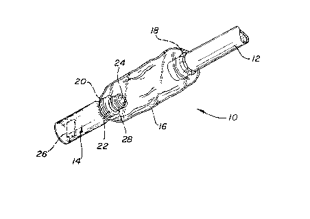
A preferred embodiment of the device to be used in
accordance with the teachinq of the present invention is
shown in Figure 1. The device indicated generally at 10
is shown attached to a ~ube 12. In the device depic~ed in
Figure 1, the means or connecting the vessel 14 to the
end of the tube comprises an e~pandable sheath 1~, one end
of which is securely attached to the vessel, and:the other
end of which comprises an elastic ring 18 making a
releasable attachment about the end of ~he tube. The
sheath 16 may be attached to the vessel in any known
manner an~, as shown in Figure 1, the sheath 16 is
attached to the vessel by a second elastic ring 20
JSU 67
~3~702
--11--
disposed over the lip 22 about opening 24 of vessel 14.
Thou~h the vessel shown is cylindrical, the vessel may
comprise any three dimensional container preferably of
semi-rigid material, having an openin~ therein. The
vessel may be made of, e.g., polyethylene, polypropylene,
glass or any other material which is nonreactive to the
antimicrobial solution of vapor. The sheath may also be
formed of polyethylene, polypropylene or other material
which is relatively nonreactive to the antimicrobial
vapor. The elastic rings may be formed of natural late~
or butyl rubber which are relatively resistant to the
sterilant vapors; however, resistivity is less critical
when the device is constructed for one time use. Disposed
within the vessel may be a substrate 26 comprising a woven
or nonwoven fabric or sponge for containing the liquid
antimicrobial solution. The vessel preferably has a means
28 associated with the opening for attaching a closure cap
over the opening prior to use in ordsr to maintain the
antimicrobial solution therein. As shown in Figure l,
means 28 comprises threads for a screw cap fitting about
the lip of the vessel.
~nother embodiment of the device of the present invention
is depicted in Figure 2 where the device is indicated
qenerally at 30. The means for connecting the vessel 34
to the end of a tubular instrument comprises a bushing 36
disposed within the open end of tha vessel. In the
psrticular embodiment shown in Figure 2, the bushing
comprises a series of rings 38 and 40 of inwardly
estending plastic flaps defininq a fle~ible aperture 32 to
receive the tubular instrument. The 1aps can be made of
any fle~ible material which is non reactive to the
antimicro~ial solution or vapor, such as polyethylene, and
of sufficient thickness that the flaps provide resistance
to withdrawal of a tube inserted through ~he aperture.
JSU 67
-12- L32~ 70~
Disposed within the vessel is a substrate 42 containing
the antimicrobial solution. Preferably, the ves~el 34 is
provided with means 44 for attaching a closure cap thsreto
prior to use. As shown in Fiyure 2, means 94 comprise
S threads for attaching a scrPw cap ~not shown) ~ithin the
opening of the vessel.
Figure 2A illustrates a variation in the design of thP
device of Fig. 2 which utilizes the same basic vessel and
means for attachment to a tubular device. In the device
shown in Fig. 2A, end 45 of the vessel opposite the open
end is provided with apPrture 46 for attaching a
disposable cartridge 47 con~aining a supply of
antimicrobial on a substrate such as a woven or nonwoven
lS fabric or sponge 48 as illustrated. The aperture 46 of
the vessel is designed i~ conjunction with neck 49 of the
cartridge to provide quick and easy attachment and release
of the cartridge and the vessel. In the embodiment shown
in Fig. 2A, aperture 46 is provided with reverse threads
for engaginq the threads of the neck 49 of the cartridge.
In this variation of the device it is not necessary for a
substrate contain the antimicrobial solution to be
disposed within the vessel since the antimicrobial
solution is provided in premeasured aliquots in the
cartridges. With the device o Fig. 2A one achieves the
convenience and accuracy of di~posable, premeasured
aliquots of antimicrobial soIution without the ~xpense
associated with the device of Fig. 2.
The followi~g table sets forth the effectiveness of the
devices depicted in Fi~ures 1 and 2 in a sterilization
procedure described below.
JSU 67
,
:L 3 ~ 2
-13-
Table II-
Effe~t of Devices on Efficacy ~f SteriIi~a~ion
I~side T~b~s
~ff ica~x_(S/So)
No Device Device
Material I~D. Len~t~ Fiq.l Fig.2A
~cm~ (cm)
Surgical Tygon 0.64 10 0
.64 20 4~4 ~ 10 5 - -
0.64 30 1.1 ~ 10 2
~.64 45 8.8 ~ 10~1 ~ 0
Rubber tubing 0.64 2S 1.7 ~ 10 1
0.64 45 7.9 ~ lo~l 0
.
The efficacy as recorded in terms of the ratio of the
number of microorganism~ sur~iving the test, S, to the
number of challenge organisms, S0 (appros. 1 s 106~,
on a paper strip disposed within the tube equidistant from
~he ends. In the sterilization procedure, 100 microliters
o~ 30% agueous H~02 solution was supplied in each of
~5 the devices. The devices w~re attached to the ends of
tubes of the indîcated length and 0.64 cm in internal
diameter.` All of the tube samples were placed within
TYVEK~MYLAR~ packaging prior to sterilization. The
packaged tubes were placed within the sterilizing chamber
and the pressure thereîn was reduced to about 0.1 torr in
about 10 minutes. Additional 30% H202 solu~io~ was
injected into the chamber to achieYe a co~c~tration o~
2.0 milligrams H202 per liter of chamber ~olume.
Followin~ inject;on of the H202, the ~ubes were
retained in the ~hamber an additional 50 minutes.
JSU 67`
: ~ : : : :
~2~02
-14-
Injection of the H202 solution raised the pressure in
the chamber to about 6 torr and the pressure was again
reduced to about 0.1 torr. During the last 10 minutes of
e2posure, low temperature gas plasma was generated in the
chamber at 300 watts. The challenge micro organisms used
in the test were Bacillus subtilis (var. globigii) spores.
As shown in Table II above, when the tube length was only
10 centimeters, sterilization was achieved without thP use
of the device according to the present invention.
However, for tubins of 20 and 30 centimeters in length, a
device of the present invention would be needed in order
to achieve sterility within the e~posur time of the
test. For tubes of 45 centimeters in len~th, total kill
was achieved during the 1 hour esposure time of the test,
using either of the devices depicted in Fi~ure 1 and
Figure 2.
A further experiment utilizin~ 1 mm medical grade Teflon
tubing 183 cm in length. The tubing was cut into three
pieces to obtain a 5 cm long center sectio~ which was
joined to the end sections by external tubing connectors.
In the e~periment, appro~imately 1.0 ~ 104 Ba~illus
subtilis ~var. globigii~ spores were deposited in the
center section of the Teflon tubing, and the tubing
assembled and subjected to sterilization with hydro~en
pero~ide vapor as described above at a concentration of
2.0 mg/liter of chamber volume. The chamber was evacuated
to a pressure of 0.1 torr before the pero~ide was injected
as a 30% aqueous solution and allowed ~o vaporize. After
20 minutes, a continuous gas plasma was generated in the
chamber at 300 watts 13.5 MgH2 and the sterilization
continued for an additional 5 minutes after which the
vacuum was released with sterile, filtered air, and the
number of surviving spores determined.
J~U 67
~3?,~ 7~2
-15-
The e~periment was first conducted without a device of the
present invention attached to the tubing, then repeated
with a device of Figure 3 as described below containing
100 ml of 30% hydrogen peroxide attached to one end of the
tubing. The e3perimental results of the tests are
presented in Table III below.
Tabl~ III
S~eriliz~a~_Qn of 1 mm Tubing
_ Ef~ica~y ~S/SQ~
~aterial 1~1 Len~h ~o ~evice ~i~. 1 Device
Teflon 1 mm 183 cm 1.9 ~ 10 1 0
The data of Table III demonstrate the efficacy of the
method of the present invention in sterilizing the lumen
of very long tubes having very small diameters as often
used in certain endoscopic procedures.
Addi~ional embodiments of the device of the present
invention are depicted in Figures 3 and 3A. The device
shown in Figure 3 indicated generally at 50, comprises a
vessel 52 in the form of a pouch constructed of a fle~ible
material. The means for connecting the vessel or pouch 52
to the end of an instrument tube comprisas a first
drawstring 54, and preferably a second drawstring 62.
These drawstrings are preferably arranged in the
con~iguration as shown in Figure 2 to be drawn from
opposite sides o the pouch. The pouch is pre~erably
provided with an airtight seal ~o maintain the
antimicrobial solution therein prior to use, and in~lu~es
a means for creating an opening in the saaled pouch so
that it may be disposed over the end of a tube. The seal~
may be created by sealing the ends 66 of the pouch, and
JSU 67
.
. .......... . ... . .. . ...
:~ 3~02
-16-
the means for opening the sealed pouch may co~prise, for
e ample, a line of weakening at 68, preferably in
combination with a notch also sh~wn generally at 68, to
. permit the pouch to be ope~ed by tearing off one end.
_ 5
Figure 3A shows a device indicated generally at 50A,
similar to device 50, but wherein the airtight seal and
the means for creating and opening the sealed pouch is a
line of fastening 64 similar to a ~zip-lock~ closure.
Optionally, opening flaps 70 may be provided on either
side of the pouch adjacent closure 64 of Figure 3~, or the
line of weakening 68 of Figure 3. These flaps are firmly
secured to the pouch. ~n use, after the sealed end 66 of
~~ the pouch o Fig. 3 has been removed along the line of
weakenin~ 68, th~ flaps when pulled oppositely from each
other will distend the opening of the pouch for disposal
around the end of an instrument tube. The flaps of Fig.
3A, when pulled in opposite directions, can be used to
open the zip-lock fastening, or if ~he fastening is
already opened, to distend the opening for disposal around
the end of an instrument tube. ~ substrate 72 such as a
woven or nonwoven fabric or sponge may be disposed within
the pouch for containing the antimicrohial solution.
In a preferred construction, the drawstrings are provided
with a locking means ~s illustrated. Though many means
for locking or catching a drawstring are known in the art
and may be used in conjunction with the present invention,
the locking means depicted at 56 at Figure 3 comprise a
catch 60 ~or a serrated edge 58 provided on the
drawstring. As shown in Figure 3, the catch, compris;ng
_ an opening for disposin~ one end of the drawstri~g
therethrough, is located at the opposi~e end o~ the
drawstring. The catch, however, may be proYided by a
flap, with opening ~herein, attached to the ~dge of the
JSU 67
.. . . . ~ . . . ~ . . . . . ..
~2~ 7, ~
-17-
pouch, provided the other end of the drawstring must also
be attached to the pouch. When two drawstrings are used,
one or both drawstrings may be provided with a locking
- means. By pulling the end 73 of the drawstring, the
fle~ible pouch is gathered and a firm fastening may be
made to a tube inserted within the pouch.
i Although the present invention has been described in terms
of specific devices for use in a preferred method of vapor
sterilization, it will be understood that various
modifications in the device and method will be apparen~ to
those skilled in the art and are within the scope of this
invention.
-
. _ .
JSU 67
,