Note: Descriptions are shown in the official language in which they were submitted.
l323~3a
METHOD AND APPARATVS FOR ANALYSING AN ELECTRO-
ENCEPHALOGRAM
This invention relates to the analysis of an electro-
encephalogram (EEG). The invention enables a computer to be
used for part of the analysis.
The EEG is a long-established techni~ue for recording
spontaneous electrical brain activity using electrodes
attached to the scalp of the subject. Its use in connection
with monitoring the development of, for example, new born
infants and especially ~remature new born infants has enabled
the brain development of the infants to be monitored during
the neonatal period. In recent years, small magnetic tape
cassette recorders, such as the Oxford Medical Ltd.'s Medilog
4-24, have been used to record up to 24 hours of EEG data on
two channels from very small sick babies, because the small
size of the recorder has enabled it to be placed in a
neonatal intensive care unit without interfering with the
other apparatus used in such units. Such recorders provide
enormous amounts of data or re~rospective analysis, at
Present performed by replaying a tape on to a visual display
unit at, for example, 20 or 60 times its recorded speed.
This enables a 24 hour recording to be reviewed in a minimum
of 24 minutes and can enable a specialist to detect
abnormalities in the EEG and to take necessary remedial
action. The EEG can also be printed whilst it is being `
displayed to enable the specialist to return to parts of the
~G which he suspects reveal abnormalities. ~n obvious
disadvantage of this system is its retrospective nature, but
it also suffers the disadvantage that the specialist is
oocupied for lonq periods of time reviewing the EEG traces. ~`
~ lthough gross abnormalities in an EEG, such as
convulsions or periods of substantially no activity can be
quickly deteated by the specialist observin~ the visual
display, the EEG can also reveal more subtle, though still
very important, information after longer and expert analysis
of the printed trace of the EEG.~ Th~ visual analysis is
.
~ ' :
.
.:
.. ' - :' . ~ '.
~323~3~
2 20648-1389
based on several well recognised features such as the frequency
and amplitude of the waveforms, ~he symmetry and synchrony of
discharges from the right and left sides of the brain, and the
presence of abnormal discharges such as convulsions.
In contrast to ~he normal adult EEG which shows
continuous electrical activity, the EEG of prematura infants is
characterized by short bursts of activity, some~imes of less than
5 seconds duration, interspersed with intervals of very reduced or
apparently absent electrical activity. The intervals between the
bursts, which may last for 60 seconds or more, are of significance
in that they can show the increaæing maturity of the infant brain
as the activity becomes more continuous with longer bursts and
shorter intervals. Prolonged intervals relative to the age of the
infant can be associated with intracranial haemorrhage or hypoxic
brain damage. Obviously ~he more rapidly ~hat the EEG can be
analyzed, the sooner remedial action can be taken when required
and possibly the severity of damage to the brain reduced.
The EEGs of older children and adults may also display
intervals of reduced or apparently absent elec~rical activlty due
to the effects of drugs, anaesthet~cs or injury.
It is an ob~ect o~ the present invenkion to provide an
lmproved method and apparatus for the analyæis of E~Gs.
According to one aspect of the present invention there
is provided apparatus for analyzing an electro-encephalogram (EEG)
in ~hich electrical signals derived from the EEG are applied to a
threshold detector and threshold output signals are examined for a
particular kind of brain activity, comprising,
input terminals for receivin~ at least two electrical
.
,
,
~3~3~3~
3 20648-1389
signals respectively representing at least ~wo channels of EEG
whlch may be derived from different sides of a patien~'s head,
wherein the two electrical signals are analyzed substantially
simultaneously; ~
timing means for measuring time intervals between
successive output signals produced by the threshold detector and
for providing output values representing the time intervals;
salecting means for selecting only those output values
that represent time lntervals longar than a predetermined minimum
time;
adding means for accumulating the selected output values
representing time intervals occurring wi~hin a section of the EEG
obtained during a time period of predetermined duration to produce
a total value; and
display means for producing a visible output including a
plurality of to~al values derived re~pectively from the BEG
channels side by side obtained during consecutive time periods of
the predetermlned duration.
When a time interval between successive output æignals
overlaps the boundary between two time periods, the parts of it
occurring in the tlme periods are allocated to khe output values,
respec~ively~ unless the part of an interval which occurs in the
~irst period i5 of short duration (e.g. less than 6 seconds) when
that part is not included in the total for the first period.
The apparatus may also include counting mean to coun~
the numbers of intervals occurring ~ithin the time periodæ. The
total values when dlvided by the numbers of intervals provide the
mean interval lengths for the periods. The vislble output may
1~,
- '
' , ~ - ' '
'-,'. '
' ' :' ' :
~323~30
4 20648-138
comprise representations of ~he mean interval lengths.
Preferably the two channels of EEG are obtained one from
the left-hand side of a patient s head and the other from the
right-hand side, to be analyzed substantially simultaneously by
the apparatus, so that the synchrony and the symmetry of the EEGs
can also be monitored by the apparatus. The representatlons of
the total values derived fro~ both EEG channels are displayed side
by side to facilitate their being compared with each other. More
than two channels of EEG may be obtained from the head, and/or
l~ ~hey may be derived from regions other than simply ~he left-hand
and rlght-hand lobes; of aourse, these E~G signals may display
~ore co~plicated relationships than those represented by synchrony
and symmetry.
The comparator means may simply compare the amplitude of
the electrical signal with a reference voltage and produce a pulse
each time the electrical signal amplitude rises to reach or exceed
the re~erence voltage, the pulses forming the indications which
are applied to the timing means.
In an alternative construction the comparator meanæ
~0 includes an analogue to dlgital converter arranged to sample the
electrical signal at a sufficiently high ~requanay to detect any
pulses likely to occur in it and to convert the magnitudes of the
samples into multi-bit digital slgnals. The comparison with a
thrashold level may be performed by comparing the multi-bit
dlgital signals wlth a digital representation of the threshold
level in a digital aomparator. Another way of effeating the
co~parison is to ahoose the reference voltages for the analogue to
digital conversion so that one of ~hem corresponds to ~he
. ' ' :
~ 3~3l~3~
~ 20648-1389
threshold level; the resulting digital signals have l s in places
at or above that corresponding to ~he threshold level only if the
electrical signal is at or above the threshold level, so that the
comparison can be effected by detectlng a "1" in the place at or
about that corresponding to the threshold level.
The timing means, selecting means and adding means may
be provided by a digital computer programmed so as to execute the
required operations on the indications produced by the comparator
means to derive from those lndica~ions the requixed total values.
If the timing means, selecting means and adding means
are provided by a sui~ably programmed digital computer, then the
multi-bit digital signals obtained from the analogue ~o digital
converter included in the compara~or means may be applied to the
computer as inputs, and the computer may also he pro~rammed to
determine uhether the digital inputs obtained from the converter
are above or below the threshold level.
Where a digital computer is presentr for example, as
mentioned ahove, it may be programmed to process the total values
obtained from the addlng means to produce a graphical display o~ a
~0 form readily assimllable by a specialist.
The EEG channel from which the eleatrical signal is
derived may be recorded on a suitable magnetic tape and replayed
at` a much hi~her speed than that at which it was obtained irom ~he
patient. For example, the EEG signal may be recorded directly
from the patient for a period of 24 hours and replayed to the
analyzing apparatus over a period of 24 minutes. Of course,
ad~ustment must be made for the compression of the time scale ln
setting the time intervals used in the analysis.
' ~
~32343~
6 206~8-1389
Alterna~ively, the E~G signal~ ob~ained from the patient
may be applied directly to the apparatus so that it can be used in
the "on-line" monitoring of the patient. The apparatus may be
arranged to generate alarm signals in response to fea~ures of the
EEG ~hich might require some kind of remedial action.
The apparatus, especially if it includes a ~uitably
programmed digi~al computer, may be able ~o proces~ an EEG
obtained directly ~rom a patient very much more rapidly than
avents are likely to occur in the EEG. Therefore EEGs from
several patients may be multiplexed to a single appara~us which
may also be arranged to produce a separate display ~or each
patient and to monitor the EEGs for features requiring remedial
action.
According to a second aspect of the present invention
there is provided apparatus for analyzing an electro-encephalogram
(~G) using the amplitude of the EEG as a criterion in assessing
the brain activity comprising,
sampling means for sampling electrical signals derived
from plural EEG channels at regular time intervals from both sides
of the head of a patient and holding the samples;
analog to digital conversion means connected to receive
the samples held uccessively by the sampling mean~ and producing
t~arefrom digital outputs representing the magnitudes of the
samples~
a digital comput~r programmed to receive the digital
outputs associated with each of said plural ~9G channels ln
su~cession from the analog to digltal ~onversion means and having
means for processing substantially simultaneously the digital
~i
,
~3~3~3~
7 20648-1389
outputs from each of the plural EEG chan~els including:
means for ~omparin~ digi~al ou~puts from each with a
threshold value;
mean~ for detarmining a time interval between current
digital outputs and immediately preceding digital outputs which
exceed the threshold value, and
means for accumulating ~otal ~ime intervals exceedin~ a
minimum duration during a particular time period and generating
signals representative of total ~ime in~ervals acaumulated during
lQ consecutlve time periods; and
display means for displaying side by side ~he
representative signals for each channel.
The representation may be o~ the mean interval duration.
Preferably the apparatus is arranged to analyze at
substantially the same time several channels o~ ~G obtained
respectively from the left and right sides of the head of a
patient and to produce on the display means corresponding displays
side by side. It may be arranged to analyze more or other
channels of BEG derived from the patiant.
~0 The reading o~ the time interval ~rom the timer may be
follo~ed immediately by resetting the timer to zero and restartlng
it when next a sample is below the threshold value.
The digital computer may also be programmed to store
lo~al maxlmum and minimum values represented by the dlgital
outputs of the analogue to digital conversion means ~or analysis
and to output to the display means data resulting from that
analysis.
The digital computer may also be arranged to calculate
- -
. ,' ,
~L323~3~
7a 20648-138g
the mean of the squares of the sample amplitudes during a time
period and count the number of time intarvals of ~he minimum
duration or longer which occur in a tlme period. Statistical
analysis of the da~a may permit, for example, ~he correlation of
the squares of the ampli~ude~ of the le~t and right EEGs and the
correlation of the squares o~ the ampli~udes with the durations of
the time intervals.
According to a third aspect of the present invention
there is provided a method of analyzing an electro-encephalogram
~BG) in which the amplitude of the E~G is used as a criterion in
assessing the brain actlvity, wherein the EEG is represented by a
plurality of digital values respectively corresponding to the
amplitude of the EEG signal at a succession o~ instants spaced
apart by predetermined e~ual time periods, comprising,
comparing sequentially the digital values with a
threshold value;
indicating whether or not the digital values exceed the
threæhold value;
measuring each time interval durln~ which successive
digital values are smaller than the threshold value;
summing the measurements of the time lntervals having at
least a certain minimum duration during an extended ~ime period to
provide a total of the measurements for each of a plurality o~
consecutive extended time period~; and
producing an output di~play dependent on ~he totals of
the measurements.
The measurements of tlme durations may b~ related to a
clock oæcillator used to control the application of the digital
~,~'` '.
~ 323'13~
7b 20648-1389
values for comparison.
The output display may include a graphical display of
the values of a plurality of the totals represanted on a
rectangular or other type of coordinate system.
The method may include the determination of the mean of
the squares of the digital values occurring during an extended
time period.
T~e method may al~o include the counting of the number
of ti~e intervals added to the total during an extended time
~eriod, and the calculation of the mean of the time tntervals
during khe period.
An example of ~he lnvention will now be desaribed with
reference to the accompanying drawings, of which:-
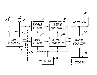
FIGURE 1 is a block diagram of one ~orm of theapparatus;
FIGURE 2 is a flow diagram o~ an example o~ so~e of the
operations performed by the computer in the apparatus of Figure l;
FIGURE 3 shows the left and right channel traces of the
EEG of a premature infant of about 28 weeks post mens~rual age;
~0 FIGURE 4 shows the le~t and right channel traces o~ the
EEG of a premature lnfant of about 32 weeks post menstrual age;
FIGUR~ 5 is a plot of the aggregates of the intervals
between burst~ ln successive periods of an EEG channel trace o~
the kind shown ln Figure 3;
FIGURE 6 is a plot o~ the aggregates of the intervals
between bursts in successive periQds of an BEG channel trace of
t~e kind shown in Figure 4; and
- , , ': ' ,, , :
~32~3~
7c 20648-1389
FIGURE 7 is a plot similar to Figure~ S and 6 showing
the change brough~ about by recti~ication of hypoxia in an in~ant
of about 32 weeks post menstrual age.
:. ~
`. ~, ` ' . '
~323~3~
Referring now to Figure 1, the apparatus shown has
probes 1 and ~ respectively for connection to the left and
right sides of the head of a Datient. The Drobes are
connected through amplifiers (not shown) and cond~ctors 3 and
4 respectively to two inputs of a magnetic tape recorder 5
for recording the channels of the EEG Picked up by the
probes. On playbackr the recorder 5 produces left and right
outputs on conductors 6 and 7 which are applied to sample and
hold circuits 8 and 9 respectively. The outputs of ~he
sample and hold circuits 8 and ~ are fed ~o analogue to
digital converters 10 and 11 respectively which su~ply binary
coded digital values to a digital computer 12. A clock 13 is
connected to supply ~iming pulses to the sample and hold
circuits 8 and 9, the analogue to digital converters 10 and
11 and the computer 12. The clock 13 may be synchronised
with the pla~back of the tape in the recorder 5 via a
connection 14. The computer 12 is connected to a keyboard 15
and to display apparatus 16 which may, for example, include a
cathode ray display tube and a printer.
Optional direct connections from the probes 1 and 2 to
the sample and hold circuits 8 and 9 are indicated by broken
lines. Such direct connections enable the apparatus to
analyse the EEGs on-line.
The use of sample and hold circuits may be unnecessary
for certain types of analogue to digital converter.
The program of the digital computer 12 includes a part
represented by the flow diagram of Figure 2. From Figure 2
it can be seen that the program is arranged to select inputs
~rom the left and right channels of the EEG alternately~ '
Both sets of inputs are treated in the same way, the ~ `
operations on the left EEG channel being shown. The
magnitudes o~ the succe~sive inputs are compared with a
threshold value Vt. If an input is less than the threshold
value the previous input relative to the threshold is taken
into account~ If the previous input is smaller than the
threshold value no further action is taken, but if it is
greater than the threshold value a timer in the computer is
started. 1he timer is stopped wh~n the next lnput havin~ a
,.
.
~ '. ` . ` ` '.', ~ ' ' `. '
.
.
.. . : . ' . . .
..
~323~3~
magnitude qreater than the threshold value occurs. The time
is then read and the timer reset to zero. The time read is
compared with a threshold time Tt and if it is less then that
time it is discarded, but if it is greater it is added to a
total time being accumulated for a period. In one example
the period is 10 minutes which as explained below may be
represented by 10 seconds of clock time within the computer.
If the interval recorded by the timer crosses the boundary of
one period into the next, the time duration of the interval
is allocated to both periods according to how it was divided
by the boundary between the periods. If, however, the part
of the interval to be allocated to the first of the periods
is less than a time St (say 6 seconds) then that part of the
interval is not included in the total for that period. The
total times accumulated for ~he periods are produced as an
output and the means accumulating the time for each period is
reset ready for the next period.
The apparatus of Figure 1 using the program represen-
ted by Figure 2 analyses left and right channels of the EEG
of a patient using measurement of the interval of time
between bursts as a basis.
The aggregates of the interval times for a succession
of periods produced in this way are displayed in a form
easily assimilated by a specialist, so that he can tell very
guickly whether or not the brain activity is normal. The
mean of the interval times for a period may be displayed
instead; for this purpose the computer could count the number
o~ in~ervals in each period.
Figure 3 shows sections of left and right EEG channel
traces for an infant having post menstrual age of about 28
weeks. Consideration of Figure 3 reveals that both left and
right EEGs consist of bursts of electrical activity followed
by intervals during which the activity is of much smaller
amplitude or is apparently absent.
The bursts in both EEG traces are similar in form and
duration and occur more or less simultaneously. If the
bursts are not similar and substantially simultaneous, this
may be an indication of some kind of disorder. EEG channels
~323~30
from other parts of the head may have other relationships.
Figure 4 shows the EEG traces for an infant 3 to 4
weeks older than that fro~ which the EEG traces of Figure 3
were obtained. It can be seen from Figure 4 that the bursts
of activity which are of longer duration sometimes continuing
for a minute or more without significant interruption and the
duration of the intervals between bursts is considerably
reduced.
~ difficulty encountered in analysing EEG channel
signals of the kind shown in Figure 3 lies in determining
when a burst of activity has ceased and when the next burst
has started so that the duration of the interval between
bursts can be determined accurately. The technique ado~ted
in the apparatus of Figure 1 involves noting every time that
the sample exceeds a threshold value, the value used
corresponds to an EEG voltage of approximately ~25
microvolts. Whenever an inpu~ first falls below the
threshold value after having been above it a counter is
started, but if an input exceeding the threshold value occurs
within a short time of the counter being started it is judged
that the input is part of the same burst of activity as the
earlier ones and the input below the threshold value did not
mark the beginning of an interval between bursts. The
threshold time used for this purpose is 6 seconds. The
counter having been started is stopped by the next input
exceeding the threshold value and if the time is less than
the threshold time the output of the timer is discarded. If,
on the other hand, the next input exceeding the threshold
occurred more than 6 seconds after the counter being started,
it is considered to be part of a different burst and the
output of the timer is retained. The retained time is added
to the total of other intervals previously occurring during a
measurement period until the end of that period. The
measurement period is, as mentioned above, 10 minutes of real
time. Each time the counter is stopped and the time recorded
is read out, the counter is immediately reset to zero.
An alternative method of measuring the interval
durations is to stop, read, reset and restart the timer at
.
:~323~3~
11
each occurrence of an input exceeding the threshold value.
This would have the effect of increasing each measured
interval duration by the time between successive samples,
which may, if desired, be compensated either by subtracting
that time from the time recorded or by delaying the starting
of the time appropriately.
~ s mentioned above, the EEG channel signals from
opposite sides of the patient's head ma~ have bursts of
activity which are similar in form and duration and which
occur more or less simul~aneously. This phenomenon is termed
"synchrony~ The apparatus can be arranged to check two or
more channels of the EEG for synchrony, by monitoring the
finish and~'or start times of the intervals between bursts
(indicating the starts and/or finishes of the bursts) in the
different channels and measuring the differences in time of
occurrence and/or duration of the bursts. If the bursts in
the channels occur within a certain time, say 2 seconds, they
are counted as synchronous and if they are separated by more
than that time they are counted as asynchronous. The numbers
of synchronous and asynchronous bursts occurring in each time
period are counted and displayed.
Occasionally a single electrical noise pulse is picked
up by a probe because of the sensitivity of the apparatus to
small voltages; the apparatus may be programmed to ignore
such a pulse in comparing the EEG channel of one side with
that of the other. On the other hand, the technique
described above for identifying the start and finish of an
interval will recognise a single pulse as a burst and divide
the interval into two. Although this can lead to a slight
reduct~on in the aggregate of interval times, it is not
likely to result in a significant change in the average value
of the aggregates of interval times as displayed~.
In order to utilise more fully the processing power of~
the computer and to enable it to analyse, for example, 24
hours' recording of EEG channel signa~s in a relatively short
period of time, the recorder 5 is arranged to replay the tape
at a much higher speed than that at which the recording was
made. Typically, the recordlngs~are r~played at 60 times the
.
.
~23~3~
12
speed at which they were made so that inside the computer the
time periods referred to above are divided by a factor of
60. Even greater speed-up of the recorded EEG sianals may be
employed, Provided that the replay system is capable of
ooeration at the speeds and the frequencies involved.
The EEG signals may be sPeeded up after sampling and
conversion to digital form by storing the samples in a random
access memory at one rate and reading them out at another
rate. This may be done instead of or additionally to the
speeding up of the magnetic tape recorder.
Although in general the clock oscillator in the
computer and the tape recorder speeds will be sufficiently
stable to permit the computer clock oscillator to provide an
accurate measurement of the real time periods in the EEG as
originally recorded and to act as a reference for the
operations executed by the computer as described above, when
the EEG has been pre-recorded and is played back by the tape
recorder it may be desirable to adjust the frequency of the
clock oscillator in accordance with the rate at which the EEG
signal is read out by the recorder. This con~rol is
represented by the broken line 14 in Figure 1 and may involve
the use of a timing track on the tape producing pulses which
adjust the ~requency of the clock oscillator 13 shown to be
external to the computer for convenience of illustration.
This oscillator determines the fre~uency of sampling by the
sample and hold circuits 8 and 9 and the operation of the
analogue to digital converters 10 and 11. The oscillator 13
provides the time reference for the counter and the other
timed operations which are carried out in the computer.
It has been found that in practice the use o~ a
voltage threshold o~ approximately +25 microvolts and a
threshold time of 6 seconds as described above to distinguish
between bursts and intervals between bursts used in the ;'
manner described above, results in a mean duration for the
intervals which corresponds very closely to that obtained by
a specialist measuring the time intervals directly from the
EEG traces. It would of course be ~o sible to use other
values for the threshold value and the threshold time,
~323~3~
although it is likely that such values will be close to the
ones given above.
The aDparatus of Figure 1 may be constructed in the
~orm oE a purpose-built microcomputer with addi~ional
circuitry for use as a monitoring unit for an individual
patient sampling continuously the EEG signals obtained
directly from the patient. In such a case the probes 1 and 2
are connected directly through amplifiers to the sample and
hold circuits 8 and 9 and the time periods in the computer
adjusted to the real time values rather than the accelerated
time values used when the computer i~ analysiny a speeded-up
replay of recorded EEG signals. The tape recorder 5 may
still be retained to record the EEG signals as well, so that
they are available for subsequent examination by a specialist
f necessary.
Advantageously but not necessarily exclusively when
the computer is used to analyse the EEG signals received
directly from the patient, the computer may be arranged to
identify alarm conditions which may occur, for example, if
the intervals between bursts become excessively long, (longer
than 90 seconds, say) or if the aggregate of the intervals
between bursts in a 10 minute period exceeds a predetermined
proportion of that period depending on the condition of the
patient. Other conditions re~uiring attention from the
specialist could also be de~ected, for example if bursts
occur in the EEG signal from one side of the head and are not
matched by bursts from the other side at substantially the
same time ~excluding isolated noise pulses)~ The samples
received by the computer can also be tested to see whether
the amplitude of the bursts is above or below a pre-set
limi~, and also to monitor the mean duration of the
intervals.
Instead of using a sample and hold circuit feeding an
analogue to digital converter to produce the digital inputs
to the computer from an EEG signal, the EEG signal m~y be
applied to a simple analogue threshold circuit producing a
~1 n output whenever the amplitude of the EEG voltage exceeds
a threshold value, say 25 uV, and a "0" output at other
~2~.~3~
14
times. The ~om~uter may be prog~ammed either to poll the
output of the threshold circuit repeatedly at short intervals
to detect the l's and O's or to receive the transitions from
"0" to "1" and "1" to "0" as interrupts. When the computer
receives a "1" to "0" input it starts the timing counter as
described above to determine whether or not it marks the
start of an interval between bursts. Such simpler apparatus
can produce a display of the aggregates of interval times and
the mean interval durations, bu~ cannot monitor the actual
voltages occurring in the EEG signals.
The form of the display produced by the computer is
preferably graphical so that, if the aggregates of the
interval times during the periods lie outside an expected
range for the age of infant concerned, this fact will be
immediately apparent to a specialist and he can initiate
remedial action without delay. Figures 5 and 6 show a
possible form for the display in which the aggregate times
are plotted as points on a graph against the minutes of the
periods to which they relate.
~ glance at Figure 5 shows that the average value of
the aggregate times varies between 100 and 2~0 seconds with a
maximum of about 360 seconds and a minimum of about 10
seconds. This appears to be fairly typical for an infant of
28 weeks post menstrual age.
The infant of which Figure 6 is a display of the
aggregate times is of 32 weeks post menstrual age~ It can be
seen that for many periods the aggregate times are zero and
the maximum aggregate time is about 150 seconds.
A particular use of the apparatus is revealed by
Figure 7 which shows a display of the same type as is shown
in Figures 5 and 6, but for an infant of about 32 weeks post
menstrual age. The normal display obtained from the
apparatus for an infant of this age is shown in Figure 6, and
it is immediately apparent that to the left of the position
marked by the arrow A in Figure 7 the aggregates of the
intervals are very much higher than they should be for a
healthy infant. This occurred when the infant was hypoxic,
and when the oxygen su~ply was increased at the time
.
.,
.
- . - . -
'' ' ' . .'' '''' '" '' "' .. :,', . '. ... . .
., .: ", , , ' . , '
indicated by the arrow ~ it can be seen that the aggreqates
immediately came down to the sort of val~es displayed by
Figure 6.
The analysed or partly-analysed data may be stored in
a semipermanent memory, such as a magnetic disc, for
subsequent display without any further processing or for
further analysis and display.
The display may take other forms and may also include
indications of the following in graphical and/or numerical
form for each side or for both sides of the brain:
1. the peak value of a burst in the EEG as
represented by a sample.
2. the length of the longest interval time in
each period.
3. the number of intervals in each period.
4. the mean of the interval times in each
period and for the entire EEG.
5. difference in amplitude and`timing between
the bursts in the EEGs of the two sides.
6. correlation between right and left mean
squares of the amplitudes.
7. correlation between the squares of the
amplitudes and the interval length.