Note: Descriptions are shown in the official language in which they were submitted.
-` 1 323537
ANGLED HOTT VENTRICULAR CATHETER
AND APPARATUS FOR MAKING SAME
TECHNICAL FIELD
The present invention relates to a ventricular
catheter having specifically angled apertures which
facilitate access to or drainage of cerebral spinal fluid and
certain methods of making and using same.
BACKGROUND OP THE INVENTION
The four ventricles of the human brain are
interconnected cavities that produce and circulate cerebral
spinal fluid (CSF). Procedures involving ventriculostomy,
i.e., placement of a catheter into the ventricular system of
the brain, form a major part of a neurosurgeon's clinical
practice. General areas of application of ventricular
catheter placement include intracranial pressure monitoring
~ICP), draining or shunting of CSF and the instillation of
pharmacological therapeutic agents.
CSF drainage is essential for patients with congen-
ital or acquired hydrocephalus. CSF drainage, which can only
be performed with an intraventricular catheter, is a life-
preserving procedure, because it can immediately reduce
intracranial pressure. The ventricular catheter, used to
drain CSF, is connected to a peripheral subcutaneous drainage
system, i.e., to the peritoneal cavity or systemic
circulation via the heart or in the case of ICP to an
external drainage collection system. Standard procedures
for ventricular catherization are disclosed in the textbook
literature. See, for example, Neurosurgery, edited by Robert
H. Wilkins and Setti S. Rengachary, Section A, Chapter 13,
Techniques of Ventricular Puncture (McGraw Hill 1984).
The most frequently chosen site for ventricular
catheterization is coronal. In most cases, a catheter is
inserted in the anterior horn of the lateral ventricle
through an orifice or burr hole drilled just anterior to the
coronal suture in the midpupillary line of the cranium, i.e.,
1 323537
-2-
in the frontal bone over the ventricle. The burr hole, only
slightly larger than the diameter of the selected catheter to
insure a snug fit and provide a seal against CSF leakage, is
placed approximately 1 cm anterior to the coronal suture,
approximately 10 to 12 cm above the nasion, and approximately
2 to 3 cm from the midline over the nondominant hemisphere.
After the burr hole is made, the dura and underlying pia-
arachnoid are opened and coagulated, for example, with afine-tipped blade after cauterizing the dural surface.
The lateral ventricles of the human brain form an
arc parallel to the arc of the cranium, i.e., the contour of
the lateral ventricles parallels the arc of the surface of
the skull. Thus, a catheter guided perpendicular to the
cranial surface at the point of entry into the cranium will
enter the ventricular system. Specifically, any line pene-
trating a burr hole in the surface of the skull at a 90
angle also bisects the lateral ventricle.
A more recently developed procedure to ensure cor-
rect catheter placement is disclosed in U.S. Patent No.
4,613,324. The apparatus comprises a guide assembly which,
when positioned over an orifice drilled in the cranium above
the anterior horn of the lateral ventricle, guides a catheter
and obturator through the orifice and into the lateral ven-
tricle at an angle normal to an imaginary plane formed by a
tangent to the cranium at the orifice, while the correspon-
ding method comprises providing an orifice in the cranium
just anterior to a coronal suture in a midpupillary line of
the cranium and inserting a ventricular catheter containi~g
an obturator through the orifice towards a lateral ventricle,
wherein the catheter containing the obturator is guided
through the orifice, by means of a novel guide assembly, at
an angle normal to an imaginary plane formed by a tangent to
the cranium at the orifice.
"-
1 323537
A wide variety of catheters are known in the priorart for the Furpose of penetrating the ventricular cavity.
Such catheters are typically in the form of a hollow tube
which is provided with a plurality of apertures at the
ventricular or inflow end to permit the passage of CSF from
the brain into the catheter and thence to the bloodstream or
peritoneal cavity of the patient or to an external drainage
system. However, malfunctions frequently occur with such a
catheter due to the blockage of the apertures in the inflow
end of the catheter. Such blockage is usually caused by the
growth of choroid plexus or ependymal tissue within the
ventricle into the apertures in the inflow end of the
catheter. This tissue may block the apertures in the inflow
end of the catheter in a relatively short period of time
after the catheter has been inserted into the ventricle
thereby rendering the cathether inoperative in relieving
excess pressure due to the build-up of CSF within the
ventricle. Furthermore, prior art catheter apertures are cut
perpendicular to the length of the catheter, thus causing
abrasion of brain tissue when the catheter is inserted.
The likelihood of ventricular catheter malfunction
by aperture plugging with brain tissue can be lessened by
angling the aperture holes in the wall of the catheter such
that there is "no see through" flow from the outside to the
inside of the lumen. Also, by positioning the rows of
apertures 120 apart there is essentially no chance for
direct ingrowth of ventricular tissue therethrough. In
addition, the apertures are angled away from the direction of
the insertion of the catheter into the brain thus lessening
the chance of brain abrasion. Further, by slightly
stretching the catheter by means of the stylet (which is
integral to the catheter and used for placement of it into
the brain) the holes will close so that no opening will be
~ :" ~''
~:
1 323537
-4-
..
visible during the placement thereof, with the holes re-
opening after the tension on the catheter is relieved by
removal of the stylet.
As such, it would be desirable to provide a
catheter which overcomes the problems of previously devised
ventricular catheters which are emplaceable within a
ventricle of a human brain to control the flow of excess
fluids to or from the brain. The present invention provides
a simple solution which resolves the problems of prior art
catheters in a novel and unexpected manner.
SUMMARY OF THE INVENTION
The present invention relates to a catheter for
placement into the ventricular system of the brain of a
subject comprising a flexible elongated body having a wall
thickness sufficient to contain and transport fluid therein.
The body has a forward end and tip for insertion into the
ventricular system and a plurality of spaced apertures
located in the forward end of the body spaced from the tip.
Each of the apertures extends through the wall thickness at
an angle such that a portion of the wall thickness is visible
when viewing the aperture perpendicular to the axis of the
body. This arrangement facilitates closure of the apertures
by slightly stretching the body with a placement stylet to
minimize abrasion of brain tissue upon insertion of the
catheter. This arrangement also helps prevent choroid plexus
tissue from growing into the catheter apertures, thereby
providing improved flow of fluid into or from the ventricular
system.
Preferably, each of the apertures extends through
the wall thickness at an angle of about 35~ with respect to
the longitudinal axis of the body, and a plurality of
apertures are aligned in a number of rows. Also, rows of
~5~ 1 323537
..
these apertures are spaced 120~ apart around the
circumference of the body for maximizing the structural
integrity of the catheter forward end.
If desired, the forward end of the body can be made
of a radioopaque material at least in the area surrounding
the apertures to facilitate monitoring of catheter placement.
The body may include means to indicate the depth of
penetration of the catheter forward end to assist in proper
placement thereof. These depth penetration indication means
may be markings of a radioopaque material to facilitate
monitoring of the placement of the catheter.
The invention also relates to method of accessing
CSF in a ventricle within a human cranium which comprises
drilling an orifice in the cranium just anterior to a coronal
suture in a midpupillary line of the cranium, and guiding a
catheter through the orifice by means of a guide assembly in
a direction perpendicular to an imaginary plane defined by a
tangent to the cranium at the orifice. The catheter thus
accurately penetrates the ventricle on the first insertion
with minimal abrasion of brain tissue. The catheter,
described above, i.e., one having the appropriate positioning
and configuration of apertures to minimize ventricular tissue
growth thereinto, is preferred so that increased flow of
fluid to or from the ventricle is obtained.
.- The catheter of this method utilizes a guide
assembly comprising tubular means and support means for the
tubular means. Therefore, the method further comprises
placing the support means so as to rest unsecured on the
human cranium in surrounding spaced relation to the orifice,
and guiding the catheter through the tubular means, into the
orifice and into the ventricle. The support means and
tubular means are related to each other such that the
catheter is guided through the orifice by the tubular means
:, : :. , .
::
- ,,, --
1 323537
in a direction perpendicular to an imaginary plane defined bya tangent to the cranium of the orifice, independent of the
orifice. To accomplish this, the tubular means is supported
through a support means comprising a plurality of legs of
equal length.
Also, this method further comprises inserting a
removable insert within the tubular means to reduce the
diameter thereof for receiving the catheter. A stylet may be
utilized to assist in the insertion of the catheter in a
manner such that the catheter is stretched so as to flatten
the apertures to further reduce abrasion of brain tissue upon
insertion therein. In a preferred arrangement, the catheter
body includes means for indicating the depth of penetration
of the catheter forward end so that the method further
comprises inserting the catheter to a predetermined depth
into the ventricle. The indicating means may be radioopaque
markings so that the placement of the catheter in the
ventricle can be monitored.
The present invention also relates to an apparatus
for making catheters having a plurality of angled holes in a
flexible, elongated body. Generally, such catheters are
hollow elongated members having a plurality of apertures near
one end. One embodiment of this apparatus comprises an
insert and a molding assembly. The insert includes rod means
- for forming the plurality of apertures of a predetermined
size and shape and means for forming and supporting the bore
of the elongated member: the rod means being positioned at a
predetermined orientation with respect-to the bore forming,
means so that the hollow elongated member receives a
plurality of apertures at a predetermined position,
orientation and dimension.
1 3~3537
The molding assembly includes means for forming and
supporting the hollow elongated member, a plurality of guide
holes in the forming means for at least partially receiving
the rod means to properly orient the insert therein, and
means for allowing a polymerizable liquid to be introduced
into the space between the insert and the molding assembly to
form the catheter by polymerization therein. Preferably, the
rod means and bore forming means of the insert are integral
and made of a material which is capable of withstanding
temperatures caused by polymerization of the polymerizable
liquid.
Generally, the forming and supporting means is
constructed in the form of a hollow elongated cylinder having
an open end and a closed end wherein the insert is introduced
into the open end in a manner such that the rod means extends
into a respective guide hole in the cylinder. These rod
means and corresponding cylinder holes can be oriented in a
spiral configuration around the circumferenfce of the bore
forming means or at predetermined stepped intervals along the
length of the bore forming means.
The invention also contemplates an apparatus for
cutting apertures in a hollow elongated member which
comprises a cutting assembly having means for cutting a
plurality of apertures of a predetermined size, and a holding
assembly. The holding assembly includes means for supporting
and substantially completely surrounding a portion of a
hollow elongated member in the vicinity where apertures are
to be made; means adjacent the supporting means for guidably
directing the cutting assembly through the supporting means
for cutting contact with the hollow elongated member at a
predetermined angle thereto; and means operatively associated
with the directing and supporting means for positioning the
portion of the hollow elongated member at a predetermined
- - :
..: , , :
,
-8- 1 323537
orientation with respect to the cutting assembly so that the
hollow elongated member can be placed into the holding
assembly in a manner to receive a plurality of apertures
therein at a predetermined position, orientation and
dimension.
The holding assembly preferably comprises a holding
block containing an elongated aperture of a size and
dimension slightly larger than that of the hollow elongated
member so that the member can be easily and removably
inserted into the elongated aperture, while the cutting
assembly comprises a plurality of elongated rods. The
directing means correspondingly comprises a plurality of
elongated guide apertures corresponding to the rods of the
cutting apparatus but being of slightly greater size and
dimension so as to allow the rods to easily and removably
pass therethrough for cutting the apertures in the hollow
elongated member.
The positioning means includes a stop member for
prevention of insertion of an end of the hollow elongated
member beyond a predetermined point in the elongated aperture
of the holding block, which is advantageously in the shape of
a cube with the elongated aperture extending along a diagonal
line passing through the center of the cube.
In the most preferred construction, the directing
means comprises three sets of elongated apertures, each set
being spaced from the others so that the hollow elongated
member is provided with rows of apertures spaced 120 apart
along its outer periphery. Thus, each of the sets of
elongated apertures of the directing means would extend aLong
a diagonal line across a face of the holding block cube to
achieve this result.
-9- 1 323537
BRIEF DESCRIPTION OF THE DRAWINGS
Preferred embodiments of the invention are des-
cribed hereinbelow with reference to the drawing figureswherein:
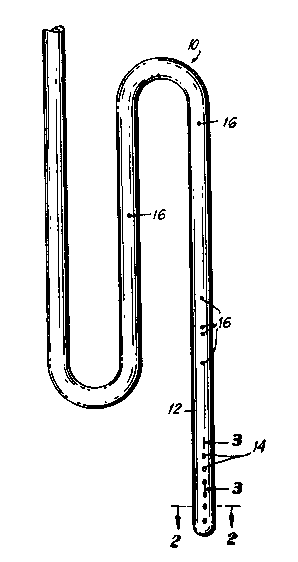
FIG. 1 is a perspective view of a catheter
according to the invention;
FIG. 2 is a cross-sectional view taken along line
2--2 of FIG. l;
FIG. 3 is a cross-sectional view taken along line
3--3 of FIG. l;
FIG. 4 is a perspective view of an apparatus for
holding the catheter during the cutting of apertures therein;
FIG. 5 is a top view of the apparatus of FIG. 4;
FIG. 6 is a section taken along lines 6--6 of FIG.
5 over which is shown an apparatus for cutting apertures in
the catheter;
FIG. 7 is an enlarged view of the cutting apparatus
piercing the catheter sidewall when the catheter is placed in
the holding apparatus of FIG. 4;
FIG. 8 is a perspective view of a symmetric molding
insert according to the invention;
FIG. 9 is a perspective view of a spiral molding
insert according to the invention;
FIG. 10 is a cross sectional view of a mold housing
for use with the insert of FIG. 8;
FIG. 11 is a cross sectional view of a mold housing
for use with the insert of FIG. 9;
FIG. 12 is a cross sectional view of the mold of
FIG. 10 taken along lines 12--12 thereof; and
FIG. 13 is a cross sectional view of the mold of
- FIG. 11 taken along lines 13--13 thereof.
.
`
:' ~ : .
-lo- 1 323537~
DETAILED DESCRIPTION OF THE PREFERRED EMBODIMENTS
Referring initially to FIG. 1 there is illustrated
catheter 10 which is intended for insertion into a ventricle
of the human brain for access to or drainage of CSF such as;
for example, would be necessary to drain excess CSF during
treatment of hydrocephalus. Since the present invention is
primarily concerned with the forward or insertion end of the
catheter, a detailed description of the opposite or out flow
end of the catheter is not provided as such details are well
known in the relevant surgical art.
This catheter 10 is a flexible, hollow, elongated
member having a sufficient wall thickness for the containment
and or transport of fluids therein and therethrough. The
forward end 12 of the catheter includes a plurality of
apparatus 14 for access to CSF in the ventricle of the brain.
By "access" what is meant is contact of CSF for removal or
drainage from the brain or, conversely, to enable medicaments
or other fluids to be directed or delivered into the brain
from the catheter through the apertures 14. These apertures
14 are positioned and configured in a predetermined manner so
as to allow for a better and more continuous flow of fluids
in and through the catheter with less chance of plugging the
holes due to ingrowth of a brain tissue when the catheter is
placed in the ventricle. Further, the design of the holes
enables the catheter placement to be made in an improved,
easier manner while causing less abrasion damage to tissue
during insertion of the catheter.
As shown in FIGS. 2 and 3, the catheter 10 is
designed with 3 sets of holes set 120 apart. These holes
are cut at an angle into the wall of the catheter such that
the angle of the cut is measured along the longitudinal axis
of the catheter in the direction of movement of the catheter
when it is inserted into the ventricle. Further, the
-11- 1 323537
diameter of each hole in the catheter is proportional to the
thickness of the catheter wall so that, as best illustrated
in FIG. 3, there is no direct linear visual access to the
interior of the catheter when the holes are viewed
perpendicular to the longitudinal axis of the catheter.
By preparing the holes in this manner, abrasion of
brain tissue is minimized upon insertion of the catheter into
the ventricle, so less brain tissue is destroyed as a direct
result of such decreased abrasion. Further, by stretching
the catheter slightly, the holes in the catheter are closed
thus preventing such tissue as may come in contact with the
catheter from entering the lumen upon insertion. The
stretching of the catheter can easily be accomplished when a
rigid placement stylet is used: the body of the catheter
being slightly pulled back from the insertion end while the
stylet is held, thus allowing the holes to be somewhat
flattened. This lack of direct access to the inside of the
catheter prevents the growth of brain cells or tissue
therein, thus resolving one of the major causes of plugging
and malfunction of prior art catheters which utilize 90 or
perpendicular apertures. The 120 peripheral offset for each
set of holes further minimizes the possibility that choroid
plexus or brain cell growth will extend across the inner
diameter of the catheter even if such growth does penetrate
.. into one or more of the holes.
Although the holes are advantageously shown as
being cut at an angle of 35 with respect to the longitudinal
axis of the catheter, it is to be noted that other angles can
also be used in this invention provided that direct access to
the inside of the catheter is prevented. These other angles
would be somewhat dependent upon wall thickness of the
catheter, since heavier wall thicknesses would allow a
greater range of angles while still preventing direct access
,, . .
- ~.- .
.
1 323537
-12-
into the catheter interior. Suitable angles for any specific
catheter construction can be det~ermined from the relationship
d tan e = t, where d is the diameter of the aperture, t is
the wall thickness of the catheter, and e is the angle
between the cut of the aperture and the longitudinal axis of
the catheter body. As shown by the relationship of these
variables, the diameter of the aperture must be less than or
equal to the wall thickness of the catheter divided by the
tanqent of the angle. To calculate suitable angles for any
particular aperture size and catheter wall thickness, the
formula would be ~ = tan 1 t , so that the tangent of the
angle, e, is greater than the quotient of the thickness
divided by the diameter.
To assist in the understanding of the invention,
direct access is avoided when the diameter of the hole on the
outside wall of the catheter does not overlap the diameter of
the hole on the inner wall catheter when viewed in a line
perpendicular to the wall of the catheter. Thus, it is
possible to utilize angles other than 35 although 35 has
been found to be particularly advantageous.
By placing the holes to avoid direct access to the
inside of the catheter, it is possible to cut the holes
larger in diameter than they would be if direct access was
provided without weakening the structural integrity of the
catheter. These larger holes allow for an increased flow of
CSF into the catheter while also making it more difficult for
any possible brain cell growth to plug the entire hole,
compared to the relatively smaller diameter holes of prior
art catheters which provide direct access into the body of
the catheter.
;: .
-13-
1 323537
The catheter of the invention can be inserted into
the ventricle of the brain in any manner currently known,
including "~reehand" or with the use of a guide. To assist
in the proper location and placement of the catheter, a
plurality of markings 16 are provided along the length of the
catheter body. These markings correspond to predetermined
insertion lengths of the catheter and enables the surgeon to
know precisely how far the tip of catheter is inserted into
the ventricle By making these markings of a radioopaque
material such as barium, the depth of placement of the
catheter can easily be monitored by conventional techniques.
Furthermore, if desired, the forward section of the catheter
in the area around the apertures can also be made of a
radioopaque material for viewing on various scanning
equipment the precise placement of the forward end and tip of
the catheter.
The improvements provided by the catheter of this
invention are significant in that the physician does not
require any guess work to determine the precise placement of
the catheter in the patient's brain. Furthermore, when so
placed, the catheter provides improved fluid delivery and/or
removal with minimal disturbance of the surrounding brain
cells while also discouraging brain tissue growth into the
catheter apertures. As mentioned above, the catheter can be
inserted in the brain in any manner commonly utilized.
Rather than a "free hand" technique, it is advantageous to
utilize a guide assembly to insure correct catheter
placement.
A preferred guide apparatus and method of insertion
of a catheter into the ventricle iB disclosed in U.S. Patent
No. 4,613,324. As shown in the patent, a stylet is used to
assist in the insertion of the ~atheter.
. ~
.,
. ~
-14- 1 323537
As noted above, the stylet can be used to stretch the present
catheters so that the angled apertures can be flattened to
minimize the abrasion of brain tissue during insertion.
Also, this flattening operation slightly reduces the overall
diameter of the catheter which further reduces such abrasion.
It is known for certain applications to utilize a
second stylet for guiding the catheter into the ventricle.
In prior art catheters, this second stylet is inserted into
one of the apertures at the forward end of the catheter.
Since those apertures are cut at 90~, an unwieldy assembly is
created. Any attempt to align the second stylet parallel to
and adjacent the first stylet and catheter causes the tip to
be somewhat bent, thus causing further difficulties in its
insertion and penetration of the ventricle. The present
invention significantly reduces and minimizes this problem
since the angled holes are more receptive to the introduction
of the second stylet in a compact orientation (i.e., in a "V"
shape, rather than an "L" shape) which greatly enhances the
manipulation of the catheter and stylets during placement in
the ventricle.
The catheters of the invention can be easily
manufactured in a highly accurate and reproducible manner by
utilizing the holding apparatus of the invention. FIG. 5
shows a holding apparatus 20 in the form of a machined metal
block or cube 22. A longitudinal extending aperture 24
extends diagonally from one corner of the cube through the
center to the opposite corner. The diameter of the aperture
24 is only slightly greater than the diameter of the catheter
10 so that the catheter is fully supported in the aperture
when the angled holes are made in the catheter wall.
FIG. 6 illustrates a cutting apparatus 30
consisting of a handle 32 and a plurality of hollow tube like
cutting elements 34 each of which have a sharpened tip 36.
-15- 1 323537
The tube elements 34 extend through guide apertures 28 on one
face of the cube 22 until contact is made with the catheter
10. As best illustrated in FIG. 7, the cutting tubes 34
penetrate the catheter wall, thus forming the appropriately
sized holes therein at the predetermined angle, position and
configuration.
Prior art catheters, as noted above, have four sets
of holes oriented 90 apart along the circumference of the
catheter. In addition to weakening the strength and
structural integrity of the catheter in the tip area, holes
on opposite sides of the catheter (i.e., those 180 apart)
are made simultaneously by a punching tool. This results in
holes on one side being larger in diameter than those on the
opposite side. Therefore, two sets of holes are large and
two are small. This non-uniformity affects CSF flow and the
smaller holes can easily become blocked by brain tissue
growth, thus causing reduced operation of those catheters.
The present invention resolves these problems by
accurately and precisely placing three sets of uniform holes
cut at the desired angle to the catheter body and spaced
apart exactly by 120. This results in increased flow
through the holes, higher strength and integrity of the
catheter body, and greater ease of insertion and placement of
the catheter in the ventricle.
FIGS. 4 through 6 illustrate the placement of guide
apertures 28 on the various faces of the cube. In a most
preferred arrangement, these guides are positioned in a
diagonal line along the top and two side faces of the cube
22, 60 that each set of holes is placed 120 apart around the
periphery or circumference of the catheter body. As noted
p~eviously, it is highly advantageous to make the holes in
the catheter 10 at an angle of 35 with respect to the
longitudinal axis of the catheter.
- 1 323537
-16-
This apparatus guarantees the accuracy of the hole
cutting at the appropriate angle as well as the precise
spacing of the holes relative to each other around the
periphery or circumference of the catheter. To cut the
holes, the user merely inserts the tubes 34 of cutting
apparatus 30 into the guides 28 when a catheter is placed in
the holding block 20. The cutting apparatus 30 after
piercing the catheter wall 10 is then removed, resulting in
placement of the holes at the precise orientation and
configuration in a simple manner which allows for repeatable
and rapid production of such angled hole catheters Further,
the precision obtained in utilizing this apparatus is very
high and reproducible to facilitate mass production.
The preceding apparatus has been found to be
suitable for constructing apertured catheters of a variety of
materials for particular applications. When very small
diameter holes in thin-walled silicone catheters are desired,
the quality of side-wall smoothness necessary to prevent
cells or tissue from binding and plugging the catheter holes
is difficult to obtain by the use of the cutting apparatus.
Accordingly, applicants have devised a molding system which
achieves all the desired results. This embodiment
illustrated in FIGS. 8 through 13, is discussed below.
Generally, a disposable insert is placed in a molding
assembly prior to the injection of the polymerizable silicone
material. This insert is retained in place until the
silicone material cures. Thereafter, the insert and catheter
are removed from the mold and the insert is discarded. This
technique enables the user to produce extremely smooth, very
small, angled holes at any orientation, position or
configuration in a relatively simple and highly reproducable
manner.
-17- 1 323537
FIGS. 8 and 9 illustrate two preferred disposable
inserts 50, 55. Each of these inserts has an elongated body ~-
portion 52, 57 and a plurality of rod like extensions 52, 54
extending from the body at a predetermined angle. As
mentioned above, it is highly advantageous to make the holes
of the catheter at an angle of 35 degrees with respect to its
longitudinal axis. Thus, the rod - like extensions 52, 59,
of these inserts are positioned at an angle preferably of 35
degrees with respect to the axis of the body member 52, 57.
Fig. 8 illustrates an insert for forming three rows of
apertures in the catheter, while FIG. 9 illustrates a spiral
orientation of such apertures about the circumference of the
catheter body.
FIGS. 10 through 13 illustrate the molding
cylinders 60, 80 for use with the previously described
inserts 50, 55. Each mold includes an open end 62, 82 which
enables insertion of the corresponding inserts 50, 55, and a
closed end 64, 84 which is used to form the insertion tip of
the catheter. These molds 60, 80 include a plurality of
guide apertures 66, 86 for at least partially receiving the
rod like members 52, 59 of the respective inserts 50, 55.
The guide holes 66, 86 extend through the wall 68, 88 of the
molding cylinders 60, 80 at an angle which corresponds to the
desired angle of the catheter holes.
In manufacturing, a plurality of molds 60, 80 and a
much greater number of inserts 50, 55 are prepared. Since
the molds 60, 80 are reusable, basically any material can be
used which would provide a useful service life. This would
include, for example, steel, stainless steel, aluminum, etc.
and certain engineering thermoplastics may also be suitable.
These materials must be sufficiently strong to retain the
insert and withstand the temperatures anticipated for the
1 323537
polymerization of the material used to form the catheter.
Such mold material must also be dimensionally stable over the
entire curing temperature range.
The insert 50, 55 should be made of a self-
lubricating material that does not bind or stick to the
polymerizable liquid used to form the catheter. Also, the
self-lubricating material must be sufficiently flexible so
that it can be easily inserted and drawn out of the mold
without damaging the catheter. At this time, the most
preferred material for the insert is an injection molded
polyamide material.
A preferred material for the catheter itself, is a
polymerizable silicon liquid which has a very low injection
and curing temperature, i.e., about lOOOF. While this
requires a relatively long curing time, high production rates
can be achieved through the use of multiple molding cavities.
The inserts are disposable so that each insert can only be
used to make one catheter. As noted above, the mold itself
can be reused an infinite number of times.
During manufacturing, after the insert is placed
within the mold, and the rod member properly positioned
within the guide holes of the mold, the polymerizable liquid
is introduced into the space between the insert and the mold.
The liquid is then allowed to polymerize and cure to form the
catheter. The insert and catheter are removed from the mold
and the insert is then destroyed to form the final catheter
product.
The preceding molding technique provides numerous
advantages, including:
.
-19- 1 323537
1) the catheter holes can be configured in any
shape or size relative to the axis of the
catheter. For example, spiral, off-set 90
degree or off-set 120 degree holes can easily be
obtained.
2) the angle of the hole relative to the axis of
the catheter can be easily changed by providing
different mold inserts and mold cavities. This
allows optimization of the hole angle compared
to the hole diameter as a function of cell
growth. This relationship is governed by the
formula given above.
3) this apparatus assures that no "flashing" will
occur on the internal bores of the catheter.
Any excess material due to "flashing", will be
visible on the exterior surface of the catheter
and can be easily trimmed therefrom.
4) this apparatus assures that each catheter can be
manufactured to very close tolerances with
little or no variation in the construction of
each device: hence, a high degree of
reproducibility in mass production is achieved.
While it is apparent that the invention herein
- disclosed is well calculated to fulfill the objects above
stated, it will be appreciated that numerous modifications
and embodiments may be devised by those skilled in the art,
and it is intended that the appended claims cover all such
modifications and embodiments as fall within the true spirit
and scope of the present invention.
, . . .
-
.: ~ ..
.
'.'' . ~ , ~
--` 1 323537
SUPPLEMENTARY DISCLOSURE
Figure 14 shows a cross-sectional view of a
further embodiment of the invention. As shown, the
catheter 100 is designed with a cylindrical exterior and
with a bore ~aving the shape of an equilateral triangle
110 with rounded corners, with three sets of apertures
114 which pass through the walls of the catheter, the
centres of which apertures coincide with the lateral mid
parts of the sides of the triangular bore. The
triangular shape is preferred over a circular shape
because it provides, for a given external diameter and
wall thickness at the apertures, 1.7 times the effective
flow area while providing additional structural integrity
since the bore will not close when the catheter is bent.
Further, this shape can easily be partitioned to allow
the formation of secondary lumens to run other implements
into the ventricle, within the main lumen while
maintaining sufficient flow area.
As in earlier embodiments, the apertures are cut
at an angle into the wall of the catheter such that the
angle of the cut is measured along the longitudinal axis
of the catheter in the direction of movement of the
catheter when it is inserted into the ventricle.
Further, the relationship of the diameter, angle of cut
and thickness of the wall is such that there is no direct
linear visual access to the interior of the catheter when
the holes are viewed perpendicular to the longitudinal
axis of the catheter. Preferably the apertures have axes
which extend at an angle of about 35 to the longitudinal
axis of the catheter.
t9
~1 ~
.
.
`