Note: Descriptions are shown in the official language in which they were submitted.
1333691
s
"Device and Method for Cryopreserving Blood Vessels"
Technic~l Fiel-l
The present invention relatcs to a device for st~bili7ing
blood vessels and more particularly to a preservation procedurc for usc
during freezing blood vessels to ultra-cold te~ a~ s whereby the
blood vessels can be preserved for extended periods of time. Also
disclosed is a method utili7ing the dcvice for frcezing and thawing of
blood vessels. Cryopreserved blood vessels arc useful for providing
grafts to patients who cannot provide their own blood vessel grafts or
where fresh blood vessels are unavailable.
R~ck~rol-nd of the ~nv~ntion
"Cryopreservation" is a technique for freezing and
storing cellular and tissue matter such as blood vessels, which include
veins and arteries, at e~ cly low temperaturcs while preserving the
viability and function of the tissue. Each year, 360,000 small vessel
coronary bypass "jumps" are performed in the U.S. alone. Another
100,000 peripheral vascular procedures, below the umbilicus, are also
performed. Of the small vessel procedures, 15% are performed on
patients who have already had a previous ope~ation reslllting in a lack
of suitable available tissue or on patients who are ~ betic or have a
disease which renders the tissue less than adequate. Clinic~lly, the only
alternative is to use less than optimal tissue or use artificial vessels
which are prone to occlusion and thus are less than ideal. R?l)se of
the successes resulting from the cryo~lesc.~adon of heart valve dssue
~,
2 13~369~
(see CDN. application Serial Number 550,943 filed Nov. 3,
1987, which may be referred to for further detail and to
date, more than 3,000 cryopreserved valves and approximately 2,200
implants, it is the intention to expand this technology to vein and artery
S dssue as well. Thus, in the clinical setdng, cryo~.~se,.ted dssue would
fill a need for the afo-e..~e-nt;oned patents and would in addition lead to
less trauma for the padent and reduce surgical dme and e~ pe,~e.
Previous attempts at the use of allograft vessels have
met with a variety of problems. The primary concern was
inconsistency in the method of harvest coupled with an inability to
freeze and store the dssue pr~,ly until its intended use. In a~itiol~
previous investigators failed to pe.rol~ the freezing process using statc
of the art techniques, and consequently, the viability of the tdssue was
low and inconsistent and resulted in early loss of patency.
Although there have been a few publishe~l reports on the
cryogenic preservation of veins and arteries, there has been no
published systematic eX~ nation for the cryobiological vPriables
involved in the preservadon ~lvccdu~. Most inves~gPt~rs have simply
infiltrated the vessel with dimethyl sulfoxidc (DMSO) and rapidly
frozen the tissue in liquid nitrogen. Several other investigPtors have
used ~ncontrolled and un~ r~s.J~~d freezing ratcs. When iicsected
from the body, blood vessel tissue has a natural tendency to constrict.
Inves~igPtio~ls to date show that under such con~litionc the endothelial
lining of the vessel may be denuded; the.efo~, if such a vessel is
tran ;pl~nted it may be prone to thrombosis.
Preservation of the endothelial lining of these vessels is
of particular importance, because the internal en~otheli~l lining of the
blood vessels actively inhibit thrombosis. Previous studies of
s~phçnous vein cryo~s~ ation in~licate that the major abnormality in
the frozen and thawed tissue was destruction and loss of this tissue
layer. A primary goal of cryopreservation of the tissue is the
pl~cnlion of ice crystals which ~l~m~e or destroy cellular ~k~
Dirr~"~,nt freezing methods are applic~ble to particular tissues; not all
tissues arc alike in their ability to with~t~nd c.~o~i~,scrvation and
3S thawing yet maintain effecdvc viability. No investigPtor is known to
13336~
have successfully applied this technology to the internal mammary artery or
other arterial tissue.
Summary of the Invention
The device of the present invention is a structure for
supporting and distending a blood vessel while permitting fluids to
infiltrate the vessel to facilitate cryopreservation.
The invention in one aspect provides a blood vessel stent
for use in cryopreserving blood vessels, comprising a substantially
straight support track means and a pair of mounting means for supporting
and distending a blood vessel.
More particularly disclosed is a blood vessel stent for use
in cryopreserving blood vessels, the stent comprising first and second
elongated stylets each having an end capable of insertion within a portion
of a blood vessel from a donor, means on the stylets operative to engage
the interior of the blood vessel and thereby facilitate fluidtight ligation
of the blood vessel on the stylets and support means receiving the stylets
in selectively adjustable mutually confronting relation whereby the blood
vessel is distended between the stylets to prevent contraction of the blood
vessel, so that the stent supports the blood vessel through the stages of
procurement and cryopreservation.
Still another aspect of the invention provides a method for
cryopreserving a blood vessel comprising the steps of placing a blood
vessel dissected from a patient into an appropriate medium with effective
amounts of at least one antibiotic, contacting the dissected blood vessel
with an effective amount of at least one cryopreservative, freezing the
blood vessel according to a freezing schedule that will maintain acceptable
levels of cell viability and storing the blood vessel at a temperature
below -100C.
Still another aspect of the invention provides a
composition comprising a tissue storage medium comprising a
cryoprotectively effective concentration of a cell penetrating
cryoprotectant and a cryoprotectively effective concentration of a
glycosaminoglycan and a segment of a blood vessel, wherein the segment of
the blood vessel is submerged in the medium.
A still further aspect of the invention provides a method
of maintaining intact, a layer of endothelial cells present in a blood
vessel during cryopreservation comprising contacting the blood vessel with
an effective amount of a cryoprotectant composition comprising a medium for
freezing the blood vessel, a cryoprotectively effective concentration of a
cell penetrating cryoprotectant and a cryoprotectively effective
concentration of a glycosaminoglycan and maintaining the blood vessel in
contact with the composition at a temperature below -100C.
C The method of the present invention using the device
1333~g~
involves the technique for preparation of the vessel prior to stenting,
removal, shipping to the processing laboratory, processing (including
freezing), thawing and dilution. A particular emphasis is made for the
preservation of the endothelium (inner lining) of the vessels in addition
to keeping the basic integrity of the vessel walls intact. This involves
a "no touch" surgical technique coupled with vasodilation, the use of a
stent and the use of a unique freezing profile that allows veins or
arteries to be frozen down to the temperature of liquid nitrogen,
approximately -196C, with minimal tissue damage due to ice crystal
formation or osmotic shock. The present invention also includes a thawing
schedule whereby the frozen tissue can be rapidly thawed with minimal
tissue damage. Vessels that are cryopreserved according to the present
invention are alive when thawed and are ideally suited for replacing
diseased or damaged vessels in patients who for whatever reason do not have
suitable vessels for cardiac or peripheral vascular reconstruction.
Accordingly, the present invention seeks to provide a
device and method for retrieval and handling of human or animal vein and
artery tissue.
Further the present invention seeks to provide a device and
method for preserving a living vessel for long periods of time.
Still further the present invention seeks to provide a
unique set of chemical constituents prior to the freezing process.
Further still the present invention seeks to provide a
device for supporting and distending a dissected blood vessel.
Further still the present invention seeks to provide a
unique cooling schedule for freezing a vessel so that the vessel cells,
including the endothelium, maintain high viability after it is thawed.
Still further the present invention seeks to provide a
method for cryopreserving a human vessel which allows rapid thawing of the
vessel while maintaining maximum cell viability.
Still further the present invention seeks to provide a
living vessel such that it is suitable for transplantation after long term
storage at super-cold temperatures and to provide a living vessel with a
specific and unique method for thawing.
These and other aspects, features and advantages of the
present invention will become apparent after review of the following
detailed description of the disclosed embodiment and the appended claims.
Brief Description of the Drawinqs
Figure 1 is a schematic representation of a stent apparatus
according to a preferred embodiment of the present invention.
Figure 2 is an exploded isolated perspective of a stent
apparatus according to a preferred embodiment of the present invention.
Figure 3 is a schematic representation of a freezing
profile for freezing a vein.
45~ Figure 4 is a representation of a thawing curve for a
133~69~
stented vein in approximately 80-90 ml of solution according to a
preferred embodiment of the present invention.
ne~ e~l nescriDtion of the Preferred F.mbodiment
Referring now in more detail to the drawings, in which
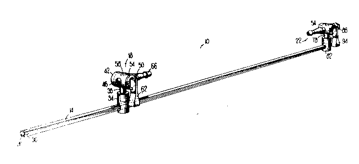
like numerals in~icatc like parts throughout the several views, Fig. I
shows a blood vessel stent 10 which is composcd of three main parts:
a ~u~ track 14, sliding plu~llal stopcock assembly 18, and fixed
distal stopcock ~csembly 22. All threc parts can bc m~nllf~lred from
any a~ liate material which (a) is freezablc to ultra low k,m~.~c5
without adverce deforming or cracking, (b) .~inti~;nc flcxibility at ultra
low t~im~-~tures, tc) is chemically inert and will not cor~ ;n~tc thc
vessel by k~hing chemic~ls such as pl~stici7~rs into the vessel, and (d)
can withstand and not react with dimethylsulfoxide ("DMSO"),
ethylene or propylene glycol, glycerol or any other chemicals or
solvents used in the cryopreservation process. Track 14 is a
substantially straight elongated membcr used to mount stopcock
~Csc.~blies 18 and 22. The track can bc any convcnient shapc, but is
prcferably a par~llrlcp;pc~ and has on one sidc a groovc 26 ~unning at
least part of its length for the p~,ose of st~bili7ing thc stopcock
ac~mblics 18 and 22 whcn ~I~o~lht~d on traclc 14. On the surfacc at the
l,l~,.hllal cnd of tracl~ 14 is located a ramp 30 with a flangc having its
larger portion facing toward thc distal end. Ramp 30 inhibits thc mount
from sliding off the end of track 14 oncc the mount is slid on the tTa~k.
~u~hllal slidable mount 34 is a cylindrical member
having a .~ccs5e~1 cylindrical opening axial at one cnd capable of
matably rcceiving a mounting pin. At the other end of thc mount 34 is
an opcning radial to the cylinder, caI~able of operatively receiving thc
traclc 14. Mount 34 is ablc to slidc along tracl~ 14 so as to bc adjustablc
for a givcn kngth of blood vesscl sc&,
F~U~ al stopcocl~ ~csembly 18 inclu~cs a ~;r~ ;e~l
g pin 38 which removably matcs CO"~ ly within thc axial holc
of thc p~al mount 34 and is prefcrably able to rotate about the
c~lind~ical axis. Pin 38 of thc pr~i,.. al mount is o.lhGæonally
1333694
integrated with the body 42, which is a hollow cylinder having a hole
46 extending through both sides of the cylinder, positioned orthogonal
to the pin. Along one end of body 42, positioned radially opposi~e each
other are a pair of protrusions 50 which act as stops. Stopcock barrel
s 54, which fits within the cylindrical interior of the body 42, is a
cylinder rotatably positioned in a lon~ituAin~l oriçnt~tiorl within body
42. A barrel hole 58 is positione~ perpen~licul~r to the ba~cl 54 axis
through thc barrel and selectively aligns with hole 46 in body 42 so as
to allow a passageway through the stopcock. Lever 62 is a
subst~nti~lly rectangular member perpen~liclJl~rly integrated with
Sl~pCOC6 barrel 54 at one end. When rotated so that the lever is parallel
with track 14, barrel hole 58 and body hole 46 are ~ ne~ to forrn a
paCs~geway through the stopcock while, when the barrel 54 is rotated
clockwise to a position perpendic~ - to track 14, the holes are not h
alignment, thereby blocking the p~s~ge of fluid or air through the
stopcock.
Stylet 66 is a hollow subst~nti~lly straight elongated
tube aligned parallel to the track 14, with a port 70 at the free end of the
stylet and with the other end of the stylet abutt ng the body 42. Stylet
66 is int~tcd with body 42 and is O~ OgOI)~l tO the pin 38. Stylet 66
is aligned with and op~s;~, hole 46. The hollow intcrior of the stylct
66 co~ niC~tes wlth the hole 46 in one side of the st~,~cocL body 42.
On the surface of stylet 66 is at least one and preferably two or more
fo,~dly tapering ramp s~l~ces 70 merging with stylet 66. Stylet 66
is ~ ~ed to açco~--n~o~l~te coupLing with a vessel which may be press
fit upon the stylet and can abut body 42. Stylet port 68 pe.~ s fluid or
air to enter the blood vessel when the stylet 66 is coupled with the
blood vessel. The stylet tube is sized so as to be essellti~lly parallel
with the internal hollow bore of the tube. Ramp surface 70 t~.-...n~tes
in a shoul~ler 74 which pc.mils p~ssu ~-dght ligPtion as ~scribe~l
more fully herdnbelow.
Fixed distal stopcoclc ~csemt~ly 22 is cssc~ y the
same as sliding p-o~i~al stopcoc~ PcSCmbly 18 except with the
following notable m~ific~tinn~ The borc of thc axial mount bole is
lirr~cnl than that of thc ~.. im&l mount hole and the ~ ,t~,~ of the
7 1333691
mounting pin 78 is correspondingly different fr~m the ~i~meter of pin
38 so that the two stopcock assemblies cannot be interchangeably
mounted. This avoids confusion of which slopcock assembly is distal
and which is proximal. Distal mount 82 is preferably perrnanently
mounted on the distal end of track 14.
Distal body 86 also has integrated on its surface in
alignment with the hole (as in hole 46) a Luer fitting 86 for ~ct~ch~bly
receiving a syringe. Alternatively, a hub 90 can be used without thc
Luer lock fitting for detcct~bly receiving a fluid delivery device such as
a syringe. Standard Luer fittings arc co.. ~nly known to thosc sl~illcd
in the art. In an alternative embo~imcn- a hub 90 can bc integrated in a
similar with the proximal ~pcoc~ ~cscmbly 18 to permit back flllshing
of the blood vessel before or after being I~IUUn~i on track 14.
Distal lever 94 rotates in a manner similar but op~osite
to that of proximal lever 62; i.e., when parallel to track 14 thc distal
lever 94 is in the "open" position and when rotated ~,~ icul~r to the
track is in the "closed" position. The sto~coc~ levers arc positiQn~ so
that when both are parallel to track 14 they point in toward cach othcr
and will not stretch the vessel during closurc. In this way thcy do not
add to the overall length of the sllucl~e~ nor does the distal levcr 94
inte.r~ ,~ with distal hub or Luer fitting 86 or 90.
The stent is used to ~uypoll and distend a ~;csccl~
vessel. The ~issec~ on ploc~lulc is dcsclibcd in detail in thc es~mrles
set forth hcreinbelow. A specially ~lesigne~ perfusion solution
con~ining a suitable vascular smooth muscle relasant drug is applied
along the length of the vessel. After a period of time, usually about 10-
15 minutes, the vessel is carefully dissected using what is con....only
termcd as "no touch" technique, whe,~. ~on an a~ph~pl;ate in~isinn is
made on the distal and proximal end of the vessel. If the donor's heart
is beating, the portion of the vessel which was tr~nce~teA will be ded
with ligature. The removablc ~tol)cocl- ~cse-~h1ies 18 and 22 of stent
s 10 are placed one on each end of the vcsscl and a po~tion of a
flushing/~istention solution is p~,r~.scd through thc vcssel. .Thc
~icse~ on can then be conlrleteA Thc p~ u~io~ can be any
ap~lo~liate medium, preferably Delbccco's Minimal ~7sse-n~isl Media
8 133369 1
("DME~"). Other media include, but are not limited to, Medium 199,
Eagle media, Hank's media, Delbecco's modified Eagle media, Iscoves
modified Delbecco's media, Defined media A2, CMRL-1066, RPMI-
1640 (also 1603, 1630, or 1634), F10, F12, alpha media, or the like.
s To this media is added serum such as but not limited to human serum,
fetal calf serum ("FCS"), serum substrate, or the like, and a vasodilator
such as but not limited to nitroprusside, dantrolene, nifedipinc,
verapamil, phentolamine, tolazoline, procardia, or the like. It is
preferable to use papaverine having a concentration of from about
lx10E-4 to about 30x10E-4M, morc preferably about 3xl0E~M. This
media solution also has certain additives: bicarbonatc, HEPES or
similar buffer, glutamine, D-glucose and sodium pyruvatc.
Before the vessel is completely removed, the stopcock
ends are attached to the support track 14 of the stent apparatus 10 in
order that the vessel does not have the opportunity to contract. It is this
natural contraction of the vessel that is deleterious to thc inner
endothelial lining of the vessel and for which thc stcnt is designed to
protect. Once the rem~ining portions of the vcsscl arc dissectcd, the
stent 10 with the ~tt~che~ vessel is ready for transport in an outcr
cont~irler.
The stent 10 contin~les to provide support and pro~tion
for thc vessel whilc progrcssing through other stages in thc
clyopIescrvadon proccss. Upon arrival of the stcnted vesscl for
cryopIese.~adon, thc stcnt will providc thc ~uppoll for continucd
flushing and inspection for tying of collatcral vcssels. During the
frcczing process the stent will be thc support neCcss~y to prc,~cnt the
vcssel sidc walls from coll~psing and at thc final phase during thawing
and dilution the stent again kecps thc vcsscl from coll~e and ~C-ilit5
thc ad~lition and removal of c~yop~clant agents.
The present invendon provides a method of freczing,
storing and thawing endothelial lined dssuc, such as a vein and artcry.
Thc dssuc that is frozen acco.~ling to thc prcsent inventdon can bc sto~ed
for long periods of timc at ultra cold t~ ues with ~;ni--~l loss of
cell viability. The present invcndon il~cludcs a uniquc frcczing profilc
that allows a tissue such as vcin and ar~cry to bc frozcn down to the
9 13336~
temperature of liquid nitrogen, approximately -196C1 with minimal
tissue damage due to ice crystal formation or solution effects due to
slow cooling. The present invention also includes a thawing schedule
whereby the frozen tissue can be rapidly thawed with minim~l tissue
S d~m~ge. Veins and arteries that are cryo~)l, scl~led according to the
present invention are biologically viable when thawed and are ideally
suited for replacing f~ se~ or me~h~nic~lly damaged vessels.
The tissue to be preserved is only as good as that which
is received into the laboratory. Consideration must be given to donor
age, health and history of vascular dise~se. Another important
co~sideration is the time bel~.~n death and the harvest of the vessels
(warm ischemia) and the time from the harvest of the vessels to
laboratory proccssing (cold ischemia). Attention must be paid to the
method of handling the tissue during pi~u~ ~nt and the "~ l;l -.. used
to ship the tissue.
A donor that can be used as a source of hum~an vessels
which are frozen according to the present invention should be in the age
range of up to about 5S years of age and the donor should not have
suffcred from significant atherosclerosis, diabetes, circulatory
disorders, severe hypertension, varicose veins, or co.. ~nicable
,lise~
All procu~emcnt is to be performed undcr sterile
condidons. Time delay bet~.ccn dcath and harvest will have a
deliterious effect on the endothelial cell layer and therefore should be
comr1ct~ ;-----~i~tely after expiration of the donor but in any case not
longer than about 10 hours post ulolle.~ For ex~ml-lç the ideal length
for coronary bypass proccdure would be to procure a vessel of
ap~o~;...~tcly 17cm with at least a di~mcter of 4mm. It is to be
understood that other diamctc.~ and lengths arc usable and are within
the scope of this invention.
Sterilization
It was discovered that many antibiotics were c.~ ,cly
toxic t~ the endothelial layer of the vessels. This toxicity is the result of
a nu.,lbcr of factors including timc, ~ v~., and mode of action. In
lo 133369~
addition to the antibiotics, the antimycotic (antifungal) agents may be
deletenous to the tissue endothelium. It is iln~.~nt to continually test
new antibiotics and fungicides for cell toxicity and ster~ ()n efficacy,
in order to improve cell viability and kill microbes resistant to previous
S agents.
A mixture of an antibiotic and an antimycotic were
found to provide suitable sterilization results. A ~ of Ihli~cn~
and Ancoban were found to be particularly suitablc. Tablc 1 shows the
effect of antibiotic inCu~tiQn on endothelial viability in vi~o.
TABLE I
Effect of Antiobiotic Incubation on Endothelial Viability In Vitro
Antibiotic Time signi~lcance
APCVL 4 hrs. NS~
Imipenem + gentamycin 4 hrs. NS
APCVL 12 hrs P< .05
PSA 12 hrs. NS
Imipenem + gentamycin 12 hrs. P< .05
Imipenem - without 12 hrs. NS
gentamycin
T~ .Y .~I + A lcol,an 18 hrs. NS
* NS = Not Significant
APCVL = Amphotericin B, 25 micrograms/ml,
Polymixin B Sulfate, 100 micrograms/ml
Cefo~itin, 240 micrograms/ml
Vancomycin, 50 micrograms/ml
Lincomycin, 120 micrograms/ml
PSA = Penicillin (SOIU/ml)
Streptomycin (SOmg/ml)
ArnphotericinB (lOmg/ml)
11 13336!3~
Freezing Media
The medium in which the tissue is frozen is of great
importance for maintaining a balanced cell en~ cnt. Time and
tclll~er~ e also contribute to whether a particular medium will be
S successful. Generally, a protcin sUspensiQrl~ such as blood serum or
artificial serum, must also bc present for ~ n...ll ccll viability.
A number of freczing mcdia can be snccessfully uscd in
practicing the present invcntion. Media, such as b~l~nce~l tissuc culture
medium or simple phosphate buffered saline, can be uscd for most
tissue types. For this particular tissue type DMEM is thc preferred
with the associatcd additive colll~nel.~ iscll~ prcviously.
The freezing mcdia is composed of the enriched DMEM
plus FCS from about 1% to 30%, more. preferably 10% fetal calf
serum; plus the range of papaverine ~liscu~se~ above, preferably about
0.012% papaverine; and, chondroitin sulrh~te having a conccn~ ion
of from about 1% to 10%, preferably 2.596 to 5%, more prefcrably
2.5%.
Dimethylsull)h.,~;dG ("DMSO") is also added dther in at
least one step of lM or preferably in three steps of .25M, .SM and lM
titrations at 4 C. Concent,~lions of DMSO can range from about 0.5 to
3 molar. The increase in molarity of DMSO should preferably be
gradual so as not to tra.. ~l;7~ the blood vessel. DMSO can be added at
higher tc~c.atures but timing bccG~es far more critical and toxicity
may result in some tissues.
An illl~l~nt innovation in endothclial ~r~ion used
to further refine and prescrve tissue integrity is to use chondroitin
sulfate. This glycosaminoglycan (GAGS) is a major co...l ol~cl~l of thc
extr~cellul~r matrix. The molecular weight of chon~vilin sulfate can
vary from 5,000 to 50,000 and it is a slllrh~ iss~ch~ride con~isting
of repeat units of D-glucuronic acid and N-acetyl-D-g~l~ct~os~mine
Currently, this material is an additivc in K-sol, a solutiol~ uscd for the
short term (4C) storage of corncas.
Examples of othcr suitable ~Iyco~ ~ o~lycans includc
but arc not limited to hyaluronic acid, ~ sulfatc, hep~nn sulfatc,
hep~fin, and the like. Other cf~opiot~tA-~Is includc but afe not limited
13336~
12
to glycerol, polyvinylpyrolidone, hydroxyethyl starch, and
polyethylene glycol, dimethylformamide, ethyl glycol, and the like.
Table II shows a series of experiments using this
additive to the freezing solution. Groups 1-6 used a protocol which
differed from Groups 7 and 8 in that a two hour 37C incubatiQ~ was
performed. The results in-lirate that the addilion of chondroitin sulfatc
to the freeze mLlcture si~nific~ntly irnproved endo~hc1i~1 viability.
These materials can be used in cryoyn,s~ ation
procedures with or without the stent app~lus 10. As a cr~o~l~t~c~lt
chondroitin sulfate or its alternatives(GAGS) can be employcd in
procedures for cryoprotection of cells, tissues and organs in ~d~lition to
blood vessels.
13 13336S~L
TABLE 11
Influence of Chon-lroitin S~ h~P l~on F.n~1n~hPli~l Cell Vi~hility
Cell Viability
(F~P~ n) T.tPCt TrP~tPAICOnIrOI
Chl~n~l~uiLin slllf~tP t ~ :I ~i~if Control :~esi RP~r~ rl
~Cs) ~ itit~n (2 ~%~
I) Without vs with 6 3 309 ~t ( 42) 51(124) 174 2.95
chondroitin sulfate
a) Sa~ h~ veinsonly 3 3.426 ~ (147) 95(391) 429 2.66
b) Femoral veins only 3 4 091 ~ ( 25) 29( 61) 71 2.44
2) Without vs with CS 4 .457 ns (217) 203(187) 171 86
in mannitol during
dilution (no CS during
freeze)
3) Without vs with CS 5 .992 ns (109) 63(60) 47 55
during 2 hr incubation
(with CS during fre~e
and dilution)
a) S "~ ~veinsonly 3 .606 ns (94) 85(89) 71 95
b)Femoralveinsonly 2 .603 ns ( 83) 40(30) 30 .36
4) Unfrozen vs frozen 4 12.171 ~ (300) 327 ( 57) 56 .19
without CS, with CS
during dilution and 2 hr
,u~ - ~r
s)Without vs with CS 6 2.069ns 69) 48 ( 32) 28 46
during all steps except
no CS during freeze
6) Unfrozenvswi~hCS 2 5.752 ~ (390)457 (177) 163 45
during dilution, no CS
during freeze and no
i~c '
All .. ~;~.,CSConc~ a~
7) Without vs with 10g% 4 2.415 ~ (118) 172 (319) 334 70
.hù~ uilhl sulfate (CS)
Withoutvsw/lg% 4 1.704 ns (20~) 131 (0) 82 ~3
~,hon~uitu~ sulfate
t ns . not si~ifir^ lt (P > .05); ~ S P .05; ~ S P .01
tt Groups 1-6 wae ~o--..o~ using Protocol II. Protocol II diffaed fiom Protocol I hv ~c
addition of post-lhaw 2 hr. 37C: n I - in culture medium
133~69~
14
Freeze Profile
The freezing profile is of critical importance to
successful cryopreservation of a tissue. A multitude of variables exist
to maximize tissue survival. For instance, the volume of fluid, the size
S of the tissue, geometry of the package and the combination of
characteristics incorporating cryop~otec~nt, tissue, and freezing media
all contribute to an optimal freezing profile. It is to bc understood that
the prior an freezing profiles available for cell sus~,.sions may not bc
suitable for freezing blood vessels, and that prior art freezing profiles
for heart valve tissue also may not be suitable. It has been dele,~ned
that each tissue has its own unique and optimal freczing profile. Thc
freezing profile required to successfully c~ ~c~se. ./c one tissuc may bc
different from the freezing profile r~li~ to successfully cryo~sc..~c
another tissue.
lS A number of factors need to bc considered when
&eezing a tissue. Among these factors arc: thc te,ll~.alu-., around thc
equilibrium point, (generally +4C, to thc temperature at thc freezing
point); release and control of the exothermic heat given off at the
freezing point; opdmum cooling rate as which is detc,~led by the
pe~nnç~bility of the cell ~ ,n,~ c to water, thc surface to volumc ratio
of the cells; the type and conrçn~ration of cl~opl~tecti~e agents in the
media; telll~.~ture and time of tA~sule to those agents, cooling rate
removal of the cryop~se.~ed tissue from the controlled rate freezer and
i~c~ig the tissue into a liquid nitrogen refrigerator, and, warming
rate and the thirl~ne5s of the tissuc.
Detail of Freezing Profile
Thus, the method of harvesting veins from a donor,
placing the vessel into a me~lium with the proper tissue preserving
characteristics for transportation, and the usc of proper
c~ ~Opl~ ation agents prior to the freezing of thc vessel ~r ~ing tO a
freezing schedule is desirable for proper c~yo~ s-.~adon. To
acco..~plish this the ch~m~ e and cooling ratc is con~olled
so as to produce the desircd effect on the sa_plc. Since the blood
vessd tissues cool at slightly dirrc.~ ratcs, and phase ch~ng~S occur at
133369~
par~cular temperatures, careful control over the rates of freezing should
be maintained.
The range of the freezing rate is also a function of the
fluid volume in the package cont~ining the blood vessel as well as the
S geo-n. l~ ~ of the p~e~ While thc freezing profiles describcd herein
are related to the volume and gcollletl~l of the pa~ ~. it is to be
understood that the present invention e~-co~ sses those mo~iffr~tio~c
of package design which result in a change in the volume and
geometry, which in turn, result in a variance in the frcczing rate.
Freezing rates can vary to a certain amount at a givcn t~ . A
suitable rangc is from about 0.01 -lOO-C/min, about 0.1-30-C/min,
preferably about 0.3-l C/min and more preferably 0.5-l C/min. In a
preferred emb~iment of the present invention, the overall rate of
freezing of the blood vessel is kept at ap~.u~;...a~Gly 0.5-C pcr mintl~e
A ~lefc.re~ freezing schedule used to cljoplcse.. ~e the veins and
arteries in the present invendon is coulpl;scd of placing the paclcaged
tissue, having a total volume of about 2.5 cm x 25 cm, into a freezing
apparatus. A typical profile for a specific fluid volume of 75-85ml
would have the inidal tcmp~,.alu~c of the cllaml;c- sct to -lO-C. The
chamber is set to cool at a rate of O.Ol C/min. until the sample(s)
reaches +4 C. At this point the tissue cools at a ratc of O.S i
0.2 C/min. until the sample reaches -2 i O.5-C, at which time a phase
change is initi~tC~ At this point, in order to prepare for thc e o~
heat of fusion, the coolir~g rate is in._.~scd to -30-C until the chsmber
reaches -70-C. ~mmc~i~tcly after the chsmk~ reaches -70-C, the
ch~mb~r iS warmed at a rate of 20-C/min. until the ch~mber reaches -
60-C, wh. .e.~pon this tc,npc,ature is held for a period of 17 minutes.
During this ~me, thc actual ratc of vcssel cooling is applv~ tely -0.5
+ 0.2 C/min. At the end of this 17 minute period, the ch~mh~ is again
wanned at a rate of lO-C /min. until thc ch~mber reaches a Icvel of -
30C. Thc level of -30C is hcld for one minutc and then thc cooling of
thc chamber comme~ccs at a rate of 0.01C/min. until thc sample
rcaches -20C. During this time, the actual rate of cQoling of the sample
is ~pr~ t~ly 0.5 i 0.2C/min. The final rate ~'jv~h~ stcp is to
condnue cooling at 0.5 i 0.2C/min. until thc sample reaches 65C or
16 1333~9~
below. The result of this freezing profile is a rate of freezing from the
start of the procedure until the end of about 0.5 i: 0.2C/min. This rate
of cooling has been optimized for vein dssue. The package cont~ining
the vein is removed from the ch~mber and placed in the storage liquid
nitrogen refrigerator at -196C. Figure 3 illustrates a typical freeze
profile of the presen~ invendon.
At such time that the vessel is requested by an
impl~nting institution, the tissue will remain in the liquid nitrogen
refrigerator.
Shipping
At the request of the impl~nting hospital, the tissue may
be returned in a suitable insulated shipping container such as the
container disclosed in U.S. Patent No. 4,597,266 whieh
may be referred to for further details, which includes a
cardboard eont~ine~ with four inehes of foam insul~tiQn~ A p,~,rc,-~d
embo~i.. entis the use of dry iee whieh has bccn storcd in liquid
nitrogen wller~ ~n liquid ni~ogen infused dry iee is plaeed around the
F~e~e col~'An-ing the vessel. The pae~e is then plaeed into the box.
Appropriate protoeols and other papers ~ essa~ to d~ cnl elinieal
impl~ntc are ineludeA. in the ~!~;p~
Upon arrival at the h~spit~l, the vessel and its acsociAt~
p~ ge are placed into a liquid nil,ogen freezer. The tissue cannot
tolerate storage at tcu~ cs above 130C since ie~t~ eycling of
teu~ s has a tendency to lessen the viability of the cells. Storage
at te~pe-atures equivalent to dry ice (-78.6C) is not considered
s~ffieiently cold to prevent enzyme rnolec~ de~hon of the tissue
and thus the storage time is signific~ntly ~.lce~
Thawinr
The thawing and ~ ting steps with an ~110~ft must be
clearly defineA~ since ice erystal growth and osi..vt;e shoel~ ean still
harm the tissue. Venous blood vessels should be thawed by being
placed in a warm water bath. It has been ~ t~ in~A that a thawing rate
of 1-1000C/min., preferably, 10-50C/min. is ap~.v~,;ate f~ these
B
17 13336!~1
vessels, depending upon the volume of the sample. Once thawed, the
cryoprotectant of choice must be removed, usually in a step-wise
fashion, to lessen the effects of osmotic shock to the cells and thus
allow for an orderly equilibration of the cell with the surrounding
medium. Time and temperature are major conci~lerations.
Immediately prior to the time that the vessel is to be
used, it is to be thawed and the cryopreservation additives arc to be
removed using a gradual dilution procedure to minimi7e osmotic
darnage. This thawing and dilution proccdurc is considered as
ilnpol~nt as the actual freezing ~loce.ll~ since ice ystal formadon
can occur during this phase of the procedure as well. In addition,
inattention to proper temperature and tirning can and will either reduce
the number of viable cells due to the toxicity of the cryoplvtcc~ant
ingredients and can on occasion actually lead to the cracking of the
vessels into unusable pieces.
The following specific cxarnples will illustrate thc
method of the present invention as it applies to harvesting, freezing to
ultra-cold ~ell,pllatures, and thawing of a blood vcssel. It will bc
appreciated that the example will be app~nt to those of O~i~ slcill
in the art and that the invention is not limited to this ~ ;rc illustrative
eY~mrlc~
F,xamDle I
The dissection is performed using sterile "no-touch"
technique. The vessel and adventitia are bathed in perfusion me~lium
(DMEM, 10% fetal calf serum and 0.12 mg of papaverine/rnl)
throughout the procedure. This medium also has as an additive:
25rnmol Hepes buffer, glutamine, 1000mg D glucose/L and sodium
- pyruvate at at pH 7.3 + 0.5(GIBCO Lab Cat. #380-2320). Following
removal of the adventitia, a caudal v~.notomy is made and ~c ~ocl~
~ssem'oly is inserted and ligated into placc. I~e vcssel is gently
perfused with the perfusion medium. Thc collateral vcsscls arc
;r.~l and ligated ayylo~ t~ly 1-2mm f~m thc main vcsscL
Before co~ letc eXcicion of the vcssel, thc support t~cl~
14 of the stent apparatus 10 is affixcd to thc distal and }~w-ilual
133369~
18
stopcock assemblies 18 and 22. The dissection is completed, the
proximal stopcock is closed and the vessel is infused with the perfusion
medium up to approximately lOOmmHg, whereupon the distal stopcock
is closed in order to keep the vessel iistçnded The vessel with the
rem~ining perfusion solution is placed into a shipping co~t~iner such as
a plastic tube and double sterile wlal)~cd. The transportation box is
commonly made of styrofoam and thc tube containing the vessel is
placed in water and ice at applo~;...~tely 4C. Hereafter, a courier
service is comrnonly used to speed the delivery to the labol~tc,l~, since
the vessel, in order to remain living, should arrive within about 24
hours after the cessation of the donor h~ l,cat.
Upon arrival at the processing laboratory, the vessel is
checked for proper packaging to verify that ice is still prcsent. In a
clean room, a sterile field is established to inspect the vessel and
complete the processing steps, the first of which is to inspect and trim
extraneous tissue. After all coll~teral branches have been che~k~ and
there are no leaks, the stented vessel is ready to start antibiotic
sterili7ation.
Sterili7~tion
In order to prophylactically sterilizc the vessels, the following critical
procedure must be observed.
1. Imipcnem (12~1g/ml) is placed into a solution of
DMEM, which is a tissue culture media;
2. Ancoban (antimycotic) (50~1g/ml) is also added
to the solution;
3. the vessel is perfused and bathed in this solution
is placed into a 37C incubator for four hours.
Following the ~I.a~ion of the cryop,~ ~nt, the vessels
are packaged in pouches which are c~pable of withst~n-ling the rigors
of ultra cold cryopreservation. Norrnally, several successive layers of
packaging are used in order to preservc sterility of the inner package
cont~ining the vessel. Finally, the vessel is ready for cryop~ ation.
The freezing medium is coull)osed of thc ennched
1333694
19
DMEM + 10% fetal calf serum + 0.12mg/ml papaverine + 2.5%
chondroitin sulphate + lM DMSO or other suitable protectant.
Detail of Freezin~ Profile
s The freezing schedule used to cryopreserve the veins
and arteries in the present invention is comprised of placing the
packaged tissue with 75-85ml of fluid volume and cylindrical shape of
2.5cm x 25cm into a suitable freezing apparatus (such as Cryomed
Model #1010 (99OC)). Te.n~lat~s provided are a~ A~e and a
certain amount of latitude must be provided to account for .. Achi~-c and
in~lulllent variance. The initial te.l,~.ature of the ch~m1~cr is set to
10C. The charnber is set to cool at a rate of 0.01C/minute until the
sample reaches +4C. At this point the tissue cools at a rate of 0.3 +
0.2C/min. until the sample reaches -2C, which initiates a phase
change. At this point, in order to prepare for the exothermic heat of
fusion, the cooling rate is increased to -30C until the chamber reaches
-70C. ~mme~ Ately after the ch~ reaches -70C, the chamber is
warmed at a rate of 20C/min. until the ch~mber reaches -60C,
whc~pon this te..,l~.fltllre is held for a period of 17 .~;n~ltes~ Dunng
this time, the actual rate of vessel cooling is al,p~v~ately 0.5 _
.2C/min. At the end of this 17 minute period, the chamber is again
warmed at a rate of 10C/min. until the chamber reaches a level of -
30C. The level of -30C is held for one minute and then the cooling of
the chamber com.l~nces at a rate of 0.01C/min. until the sample
reaches -20C. During this time, the actual rate of cooling of the sample
is ap~,o~imately 0.5 i 0.2C/min. The final rate adjustment step is to
continue cooling at 0.5 + 0.2C/min. until the sample reaches -65C or
below. The result of this freezing profile is a rate of freezing from the
start of the procedure until the end of 0.5 i 0.2Clmin. This rate of
cooling has been optimized for vein tissue. The package cont~ining the
vein is removed from the chamber and placed in the storage liquid
nitrogen refrigerator at -196C.
Until such time that the vessel is requested by an
imrlan~ing institution, the tissue will remain in the liquid nitrogen
refrigerator.
1333694
Shippin ~
The frozen vessel is shipped in an appropriate container
in the manner as described above.
s
Thawin~
The thawing and ~1ilution procedure can bc ~.Ço,l,lcd as
follows:
1. In a sterile field and with all sterilc co",pol-e~
at least two liters of sterile water warmed to about 37- 42C arc placed
into an in~ lcnt tray or other vessd to accG---, no~ate thc length of the
stented tissue.
2. The packaged vessel is removcd from thc
protec~i~/e cardboard box and placed into the water bath. Thc package
is to remain in this bath, wherc it is manually or ~uto-m~tic~1ly ~gitated
for al,l"v~ lately eight .~;nutcs, or until such timc that gcntlc F~1~tion
of the package reveals that no further icc crystals arc prescnt.
3. Once it has bccn dct~,~...;ned that no icc crystals
are prcscnt, the packagc is rcmoved from the bath and the outer foil
p~cl~ ç is carcfully wiped dry in thc area bct.. ~n the two notches and
with a pair of scissors, the foil p~ge is cut ~t~ ccn the two not~llcs
Sterilc forceps are used to rctricve thc inner clear pouch from the foil
pouch and the process is l~atcd using sterile scissor to cut the inner
pouch.
4. The stented vessel is carefully removed and
placed into a clean sterile ins~ ,ent tray or other suitable container
whe.~.~pon the first of the dilution steps takes place. A syringe is used
to add to the vessel 50cc of a solution ("Bottle A") cont~ining:
0.5M m~nnitol;
10% fetal calf serum; and
DMEM.
The vessel is gently perfused with this solution and thc washout
material is allowed to rest in the tray wh~ thc vessel is allowed to
soak for fivc minutes. Instead of m~nni~ol, any non-pc"~lc~ble
35 - biocompatible sugar can be substituted, such as but not linnted to,
133369~
21
sucrose, sorbitol, trehalose. glucose or the like. llle dilution of DMS0
concentration should be in decreasing steps of no more ~han 1/2 the
molarity of the previous step. Thus, if the original DMS0
concentration is lM, the first dilution step should be 1/2M sugar,
S followed by l/4 M sugar and finally zero molar sugar.
5. At the completion of step 4, the solution in the
tray is discarded into another suitable container and the following is
added to the tray and mixed:
50cc remaining in the bottle A from step 4; plus 50cc from a
solution ("Bottle B") cont~ining
10% fetal calf serum; and
DME~I
The mixture is now 0.25M mannitol. A syringe is used to gently
perfuse the vessel with this mixture, and it is bathed in this solution for
approximately 5 minutes.
6. At the completion of step 5, the contents of thc
tray are again discarded and the rem~indcr of bottle B is addcd to thc
tray. The vessel is gently perfused and ~gi-~c~ in this solution for fivc
minutes~
7~ The vessel is now ready for transplant, but
should not be removed from thc stent until just prior to its in~en~e~ use.
Fig. 4 illustratcs a typical thawing curvc bascd upon an
85 ml (lM DMS0 + DMEM) sample containing a 20 cm. segment of
vein bcing placed in S liters of 42 C water and package was agit~ted by
hand twicc per minute for 15 seconds for each agitation until the vessel
is thawed. The water bath was allowed to cool as the thaw progrcssed.
The thawing rate was approximately 25'C/min. A typical thawing timc
is about eight rr~inutes.
T~nsplant
The vessels preserved by the method just describcd are
intended for use as arterial substitutes for the coronary arterics or for
peripheral vascular reconstruction. Accordingly, since thc graft tissue
is antigcnic tissue several precautions and reco~en~tiorls are
suggci,t~d:
1333694
22
1. The donor/recipient blood groups should be
compatible;
2. A postoperative course of antiplatelet therapy
may include but not be limited to Dipyricl~mole or aspirin; and
3. Low dose short duration immunosuppression
which may include but not be lirnited to cyclosporin, pr~dnisolone, and
azathioprine to minimi7e the possibility of graft rejection.
By use of the above technique, platelet deposition is de~lesscd, thus
lessening the possibility for thrombus formation and eventual loss of
patency. Other forms of vessel pretreatment to lessen immunologic
effects such as incubation in an immuno~upplcss,~/c agent could bc
considered.
Studies to Support the Clairn that Vessel Endothelial Cells are
1 5 Preserved
An endothelial cell line was comprised of clonal bovine
endothelium (BFA-lc). These cells were cryo~sel~ed in sin~ while
att~ched to plastic tissue culturc substrate. Viability was ~le~....;l~e~ by
using a combination of acridine orange and propidium iodide, a dye
inclusion/exclusion assay. Dimethylsulfoxide was found to be more
effective than other Cryoprote~,Live agents tested including glycerol,
hydroxyethyl starch and polyvinylprolidone. It was determined that
lM DMSO was optimal when used in combination with slow cooling
rates. SeeTable m.
23 1 3 3 3 6 9 !~
TABLE Ifl
Studies on Freezing P~cedures for Veins
~using limiting dilution assay on endothelial cells)
ICell Viability
n~eprecsion) T~ t ~at~/Con~l
Fre~7in~ ri"~ 1 ~ ~ ~ Reer~s~ion
1) Unfirozen vs 0 5/rnin, 6 6 696 ~ (258) 338(153~ 138 .59
3 step DMSO addition
2) Unfrozen vs3/min, 3 3 554 ",(248)228 ( 96) 56 .39
3)0.5vs3/rnin, 1679 (119) 95 ( 96) 56 .81
4)Withoutvs3with) MSO3 4,512 ( 20) 18 (109) 84 545
1. ns, not significant, p ~ 0.05
^ - p s .05
-=ps .01
24 133369~
At 0.5C/min., more than 70% of the endothelial cells survived. as
compared with a cooling rate of 10C/min. or more where less than
~0% of the cells survived. See Table IV.
TABLE IV
Influence of Cooling Rate Upon Sulvival of a
Bovine: :ndothelial Cell _ine Cooling Rate C/min
Experirnent # 1/2 3 10 30
Viable Cells 1 68.8 58.7 17.5 15.3
(% Total) 2 78.9 63.1 3.1 16.1
3 76.7 56.4 17.5 10
4 80.3 28.4 24.5 S.3
Mean plus one 75.68 51.65 ls.qs 11.68
standard deviation 5.1 13.64 9-0 5.6
s
The previous e~clilllent was repeated using actual saphenous vein
vessels and utilizing the protocol for preparation and titration of the
DMSO discussed earlier. In essence~ DMSO is added in three steps of
ten minutes each at 4C, such that the conce .l~ ~ion increase from 1/4,
1/2 to lM. The DMSO is mixed with DMEM containing 25mM
HEPES buffer and 10% fetal calf serum. The freezing rate was varied
from either 0.5 or 3C/min. The experiments show that the freezing
rate of 0.5C resulted in a higher percentage of endothelial integrity than
the faster rate of 3C. See Table V.
2s 133369~
TABLE V
Endothelial Integrity After Cryopreservation at 0.5 or 3C/min.
Endothelial Inte&~rity (%l
Rate (C/min) Sample # Vein # Mean +/- se % of Control
Control 11 4 38 86+/-1
3 10 4 62+/-7 72.1
0.5 20 7 70+/-5 81.4