Note: Descriptions are shown in the official language in which they were submitted.
- 1 334035
METHOD AND APPARATUS FOR IMAGING THE ANATOMY
BACKGROUND AND DISCUSSION OF THE INVENTION
This application, being a divisional of
Canadian Patent Application Serial Number 581,885 filed
November 1, 1988 discloses subject matter claimed in both
serial number 581,885 as well as in this application.
Diagnostic techniques that allow the practicing
clinician to obtain high fidelity views of the anatomical
structure of a human body have proved helpful to both the patient
and the doctor. Imaging systems providing cross-sectional views
such as computed tomographic (CT) x-ray imagers or nuclear
magnetic resonance (NMR) machines have provided the ability to
improve visualization of the anatomical structure of the human
body without surgery or other invasive techniques. The patient
can be subjected to scanning techniques of such imaging systems,
and the patient's anatomical structure can be reproduced in a form
for evaluation by a trained doctor.
The doctor sufficiently experienced in these techniques
can evaluate the images of the patient's anatomy and determine if
there are any abnormalities present. An abnormality in the form
of a tumor appears on the image as a shape that has a discernable
contrast with the surrounding area. The difference in contrast is
due to the tumor having different imaging properties than the
surrounding body tissue. Moreover, the contrasting shape that
represents the tumor appears at a location on the image where such
a shape would not normally appear with regard to a similar image
of a healthy person. ~
~ 334035
Once a tumor has been identified, several methods of
~reatment are utilized to remove or destroy the tumor including
chemotherapy, radiation therapy and surgery. When chemotherapy is
chosen drugs are introduced into the patient's body to destroy the
tumor. During the course of treatment, imagers are commonly used
to follow the progress of treatment by subjecting the patient to
periodic scans and comparing the images taken over the course of
the treatment to ascertain any changes in the tumor
configurations.
In radiation therapy, the images of the tumor generated
by the imager are used by a radiologist to adjust the irradiating
device and to direct radiation solely at the tumor while
minimizing or eliminating adverse effects to surrounding healthy
tissue. During the course of the radiation treatment, the imaging
system is also used to follow the progress of the patient in the
same manner described above with respect to chemotherapy.
When surgery is used to remove a tumor, the images of
the tumor in the patient can guide the surgeon during the
operation. By reviewing the images prior to surgery, the surgeon
can decide the best strategy for reaching and excising the tumor.
After surgery has been performed, further scanning is utilized to
evaluate the success of the surgery and the subsequent progress of
the patient.
1 334035
A problem associated with the scanning techniques
mentioned above is the inability to select and compare accurately
the cross section of the same anatomical area in images that have
been obtained by imagers at different times or by images obtained
essentially at the same time using different image modalities,
e.g., CT and MRI. The inaccuracy in image comparison can be
better appreciated from an explanation of the scanning techniques
and how the imaging systems generate the images within a cross-
sectional "slice" of the patient's anatomy. A slice depicts
elemental volumes within the cross-section of the patient's
anatomy that is exposed or excited by a radiation beam or a
magnetic field and the information is recorded on a film or other
tangible medium. Since the images are created from slices defined
by the relative position of the patient with respect to the
imager, a change of the orientation of the patient results in
different elemental volumes being introduced into the slice.
Thus, for comparison purposes two sets of approximately the same
anatomical mass taken at different times, do not provide
comparable information that can be accurately used to determine
the changes that occurred between two images in the sets, since it
is unknown to what extent the two images selected from the
respective sets share common views.
The adverse effects on the medical practice of such
errors is exemplified by diagnostic technlques utilized by the
surgeon or others in diagnosing a tumor within a patient. If a
patient has a tumor, its size density and location can be
1 ~4035
~etermined with the help of images generated by a scanning device.
~or the clinician to make an assessment of the patient's
treatment, two scanning examinations are required. The patient is
subjected to an initial scan that generates a number of slices
through the portion of the anatomy, for instance the brain, to be
diagnosed. During scanning, the patient is held in a
substantially fixed position with respect to the imager. Each
slice of a particular scan is taken at a predetermined distance
from the previous slice and parallel thereto. Using the images of
the slices, the doctor can evaluate the tumor. If, however, the
doctor wants to assess changes in the configuration of the tumor
over a given period of time, a second or "follow-up" scan has to
be taken.
The scanning procedure is repeated, but since the
patient may be in a position different from that in the original
scan, comparison of the scans is hampered. Slices obtained at the
follow-up examination may be inadvertently taken at an angle when
compared to the original slices. Accordingly the image created
may depict a larger volume than that which was actually depicted
before. Conseguently, the surgeon may get a false impression of
the size of the tumor when comparing scans taken at different
periods. Because of this, slice-by-slice comparison cannot be
performed satisfactorily.
Similarly for certain surgical techniques it is
desirable to have accurate and reliable periodic scans of
I ~S4U3S
identical segments of the tumor within the cranial cavity. If the
scans before and after surgery are inaccurate, the doctor may not
get the correct picture of the result of surgery. These same
inaccuracies apply to other treatments such as chemotherapy
discussed above.
Additionally, with regard to imaging systems and the
integral part they play in surgical and other tumor treatment
procedures, there is a dearth of methods currently existing that
allow a determination of a desired location within the body at a
given time. For example, U.S. Patent 4,583,538 to Onik, et. al.
discloses a localization device that is placed on a patient's skin
which can be identified in a slice of a CT scan. A reference
point is chosen from a position on the device which exactly
correlates to a point on the CT scan. Measurements of the
localization device on the CT scan is then correlated to the
device on the patient.
Exterior devices have been utilized in an attempt to
solve some of these problems with accuracy such as that shown in
U.S. Patent 4,341,220 to Perry which discloses a frame that fits
over the skull of a patient. The frame has three plates, each
defining a plurality of slots on three of four sides. The slots
are of varying lengths and are se~uentially ordered with respect
to length. Frame coordinates defined and found on the frame
correspond to the varying heights of the slots. When slices of
the skull and brain are taken by an imaging device, the plane
~ 334035
formed by the slice intersects the three plates. The number of
full slots in the slice are counted with respect to each plate to
determine the coordinate of a target site with the brain.
Accordingly, only one CT scan is needed to pinpoint the
coordinates of the target.
Other attempts have included the use of catheters for
insertion into the anatomy. For example, U.S. Patent 4,572,198 to
Codington discloses a catheter with a coil winding in its tip to
excite or weaken the magnetic field. The weak magnetic field is
detectable by an NMR device thus pinpointing the location of the
catheter tip with respect to the NMR device.
Applicant's invention largely overcomes many of the
deficiencies noted above with regard to imagers used heretofore.
The invention relates to a method and apparatus for insuring that
scans taken at different times produce images substantially
identical to those of previous scans even if they are from
different image modalities at different times. This insures that
a more accurate assessment of any changes in anatomy is obtained.
As a result, the doctor can be more certain as to the size,
location and density of the tumor, or a section thereof, that is
located in the cranial cavity.
This ability will enhance the use of surgical techniques
in removing or otherwise eliminating the tumor in particular by
those noninvasive techniques such as laser technology. By having
~ 334035
the ability to define accurately the tumor location and size,
laser beams can be focused directly on the tumor. Intermittently,
as part of surgical techniques, scans can be made to determine if
the tumor has moved or substantially changed in size as a result
of the surgery. The laser or other surgical instrument can be
adjusted accordingly. Because of the accuracy of the imaging
techniques produced by the invention, the doctor can be confident
that the amount of healthy tissue destroyed during surgery is
minimized.
A method adopted by the invention disclosed herein
utilizes fiducial implants or implants to define a plane which
cooperates with the imager, or other computer, and particularly
the data processing capabilities of the imager to insure that
subsequent scanning results in slices substantially parallel to
those taken during the initial scan. The fiducial implants are
implanted beneath the skin into the calvania and are spaced
sufficiently from one another to define a plane. The patient with
these implants implanted is placed in the scanning device in the
conventional manner and scanned to provide the images of
consecutive parallel slices of a given thickness along a
predetermined path through the cranial cavity.
As the scans are taken, one or more slices will be
needed to accommodate part or all of each fiducial implant. The
computational features of the imager or other computer will take
into account the spatial relationship between any selected plane
~ 334035
of a slice and that plane defined by the fiducial implants.
Because of this capability, images taken in subsequent scans at
different points in time, at different angles can be reconstructed
to be substantially identical with the slices taken originally.
Fiducial implants for this purpose are specially
configured and made of material that enables their implantation
into the skull and the ability to be detected by scanning devices.
The fiducial implant as disclosed herein is configured to insure
that during implantation it does not have adverse effects on the
skull such as cracking or extending through to the cranial cavity.
Nor is it sufficiently exposed between the skull and the skin to
distort any external features of the anatomy. Furthermore, the
fiducial implant is positioned at least on a portion of the skull
at the interface of the skin and the bone of the skull to
facilitate its imaging by the imager. At least a portion of the
implant is symmetrical in cross-section such that slices taken of
the cranial cavity for example can be used to locate the center of
mass of the implant. This insures accuracy in using the implant
image as a reference point to transform the subsequent slices of
the follow-up examination into the proper position and
orientation.
The above has been a description of certain deficiencies
in the prior art and advantages of the invention. Other
advantages may be perceived from the detailed description of the
preferred embodiment which follows.
~ 334035
BRIEF DESCRIPTION OF THE DRAWINGS
A more complete appreciation of the present invention
and many of the attendant advantages thereof will be readily
obtained, as the same becomes better understood by reference to
the following detailed description, when considered in connection
with the accompanying drawings, wherein:
Figure 1 is a side and overhead view of fiducial
implants.
Figure 2 is a side and overhead view of a preferred
positioning scheme of fiducial implants in the skull.
Figure 3 is an offset view of two coordinate systems
that have undergone translation with respect to each other.
Figure 4 is an offset view of two coordinate systems
that have undergone rotation with respect to each other.
Figure 5 and Figures Sa, 5b and 5c are offset views of
two coordinate systems that have undergone translation and
rotation with respect to each other.
1 334035
Figure 6 is a flow chart with respect to determining the
-same point P~at two different times in an internal coordinate
system to the body.
Figure 7 is a side view of a preferred embodiment of the
present invention.
Figure 8 is a flow chart with respect to determining the
location of a point P in an internal coordinate system with
respect to an external coordinate system.
DESCRIPTION OF THE PREFERRED EMBODIMENT
In FIG. 1, there is shown a fiducial implant lO for the
human body that is detectable by an imaging system. The fiducial
implant comprises a first portion 12 and a second portion 14. The
first portion 12 is configured to be detected by an imaging system
(when place beneath the skin.) The second portion 14 is
configured for fixed attachment to the bone beneath the skin
without penetrating entirely through the bone and without
fracturing the bone. The first portion 12 is sufficiently large
and comprised of a material for detection by an imaging system and
sufficiently small to provide minimal distortion of the skin when
placed at an interface between the skin and the bone. First
portion 12 also has at least a portion which is spherical and
defines a surface for cooperating with a tool for securing the
second portion 14 to the bone. Additionally, the placement of
--10--
1 334~35
-
'three fiducial implants 10 into a portion of anatomy of the human
body allows for the recreation of a particular image slice of the
portion of the anatomy taken by an imaging system in order to
duplicate images taken at the first time period, that is, at the
initial examination. This provides a doctor with the ability to
accurately follow the progress of treatment on selected slices
representing the anatomy of interest.
Moreover, the existence of three fiducial implants 10
allows a target (a tumor for instance) to be identified relative
to an external coordinate system. The portion of anatomy with the
target may then be operated on, for instance, robotically, or
precisely irradiated.
To allow for the accurate comparison of image slices
from at least two distinct periods of time, the three fiducial
implants 10 are first implanted into a body of a patient at a
desired region of interest. The patient is then placed in an
imaging system and images of a series of cross-sectional slices
are obtained that include, for example, the volume of the tumor
which is the primary target of interest. From the imaging data
obtained, the three fiducial implants are located and an internal
coordinate system is defined with respect to them. If it is so
desired, the image data may be further reformatted to show image
slices whose direction is different from that obtained originally
during the imaging period. Depending on the diagnostic
information that these image slices reveal, appropriate decisions
--11--
~ 334035
with regard to surgery, chemotherapy or radiation therapy on a
patient may be made. The imaging data can also be used from
several different types of images, such as CT, PET or NMR to
obtain the same view of the anatomy but with different qualities
stressed.
If it is decided to obtain further imaging data at a
later time, then the patient is returned to the imaging system and
the procedure for obtaining image data is repeated. The fiducial
implants 10 are located with respect to the second imaging session
and the same internal coordinate system is defined relative to the
implants 10. Once the same internal coordinate system is defined
with respect to the second imaging session, the translation and
rotation of the internal coordinate system and the images with it
is determined with respect to the coordinate system established at
the first imaging session. An image slice identified from the
first imaging session that is to be used for diagnosis, is
recovered from the second imaging session. The two image slices,
one from the first image session and one from the second image
session, are then compared to determine what changes, if any, have
occurred in the anatomy of the patient.
More specifically, a 3-dimensional noncollinear
coordlnate system requires three distinct noncollinear points to
be fully defined. If there are more than three identifiable
points, the system is over-determined and three points have to be
chosen to define the coordinate system. If there are less than
-12-
1 334035
three identifiable distinct points, the system is undetermined and
a position relative to the one or two identifiable points will not
be defined.
The known location of three distinct points identlfies a
plane upon which an orthogonal coordinate system can be
established. If the three points are fixed in place relativé to
each other over time in the body, a coordinate system can be
established that is also fixed in time. The ability to define a
fixed internal coordinate system to the human body over time has
important ramifications. A fully defined internal coordinate
system that is fixed in place over time with respect to some
location in the body permits comparison of subsequent images of
the body taken into imaging systems such as CT scans, NMR scans or
PET scans, to name a few. More precisely, these comparisons will
allow a diagnostician to see what change, if any, has occurred
within the body at a predetermined location.
By utilizing a fixed coordinate system relative to the
body, the same coordinates can be compared over time. However,
the tissue or body material is not necessarily fixed in place
relative to a predetermined set of coordinates over time. After
the passage of time, the tissue may have shifted, a change not
uncommon following surgery. Nevertheless, the ability to compare
various properties (depending on the type of images) of the tissue
at the same coordinates and at different times is a great
advantage for diagnostic purposes.
-13-
~ 334035
In principle, the three points-(that are necessary) to
define a coordinate system can be chosen in a variety of ways. In
one embodiment with respect to the brain or head region, the two
ears and a tooth, or the two ears and the nose may comprise the
three points. Alternatively, an image slice of the skull could
provide a set of points from which the three points would be
chosen to create the coordinate system for the body. Preferably,
three fiducial points that are implanted into the body, and create
high contrast images during scanning, provide the most reliable
way to define a coordinate system. Ideally the three points
should be in the same approximate area of the body that is under
analysis, and also should be identifiable and measureable by
different imagery systems, such as CT imagers and NMR imagers.
To create a fully defined coordinate system the
detection of three distinct noncollinear fiducial points is
required. With respect to creating a fully-defined coordinate
system anchored to the human body, the requirement of detection
dictates the need that fiducial implants 10 are made of a material
that is detectable by a system imaging the human body. The
fiducial implant 10 has a first portion 12 that provides means for
marking a predetermined position within a body. See Figure 1.
First portion, or marker 12, ideally provides a high contrast in
an image compared to the surrounding material. The material
marker 12 is made of also provides as little distortion as
possible to the image so the appearance of artifacts is kept to a
-14-
1 334035
minimum. Marker 12 is also safe for use in the human body and is
unobtrusive so no discomfort or sel,f-consciousness is experienced
by a wearer.
Marker 12 exhibits symmetrical integrity to facilitate
its location by the imaging system. When marker 12 is scanned,
the symmetry insures that any plane through the implant provides
essentially the same image and the ability to locate its center of
mass. The importance of being able to identify the center of the
marker 12 lies in the fact that the same exact point can be
reproductibly found for use in defining the coordinate system.
Error is thus minimized from subsequent recreations of the same
coordinate system due to displacement of the coordinate system
from a previous alignment. Eor instance, a sphere is the ideal
shape for a marker 12 with respect to symmetrical integrity since
the image of any plane of the sphere is always a circle.
By knowing the radius of the spherical object and
applying standard algorithms, the center can be determined of the
spherical marker 12 from any plane passing through the sphere.
The algorithm for determining the center of a sphere may require
operator interaction to mark the approximate location of the
implant. The center of mass can be determined with successful
approximation from the boundary of the circular profile identified
through the operator's interaction. For instance, by having a
Priori information about the density distribution of the fiducial
implant's image and assuming it has spherical symmetry, then scan
1 334035
profiles through its image result in bell-shaped distributions,
the boundaries-of which can be determined therefrom. F~om the
boundary points of the center of mass is computed. This may
require additional slices depending on the size of the fiducial
implant and its relative position with respect to adjacent slices,
particularly when the physical size of the implant exceeds that of
the scan slice.
When the centers of mass of the 3 fiducials (lOa, lOb,
lOc) are determined, then two of them (lOa, lOb) define for
instance the x-axis vector of the coordinate system and the vector
product of vectors lOa, lOb and lOa, lOc fully determine the
coordinate system as shown in Figure 5a which is described more
fully below.
Marker 12, which is 1 to 10 and preferably 4 millimeters
in diameter, can be made of, for example, titanium in the form of
a hollow sphere. The hollow of the sphere can be, for example,
filled with agarose gel having various desired dopants, the choice
of which depends on the imaging system used to best accent or
highlight the marker 12. Marker 12 is intimately connected to a
second portion 14 of the fiducial implant 10.
The second portion 14 provides means for anchoring 14
the implant means 12 into the body The site of preference for
anchoring the marker 12 in the body is bone, since it provides a
good material to hold the implant means in place and also because
-16-
1 ~34035
bone stays in a fixed position over time in the body. Anchor 14
is long enough to penetrate into the bone to which it is anchored,
and long enough to be firmly embedded without fracturing the bone
Anchor 14 is 1 to 10 and preferably 3 millimeters long.
Preferably the anchor 14 should be screwed into the bone, rather
than driven with an impact tool to lessen the chance of fracturing
the bone. Anchor 14 can also, for example, be made of titanium.
The fiducial implant 10 also has means 16 for receiving
force so the anchoring means 14 can be fixedly secured to the
body. Where anchoring means 14 is a screw, preferably an
indention 16 in the shape of a polygon recess to receive an allen
wrench is located in implant means 12. The use of an allen wrench
with the associated polygonal recess has more symmetrical
integrity than the cross shaped receptor site for a phillips screw
driver or a single groove receptor site for a standard screw
driver.
The implantation of a fiducial implant 10 having an
anchor, in this case a screw 14, preferably utilizes a trocar not
shown, to penetrate the skin and reach a desired bone site. The
trocar is first placed on the skin over the desired anchoring site
and a piercing rod therein is forced through the skin. The
piercing rod within the trocar is then removed while the trocar is
kept in place. A rod with an allen wrench head fitted to the
polygonal indentation 16 in the implant means 12 of the implant 10
is inserted into the trocar until the screw 14 portion of the
1 334035
implant 10 contacts the anchoring site, for instance bone. Force
is then applied to the por`tion of the rod extending out the trocar
until the implant 10 is embedded into the bone. Such a procedure
is accomplished under local anesthesia and should only be about 5
minutes in length.
The placement of the three fiducial implants 10 depends
on the portion of the anatomy to be evaluated. Essentially, three
fiducial implants 10 are placed in three locations such that they
are readily identifiable and the locations are fixed with respect
to each other over time. If, for example, a study of the skull
and brain is to be undertaken, preferably an implant lOA is placed
on the midline of the skull 18 just above the hairline, with the
other two implants lOB, lOC being placed on the right and left
side, respectively, of the midline in a posterior position to the
midline implant lOA. See Figures 2a and 2b which are a frontal
and overhead view of the skull 18, respectively. Another example
of an area of interest could be the torso, with one fiducial
implant 10 placed on the midline of the sternum and the other two
fiducial implants 10 placed laterally thereto on the right and
left side, respectively, and in a rib. Or, one fiducial implant
10 can be placed in the spinous process of a vertebra in the
midline and the other two fiducial implants placed in the right
and left illiac crest, respectively.
Imaging apparatus provides a fixed axis relative to
which any other position in space can be located. As a result,
-18-
- ~ 334~
the position of the fiducial marker and the coordinate system
these markers define can be located relative to the imaging
apparatus. The features of the invention permit the location of
the markers relative to the imaging apparatus to be recorded for
future reference. In subsequent scans, the patient's orientation
may change relative to the imaging apparatus. This new
orientation can be measured by locating the fiducial markers in
relation to the image apparatus and comparing it to the previously
recorded location. The comparison technique permits re-orienting
images of subsequent scans to a position corresponding to the
earlier recorded scan so that image slices are always at generally
the same cross-section of the earlier recorded slices.
In actual operation, these positions are defined by
coordinate system and it is the position of these systems that is
accomplished translation on rotation as discussed below.
Once the fiducial implants 10 are in place and a
coordinate system defined, subsequent images of the same
anatomical volume area can be compared. If, for example, images
of the brain are being taken, a person's head may be placed below,
above or to the side (see Figure 3), of its location at a previous
imaging session. The head might be rotated (see Figure 4), as
compared to its orientation during an earlier imaging session.
The head might have undergone rotation and translation as compared
to a previous imaging session, see Figure 5. Regardless of the
reason why the head is oriented differently, by taking advantage
--19--
~ 334035
of the fixed fully-defined internal coordinate system in the brain
a previous point or slice image of the brain can be obtained from
subsequent image information. This is accomplished as shown in
Figure 6, by comparing the location and direction of the plane
defined by the three fiducial points at the first examination with
the location and direction of the same plane defined by the three
fiducial points at the time of the second examination. For
simplicity, the origin of the coordinate system is located at a
given fiducial point. By measuring the distance in say, the x, y
and z directions between the same fiducial point (the origins) at
the two different times, the translation of the origin of one
coordinate system with respect to the other can be obtained.
Preferably, one can carry out the transformation with
respect to rotation from a given cartesian coordinate system to
another by means of three successful rotations performed in a
specific sequence. Three angles known as the Eulerian angles are
then defined. These three Eulerian angles are the three
successful angles of rotation that are required to carry out the
transformation. The determination of the Eulerian angles is
accomplished by first computing the intersection of two planes
determined by the fiducial implants, then computing the angle
between the fiducial x-axis and the line of intersection (psi),
then computing the angle theta; and then computing the angle phi
At this point the three Eulerian angles are determined. For the
example given in Figures 5a, 5b and 5c the sequence that is
required to carry out the transformation is started by rotating
-20-
- 1 334 035
the initial systems of axes, xyz, by an angle phi counterclockwise
about the z axis as shown in Figure 5a. The resul-tant coordinate
system is labelled the xi,eta,zeta axes . In the second stage,
the intermediate axes, xi,eta,zeta, are rotated about the xi axis
counterclockwise by an angle theta to produce another intermediate
set, the xi',eta',zeta' axes as shown in Figure 5b where the third
fiducial implant lOc is not shown to simplify understanding. The
xi' axis is at the intersection of the xy and xi'eta' planes and
is known as the line of nodes. Finally the xi',eta',zeta' axes
are rotated counterclockwise by an angle psi about the zeta' axis
to produce the desired x'y'z' system of axes as shown in Figure
5c. The Eulerian angles theta, phi and psi thus completely
specify the orientation of the x'y'z' coordinate system relative
to the xyz coordinate system and can therefore act as the three
needed generalized coordinates.
The elements of the complete transformation A can be
obtained by writing a complete transformation matrix as the triple
product of the separate rotations, each of which can be written in
matrix form. Thus the initial rotation about the z axis can be
described by the matrix D:
xi = Dx
where xi and x stand for column matrices. Similarly, the
transformation from xi,eta, zeta, to xi',eta',zeta' can be
described by the matrix C:
~ 334035
X' = Cxi
nd the last rotation to x'y'z' by a matrix B
x' = Bxi'hus the matrix of the complete transformation can be written as
x' = Axhich is the product of the successive matrices:
A = BCD
The matrix D can be written as
cos phi sin phi `R
D= -sin phi cos phi `R
`R `R
The matrix C can be written as
1 `R `R
C= `R cos theta sin theta
`R -sin theta cos theta
The matrix B can be written as
cos psi sin psi `R
B= -sin psi cos psi R
`R `R
he product matrix A = BCD is then obtained with the help of thebove expression. The order of the matrix multiplication depends
-22-
~ 334035
upon the task identified; in the present case it defines the
transformation from the xyz set of axes to the x'y'z' set of axes.
Once the Euler angles are determined, the problem of
orientation is solved, at least, in principle. A major
simplification of the computation can, however, be achieved if
Euler's theorem is implemented.
Euler's theorem on the motion of a rigid body states:
that the general displacement of a rigid body with one point fixed
is a rotation about some axis.
If the fixed point is taken as the origin of the body
set of axes, then the displacement of the rigid body involves no
translation of the body set of axes, the only change is the
orientation. The theorem then states that the body set of axes
can always be obtained as a single rotation of the initial
coordinate system. It is characteristic of rotation that it
leaves the direction of rotation unaffected by the operation. In
other words, any vector lying in the direction of the axis of
rotation must have the same components before and after the
rotation. A necessary condition is that the magnitude of the
vector should be unaffected and is automatically provided by the
orthogonality conditions. Thus Euler's theorem can be proven if
it is shown that there exits a vector R having the same component
before and after the transformation, that is, in both systems.
From this it follows, that
1 334035
R' = AR = R
The above is an eigenvalue problem that can be written as
AR - kR
where k is constant. The values for which k is soluble are called
eigenvalues of the matrix.
The eigenvalue equations may be written
(A - kl)R = `R
This equation comprises a set of three homogeneous simultaneous
equations for the components X,Y,Z of the vector R. Because of
this they can never provide the definite values of the three
components, only their ratios. Thus the magnitudes of the
components remain undetermined. For homogeneous equations the
determinant of the above equation has to vanish, and the solution
provides the values of k. For the real, orthogonal, matrices the
equation must have k=+1.
In general the equation has three roots corresponding to
three eigenvectors. The consideration lead to diagonal matrix of
k
kl `R `R
k= `R k2 `R
R `R k3
The matrix equation can then be written
AR = Rk
or multiplying from the left by R**(-1)
R **(-l)AR = k
This equation provides a useful approach to the problem: seek a
-24-
1 3~4~35
matrix that transforms A into a dlagonal matrix, the elements of
which are the desired eigenvalues.
Finally the angle of rotation has to be determined. The
direction cosines of the axis of rotation can be obtained by
setting k=1 in the eigenvalue equation and solving for the
components of R. It can be shown that the trace of the matrix A
can be used to determine the angle of rotation. One has to
compute the trace of A, i.e. T, that is,
T= 1 + cos W
from which W can be determined.
For the rotations described above to have any meaning,
the fiducial implant lOA, or some point, must be at the same place
for the two coordinate systems that are being aligned. This
requires a translation of the fiducial implant lOA at a location
corresponding to one coordinate system into the location of
fiducial implant lOA at the other coordinate system. By simply
moving the desired coordinate system the linear amounts of x, y
and z, with respect to a cartesion coordinate system, the fiducial
implant lOA is situated at the same location. For a more complete
discussion of the transformation of a cartesian coordinate system
into another, see Herbert Goldstein, Classical Mechanics, Addison
Wesley, Reading, MA, 1965, pp. 107-109.
-25-
1 3~4()35
Thus, any point can be obtained with respect to
translation and rotation of a given cartesian coordinate system.
Since any point can be obtained, any plane can also be obtained,
because a plane is comprised of a set of points. For example, if
a given point is desired to be looked at over time, then the
coordinate of the point is identified with respect to a first
time. The translation and rotation information corresponding to
the coordinate system at the first time with respect to the second
time is then applied to the point at the first time to indicate
the coordinates of the identical point in the coordinate system at
the second time. The imaging data pertaining to the second time
is then searched to find the desired point. This is but one way
of many possible ways to obtain the same point in the coordinate
system as a function of time.
Similarly, for a plane or slice image, the same
procedure is applied to each point of the set of points that make
up the slice image. The desired points are then searched for in
the image information corresponding to the coordinate system at
the second time. Once all the points, with their associated image
information are identified, they are reformatted to produce an
image slice as close as possible to the desired image slice
pertaining to the coordinate system at the first time. Of course,
the position of the slice selected by the physician from the
initial image slices has to be determined with respect to the
fiducial implants. To this end, preferably, the z coordinates or
the elevation coordinates of the system have to be introduced.
-26-
1 S34U35
This can be done with respect to any slice in the image set. Eor
instance, the slice containing the first fiducial implant can be
chosen.
Ideally, the reformatting step takes image points from
image slices of the second time and aligns them together and
produces an image slice as similar as possible to the desired
image slice of the first time. In practice, however, quite often
a point that is necessary for the creation of a reformatted image
does not exist because image slices were taken for instance above
and below the point. In this case interpolation must be used to
estimate the attributes of the missing point so a desired image
slice can be prepared. For example, one simple method of
interpolation utilizes the two closest known points to the
nonexistent desired point. These two known points are also as
nearly opposite each other as possible with the desired point
therebetween, and averages their image value. For example, if the
intensity of the image associated with one point is 6 units on a
scale of 1 to 10 units and that of the second point is 4 units,
and the two points are essentially equal in distance from the
desired point, the desired point is assigned an image intensity
value of 5 units. See Figure 6 which shows the flow chart
describing the above overall process.
Interpolation could be avoided if the internal
coordinate system is positioned identically at the different times
the imaging data is obtained. This could be accomplished by
-27-
1 334035
causing the three fiducial implants 10 to be at exactly the same
position-whenever imaging data is obtained.- By having, for
instance, an X-ray machine, or following the method discussed
below that reveals the location of the fiducial implants in the
body with respect to an external coordinate system, and knowing
where the implants were positioned at the first time that imaging
occurred, the body could be moved to be in the same exact
location. One way of moving the body in position is with a table
or platform that has 3 dimensional movement. Then, knowing where
the coordinate system is in the body with respect to the platform,
the platform could be moved up, down, forward, backward and/or
rotated so the internal coordinate system is positioned exactly
the same way it was the first time imaging data was obtained.
To summarize, and referring to Figure 6, the procedure
consists of the following steps:
1. Locating the fiducial implants in the initial
examination image set, and establishing the internal
coordinate system;
2. Selection of the slice(s) of interest in the initial
set;
3. Determination of the translation distance between
the coordinate system determined by the fiducial
implants and the selected slice;
-28-
1 3 j40:35
4. Localization of the fiducial implants in the follow-
up study;
5. Determination of Eulerian angles in the coordinate
system;
6. Determination of the coordinates of each point in
the transformed slice corresponding to the selected
slice in the initial system;
7. Determination of the intensity values at each point
using interpolation in the axial direction. (Axial
direction is defined as the direction of motion of the
imager table).
Although there are many different hardware and software
embodiments to implement processing of the image data, each can be
divided according to its functioning as follows:
(1) hardware that facilitates fast reconstruction of
the cross sectional image;
(2) operator-interactive image display;
(3) storage device for images;
-29-
1 33403S
(4) hardcopy capability for images.
One embodiment utilizes the existing computer and its
peripherals to generate the reformatted images.
Another embodiment utilizes a stand-alone system, in
which the images are fed from the respective imager, and then
perform the comparative analysis in the stand-alone system. The
whole computer part of the imager must be duplicate essentially,
plus various options for data input supplied, in order to
accommodate images of all types. Hardcopy capability is also
desirable therein, such as a matrix camera, because permanent
records are invaluable to the diagnostician.
Whether a stand-alone system or an existing system is
modified for implementation of the above described reformatting,
the images are preferably stored as files having two parts: (1)
the header that contains the patient's demographic data and
information on the examination itself, that is, technical
parameters of the exposure or image procedure; and (2) the image
matrix. These two parts are preferably stored temporarily (for a
couple of days, usually) on magnetic disk drives, and then moved
to permanent storage medium, such as magnetic tape or floppy disk.
In addition to this file structure a subfile may be added
containing the results of the computation (the Euler angles may be
added, for instance).
-30-
1 334(~35
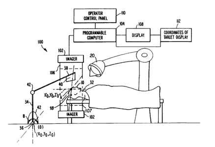
An apparatus 100 carries out the imaging, signal
processing and display necessary to provide images of essentially
the same coordinates in the human body which can be compared over
time, or to provide the location of targets, such as tumors is
shown in Figure 7. Such an apparatus 100 is comprised of an
imag,er 102 that supplies imaging data and is controlled by a
programmable computer 104. The imaging data is obtained from a
source 106 in the imager 102 that is approximately placed about a
patient 107 as is well known in the art. The imaging data
experiences signal processing, as described above, and the desired
images are displayed on display 108. Additionally, operator
interaction can be achieved through an operator control panel 110
and the coordinates of a target can be displayed in the
coordinates of the target display 112 for radiation therapy
applications.
An application that takes advantage of a fully-defined
internal coordinate system of the body relates to radiation
therapy. For radiation therapy the location of a radioactive beam
of an external coordinate system must be related to the internal
coordinate system. See Figure 5 where the external coordinate
system can be considered the unprimed system and the internal
system the primed system. The point P can represent the location
of a point of a tumor. In this situation the actual distances and
locations of the point P in the primed coordinate system, and the
location of the origin `s of the primed coordinate system are
important. If the point P is known with respect to the internal
~ 334035
or primed coordinate system, and the primed coordinate system is
known with respect to the external or- unprimed coordinate system
and the Euler angles of rotation are known, then the location of
point P is known with respect to the external coordinate system.
For example and referring to Figure 7, in radiation therapy o`r
surgery knowing where the internal coordinate system A is with
respect to an external coordinate system B has many uses. In
radiation therapy if the location of a tumor is known with respect
to the internal coordinate system and the internal coordinate
system is known with respect to an external coordinate system
having a radiation source 20, such as an x-ray machine for killing
cancer cells, then radiation can be applied only to the tumor
provided it can concentrate on the volume of the tumor only. This
would remove the guess work of a radiotherapist looking at various
images of a tumor in a body and estimating where to aim the
radiation source so, hopefully, only the tumor is irradiated. The
location of a tumor in an internal coordinate system can be
identified for instance, by a first imaging session. The data
therefrom is stored in a medium that allows its recall when the
tumor position is desired to be known and it is not desired to
have to retake images of the anatomy.
One way to accomplish the irradiation of a s~ecific
location in the body 32, where, for instance, a tumor is located,
involves the use of a robot arm 34 whose base 36 can be chosen as
the origin (0,0,0) of the external coordinate system B. At the
tip 38 of the robot arm 34 is located a sensor 40. The sensor 40
-32-
1 334U35
can be a metal detector or an ultrasonic detector or any
instrument that can sense the position of a fiducial implant 10 in
a body 32. If the fiducial implants 10 are placed in a skull 18
and there is a tumor therein, the sensor 40 in the tip 38 of the
robot arm 34 is moved by the arm 34 until it contacts a fiducial
implant 10 in the skull 18. The movement of the robot arm 34 is
tracked by a computer (not shown) so the position of the sensor 40
relative to the arm's 34 base 36, the origin O of the external
coordinate B, is known. The means to track the arm is well known
and is accomplished by sensors (not shown) in critical locations
of the arm 34, detecting rotation or movement of the joints 42 of
the arm 34. By supplying this information to a computer along
with the information of the fixed lengths of the structure of the
robot arm 34, the tip 38 location of the arm 34 is always known.
When the tip 38 of the arm 34 rests on the fiducial implant 10 in
the skull 18, the location of the internal coordinate system A
defined by the fiducial implants 10 is known with respect to the
external coordinate system B. Supplying the Euler angles of
rotation and the location of the tumor which is known relative to
the internal coordinate system A to the computer, provides the
ability to determine the location of the tumor in the external
coordinate system B. The location of the tumor is known relative
to the internal coordinate system through for instance the image
data already stored, and the fact that the fiducial implants 10
are also fixed relative to each other once they are in place. The
radiation source 30 and where it is aimed is known by the computer
relative to the external coordinate system B. The computer,
-33-
~ ~3403S
having the information where the tumor is located in the external
coordinate system B, can aim the radiation source 30-to precisely
irradiate the tumor site in the brain. In general, the location
of a point P in the internal coordinate system relative to the
external coordinate system is determined when the distance between
the origins of the two coordinate systems is known and the Euler
angles are known, as described above.
In surgery, the internal coordinate system defined by
the three fiducial points can allow, for example, a laser to be
followed as it cuts through tissue to a tumor. An imaging system
present in the operating theater would be positioned to
continually take imaging data that is provided to a computer
system which also guides the laser based on the inputted data. As
the laser cuts through the tissue, the change in the tissue is
apparent through the imaging system and can be followed with
respect to the fixed internal coordinate system. When a
predetermined position is reached by the laser, or a predetermined
portion of tissue has been removed by the laser, the computer
controlling the laser and processing the imaging data would
discontinue the operation of the laser.
In the operation of the invention, after the fiducial
implants are in place in a patient, imaging data is taken at a
first time and stored. At distinct intervals in time, for
instance about every year thereafter, the patient returns to the
location of the imaging system or one similar to it, and undergoes
-34-
~ 334035
lollow_up imaging. The most recently received imaging data is
then reformatted, as described above, to obtain high fidelity
images of the same cross-sections on the body as attained in the
earlier session. The images from the latest session are then
compared with the earlier session (if there are many earlier
sessions they can all be used for comparison purposes) to
determine if there have been any significant changes such as
progression or regression of an abnormality, such as a tumor. The
imaging data collected from various imaging sessions taken at
different time intervals can, of course, be compared many ways
such as by reformatting images taken at earlier sessions to show
an image slice of interest chosen from the latest session, instead
of just comparing image slices of a latest session to those of an
earlier session. The purpose of the comparisons, as stated
earlier can be multifold: (a) either a simple follow-up of the
growth of the tumor, without therapy; or (b) verification of
therapeutic treatment, such as radiation or chemotherapy or (c)
follow-up of surgical treatment.
In the operation of the invention with regard to
radiation therapy, the tumor is first identified in the patient's
body. The patient is then positioned in the imaging system such
that at least the tumor area can be imaged. The imaging system is
used to locate the position of the tumor in the internal
coordinate system. The image data can, for instance, then be
stored for later use so the tumor position is identified without
new images having to be obtained every time radiation therapy is
~ 334035
performed. The patient can then be placed before a radiation
source, and each time radiation therapy occurs, the information
from the imaging session that is stored is supplied to the
computer operating the radiation source. The internal coordinate
is located with respect to the external coordinate system, for
instance by locating one fiducial implant, as described above,
with respect to a known position in the external coordinate
system. Once the position of the internal coordinate system is
known with respect to the external coordinate system, the tumor
position is known with respect to the external coordinate system,
since the tumor position is already known with respect to the
internal coordinate system from the stored imaging information. A
radiation source is then aimed, for example by a computer
receiving the imaging and position data, at the tumor in the body.
With respect to surgery, the procedure that is followed to take
advantage of the fiducial implants is similar to the procedure
described above for radiation therapy. Once the tumor is located
with respect to the internal coordinate system, and the location
of the internal coordinate system is known with respect to the
external coordinate system, the tumor is located with respect to
the external coordinate system. Surgical instruments can then be
guided to the tumor by the computer with the imaging system placed
in an interactive mode therewith. The imaging data that the
imaging system constantly feeds the computer allows the computer
to track the progress and the extent of the surgery.
-36-
1 3S403~
- Obviously, numerous (additional) modifications and
variations of the present invention are possible in light of the
above teachings. It is therefore to be understood that within the
scope of the appended claims, the invention may be practiced
otherwise than as specifically described herein.
-37-