Note: Descriptions are shown in the official language in which they were submitted.
~ ANIMAL MARKER IMPL~NTING SYSTEM
1 33~3~
B~CKGROUND OF THE INVENTION
This invention relates, in general, to a system for
implanting an identification marker in an animal and, in particular,
to a system for facilitating implantation and retention of an iden-
tification marker into a laboratory animal.
Heretofore, the marking of animals for tracking and testing
purposes has involved marking the animal externally, i.e., tatooing,
branding or tagging. These external markers are difficult to read
when identifying the animal and are extremely limited in the amount
of information about the animal that can be carried by the external
marker.
In order to overcome the disadvantages noted above with
external markers, a system has been proposed whereby markers carry-
ing information that can be read by an external detector can be
implanted in a test animal. However, such a system requires an
instrument that permits a marker to be delivered into the animal
without difficulty and wherein the marker will remain securely
embedded in the lab animal for a considerable length of time.
SUMMARY OF THE lNv~NlION
Generally speaking, in accordance with the invention, an
improved apparatus for implanting a marker into an animal is pro-
vided. The apparatus includes a hollow tube having an opening at
each end. An entrance end of the hollow tube is supported within a
housing. An exit end of the tube is sharp to allow subcutaneous
penetration of the tube underneath the skin of a laboratory animal.
A plunger is slideably mounted within the housing. The plunger is
adapted to displace the marker from a first position in the tube out
of the exit end of the tube.
In an exemplary embodiment, an electronic transponder
containing information about the animal, such as identification
numbers, is placed in the marker. When the tube is inserted below
the skin of the animal and the plunger is displaced, the marker
,1 ~
~ 2- 1 334366
containing the electronic transponder is forced through the tube,
lodging it underneath the skin of the animal.
An object of this invention is to provide an improved appar-
atus for implanting markers in laboratory animals.
A further object of this invention is to provide an implant-
ing system for facilitating identification of laboratory animals.
Still a further object of this invention is to provide an
easy to use implanting instrument for implanting a marker into a
laboratory animal.
Yet a further object of the invention is to provide an
implanting system for implanting a marker subcutaneously in the
animal so that the marker will be retained within the animal.
Still other objects and advantages of the invention will in
part be obvious and will in part be apparent from the specification
and the drawings.
The invention accordingly comprises features of construc-
tion, combination of elements and arrangement of parts which will be
exemplified in the construction hereinafter set forth and the scope
of the invention will be indicated in the claims.
BRIEF DESCRIPTION OF THE DRAWINGS
For a fuller understanding of the invention, reference is
had to the following description taken in connection with the
accompanying drawings, in which:
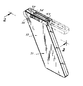
FIG. 1 is a perspective view of an animal marker implanting
instrument constructed in accordance with a preferred embodiment of
the instant invention;
FIG. 2 is a partial sectional view taken along line 2-2 of
FIG. l;
FIG. 3 is a partial sectional view taken along line 2-2 of
FIG. l;
FIG. 4 is a partial sectional view taken along line 4-4 of
FIG. 3;
FIG. 5 is a partial sectional view taken along line 5-5 of
FIG. 3;
_3_ ~ 33436~
FIG. 6 is a partial sectional view taken along line 2-2 of
FIG. 1, when the needle assembly is inserted therein;
FIG. 7 is a sectional view taken along line 7-7 of FIG. 6;
FIG. 8 is a perspective view of the implanting instrument
illustrated in FIG. 1 in use;
FIG. 9 is a plan view of the implanting instrument illus-
trated in FIG. 1 in use;
FIG. 10 is a sectional view taken along line 10-10 of FIG.
9;
FIG. 11 is an enlarged partial sectional view of the implan-
ter instrument illustred in FIG. 10;
FIG. 12 is a sectional view of the implanting instrument
taken along line 12-12 of FIG. 11;
FIG. 13 is a perspective view of the needle assembly;
FIG. 14 is a sectional view of the marker depicted in FIG.
11;
FIG. 15 is a sectional view depicting a sectional view taken
along line 15-15 of FIG. 13; and
Fig. 16 is a perspective view of a cylinder to be used as
part of the instant invention.
DETAILED DESCRIPTION OF THE PREFERRRED EMBODIMENTS
Reference is initially made to FIGS. 1 through 15, wherein
an animal marking system including an implanting instrument, gener-
ally indicated at 10 (FIG. 1), a needle assembly, generally indicated
as 19 (FIG. 13) and an animal marker, generally indicated as 30
(FIGS. 11, 12 and 14) is depicted. As is explained in greater detail
below, the cooperation of the needle assembly, marker and implanting
instrument permits the facile implantation of a marker into a
laboratory animal and the retention of the marker within the animal
during long periods of laboratory monitoring and testing.
Reference is now particularly made to FIGS. 1 through 5,
wherein instrument 10 is illustrated in detail. Instrument 10
defines two opposed half walls 27 which are molded in mirror image
and secured together to define a unitary housing in the shape of a
handle 31. Each opposed wall 27 is defined by a substantially
parallelogram shaped configuration including lengthwise mating
~4~ 1 3 3 4 3 6 6
walls 27a and lateral mating walls 27b and 27c. As is explained in
greater detail below, mating walls 27a are inclined with respect to
lateral mating walls 27b to define handle 31 and to facilitate
storage therein of a plurality of needle assemblies. A cap 34 is
slideably mounted to the housing defined in opposed wall 27. In an
exemplary embodiment, at least one of the opposed walls 27 can be
transparent or translucent to allow the user to view the needle
assemblies 19 disposed within the handle.
Cap 34 is normally disposed in a closed position, and can
be displaced in the direction A (FIG. 2) from a closed position
(solid lines in FIG. 2) to an open position (phantom lines in FIG.
2). As is illustrated in FIG. 7, cap 34 includes side walls 34a and
gripping walls 34b which are disposed in elongated slots 27' formed
in opposed walls 27. Cap 34 includes ribs 35 on the side for
permitting the cap to be easily gripped and can be displaced between
an open and closed position. An arrow 37 or other indicia can be
imprinted on cap 34 to indicate the proper directions for sliding.
Opposed lateral walls 27b are covered by cap 34 when cap 34
is in a closed position. Opposed lateral walls 27b are configured
to define opposed recessed walls 38a and an open chamber, generally
indicated at 38, for receiving a needle assembly and for permitting
each needle assembly to be dispensed through the opening from the
interior of the housing when cap 34 is displaced to an open position.
Opposed lateral walls 27b are further configured to define a channel
39 which orients the needle assembly when it is positioned in chamber
38.
Referring particularly to FIGS. 10 through 15, needle
assembly 19 is formed from a stainless steel hollow tube 20 having
an exit opening 21 and an entrance opening 23. Exit opening 21 is
formed in the shape of an inclined edge 22 which forms a sharp point
for permitting the tube to easily penetrate an animal's skin. The
side of tube 20 having entrance opening 23 is molded in a plug 24.
Plug 24 includes a sleeve 25 integrally formed therewith and pro-
jecting about tube 20 to extend along a portion of the tube's length.
Plug 24 includes arcuate end walls 24a for facilitating the posi-
_5_ 1 334366
tioning of the plugs in chamber 38 in a manner that will be discussedin detail below. As is particularly illustrated in FIG. 15, marker
30 is positioned in tube 20 near the exit opening 21 thereof. A drive
pin 16 is used to position the marker within the tube. Drive pin 16
includes a sealing disc 17 that is integrally molded therewith.
Sealing disc 17 has an outside diameter that is sufficient to
interference fit with the inside diameter of the tube 20 and prevent
displacement of the drive pin during normal stoage and handling of
the needle assembly. Drive pin 16 aids in positioning the marker in
the tube. However, it has been found necessary to facilitate
positioning of the marker in tube 20 particularly when the marker is
a glass capsule in order to prevent the marker from slipping out of
the exit opening of the tube.
Reference is now made to FIGS. 11 and 12 wherein a projec-
tion 29 integral with sleeve 25 extends through opening 28 in order
to prevent the marker from slipping or moving in the tube prior to
the discharge of same into the animal. This projection can be easily
formed during assembly of the hollow tube within plug 24 by molding
the plug about the tube and permitting the resin used to form the tube
to enter aperture 28. Projection 29 is intended to frictionally
engage marker 30 when the marker is positioned within tube 20 to
prevent the marker from sliding in the tube. Projection 29 will hold
the marker in place until a force sufficient to push marker 30
through tube 20 is applied to a plunger and, in turn, to the marker.
In an exemplary embodiment, tube 20 is stainless steel.
However, tube 20 may be made from other rigid FDA approved materials,
such as Ultem, manufactured by General Electric. Also, as afore-
noted, sleeve 25 and plug 24 can be integrally formed by injection
molding a plastic resin about the entrance opening of tube 20. Also,
the sleeve and plug may be formed of rigid materials other than
plastic.
Needle assembly 19 is easily positioned in chamber 38 when
cap 34 is displaced into an open position. Moreover, needle assembly
19 is tightly secured within chamber 38 by returning cap 34 to a
closed position. This prevents any wobbling of the needle 19
assembly during use.
_ -6- 1 3 3 4 3 6 6
Plunger 18 includes a rod 41 and a knurled surface 43 inte-
grally formed at one end of rod 41. Plunger 18 is slideably mounted
within elongated channel 39 formed by lateral walls 27b formed in the
top of the housing. Knurled surface 43 projects through elongated
opening 44 in cap 34 and permits the plunger to be displaced between
a start position and an implanting position. Channel 39 is coaxially
aligned with the entrance opening 23 of tube 20 of the needle
assembly 19 and alignment channel 49 to form a continuous pathway for
rod 41 when needle assembly 19 is retained in chamber 38. Knurled
surface 43 extends through elongated opening 44 (FIG. 9) in cap 34,
allowing displacement of the plunger 18 by pushing knurled surface
43 from a start position to an implanting position. Plunger 18 also
includes seats 18a projecting therefrom which rests against stops
27e formed by opposed half walls 27. Stops 27e and seats 18a
cooperate to normally maintain the plunger at the start position
depicted in FIG. 6.
The distance of the placement of the marker in the tube from
the exit opening and the length of elongated opening 44 have relative
lengths with respect to each other. When cap 34 is displaced in the
direction A into an open position, it will capture knurled surface
43 if it is not already in a start position and displace the plunger
to a start position so that rod 41 is entirely displaced outside of
the entrance opening 23 of the needle assembly 19. Furthermore, the
distance of the placement of the marker from the exit opening
determines the distance through which the rod will be displaced and,
hence, the preferred distance of elongaged opening 44. Moreover,
this distance further assumes that rod 41 is entirely displaced
without the entrance opening of the tube when plunger 18 is in a start
position. This permits placement of needle assembly 19 in chamber
38.
When cap 34 is returned to a closed position, stop 27e helps
maintain plunger 18 at its start position so that the plunger is not
unintentionally pushed forward. If slideable cap 34 is not pushed
entirely into a closed position, knurled surface 43 is prevented from
being pushed forward sufficiently to cause plunger 18 to eject the
1 334366
marker 28 from the needle assembly 19. This configuration prevents
use of the instrument unless the needle assembly 19 is fully secured
within chamber 38 and is securely captured by cap 34 being displaced
into a closed position. Also, since knurled surface 43 of the
plunger 18 comes in contact with the cap at the limits of elongated
opening 44, the plunger 18 is automatically positioned by manipulat-
ing the cap.
Reference is now also made to FIG. 8, wherein operation of
the instant invention is depicted. In an exemplary embodiment,
marker 30 is stored within tube 20 and is retained therein by a
projection 25. Cap 34 is then slid into an open position. Needle
assembly 19 is then pivotably displaced into chamber 38. Cap 34 is
then displaced forward into a closed position supporting and anchor-
ing needle assembly 19 securely in place within chamber 38 and
channel 49.
Next, a test animal, such as a mouse 46, must be stabilized.
As illustrated in FIG. 8, a mouse can be picked up in the user's one
hand and the implanting instrument held in the user's other hand.
However, as is illustrated in FIG. 16, in an exemplary embodiment,
a cylinder 80 that is open at both ends can be utilized to render the
head of the mouse immobile. By inserting the mouse's head in a
cylinder the mouse cannot turn its head and bite the user's hand or
otherwise interfere with the procedure. Furthermore, once the
mouse's head is immobilized in the cylinder it permits the hand of
the user to be used to stretch the animal's skin and thereby
facilitate manipulation of the mouse during subcutaneous implanta-
tion. Accordingly, the implanter systems of the instant invention
contemplates the use of different sized tubes to accommodate the
distinct differences in the size of the laboratory animals. Once the
animal is immobilized, the user is prepared to insert tube 20 into
the laboratory animal.
Exit end 21 of tube 20 is inserted subcutaneously into mouse
46 until the animal's skin 48 reaches the edge of sleeve 25. This
automatically places marker 30 at the desired position beneath the
skin. Knurled surface 43 of the plunger 18 is then pushed forward,
`~ -8- 1 3 3 4 3 6 6
preferably with the user's thumb 50, with enough force such that
plunger 18 engages drive pin 16. Knurled surface 43 is displaced
until knurled surface 43 is disposed into an implanting position so
that rod 43 comes in contact with the end of opening 44 in cap 34.
At this point, plunger rod 41 of plunger 18 has engaged drive pin 16
and extends far enough within tube 20 to have forced drive pin 16 to
eject marker 30 from tube 20 underneath the animal's skin. Next, the
implanting apparatus is removed from animal skin 48, cap 34 is pulled
back and needle assembly 19 is removed and discarded. The process
may then be repeated for another animal.
In an exemplary embodiment, marker 30 is a glass capsule
having therein an electronic transponder containing identification
information about the animal. This is used by way of example only.
This process is adaptable to the implantation of any type of marker.
Marker 30 is formed by embedding an electronic transponder (not
shown) in a glass capsule. By using an electronic transponder, the
amount of storable information is greatly increased, especially when
transponder information can be directly linked to computer systems
containing further information and processing software. Because the
capsule is glass, it tends to slide easily in stainless tube 20. It
is for this reason that projection 29 is used to interference fit the
capsule in the tube and prevent same from moving within the tube
during storage and handling of the needle assembly.
Each needle assembly 19 is sealed within a sanitary sleeve
33 which can be easily removed when the needle assembly is displaced
into chamber 38 for use in the manner described above. Moreover,
after sanitary sleeve 33 is used to cover the exit opening of the
tube, a sterilant gas can be injected into a chamber defined by tube
20, drive pin 16 and sealing disc 17 and the sleeve 33. By
introducing a sterliant gas, the marker can be sterilized and remain
sterilized until the needle assembly is ready for use. Furthermore,
as is illustrated particularly in FIGS. 2 through 5 and 13, the
opposed side walls 27 of the housing and the plug 24 of each needle
assembly are configured in a manner discussed below to permit each
needle assembly to be stored in the handle and removed therefrom for
easy use.
- 9 -
1 334366
Specifically, a pair of opposed ramps 45 are formed in each
wall 27. Ramps 45 are formed in mirror image on each wall so that
they are disposed in registry with each other when walls 27 are
brought together to form the housing defining handle 31. Further-
more, each ramp is disposed in parallel with lateral wall 37c and at
an angle with respect to the lengthwise extent of the handle. Ramps
45 are spread a sufficient distance apart to permit two rows of
needle assemblies to be stored in the handle.
The plug of each needle assembly includes positioning
grooves 26 found in opposed surfaces, the grooves being disposed on
an angle with respect to the lengthwise extent of the plug and
diagonally opposed with each other to facilitate placement of each
needle assembly in the housing during assembly of the product. As
is illustrated with some particularity in FIGS. 3 through 5, each
needle assembly can be positioned within handle by racking the plug
onto a first ramp 45 so that the ramp is positioned within the
positioning groove 26. The groove 26 and ramp 45 prevent any
substantial lengthwise displacement of each needle assembly during
storage and use of the instrument. The opposing ramp assists in
positioning the plug by pressing against the plug. Each ramp 45
includes a positioning ramp 45a that is parallel with the lengthwise
side walls 27a of handle 31. Positioning ramp 45a does not protrude
as far as ramp 45 and is provided to assist in preventing the plug
from sliding laterally and to further assist in guiding the needle
assembly through the opening in chamber 38 when a needle assembly is
to be removed from the handle.
In an exemplary embodiment, ten needle assemblies are
stored on each ramp 45. As noted above, wall 27a and ramp 45a
facilitate delivery of each needle assembly to the operator.
As is illustrated in FIG. 2, the opening in chamber 38 is
sufficiently large to permit the needle assembly to be removed
therethrough. Accordingly, when a needle assembly is needed, cap 34
is displaced from a closed position to an open position. By
manipulating the orientation of the housing, a needle assembly
positioned closest to chamber 38 will then slide out of the housing
-lo- 1 3 3 4 3 6 6
through opening 38. As aforenoted, such manipulation can be facili-
tated by forming one of the opposed walls 27 forming handle 31 out
of a transparent or translucent material. It is then a simple matter
to position plug 24 of the needle assembiy in chamber 38, slide cap
34 to a closed position and remove the sanitary sleeve 33, so that
the user is ready to begin implantation of the marker in the manner
discussed above.
Reference is now made to FIG. 14, wherein a marker 30 is
formed of a smooth material 81, such as glass. As aforenoted, the
use of a glass marker can be problematical. First, when the needle
assembly does not include a projection 29, marker 30 is not secured
in the tube and, hence, the marker may slide out of the tube of the
needle assemlby. Also, it has been observed that when a glass
encapsulated transponder is implanted in a laboratory animal, migra-
tion of the transponder out of the wound of the animal can occur.
Accordingly, in a preferred embodiment, one-half of marker 30 is
coated with a layer 83 having a high coefficient of friction. For
example, Silastic~, manufactured by Dow Corning, has been success-
fully used. Also, polypropylene has been used as a coating. By
utilizing a layer coating marker 30, projection 29 can be eliminated,
thereby allowing for a thinner tube 20 having a greater inner
diameter than the embodiments containing projection 29.
The instant invention further contemplates a method of
forming layer 38 about a glass marker. Specifically, markers are
partially inserted into a mold cavity. Thereaftrer, a polypropylene
resin is injected into the mold cavities and cured about the marker
to define a suitable non-slippery surface.
In a further embodiment, the outer surface of glass of
marker 30 can be etched. Although etching of the outer glass coating
prevents migration in the animal, projection 29 is still needed to
hold marker 30 in place in the tube 20. However, etching has been
found to weaken the marker and although experimentally viable, does
not appear to offer the same efficiency as the use of a coating on
the glass capsule.
1 3343b~
It will thus be seen that the objects set forth above, and
those made apparent from the preceding description, are efficiently
attained and, since certain changes may be made in the above
construction without departing from the spirit and scope of the
invention, it is intended that all matter contained in the above
description or shown in the accompanying drawings shall be interpre-
ted as illustrative and not in a limiting sense.
It is also to be understood that the following claims are
intended to cap all of the generic and specific features of the
invention herein described, and all statements of the scope of the
invention which, as a matter of language, might be said to fall
therebetween.