Note: Descriptions are shown in the official language in which they were submitted.
1337C44
-
DEVICE AND PROCESS FOR CELL
CAPTURE AND RECOVERY
Thi~ is a divisional application of CAnA~;an
patent application no. 613,945 filed September 28, 1989.
Technical Field
The subject field concerns cellular
separations employing devices having specificity for
cell surface proteins. Particularly, the cellular
source will be blood, spinal fluid, bone marrow, tumor
homogenates, lymphoid tissue and the like.
Background
There are numerous situations where it is of
interest to isolate a specific class of cells or to
remove a particular set of cells from a mixture of
cells. Techniques which have been employed include
fluorescence cell sortinq, magnetic immunobeads,
complement-mediated lysis, affinity chromatography,
centrifugal elutriation and polystyrene panning of
cells. Cells having substantial density differences,
such as that between platelets and red cells can be
grossly fractionated by gradient centrifugation
methodologies. However, mammalian cells with
equivalent densities, such as tumor cells, lymphocyte
subsets, granulocytes, or stem cells, require some form
of separation using molecular recognition of surface
markers which correlate with their phenotype
Similarly, such molecular mechanisms are required to
separate viruses and bacteria from one another in
complex mixtures. Each of the aforementioned methods
have serious drawbacks for many applications, where
there is interest in isolation or removal of particular
133764A
~_ 2
subsets of cells.
Disadvantages of fluorescence activated cell
sorting for recovery of viable sorted cells are the
slowness of the procedure, the fact that the isolated
cells are coated with antibody, and the limited amounts
of cells which can be obtained by the procedure.
With immunobeads, it is difficult to recover
the cells from the beads after separation; the cells
are frequently coated with antibody and magnetic beads,
and distinct separations are only difficultly achieved.
Complement mediated lysis is problematic for two
reasons: first, depletion of target cells is
incomplete, and second, non-target cells can be
-adversely affected by exposure to complement and the
toxic by-products of target cell lysis. Affinity
chromatography of cells using, for example, Sephadex
G-10 coupled to antibody, suffers from poor recovery
and inefficient depletion of target cells. Centrifugal
elutriation is not capable of separating different
phenotypic subpopulations of cells of like size.
The last methodology, panning developed by
Wysocki and Sato, PNAS, 75:2844, (1978), utilizing
passively adsorbed antibody on polystyrene, is
particularly inadequate. Only low recoveries can be
achieved, and the process suffers from lack of
specificity and contamination of the separated cells
with antibody.
As the immune defense system becomes
elucidated, it is increasingly evident that subsets of
cells can have relatively narrow ranges of
activities. Thus, subsets can be specialized for
response to a particular disease, such as neoplasia,
infection, viral or cellular, etc., response to
transplants, and the like. It is therefore of great
interest to be able to identify and purify these
subsets of cells, not only to understand their action,
but also to use these cells for prophylactic and
1337644
~_ 3
the~apeutic purposes. In orde~ to achieve the desired
result~, lt 1~ necessa~y that substantl~lly pure
populations o~ the desl~ed subset o~ subsees o~ cells
can be obtdined. ~urthe~more, the cells should be:
~1) free of antibodies on their surface, (2) viable,
(3) capable o~ ful~illin9 their normal function and (~)
responsive to activation by biologicals ln the same
manner as normal cells ln their normal en~onment
SU~MARY OF THE INVENTION
Methods, compositlons and devices are provided
~o~ the selective capture and release of biological
particles capable of replication, partlcularly virus
particles and cells. A medium containing the particles
15 ~ i~ contacted with a solid support havlng a h~gh density
of specif~c receptor sites, whereby particles having
the complementary liqand become bound to the surface.
The biolo~icals may be released from the recepto~s by
either blologlcal activation ~esulting ln ligand
shedding and release o~ physical means such as
pipetting, mechanical vibratlon or ultrason~c sound, aQ
app~opriate. The cells can be numerlcally expanded, -
subjected to biologicals or other factors for
differentiation and/or activation, or the like, and may
be used for research, diagnostic, prophylactic and/o~
therapeutic pu~poses.
This invention provides a method for chanqing
the composition of a mixture of biological particle~,
employing receptors specific for at least one ligand
present on said particles, wherein said receptors are
covalently bound to a surface in a substantially
uniformly dense distribution to provide at saturation for
a substantially uniform layer of particles, said method5 comprisings
contacting 6aid surface with said mixture of
biological particle~ for sufficient time for biological
particles having the ligand to which the receptors
specifically bind to bind to said surface; and
133764~
3a
removing non-specifically bound bioloqical particles
without significantly disturbing specifically bound
biological particles.
This invention also provides a method for
changing the composition of a mixture of hematopoietic
cells, employinq monoclonal antibodies specific for at
least one cell surface protein present on said cells,
wherein said monoclonal antibodies are covalently bound
to a surface in a substantially uniformly den-~e
distribution to provide at saturation for a substantially
uniform layer of cells, said method comprisinq:
contactinq said surface with said mixture of
hematopoietic cells for sufficient time for said cells
having a cell surface protein for which the covalently
bound antibodies have specificity to bind to said
surface;
removing non-specifically bound cells without
significantly disturbing specifically bound cells.
This invention also provides a method for
preparing a therapeutic cellular composition for use in
the treatment of a patient, said method comprising:
contacting cells with monoclonal antibodies specific
for at least one ligand on the cells, wherein said
monoclonal antibodies are covalently bound to a surface
in a substantially uniformly dense distribution to
provide at saturation for a substantially uniform layer
of cells, whereby cells having ligands for which the
covalently bound antibodies have specificity become
specifically bound to said surface;
removing non-specifically bound cells from said
bound cells;
releasing bound cells substantially free of
monoclonal antibodies by physical methods or mitogenic
release to provide a substantially homogeneous phenotype
population having activity for use as a therapeutic
agent.
1337644
3b
This invention also provides a method for
preparing a substantially homogeneous population of
lymphokine activated killer cell~ (LAR cells), said
method comprising:
contacting hematopoietic cells from a patient with a
neoplasm with receptors comprising monoclonal antibodies
specific for CD3 or CD5, wherein said antibodies are
bound to a surface in a substantially uniformly dense
distribution to provide at saturation for a substantially
uniform layer of cells, whereby CD3' or CD5' cells become
bound to said surface and a population of cells depleted
of CD3 or CD5 is nonbound;
contacting the CD3 or CD5 depleted nonbound
population with receptors comprising antibodies specific
for CD14, CDl9, or CD20, wherein said CD14, CDl9, or CD20
antibodies are covalently bound to a surface in a
substantially uniformly dense distribution to provide at
saturation for a substantially uniform layer of cells,
whereby cells positive for at least one of CD14, CDl9, or0 CD20 become bound to said surface; and
culturing unbound cells depleted for CD3 or CD5, and
CD14, CDl9, or CD20 in a lymphokine containing culture
for sufficient time to expand the cell population or
activate the cell population to provide a substantially5 homogeneous LAK cell population having cytolytic activity
for neoplastic cells.
This invention also provides a method for
preparing a substantially homogeneous population of T-
cells, said method comprising:
contacting hematopoietic cells with receptors
comprising monoclonal antibodies specific for T-cells
wherein said monoclonal antibodies are covalently bound
to a surface in a substantially uniformly dense
distribution to provide at saturation for a substantially ,~
uniform layer of cells, whereby T-cells to which the
covalently bound antibodies specifically bind become
bound to said surface;
1337~4~
-
3c
releasing said bound TJcells substantially free of
monoclonal antibodies by physical methods or mitogenic
release from said antibodies, whereby said cells are
substantially free of antibodies to T-cells, to provide a
substantially homogeneous population of T-cells.
This invention also provides substantially
homogeneous populations of cells prepared according to
the preceding methods.
This invention also provides a method for
protecting a polymeric surface from degradation when
subjected to ionizing radiation (such as the plastic
surface of the preceding methods), wherein the surface
comprises a biologically active agent (such as the
receptors of the preceding methods) coupled thereto,
which method comprises:
applying to the surface an amount of a protective
composition contAin;~g a surface stabilizing agent and an
oxygen radical scavenger effective to preserve the
activity of the biologically active agent when subjected
to sterilizing amounts of irradiation.
This invention also provides a method for
preparing a polymeric surface which comprises:
providing a polymeric surface comprising a
biologically active agent coupled thereto, applying to
said surface a protective composition containing a
surface-stabilizing agent and an oxygen radical
scavenger, whereby the activity of the biologically
active agent is preserved upon subjecting said surface to
sterilizing amounts of ionizing radiation.
This invention also provides the preceding
methods for protecting or preparing a polymeric surface,
wherein the surface is dried and wherein the surface is
subjected to sterilizing amounts of ionizing radiation.
The surface stabilizing agent is a relatively high
1337644
'_
3d
molecular weight agent such as is conventionally used in
drying biologically active substances, such as a
polypeptide or polypeptide moiety, including an albumin
such as human serum albumin (HSA). The oxygen radical
scavenger is generally a polyol or a polyol moiety, such
as is provided by a saccharide, including sucrose.
This invention also provides a polymeric
surface prepared according to the preceding methods and a
method to conduct extracorporeal treatment of body fluid
which comprises passing said fluid over a surface coupled
to a biologically active agent prepared according to the
preceding methods and which has been subjected to
sterilizing amounts of ionizing radiation.
BRIEF DESCRIPTION OF THE DRAWINGS
Fig. 1 is a schematic drawing of a process to
prepare cells from peripheral blood or bone marrow for
the captured device according to this invention; and
Fig. 2 is a diagrammatic view of the inside of
a cell capture device.
DESCRIPTION OF THE SPECIFIC EMBODIMENTS
Methods, compositions and devices are provided
for isolating a particular population of biologically
1337644
replicatable particles, particularly cells, from a
mixture of particles by binding the population to a
solid substrate through the intermediacy of one or more
specific receptors. Optionally, the particles may then
be treated in a variety of ways to affect the popu-
lation size and/or characteristics of the captured
particles. The cells may then be released from the
support substantially free of receptor.
The method will involve contacting a source of
particles with receptor bound to a support in a
collection device, where the population of the receptor
on the surface provides for a high binding density for
the ligand(s) of interest. The conditions for the
contact are such as to allow for sufficient time and a
low degree of turbulence to permit the particles to
specifically bind to the receptor. After sufficient
time for binding to occur, the medium may be removed
and the surface washed to remove non-specifically bound
particles. Since the particles will normally be bound
to the surface at a plurality of sites, so as to have a
high binding avidity, the washing may be fairly
vigorous to insure the substantially complete removal
of all non-specifically bound particles. The particles
may then be subjected to a wide variety of conditions
or treatments, usually involving contact with one or
more reagents. Optionally, the medium containing the
reagents may then be removed and the particles released
from the receptors. Release may be achieved either by
treatment with a combination of a mitogenic agent and a
lymphokine or physical means such as pipetting,
vibration or sonication. The particles may then be
isolated and used for their intended purpose.
For the most part mixtures of cells will be
employed and, therefore, the remaining discussion will
be directed to cells. However, substantially the same
procedures may be used for the isolation of virus and
bacterial particles and in referring to cells, it
~ 1337644
s
should be understood that viruses and bacteria may
usually be substituted therefore.
The subject device finds application in a
number of different situations. The first is a
situation in which one wishes to capture and remove
undesirable cells from physiological fluids, thereby
depleting the fluid of the undesirable cells for
therapeutic benefit, or diagnostic or research
applicatioQs. This may be illustrated by depleting
bone marrow of certain T-lymphocytes to diminish graft-
versus-host disease. The bone marrow, depleted of the
unwanted cells, is immediately prepared for transplan-
tation into the marrow recipient.
A second situation is to capture and recover
certain cells from physiological fluids for research
and diagnosis. Diagnostic applications may include
capture and subsequent enumeration and description of
captured (1) malignant cells from blood or other
tissues, (2) viral or bacterially infected mammalian
cells, (3) viruses or bacteria or parasites themselves
from physiological fluids, (4) human fetal cells for
karyotypic analysis from blood, (5) transplanted cells
from blood as an index of recovery from bone marrow
transplantation, and (6) immune competent cells with a
particular surface marker, such as the presence of the
IL-2 receptor, indicating a state of activation. For
research, one may be interested in (1) the genetic
analysis or modification of captured cells, (2)
analysis and modification of the physiology of certain
classes of cells that may be activated or suppressed by
a particular disease process and ~3) at the molecular
level, the surface membrane compositional analysis and
modification of cells involved in the pathogenesis of a
particular disease.
The third situation lies in the capture and
recovery of cells from physiological fluids for
modification (activation) and return-to the patient of
~i 6 13376~
origin for a desired therapeutic effect. The process
involves cell capture and recovery, processing of the
captured cells or depleted cell population which may
result in numerical expansion and/or biological
activation, and the subsequent recovery and use of
these cells.
The third situation may be further divided
into three levels of application. The first level is
biological activation of the captured/recovered cells
themselves. Activation is performed without further
fractionation of the cells. For example, in the case
of an AIDS patient, CD8 positive cells captured from
peripheral blood can be expanded and activated for
sub~sequent return to the patient of origin. The second
level is selective activation. Captured and recovered
cells are further processed or fractionated to provide
a subset of captured cells, identified for example by
antigen specificity, which are then activated and/or
expanded. For example, certain antigen restricted
subsets of the captured CD8 positive cells can be
selected by certain co-culture conditions and
concentrations of lymphokines, which allow only the
desired subset to be expanded and activated. A third
level is in vitro generation of antigen specific
patient-unique cells for activation or suppression of
the immune function. Exemplary of this situation is
monocyte or B-cell capture and exposing the captured
monocytes or B-cells to a patient-specific immune
complex or other antigens under conditions which
augment monocyte or B-cell antigen uptake, processing,
and presentation along with increased surface MHC
expression. One would then add a subset of effector or
regulatory cells captured from the same patient to
interact with the antigen-primed monocyte or B-cell.
The process of antigen specific T-cell activation would
occur, much in the manner that occurs in the lymph node
of an intact animal or human. An additional example of
7 133~644
cell modification made possible by the subject method
is the introduction of exogenous genes via viral or
other vectors or other means into the captured cells
and the subsequent capture in a second device of the
subpopulation of cells which express the exogenous
gene.
The cellular source may be any mixture of
cells. However, it is desired to have a predetermined
population which may be defined by single or multiple
markers or plurality of markers or ligands. Cellular
sources of interest from animal hosts may include
organs, such as blood, brain, kidney, spleen, heart,
intestine, bone marrow, cerebral spinal fluid, lymphoid
tissue, or the like, or neoplastic cells from any of
the above organs. Other sources may be parasites,
viruses or bacteria mixed with animal cells. The cells
are employed in a flowable form, conveniently as a
dispersion. Where the cells are not held together, as
in blood, the blood will usually have red cells,
platelets and plasma removed to provide for a mixture
of white cells. Where the cells are held together by a
membranous or other connecting material, the cells may
be dispersed either mechanically or enzymatically in
accordance with conventional techniques. The
individual cells may then be dispersed in their
appropriate nutrient medium for separation by the
subject method.
With blood, red blood cells may be removed by
agglutination, lysed with ammonium chloride, removed
with lectins or by centrifugation in accordance with
known ways. Platelets and red blood cells may also be
removed by gradient density centrigation, employing
Ficoll or Leukoprep and isolating the buffy coat, by
centrifugation or the like.
The various cell sources may be subjected to a
variety of treatments in addition to those described
above. In some situations, it may be desirable to
~~ 8 133764~
concentrate the cells by any convenient means, followed
by dispersion in an appropriate nutrient medium. In
some situations it may be desirable to expand a
particular population, where one can selectively expand
one group of cells as against another group of cells.
For expansion, various mitogenic agents may be employed
or interleukins, growth factors, or the like. These
cells will then usually be concentrated by any
convenient means to substantially remove the medium in
which they have been isolated or maintained. Usually,
these cells will comprise at least about 10 vol % of
the dispersion to be used and not more than about 90
vol %, so as to provide a flowable dispersion. The
concentration of cells introduced into the device is
conveniently based upon the surface area of the
derivatized polystyrene surface and will vary widely,
depending upon the frequency of the target cell in the
input cell suspension. Usually, the concentration will
be at least lx103 cells/cm2 and not more than lx101
cells/cm2, usually from about lx105 cells/cm2 to
lxlO7/cm2 .
The separation device may take a wide variety
of forms. For the most part, the device will be
comprised of polystyrene surfaces, where the
polystyrene is normally substantially free of cross-
linking, less than about 0.5%, usually less than about
0.1%, preferably molded or extruded, so as to have a
very smooth surface. Polystyrene surfaces of this
nature allow for substantial uniformity of derivati-
zation, where the orientation of the receptor providesfor a high level of accessibility of binding sites.
(It should be understood in referring to receptor, the
term is entirely arbitrary. By receptor is intended a
molecule which is able to specifically bind to a
complementary molecule. Thus, for the purposes of this
invention, the receptor may be a ligand, which includes
both haptens and antigens, or a surface membrane
1337644
~_ g
protein which specifically binds to another molecule,
such as an immunoglobulin, T-cell receptor, insulin
receptor, etc., or a molecule which is found
intracellularly, such as a steroid binding protein, or
molecules which are found in body fluids, such as
thyroxine binding globulin, lipoproteins, etc.
Therefore, the membrane protein which binds
specifically to the surface bound "receptor" is
referred to arbitrarily as the "ligand." For
convenience, they will be referred to jointly as
complementary members of a specific binding pair.)
The functionalized polystyrene surface may be
the surface of a wall, partition, sheet, hollow fiber,
bead, particle, or the like. For the most part, it
would be desirable to use a flat surface, although in
some situations other surfaces may find application.
The device may take the form of a bottle, standard T
flask, sheets, e.g., a bag or box with multiple
separated sheets, cylindrical or serpentine sheets in a
container, rectangular box, or the like. The choice of
the device will depend upon convenience, the purpose of
the separation, the interaction with other devices, the
cell population of interest, the intended treatment,
whether the population of interest is as a result of
positive or negative selection, or the like.
The surface will be derivatized by substi-
tution of the benzene ring of the polystyrene with an
electrophilic reagent, particularly by a Friedel-Crafts
reaction in a solvent which does not soften or dissolve
the polystyrene. For this purpose, sulfolane finds
particular application. Relatively mild conditions may
be employed and the benzene may be derivatized with a
variety of agents, such as nitro, which may be reduced
to amino, halomethyl, which may be used to form an
amino, hydroxy, or thiol group, or a substituted N-
hydroxymethyl acetamide where the substituent is an
active halogen or pseudohalogen. A description of the
~_ 10 133764~
reaction may be found in EPA 88-304516.3.
The derivatized polystyrene surface may then
be reacted with the receptor. Under the conditions of
derivatization, it is found that a high percentage of
the benzenes at the surface are derivatized, so that
one may obtain a high density of receptor at the
surface.
Depending upon the nature of the receptor,
various reactions may be performed for bonding the
receptor to the surface. Of particular interest is the
bonding of proteins to the surface. Proteins can be
bonded by contacting the proteins in an aqueous medium
with the functionalized surface, having active halogen,
activated carboxy groups, e.g., esters, or the like,
under mild conditions for sufficient time for complete
reaction. Any remaining unreacted functional groups
may be blocked by using an appropriate small molecule
blocking agent. For example, active halogen may be
blocked with aliphatic amines, thiols with maleimide,
or the like. In some situàtions, there may be no need
to block excess reactive groups, since they will not
interfere with the subsequent steps in the process,
The surface may then be washed to remove the non-
specifically bound receptor and evaluated to insure
that appropriate receptor binding has occurred.
Depending upon the nature of the collection
device, the contact with the cell containing medium
will be varied. For example, with a roller bottle, one
may introduce the medium into the roller bottle and
then allow for slow revolution of the bottle over
sufficient time for the cells to become bound. With a
T-flask, or plates-in-a-bag/box configuration, one may
allow the device to stand on a level surface or be
slightly agitated on a shaking platform, followed by
turning the device over and repeating the process on
the other side. Similar techniques may be employed
with other types of containers. Additionally, the
~_ 11 1 337Cq4
device may be centrifuged to press the target cells to
the contact surface.
Of particular interest, is a device which will
be referred to as a collection bag/box. The bag/box
will be a container of rigid or flexible walls
containing polystyrene sheets superimposed or stacked
one upon the other and separated from each other to
allow for flow between the sheets. Packed cells as a
result of concentration, e.g., gradient density
centrifugation or centrifugation, would be allowed to
flow into the bag/box which would be maintained in a
horizontal position. The cellular dispersion would
spread through the bag/box, so as to be in contact with
substantially all of the receptor-coated polystyrene
lS surface in the bag/box. After sufficient time for the
cells to bind, the bag/box may be turned over so as to
allow cells which are still dispersed or unbound to
settle on the film surfaces which are now below them,
so as to provide for efficient utilization of the
surface. Alternatively, the bag/box may be
centrifuged, once on each side, to press the cells to
the contact surfaces.
The contact time will vary widely, depending
upon the concentration of the ligand on the cell
surface, the binding affinity of the receptor, the
concentration of cells in the medium, the nature of the
collection device, and the like. Usually contact times
will be at least about 5 min and not more than about
120 min, usually from about 15 to 60 min.
The cellular dispersion may be moved through
the collection device by any convenient means. A
pressure differential may be achieved through the
collection device by means of pumping. Alternatively,
gravity flow may provide for an appropriate flow
rate. Any convenient technique which allows for a rate
of flow of the cells permitting binding to the surface
without significantly affecting their viability may be
12 1337 644
employed.
The subject devices can be sterilized using
gamma or electron beam radiation, without adversely
affecting the properties of the collection device.
That is, the activity of the receptor is retained,
while at the same time retaining the covalent nature of
its bonding to the surface. Thus, when the collection
device is in use, substantially none of the receptor
bound to the surface is lost.
Once the cells have become bound to the
surface, the collection device may be subject to a wide
variety of treatments. Vigorous washing may be
employed to remove non-adherent cells, since the
adherent cells are bound firmly to the surface at a
plurality of contacts. The wash medium may be pumped
in and out,lligands flowed through the device, or other
means of mild but relatively vigorous agitation. The
wash solution may be deionized water, saline, phosphate
buffered saline, nutrient medium, or the like. The
particular wash solution which is employed will usually
depend upon how the cells are to be used.
Where the cell isolation is concerned with
removal of cells from the cell population, (cell
depletion), the captured cells may be discarded and the
depleted cell population harvested, subjected to any
additional treatments, and then used for its intended
purpose.
For the most part, the subject invention finds
particular application for cells which have been
isolated for subsequent use. Depending upon the
intended use, as well as the nature of the cells, the
cells may be subjected to a wide variety of treatments.
Particularly, where one is concerned with the lymphoid
or myeloid lineages, these cells may be treated to
expand or modify the activity of a particular set or
subset of cells. Thus, various factors may be added
which result in the proliferation of the cells,
~~ 13 1337644
activation of the cells, enhancement or reduction of
one or more surface membrane proteins, and the like.
Depending upon whether one wishes to have all cells
bound during the treatment or allow for the formation
of free cells, one can provide for an appropriate ratio
of receptor to bound cells in the container. By having
a large number of receptors compared to the initially
bound cells, any progeny will also become bound and
retained on the surface. This may serve as an
additional resolution, since other cells which may have
been present and expanded will not become bound and may
be removed from the collection vessel.
~ or the most part, the cells of interest will
be obtained from blood, bone marrow, solid tumors and
lymphoid tissue. These cells may be divided into the
lymphoid and myeloid lineages. The first lineage to be
considered will be the lymphoid lineage. This Iineage
may be further broken down into categories of B-cells
and T-cells. B-cells are identified by having sIg as a
surface marker and rearranged germline DNA at the
immunoglobulin locus. T-cells, for the most part, have
CD2 and/or CD5 as surface markers and rearranged
germline DNA at the T-cell receptor locus. The B- and
T-cells will also include specific progenitor cells,
- 25 although pluripotent stem cells will be discussed
separately, and in the case of B-cells, plasma cells
are also included.
Other specialized lymphoid cells which may be
isolated by markers include: lymphokine activated
killer (LAK) cells, natural killer (NK) cells, tumor
infiltrating lymphocytes (TIL), antibody dependent
cytotoxic cells (ADCC), cytotoxic T lymphocytes (CTL),
etc.
In the myeloid lineage, one may be interested
in isolating monocytes, macrophages, eosinophiles,
basophils, polymorphonuclear leukocytes, dendritic
cells, etc.
1337644
14
The B-cells may be expanded by treatment with
various of the interleukins, 1-7 or others, when
discovered, particularly IL-l, -2, -3, or the like.
The B-cells may be selected by surface bound antigen,
surface markers (e.g., CD20) or by specific binding to
a soluble antigen, where such antigen may be added to
the cells, so that those cells having a surface
immunoglobulin which recognizes the antigen will bind
the antigen to form a complex which is endocytosed and
processed. A fragment of the antigen with the cell's
MXC antigen will then be presented. By adding T-cells
to the medium which are restricted by the B-cells,
T-cells which recognize the antigen fragment will
secrete lymphokines, resulting in proliferation of the
B-cells. By providing for an excess of receptor on the
solid surface or after release of the B-cells
separating the cell population in a second collection
device, one can substantially augment the number of
B-cells and plasma cells which recognize the antigen of
interest.
Alternatively, B-cell fusion partners
(hybridoma cells) or other B-cells from any source can
be selected by binding to a polystyrene surface which
bears covalently bound antigen. Desired hybridoma or
other B-cells bearing sIg reactive with polystyrene
bound antigen will be captured on the polystyrene
surface, allowing for antigen-specific selection of
specific hybridoma or other B-cells. Captured cells
can then be recovered and expanded according to the
procedures described in the subject method.
Alternatively T-cells or any cell containing a specific
surface receptor can be captured by the polystyrene
surface when said polystyrene surface contains said
antigen covalently bound.
Where one wishes to deplete a specific subset
of B-cells, one may add the antigen conjugated to a
toxin, employ antibodies specific for the surface
~ 15 1 3 ~
immunoglobulin and complement or other selective cyto-
toxic capability. In this way, one may selectively
diminish the cells responsive to a particular antigen.
Alternatively, antigen or a B-cell marker (e.g., CD-20)
S can be immobilized on the polystyrene and the targeted
B-cell population captured on the surface.
Particularly, where memory cells exist, one can reduce
the humoral response by substantially depleting the
memory cell population to a particular antigen.
The T-cell population is more varied than the
B-cell population as to function. One may divide the
mature T-cell population into CD4 MHC Class II
restricted cytotoxic, helper or suppressor cells and
CD~ MHC Class I restricted cytotoxic and suppressor
lS cells, where the cells have different functions and
their expansion-and depletion may be of interest.
For either T-cell population (CD4 or CD8), it
may be desirable to activate the T-cells which
recognize a specific antigen. Many strategies can be
used for this purpose. B-cells specific for a
particular antigen may be exposed to that antigen and
then used as antigen presenting cells to activate the
particular antigen restricted T-cell subset.
Alternatively, monocytes or macrophages may be employed
as the antigen presenting cells. Macrophages may be
preferred since they do not have the specificity of the
B-cells for a particular antigen. Therefore, one would
introduce monocytes and/or macrophages, which have been
pre-treated or treated concomitantly with the antigen,
to the bound T-cells to provide for expansion of those
T-cells which recognize the antigen fragment when
presented by the monocyte/macrophage in the context of
the MHC.
Biological activation of cells may be achieved
as a result of a particular soluble or immobilized
lymphokine, e.g., IL-2, or by use of a specific binding
compound, such as an antibody. For example, T-ceils
~ 16 13376~4
may be selected using an anti-T-cell (e.g., CD-5)
surface. The CD-5+ captured cells may then be released
and introduced onto an activating anti-CD3 surface, or
to a surface to which a lymphokine has been covalently
bound. The cells will bind and become activated.
After activation, the cells may be released by
sonication, mechanical agitation or other convenient
means and harvested.
Of particular interest are stem cells, which
may be obtained from bone marrow or peripheral blood.
These stem cells may serve as the progenitors of one or
more of the blood cell lineages. Isolation of the stem
cells may be as a result of both depletion (negative
selection) and/or positive selection. Thus, one may
provide for a series of devices or device subsections
where initially the receptors will bind to undesired
cells for their removal of cells (negative selection)
from the medium. The unbound cells may then be
isolated, freed of the captured cells and further
selected (positive selection) for cells with different
markers associated with stem cells, leaving a bound
population of cells, which may then be freed followed
by further positive selection for a marker specific for
a population which includes the stem cells. In this
way other cells having the analogous final marker may
be removed by the previous process step.
Where cells other than blood cells are
involved, cells of interest for isolation may include
islets of Langerhans, glial cells, astrocytes, neurons,
endothelial cells, epithelioid cells, stromal cells,
stem cells, squamous cells, or the like.
Substantially homogeneous populations, greater
than about 95%, usually 98~, of cells may be achieved,
where the cells may be in a quiescent or activated
state. Cellular compositions may include any cellular
population expressing a surface marker (ligand)
recognized by the immobilized receptor. Such
~_ 1337~14
17
composition~ include cel 18 bearing any of the recognized
leukocyte antigens of the CD (cluster designation series)
or others recognized by monoclonal antibodies to specific
cell surface ligands. Such compositions may include other
blood cells, tumor cells, bacteria, viruses, or parasites
similarly sharing a common surface marker. Virtually any
cell population whose members share a surface ligand
recognized by the immobilized receptor can constitute 10
such a cellular compo~ition.
A great variety of autoimmune, neoplastic, infectious,
metabolic, hematologic and immunologic diseases and
conditions ~the disease field) may be treated in accordance
with this invention. Among autoimmune diseases are
diabetes, lupus erythematosus, and rheumatoid arthritis.
Among infectious diseases are localized and systemic
infections due to gram positive cocci, gram negative cocci,
gram negative bacilli, anaerobic bacteria, mycobacteria,
fungi, viruses, protozoa, etc. Among neoplastic diseases
are all solid and hematologic malignancies. Among
metabolic diseases are atherosclerosis and septic shock.
Among hematologlc diseases are sickle-cell anemia and
familial hypercholesterol anemia. Among immunologic
diseases and conditions are organ transplantation and
immunodeficiency conditions.
These and other diseases or conditions may be
addressed by the subject process as follows. By an
alternative process, one may isolate immune complexes
associated with the autoimmune infectious or neoplastic
disease ~Qee Canadian Patent Application Serial No. 5g8,875
filed May 5, 1989). One can use the antigen obtained from
the complexes to select for both B- and T-cells as
described above which are activated by the particular
antigen. Thus, one can remove blood from the host
suffering from the autoimmune or neoplastic disease and
either selectively deplete B- and/or
133~
18
T-cells associated with the disease or activate T- or
B-cells to suppress the autoimmune disease or to detect
and eliminate the neoplastic cells. In this way, one
may provide for a remission, halt the proqress of the
5 disease, or the like.
Alternatively, in cases of infection,
autoimmune or neoplastic disease, one may provide for
selection of 8- and T-cells reactive with the
particular pathogen or disease antigen. In this case,
10 one would wish to enhance the concentration of the B-
and T-cells associated with the immune defense. Thus,
complexes or antigens associated with the pathogen,
autoimmune or neoplastic disease or the pathogen,
autoreactive or neoplastic cell itself may be used to t
15 enhance the lymphoid cellular population associated
with the defense against the disease. One may isolate
the pathogen, autoreactive or neoplastic cell using the
subject device, and use the isolated pathogen or cell
as the immunogen or receptor, as defined above to
20 capture the appropriate T- or B-cells active in the
defense against the disease. Thus selected, these
cells could be recovered, expanded, activated as
described above for a subsequent return to the patient
of origin. This technique may be used with a wide
25 variety of diseases associated with viruses, e.g.,
AIDS, HT~V-I, or II, bacteria, protozoa, fungi,
helminths, and the like.
In addition, the subject method may be used
for prophylactic and diagnostic purposes in the disease
30 field. The subject method will also find use in
research for detecting B- and T-cell responses,
investigating immune responses, identifying epitopes
associated with autoimmune diseases, and ultimately
used for gene therapy.
One may also use the subject device for
producing monoclonal antibodies by activating B-cells,
followed by immortalization of the B-cells usually by
lg 13376~
fusion with an appropriate fusion partner. In this
way, one can immunize human lymphocytes against
antigens one could not normally administer to a human
host and provide for double selection, initially for
B-cells generally, followed by selection for those
specific B-cells which are capable of binding to the
antigen. Thus, one can greatly concentrate B-cells
specific for the antigen to greatly enhance the
probability of obtaining monoclonal antibodies specific
for the antigen.
The cells may be isolated from the collection
device by different ways. Of particular interest is
the use of a mitogenic agent, such as phytohemagglu-
tinin (PHA), in conjunction with a compound having
growth factor-like activity such as an interleukin or
growth factor, e.g., interleukin-2 (IL-2), GM-CSF, etc.
which results in release of the cells by shedding of
the ligands on the cell surface bound to the receptor.
The medium may be a standard tissue culture medium
containing about 20 to 1000 units/ml IL-2 and about 0.1
to 5.0 ~g/ml of phytohemagglutinin. Alternatively, the
cells may be released by physical methods such as
mechanical disruption, particularly shearing, such as
by vibration, vigorous pipetting or by sonication using
an ultrasonicator and placing the collection device in
a water bath. Conveniently, a Crest ultrasonics model
may be employed. See Menssen, et al., J. Immunol.
Methods ~1987) 104:1-6.
In order to further understand the invention
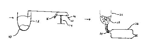
the figures will now be considered. ~ig. 1 is a
diagrammatic flowchart of a process according to the
subject invention using blood as the source of target
cells. The drawing involves a first stage involving
the separation vessel 10, where red blood cells and
platelets are removed to provide a supernatant. The
supernatant 12 is then transferred to a centrifuge 14
having tubes 16, where the supernatant 12 is
1337~4~
centrifuged to concentrate target cells 20 in the tubes
16. The target cells 20 are then transferred to a
feeding vessel 22, which feeds the target cells through
valve 24 into cell capture device 26, also depicted in
Fig. 2. This process is not limited by the example
cited. Any commonly used method to remove red blood
cells, platelets and plasma can be used to achieve
target cell population 20 from peripheral blood or bone
marrow. Alternatively, solid tissue may be
disaggregated by enzymatic or physical means to achieve
target cell population 20.
Cell capture device 26 has a plurality of
polystyrene films or sheets 30 separated by supports
32. On the upper films or sheets 30 are indicated the
presence of receptors 34 desiqnated as R. The
receptors 34 are only indicated on a few of the films
or sheets, indicating that the receptors are on both
sides of the film or sheet, although it should be
understood that all of the films or sheets are coated
on both sides with receptors. Alternatively, the cell
capture device can be a T-flask, microtiter plate,
multiwell plate, roller bottle, cell farm or any other
polystyrene vessel all or part of whose internal
polystyrene surface has receptor immobilized to it.
The cells enter the cell capture device 26
through conduit 36. When cell capture device 26 is
substantially full, it is allowed to stand for
sufficient time for the cells to settle and contact the
receptors on the film or sheet below the liquid
layer. After sufficient time for the cells to have
settled and become attached, the cell capture device 26
may then be turned over so that cells which have not
become specifically bound may settle on the reverse
side of the films or sheets 30 and become bound to the
receptors on that side. The cell capture device may
then be washed by introducing a wash solution through
conduit 36 and allowing it to exit through conduit 40,
21 1337~
so as to remove non-specifically bound cells.
One or more treatment solutions or cell
suspensions may then be introduced to expand the number
of captured cells, activate the captured cells, deplete
a subset of the captured cells, introduce exogenous
genes into the captured cells, or the like. After the
treatment has been completed, the vessel may then be
washed again to remove the treatment solution, cellular
debris, or the like and an appropriate medium
introduced to maintain the viability of the bound
cells. The cells may then be released by adding a
medium containing interleukin-2 and a mitogenic agent,
or by taking the cell capture device 26 and introducing
it into an ultrasonic bath or subjectin~ it to
mechanical vibration or vigorous pipetting. After a
short period of such physical treatment or under
relatively mild sonic vibration, the captured cells are
released and may be harvested.
The following examples are offered by way of
illustration and not by way of limitation.
EXPERIMENTAL
I. DEVTCE AND PROCESS VALIDATION
A. Synthesis of N-(hydroxymethyl)
2-bromoacetamide (H~BA) and generation of the
bromoacetamide polystyrene surface (BA-PS).
HMBA is synthesized by conventional means
(Leonard et al., J. Org. Chem. 50:2480 (1985)) from 2-
bromoacetamide, available from commercial sources, in
the presence of formalin at pH 10, which provides a 93%
yield of the starting reactant, N-(hydroxymethyl) 2-
bromoacetamide (HMBA).
The second step involves the generation of the
bromoacetamide polystyrene surface (BA-PS). In this
step, 2M triflic acid and 0.2M HMBA, both in
tetramethylene sulfone (sulfolane), are mixed 1:1 in a
22 13371i~4
volume sufficient to cover the inner surface of a
polystyrene vessel being activated. The reaction is
allowed to proceed at 27C ~or 3 hours. The reaction
solution is drained, the device washed with water,
followed by ethanol, and the activated polystyrene
chambers are air dried. The resulting bromoacetamide
polystyrene surface is stable in room air for six (6)
months.
B. Cell capture surface preparation,
stabilization and sterilization.
The next step is the receptor capture (the
monoclonal antibody one wishes to covalently bind to
the bromoacetamide-polystyrene surface). The mono-
clonal antibody of interest is diluted to approximately0.01 - 0.05 mg/ml in phosphate buffered saline, pH
7.4. The appropriate volume of diluted monoclonal
antibody is introduced into the polystyrene chamber and
the reaction is allowed to proceed for from about two
to twenty, preferably about 2 to 4 hours, at 27C with
rotation. The antibody remaining after the reaction is
decanted and can be re-utilized up to 10 times in
subsequent coating reactions.
The antibody bound device is then washed ten
times with phosphate buffered saline ~PBS), p~ 7.4, and
the surface is then stabilized by the addition of 2%
sucrose/0.2% human serum albumin (HSA), medical grade,
to each device. The sucrose/albumin solution is
allowed to coat the surface, after which the excess
sucrose/HSA solution is decanted and the stabilized
polystyrene chambers dried 24-96 hours in a vacuum
(<0.10 Torr) at 25C. After drying, the vacuum is
broken with dry nitrogen and the device is flushed with
inert, dry gas and capped tightly. The device is
sealed and then sterilized. Sterilization is achieved
by irradiation with 2.7 + 0.2 megarads of electron beam
or gamma irradiation. Sterility tests showed that the
~ 23 13376~4
flasks were sterile after a 14 day in situ media
incubation.
C. Density of cell capture surface receptor
A variety of surface functionalization groups
were employed and tested for the stability of binding
of antibody to the surface. The polystyrene was
functionalized using N-~hydroxymethyl)2-haloacetamide,
where the halo group was chloro, bromo or iodo;
diazonium and sulfonium. After monoclonal antibody
attachment using these surfaces, the flasks were each
washed 10 times with PBS and once with 1~ SDS at 55C
for 14 hours. The plastic surface was then assayed for
radioactivity of the labeled monoclonal antibodies and
the results expressed as surface density for monoclonal
antibody in ng/cm2. ~he bromoacetamide has a surface
density of about 250 ng/cm2 of antibody, more than 2.5
times that achieved by adsorption on an Immulon-2~
(Dynatek) surface. While the bromoacetamide provided
the highest surface density, the surface density for
the other functionalities fell between 200 and about
240 ng/cm2.
D. Stability of capture surface receptor.
The stability of the antibody binding was
determined by coating the surface with 0.02 mg/ml of
(35S) human IgG. The flasks were washed 5 times with
borate-carbonate buffer, once with borate-carbonate
buffer for 8 hours and twice with borate-carbonate
washes over night. Aliquots of each wash were saved
and assayed for radioactivity. After the second wash,
there was no evidence of any antibody leaching. In a
second study, using an ELISA assay for the antibody
bound to the surface, the results observed showed that
the amount of extractable antibody was less than the
detection limit of the assay, (7.7 ng/ml).
24 133764-4
E. Density of cell capture by
derivatized polystyrene surface.
Because the receptor-derivatized polystyrene
surface retains its optical clarity, the density of the
captured cells per unit area of derivatized polystyrene
can be calculated by direct microscopic visuali-
zation. For most cell capture applications, the
density of bound cells approaches the closest packing
of spheres on a monolayer. For lymphocytes of mouse or
human origin, the binding density is from 0.5x10⁶
cells/cm2 to 1x10⁶ cells/cm2. Depending upon the
frequency and size of the target cell in the input cell
suspension, the density of bound cells can vary widely,
lS from lx103 cells/cm2 for large, rare target cells to
greater than 101 cells/cm2 for small, abundant cells
or particles, such as bacteria or viruses.
F. Specificity of cell capture by
the derivati2ed polystyrene surface.
Cell binding to the functionalized polystyrene
surface is specifically determined by the receptor
bound to the polystyrene. The following experiment
illustrates the specificity of cell binding.
Mononuclear cells were prepared from peripheral blood
by standard histopaque centrifugation and diluted to
3X10⁶ cells/ml of PBS. An aliquot of the input was
reserved for phenotype analysis by flow cytometry. The
cell suspension was added to T-25 flasks which
contained on their internal bottom surface, a purified
monoclonal antibody covalently bound by the subject
method with specificity for either (1) Thy 1.2 (a
murine T-cell marker), (2) CD5, (3) CD8, or (4) an
equimolar mixture of CD8 and CD5 antibodies (human
T-cell markers). Cells were allowed to incubate for 30
min, then rocked gently and allowed to settle for an
additional 30 min. Non-adherent cells (those not
13:37~4~
attached to the flask surface) were recovered by
decanting and aliquots were also reserved for phenotype
composition analyzed by flow cytometry. In all cases,
except the Thy 1.2 flask in which no cells were seen
bound to the flask, microscopic examination of the
flasks showed confluent cell binding, at a density of
0.5x10⁶ cells/cm2. Flow cytometric analysis was
performed on all input and efflux (non-adherent) cell
populations for the markers CD5, CD8 (human T-cells),
CD14 (human monocytes), CD16 (human NK-cells) and CD20
(human B-cells) and the relative frequencies of these
markers in input and efflux compared for each flask.
The results show no differences between input and
efflux for the Thy 1.2 flask. The CD5 and CD8 flasks
showed respectively, greater than 98% depletion of CD5
and CD8 cells in the efflux, and the CD5/8 flask showed
depletion of CD5 and CD8 cells to a degree equivalent
to that of either the monospecific CD5 or CD8 flask.
The markers for monocytes and NX-cells and B-cells
showed relative enrichment in the efflux as they were
not captured by the flask. These data show that (1)
cells are quantitatively and specifically captured by
the cell capture device, (2) the functionalized surface
does not exhibit non-specific cell binding, and t3)
more than one cell phenotype can be captured
simultaneously by a bi-derivatized polystyrene surface.
G. Process for cell recovery
from the capture device
Two techniques were employed to recover cells
from the capture device. Both show quantitative cell
recovery, good viability, absence of monoclonal
antibody on the surface of the recovered cells and full
biological activity for both replication and
function. The first method, called lymphokine release,
was tested with CD8+ cytotoxic T-cells captured from
normal human peripheral blood according to the subject
26 133764~
method described above. After decanting of the non-
adherent cells and verifying confluent binding by
microscopic observation, standard tissue culture media
supplemented with recombinant IL-2 (300 units/ml)
(usually between 20 and 1000 units/ml) and phytohem-
agglutinin (PHA:Gibco 0.1 mg/ml) (usually between 0.1
and 5.0 mg/ml) were added. After 48 to 72 hours of
culture, the captured CD8+ cells spontaneously detach
from the flask, leaving all the monoclonal antibody
covalently attached to the polystyrene surface.
The captured CD8 cells were shown to be free
of surface-bound monoclonal antibody by flow cytometry
analysis using fluoresceinated anti-mouse antibody.
None of the released CD8+ cells were positive for
surface mouse IgG. Furthermore, the flask can be
re-used for cell capture by washing in PBS containing
4M MgCl which regenerates the capture surface. Such
re-used flasks perform consistently for 4-6 cycles
after which repeated washing reduces the bound antibody
activity. Further proof of retention of the antibody
by the polystyrene surface is provided by in situ
polystyrene blotting studies in which radiolabeled
anti-mouse antibody is reacted with the derivatized
polystyrene, washed vigorously and the surface either
assayed by autoradiography or by direct scintillation
counting. In both sets of experiments, the polystyrene
surface is fully saturated with bound monoclonal
antibody indicating retention of the antibody in the
device.
The detached cells, recovered by decanting,
can be expanded numerically in standard tissue culture
chambers supplemented with IL-2 and phytohemagglu-
tinin. Viability by Trypan blue exclusion was shown to
be greater than 98~ and the recovered, homogeneous cell
population could be expanded by two orders of magnitude
over a period o~ about 10 days.
The second method for cell recovery, called
~ 27 1331644
ultrasonic release, utilizes an ultrasonic bath ~Crest
Ultrasonics model ~H-4HT-1014-6) with an output of 40
to 90 kHz sonic output (main frequency at 40 kHz)
evenly distributed through a water bath by means of the
S Crest Vibra-bar. The power supply delivers 500 watts
at 40 to 90 kH2. The ultrasonic bath has an immersion
tank of 10 x 14 inches, holding a volume of 6 gallons
of fluid which contained one litre (0.5" from the tank
bottom) for sonication in the subject studies.
Immersion tanks of various sizes are commercially
available. The capture device containing the bound
cells is immersed in the one litre of fluid in the
ultrasonic bath and the power supply and power
application time experimentally determined. Depending
upon the cell phenotype, times and powers varied: ~or
example, CD4+ T-cells: 78% max power, 17 sec; CD8+ T-
cells: 30% max power, 20 sec; Leu 19 cells: 75S max
power, 10 sec, etc.
To demonstrate that the cells recovered by
sonication were still viable and retained their
physiological activity, CD16~ NK-cells were recovered - j
by sonication at maximum power for 15 to 20 seconds.
The cells recovered by sonication (1) were greater than
85% viable by Trypan blue exclusion, and (2) were
extremely active in a lytic assay routinely utilized to
quantitate NK-cell activity. Using flow cytometric
analysis, cells recovered by sonication were shown to
be free of monoclonal antibody, as were cells recovered
by the mitogen/lymphokine drive method described
above. Thus, in cells recovered by both methods, the
antibody remains behind when the cells are recovered,
providing viable, homogeneous, fully functional cells
free of monoclonal antibody.
H. Phenotypic homogeneity of released cells.
Previously (section F) analysis of input and
efflux (non-captured) cell phenotypes showed that cell
13376~14
~_ 28
binding by the cell capture device is specified by the
monoclonal antibody covalently bound to the device
surface. In this section, data are presented to
confirm and extend these findings by analyzing by flow
cytometry the cells recovered from the device by
lymphokine release. Mononuclear cells from peripheral
blood of normal human volunteers were prepared by
standard histopaque centrifugation as described and
introduced at a concentration of 3X10⁶ cells/ml PBS
into cell capture devices containing either CD8 or CD4
monoclonal antibody covalently bound to the inner
surface. After standard incubation and decanting of
non-attached cells, the captured cells were recovered
by incubation for 48 hours with IL-2 and PHA as
described in section G. The recovered cells were then
phenotyped by flow cytometry and cultured in standard
culture media supplemented with IL-2 and P~A. Aliquots
o cells were sampled periodically over 6-25 days in
culture for flow cytometric analysis. The data show
greater than 95% homogeneity for CD4+ and CD8+ surface
markers on recovered cells from the CD4 and CD8
devices, respectively, at time zero (immediately after
recovery from the device). More importantly, as the
recovered cells logarithmically grow in culture, their
phenotypic homogeneity is preserved, with cultures
maintaining greater than 95% purity for CD4 and CD8
markers, respectively, over the 6-25 day culture
period. Released cells are therefore homogenous in
phenotype and their homogeneity is maintained during in
vitro logarithmic growth.
I. Numerical expansion of released cells.
CD8+ cells recovered by lymphokine drive from
a CD8 capture device using human peripheral blood
mononuclear cells from six (6) different individuals
were cultured in standard culture media supplemented
with IL-2 and PHA (300 units/ml and 0.1 ~g/ml,
1337644
29
respectively) in standard culture vessels in a
humidified incubator at 37C. Cells were sampled for
viability by Trypan blue exclusion and cell number by
hemocytometer counting periodically over 25 days of
culture. Each individual's cells were kept separated
from the others. The data show greater than 95% cell
viability and a two log increase in cell number over 20
days. These data demonstrate the capability of cells
recovered from the capture device to exponentially
expand in number in standard tissue culture.
J. Induction of proliferation of recovered
cells by immobilized CD3 monoclonal antibody.
In this study, CD8+ cells harvested from
peripheral blood of normal volunteers were captured in
the subject device containing CD8 antibody and
recovered by lymphokine drive. The recovered cells
were then cultured in either standard tissue culture
flasks using standard tissue culture medium
supplemented with recombinant IL-2 and PHA, or cultured
in the subject device with covalently-bound anti-CD3
monoclonal antibody using standard medium without
supplementation with either recombinant IL-2 or PHA.
Duplicate flasks with the anti-CD3 monoclonal antibody
were employed. At time zero, equal numbers of cells
were loaded, respectively, into flasks A, B and C
(A=standard tissue culture flask with IL-2/PHA
supplemented media; B and C=CD3 subject device without
IL2 or PHA). After five days in culture, each culture
was split into two aliquots and replated in identical
flasks under identical culture conditions. Cells were
then recounted at day 9, resulting in the following
fold-expansions between days 5 and 9: A:2.7; B:2.55;
C:6.75. Control cultures in which CD8+ cells were
cultured in standard tissue culture vessels without
IL-2 or PHA supplement failed to grow at all. Thus,
cell expansion was achieved at the same or greater
13376~4
~_ 30
multiple using immobilized anti-CD3 antibody and the
subject device as compared to IL-2/PHA supplemented
media in a standard tissue culture flask. These data
demonstrate that by immobilizing a T-cell activating
monoclonal antibody (CD3) to the polystyrene surface
according to the subject method, T-cell activation/
proliferation can be achieved by the immobilized
monoclonal in the absence of soluble activation factors
~IL-2/PHA) in the culture medium.
I I . SPECIFIC EXAMPLES
A. Bone marrow transplantation.
In this first example, T-cell depletion for
bone marrow transplantation is exemplified. Data
indicate that the CD5+ and CD8~ T-cells which are
present in bone marrow material cause graft-versus-host
disease. A device as described above was prepared
using monoclonal antibodies to CD5 and CD8 positive
human T-cells. Aliquots of human bone marrow obtained
from normal human volunteers were introduced into a
subject device and the cells incubated as described.
Non-adherent cells were recovered, phenotyped and
subjected to in vitro cultures to quantitate enrichment
for progenitor cells compared to input non-fractioned
marrow. The following tables indicate typical results.
TABLE 1
Depletion of T-cells
(% depletion of input)
CD5 CD8CD4 CD14 CD16 CDl9
91 9665 -129 -29 4
1337~44
31
TABLE 2
Enrichment of Progenitors ~-
(~ enrichment over input)
CFU-EU~FU-E CFU-GM C~U-M CFU-G
513 633 376 311 24g
CFU-EU = colony forming units, erythroid units
BFU-E = burst forming units, erythroid
CFU-GM = colony forming units, granulocyte-monocyte
CFU-M = colony forming units, monocyte
CFU-G = colony forming units, granulocyte
,::
The data in the tables show specific depletion
of CDS+ and CD8+ cells (CD14+, CD16~ cells are
enriched, CDl9+ cells are unchanged) and 2-6-fold
enrichment for progenitor cells. These data illustrate
20 the use of the subject method to specifically deplete
cells causing graft-versus-host disease while enriching
for the desired progenitor cells.
In the second example of bone marrow
transplantation applications, the ability of the
25 subject device to concentrate a particular rare cell F
population in a mixture of cells from bone marrow or
peripheral blood is demonstrated. The cells to be
concentrated are progenitor stem cells from human bone
marrow. In this example, the subject device
30 incorporates a CD34 monoclonal antibody covalently
bound to the polystyrene surface. In the first case of
this example, human bone marrow samples were introduced
into the CD34 subject device, the cells incubated as
described, the non-adherent cells recovered by
35 decanting and the captured cells recovered by
sonication. The three fractions, input, non-adherent
and adherent cells, were assayed for CFU-C, a standard
1~376~
32
assay for progenitor cells. The following table shows
the results:
TABLE 3
CFU-C/25,000 Cells
Input cells 3
Non-adherent cells 0
Adherent cells 44
The data indicate a 15-fold increase in
progenitor cells achieved by the subject device and the
subject method of cell recovery by sonication.
In the second case of this example, peripheral
blood mononuclear cells were introduced into another
CD34 subject device. Non-adherent cells were recovered
by decanting, adherent cells were again recovered by
sonication. Aliquots of the input and adherent cells
were analyzed for CD34+ phenotype. The input cells
were less than 0.1% positive for CD34+ cells. The
adherent cells recovered by sonication were 15% CD34+
indicating the utility of the subject device and method
for recovering viable progenitor cells from peripheral
blood.
B. Anti viral cellular therapy, e.g., AIDS
In the next example, a process for the
treatment of viral infection, e.g., AIDS, is
exemplified. The technique is to expand CD8+ cells by
capturing CD8+ cytotoxic T-cells from peripheral blood
mononuclear cells. The captured CD8+ cells are then
recovered by a brief culture in medium containing a
~_ 13376~4
33
lectin and recombinant IL-2 (lymphokine drive),
followed by expansion of the detached cells in standard
tissue culture vessels for 14 to 28 days prior to final
washing and collection for reinfusion into the patient
of origin.
Specifically, peripheral blood human
mononuclear cells ~PBMC) concentrated with Ficoll-
Hypaque were introduced into a T-150 polystyrene flask
with a anti-CD8 monoclonal antibody covalently
attached. After one hour of incubation, the blood was
decanted and tissue culture medium supplemented with
IL-2~300 unit/ml) and PHA ~0.1 ~g/ml). After 48-72
hours of culture, the CD8+ cells spontaneously detach
from the flask leaving the antibody covalently attached
to the surface of the polystyrene as demonstrated by
flow cytometric analysis showing the absence of
monoclonal antibody on the surface of the detached
cells. The detached cells are then expanded in a
standard tissue culture chambers supplemented with IL-2
and P~A as above.
Analysis by flow cytometry showed the
population to be 100% positive for CD3 and 98% positive
for CD8 cell surface markers. The phenotype of the
captured cells is consistent with the description
reported for cytotoxic lymphocytes bearing the CD8
surface marker.
Captured CD8+ cells from six healthy donors
were shown to grow logarithmically for up to 15-36 days
in culture with the media containing IL-2 and PHA as
above. Analysis of the cells during growth at days 7,
10, 15 and 25, show that the CD3+, CD8+ phenotype was
persistent (greater than 98% positive) throughout the
25 days expansion. In a lectin-dependent cellular
cytotoxicity assay using concanavalin A-coated-CEM
cells, the composite lytic activity of cells from five
different normal donors was determined. Substantial
lysis was observed at effector-to-target ratios ranging
34 13376~4
from 2.5 to 10. These same CD8+ cells after expansion
show no lysis of normal autologous PBMC from healthy
donors. Thus, these cells have normal cytotoxic r
activity to appropriate target cells, while lacking
5 autoimmune cytotoxic activity.
These cells were investigated to determine
whether they had undergone changes which might make
them susceptible to immune attack by autologous PBMCs.
The results of two experiments from different donors in
10 which the donor PMBC response to chromium-labeled self
and non-self CD8 cells were examined showed that lysis
occurred only for non-self CD8+ cells after in vitro
priming with non-labeled, non-self CD8+ cells. Thus,
the CD8+ cells do not undergo surface phenotype
15 alterations which after reinfusion into the patient of
origin, might render them targets for an autoimmune
process.
These cells were shown to retain antigen-
specific, MHC restricted cytolytic activity after
20 isolation and expansion. CD8+ cells were harvested
from EBV ~Epstein Barr virus)-positive healthy donors
and tested for specific cytotoxicity against chromium-
labeled EBV-infected mitomycin C-treated autologous
B-cells. During co-cultivation, reduced doses of IL-2
25 were added to the medium to allow for the selective
expansion of CD8+ cells with specific reactivity
against EBV-infected MHC restricted autologous
B-cells. The protocol for this assay was to include a
control in which CD8+ cells were grown but not primed
30 and then subjected to the chromium release assay on day
9 and thereafter. The experiment included: (1) an
aliquot of cells subjected to the chromium release
assay on day 0 before priming, (2) another aliquot
primed on day 0, primed again on day 7, and then
35 subjected to the chromium release assay on day 9 and
thereafter. The results were as follows: (1) CD8+
cells not exposed to EBV-transformed autologous B-cells
~ 35 13376~
showed no lytic activity, (2) control cultures
utilizing CD8+ cells ~rom EBV-sero-negative healthy
donors also showed no lytic activity whether primed or
not primed, ~3) with primed CD8~ cells from EBV-sero-
S positive donors, at effector:target ratios in the rangeof 3 to 12.5, percent specific lysis ranged from 25 to
about 45. These results demonstrate that the CD8+
cells harvested and expanded after 14 days of cold
culture demonstrate antigen-specific MHC restricted
cellular cytotoxicity appropriate to the antigenic
milieu of the host.
In the next study, CD8+ cells were obtained
from HIV-positive volunteers. The cells were harvested
from Ficoll-Hypaque PBMC as described above, captured
lS with a CD8 subject device and recovered by lymphokine
release as described. Logarithmic growth for 18 days
in culture with nearly 100% viability was achieved with
CD8+ cells from HIV-positive donors. The CD8+
phenotype was substantially retained (greater than 95%
positive CD8) during in vitro expansion. These cells
exhibited appropriate cytotoxicity against lectin
coated CEM cells and exhibited no NK-like lytic
activity against K562 targets. In addition, the CD8+
cell showed no suppressor activity in a B-cell
immunoglobulin synthesis assay. The cells are not
transformed, requirinq constant IL-2 to remain in
growth phase. The cells do not produce HIV virus and,
after washing, are lymphokine, PHA and monoclonal
antibody free.
The expanded CD8+ cells showed stable
phenotype, normal lytic activity, maintained the
absence of markers for other types of cells, and were
capable of cytolytic activity against appropriate
target cells. Most importantly, these CD8+ cells
exhibited an inhibition of autologous HIV virus
replication in vitro. This was established by
combining CD4+ cells infected with HIV with autologous
13376~4
36
expanded CD8+ cells. Complete repression of HIV
replication was achieved at as low a CD4:CD8 ratio as
1:0.25 after 7 days. Different time periods and
different CD4:CD8 ratios were involved with different
donors, but in all cases, HIV repression was complete
in the autologous setting, lasting for up to 35 days in
culture (the longest period tested).
In summary, these data show that CD8+ cells
captured from PBMCs by the subject method and recovered
by lymphokine drive: (1) are phenotypically pure, (2)
are capable of exponential growth in vitro, (3) are
phenotypically stable during exponential growth, (4)
are capable of potent, appropriate cytotoxic activity,
(5) are capable of repressing HIV replication in
autologous CD4+ cells when the CD8+ cells are captured
from HIV seropositive donors, (6) show, in general, MHC
and antigen restricted cytotoxicity, (7) show no
autoreactivity, (8) show no auto recognition, (9) show
no suppressor cell activity, (10) are not transformed,
(11) do not produce HIV virus, and (12) do not retain
residual biologicals derived from the culture process
after washing. These cells are suitable for a variety
of therapeutic applications, including AIDS, cytome-
galovirus infections, EBV infections, toxoplasmosis
infections, etc. Furthermore, the CD8+ cells, when
isolated by the subject method from tumors or lymphoid
homogenates from cancer patients, show substantial
anti-cancer activity, as shown in the next example.
C. Tumor infiltrating lymphocyte.
Cell suspensions obtained by enzymatic
digestion of tumors or lymphoid tissue from cancer
patients were introduced into devices containing CD4 or
CD8 monoclonal antibody bound to the surface. After
capturing the CD4+ or CD8+ cells and recovering them by
either sonication or lymphokine drive, the recovered
cells were shown to be greater than 98% viable and
1337644
~_ 37
greater than 95% phenotypically pure CD4+ or CD8+
cells, respectively. In all cases examined, either the
purified CD4+ or purified CD8~ cells exhibited at least
as much autologous tumor cytotoxicity as the
unseparated starting tissue suspension. The purified
population did not exhibit non-specific killing of
allogeneic tumor. The purified population were capable
of logarithmic growth, maintaining viability, pheno-
typic homogeneity and autologous tumor cytotoxicity.
The phenotype of the cytotoxic cell varied among tumor
types, CD8+ predominating for melanoma and squamous
cell carcinoma, CD4+ predominating for renal cell
carcinoma.
D. Lymphokine activated killer cells (LAK).
In the next example, anti-CD5, anti-CD14 and
anti-CD20 monoclonal antibodies were employed in
subject devices to deplete PBMCs to enrich by negative
selection for NK-cells. The antibodies were shown to
be capable of depletion of the CD5+ phenotype by
greater than about 98%, the CD14~ phenotype by greater
than 50% and the CD20+ phenotype by greater than 90%.
To obtain LAK cells, 1.5 X 10⁸ mononuclear cells were
added to the collection device containing CD5
monoclonal antibody. After incubating for 30 min, the
non-adherent cells were recovered by decanting, and
transferred to a device containing covalently bound
anti-CD14 antibody. After incubation, the non-adherent
cells were again recovered by decanting and transferred
to a third device, this one containing CD20 monoclonal
antibody covalently bound to the surface. Incubation
was again for 30 min at room temperature. The third
population non-adherent cells were then cultured in
IL-2 for 48 to 72 hours and their lytic activity
assayed in standard 4-hour chromium release assays
using K562 for NK activity and COLO-205 cells for LAK
activity. The percent specific lysis for different
~ 13~7644
38
effector to target ratios was determined where the
effector to target ratio varied from 2.5 to 20. Using
cells from several different normal donors, the
enrichment of LAK activity varied from 50% to 300% over
S input cells cultured under identical conditions.
The cell population derived from the subject
device was shown to be substantially enriched for LAK
precursors, virtually free of T- and B-cells and
significantly depleted of monocytes. The phenotype of
the LAK precursor purified by the subject device was
found to be CD3 , CD16+ and Leu 19+.
In the next study, the question of whether the
lytic unit activity was increased out of proportion to
the phenotypic enrichment of NK cells in the purified
samples was addressed. The lytic units were calculated
for the input and purified fractions per 106 NK
effector cells. A 2 to 50-fold increase in lytic units
per 106 NK effector cells is achieved with the three
step monoclonal antibody negative depletion method
described. This established that the monocyte, B-cell,
T-cell depletion protocol increased by a factor of 2 to
50, the lytic unit activity expressed per NK effector
cell. This increase is achieved by the removal of
other cells that directly or indirectly exert
inhibitory influences on LAK activity. See Nii, et
al., Int. J. Cancer (1988) 41:33-40; ~oyer, et al.,
Cancer Res. (1986) 46:2834-2938. These authors report
the down regulation by activated autologous monocytes
of human lymphokine IL-2 activated killer cell
activity. ~he subject procedure achieved a 90~
reduction in total cell number resulting in the saving
of culture resources required to perform the NK
activation; and a 2 to 50-fold augmentation in lytic
activity expressed on a per-NK effector cell basis.
The data presented above demonstrate that the
subject methodology improves the efficacy of IL-2/LAK
therapy, decreases the cost of the therapy, and reduces
39 1337644
the toxicity of the procedure by lowering the total
number of cells obtained by leukapheresis necessary to
generate the targeted total lytic activity for
re-infusion after IL-2 activation.
E. Suppressor-inducer cells.
In a similar manner to the previous proce-
dures, the monoclonal antibody 2H4 which binds to the
suppressor/inducer cell, a cell which induces specific
suppressor cells, can be utilized to harvest, recover,
activate and expand suppressor/inducer cells to treat
autoimmune disease. The suppressor/inducer cell would
be positively selected from P8MCs, recovered from the
collection device by sonication, expanded and activated
numerically according to the prior procedures. These
expanded and activated suppressor/inducer cells could
then be reinfused to the patient of origin with the
autoimmune disease in question, which would result in
the induction of suppressor cell activity appropriate
to the patient's pathophysiology.
The above results demonstrate the power of the
subject device and process in isolating a wide variety
of cells with different surface markers. The
procedures may be used in research, diagnosis and
therapy. Furthermore, the procedure allows for
collection, expansion and activation of cells while
retaining a very high percentage of viability of the
cells. In addition, antigenic components of blood or
tissue may be taken from a patient, such as immune
complexes or tumor cells or normal tissue and used to
activate or deactivate specific responses to an antigen
or cell. Thus, cellular responses may be provided to a
wide variety of diseases, including: genetic diseases,
where stem cells may be transfected so as to modify
their phenotype; autoimmune diseases, where suppressor
cells may be used to suppress an immune response;
cancer and viral diseases where killer cells may be
1337644
~__ 40
used in their treatment; and pathogen derived diseases
where helper and B-cells may be used in protection
against a wide variety of pathogens.
Although the foregoing invention has been
described in some detail by way of illustration and
example for purposes of clarity of understanding, it will
be readily apparent to those of ordinary skill in the art
in light of the teachings of this invention that certain
changes and modifications may be made thereto without
departing from the spirit or scope of the appended
claims .