Note: Descriptions are shown in the official language in which they were submitted.
1337986
PADIO LABELED DIHEMATOPORPHYRIN ETHER AND ITS
USE IN DETECTING & TREATING NEOPLASTIC TISSUE
Background of the Invention
Early detection of malignant neoplastic
tissue is absolutely critical for successful treatment
of many types of cancer. Various methods have been
used to detect neoplastic tissue, but to date non-
invasive methods of detecting such tissue have shown
only limited usefulness. Many attempts have centered
around discovering imaging agents which localize in
neoplastic tissue.
There are materials such as certain
porphyrins which do localize in neoplastic tissue.
The porphyrins are complex tetrapyrrole compounds
normally found in plants and animals. Many of these
porphyrins fluoresce when exposed to an appropriate
light source. One particular porphyrin preparation
which selectively localizes in neoplastic tissue is
hematoporphyrin derivative (HPD) prepared by treating
hematoporphyrin with concentrated sulfuric acid,
resulting in a crude mixture of several porphyrins.
(Lipson et al J. Natl. Cancer Institute. 26:1-11,
- lt
133 7986
--2--
1961) When injected into tumor bearing animals it
localizes in tumors and produces a brilliant red-
orange fluorescence when exposed to ultraviolet light.
It has been found that dihematoporphyrin ether (DHE)
s (see formula I) is the active component of hemato-
porphyrin derivative responsible for tumor localizing
properties.
Although HPD and DHE localize in neoplastic
tissue and can be detected by photodynamic methods,
the usefulness of these compounds is limited. This is
primarily due to the fact that these photodynamic
methods require invasive procedures. The HPD and DHE
must be activated 1n situ by exposure to appropriate
wavelength light. Direct observation of tissue
fluorescence at best is qualitative and subjective,
and varies widely between different investigators.
Quenching of the fluorescence by normal tissue, body
fluids, and blood is another major obstacle in
achieving significant reliability and reproducibility
in the use of this technique.
HPD has been radio labeled in an attempt to
eliminate the major problems encountered by the
photodynamic technique. Nuclear scintillation imaging
procedures employing radio pharmaceuticals are simple
and not invasive. Following parenteral administration
of the radio labeled HPD, the radiopharmaceutical
1337986
--3--
concentrates in the tumors to be detected and is
imaged using appropriate nuclear medicine imaging
devices. Past attempts have met with only limited
success. Protoporphyrin and hematoporphyrin labeled
with 64Cu were shown to cbncentrate in mouse tumors in
vitro but failed to achieve significant tumor uptake
in vivo. Similar results were obtained with
57Co-labeled hematoporphyrin. More recent studies
indicate that 99Tc and l11In labeled compounds
localize in neoplastic tissue but have no therapeutic
value.
Summary of the Invention
The present inventior. is premised upon the
realization that the following compound
HO2C(cH2)2 CH3 C ~ CH2)2cO2H
HO2C(cH2)2 ~ CH3 H3C ~ ~ (CH2~2C~2H
l NH HN ~H Hl ¦ NH HN
20~3c~ ~ ~ `!CcH3 - CCH' ~ ~CH3
HO CH CH3 H3C llCOH
CH3 CH3
Formula I
when labeled with a radionuclide at one or more of the
four carboxylic acid groups or two hydroxyl groups
still localizes in neoplastic tissue and can be
detected by non-invasive radio scintillation imaging.
_4_ 1337986
Two preferred radionuclide tagged compounds are
histamine and tyrosine which may be labeled with
radioactive halogens such as 123I 125I 131 132
133I, 135I, 77~r and Br (hereafter generally
referred to as radionuclide halogens).
These radiolabeled DHE compounds, when
injected into a mammal, provide a means to detect
neoplastic tissue. Further, when the radio labeled
compound has an adequate component of particulate
radiation, such as a labeled compound wherein the
radionuclide is iodine 131 (a strong beta emitter~,
then the compound can be used as a therapeutic agent
in the treatment of neoplastic tissue. The DHE
localizes in the neoplastic tissue and the radiation
emitted by these particular radio pharmaceuticals will
act to destroy or reduce the mass of neoplastic
tissue. Further advantages of the present invention
will be appreciated in light of the following detailed
description.
Detailed Description of the Invention
Dihematoporphyrin ether (DHE) is one of
several components contained in hematoporphyrin
derivative (HPD). Hematoporphyrin derivative (HPD) is
prepared by the method of Lipson (Lipson, R. L., et
al: J. Nat. Cancer Instl. 26:1, 1961). According to
this method, hematoporphyrin hydrochloride is dis-
solved in a mixture of 19 parts glacial acetic acid
and one part concentrated sulfuric acid and allowed to
- 1337986
--5--
stand at room temperature for 5-10 minutes. HPD is
- precipitated out of solution by the addition of 20
volumes of 3% sodium acetate solution. The precipi-
tate is removed by filtration, thoroughly washed with
distilled water and allowed to dry in the dark at room
temperature overnight. The yield is approximately 80%
HPD. HPD crystals are dissolved in normal saline and
alkalis to a pH of 11.5 with 1 N NaOH. After complete
dissolution, the HPD solution is quickly brought down
to pH 7.4 with 1 N HCl. It is essential that the pH
of the HPD solution be maintained above pH 7.4 to
avoid reprecipitation. The neutralized HPD solution
is sterilized by ultrafiltration techniques and
packaged in a dark amber colored ampule in concentra-
tion of 5-10 mg/ml. Any form of pharmacologically
acceptable buffers having a pH above 7.4 such as
phosphate, citrate or bicarbonate buffer systems can
be used to stabilize the HPD solution.
Dihematoporphyrin ether (DHE) is separated
from the HPD solution by liquid chromotography, gel
filtration or electrophoretic methods. If P-10 gel
filtration is used the DHE can be recovered in the
void volume. As described in Porphyrins in Photo-
therapy ed. A. Andrium & R. Cybeddu, Plenum Publishing
Corp. (New York), 1984 pp.23-35, DHE can be separated
from HPD by gel filtration using a Bio Gel P-10,
100-200 mesh packed column. (Bio Rad., Richmond,
California). HPD is eluted with distilled water
`- 1337986
--6--
(pH 7-8). DHE was eluted at the exclusion limit of
the column. DHE is also supplied by Johnson & Johnson
under the name Photoprin II.
Radiolabeled DHE according to the present
invention has the following general formula:
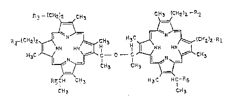
R3 _1CH2)2 C~3 C~3 ~C~2)2- R2
==~ ~=
P~4-(C~2)z~¢ ~N~CH3 H3C~' ~,~(c~2)2
l NH HN H H ¦ N ~I HN
H 3C ~ /--~ ~ CH3 --Cc H. ;~ ~ C~3
R5CH CH3 H3C HCI'R6
lS CH3 Cff3
Formula II
In this formula R1-R4 represent a carboxylic
acid group (-COOH) or C1-C10 alkyl ester derivative
thereof, a radio labeled amide or a radio labeled
ester. R5 and R6 can represent hydroxyl (-OH) or the
radiolabeled reaction product of a cyclic anhydride
such as succinic anhydride with the hydroxyl group.
This would form mono or di succinyl DHE having a
carboxylic acid group which in turn can react with a
radiolabeled amine or alcohol. At least one of R1-R6
must be radiolabeled.
~'
-
1337986
--7--
Preferred radio labeled amide groups include
amide groups substituted with 123I, 125I 131I 132I
133I, 135I, Br or 82Br. Preferably at least one of
Rl-R4 and more preferably three of the Rl-R4 groups
represent -CO2H. Preferably R5 and R6 represents
hydroxyl group. The preferred radio labeled compound
is one where one of Rl-R4 represents radio halogenated
histamine or tyrosine.
According to a first method, radiolabeled
DHE is prepared by reacting a radiolabeled compound or
precursor with DHE under suitable reaction conditions.
Preferably for use in the present invention the
radiolabeled precursor will be a radiolabeled amine or
radiolabeled alcohol which can form an amide or an
ester with the DHE. Suitable radiolabeled amines
would include imidazol substituted alkyl amines,
phenol substituted alkyl amines, sulfide substituted
alkyle amines. Suitable alcohols would include
imidazol substituted alkyl alcohols, phenol
substituted alkyl alcohols and sulfide substituted
alkyl alcohols.
For example the following precursors should
be suitable for use in radio labeling DHE:
X X
H -N-R I _ ~ HO-R
~ H
imidazole sub- imidazole sub-
stituted alkyl stituted alkyl
amine alcohol
C
1337986
-8- ~ %
H2 N R7 ~ 0 Ho-R7 ~ 0 H
phenol sub- phenol sub-
stituted alkyl - stituted alkyl
amine alcohol
H2-N-R7-sx H-R7-SX
sulfiae sub- sulfide sub-
stituted alkyl stituted alkyl
amine alcohol
wherein R7 represents Cl-C10 alkylene and X represents
a radionuclide halogen. Particular suitable radio-
labeled amines include:
l3
H2 CH2 C 2l -
(131 iodohistamine)
C ~3
o
1 l3
C =
H2-N-CH-CH2~)--0 ~
(131 iodo tyrosine methyl ester)
Preparation of radiolabeled precursors is
well known. For example the method of radio halogena-
tion is reported by Greenwood F. C., Hunter W. M.,
1337986
g
Glover, J. S., The Preparation of 131Iodine Labeled
Human Growth Hormone of ~iqh Speci~icity, Bio. Chem.
Journal, vol. 89, p.114, 1963. Basically this method
calls for the oxidation of, for example, sodium
iodinel31 with Chlorimine T in the presence of an
alkyl amine substituted with an imidazol, a phenol, or
sulfide group to produce an iodinel31 radical sub-
stituted on one of the imidazol, phenol or sulfide
groups. Another preferred method incorporates the use
of iodogen as a oxidizing agent in place of the
Chloramine T. This method is reported by Pamela J.
Fraker, et al Vol. 30 Biochemical and ~iophysical
Research Com.munication pp. 849-857 (February 28,
1978).
Conjugation of one of the amine radiolabeled
precursors with DHE to form radiolabeled DHE is
carried out in an aqueous tetrahydrofuran solution in
the presence of carbodiimide reagent for amide
formation. Isolation and purification of the con-
jugated products is achieved by selective
precipitation and gel filtration ion exchange
chromotography. Biogel P-10 is a suitable medium for
gel filtration chromotography in which the aggregate
of radio labeled DHE conjugates in an aqueous medium
can be excluded completely from the gel while lower
molecular weight radio labeling compounds such as
histamine or tyrosine can be retained on the column.
*Trade-mark
1337986
1 o--
Esterification of one of the alcohol pre-
cursors can be conducted by simple raction of the
alcohol with DHE in the presence of a mineral acid.
Due to steric hinderance, this reaction may proceed at
a relatively slow rate. Accordingly, labeling by
formation of the amide is preferred.
The precursor compounds, both amines and
alcohols, can also be bonded to either of the two
hydroxyl groups. However, to facilitate this reaction
the hydroxyl group must first be reacted with a cyclic
anhydride or a diacid. The anhydride or acid react
under acid pH is an aqueous medium to form an ester
with a free carboxylic acid fur.ctionality. In turn,
this carboxylic acid funçtionality can react with the
amine or hydroxyl group of the precursor to form an
amide or ester, respectively. Thus R5 and R6 can
represent
H
/X
-O-C-R8-C-N-R7- 1_ f
N;~, N
O O H X
-O-C-R8-C-N-R7 ~ O~
1337986
--1 1--
o o
-O-C-R8-C-W-R7-SX
O O
Il 11 X
-O-C-R8-C-O-R7-
O O %
-O-C-R8-C-O-R7~ 01
O O
Il 11
-O-C-R8-C-O-R7-SX
wherein R8 represents C2-C10 alkylene-
The radio labeled DHE can be used for
diagnostic purposes by injecting an effective amount
of the radiolabeled compound and observing localiza-
tion of the compound using radio scintillation methods
after about 30 minutès to 72 hours (preferably about
24 hours) to allow the DHE to clear the blood. The
methods of imaging using nuclear medicine imaging
~ - 1337985
-12-
techniques are well known and can be conducted, for
example, using a gamma camera which detects gamma
radiation emitted by the radionuclide. Positron
detectors can also be used with 77Br.
The administered activity will vary
depending on the subject. Examples provide dose
information for smaller mammals. For use in, for
example, a 70 kilo human the dose range will vary from
about 25 microcuries to about 2 millicuries depending
on the purpose of the examination. The labeled DHE is
applied parenterally and preferably intravascularly.
The labeled DHE can be carried in any therapeutically
acceptable carrier or vehicle such as saline.
For therapeutic uses the radio labeled DHE
must have a strong component of particulate radiation,
for example, a strong beta emitter. Accordingly, the
radio emitting compound must be 5I, 3 I, 132I,
I, I, Br. For therapeutic applications the
administered activity should be, for example, from
about 500 microcuries to about 200 millicuries for a
70 kilo adult applied intravascularly. The adminis-
tered activity will of course vary depending on the
stage of the cancer, the age and health of the sub-
ject, and radiation dose response considerations.
The invention will be further appreciated in
light of the following examples.
-13- 1337986
Example I - Preparation of Iodinated Histamine
- Histamine (0.4 mg in 100 microliters of
aqueous phosphate buffer) was added to an iodegen
plated (4 micrograms) polypropylene test tube together
with 10 microliters of Na I (20 millicuries). This
was left at room temperature for thirty minutes. This
produced an aqueous solution of 131-Iodohistamine,
which can be used directly in the conjugation reaction
with DHE described in Example II.
Example II - Preparation of Iodinated Histamine DHE
DHE (8.5 micromoles) was radiolabeled by
coupling at least one of the four carboxylic acid
groups with 125I-iodohistamine (2.13 micromoles) in
90% tetrahydrofuran (THF) in the presence of tri-
ethylamine (2.13 micromoles) and 1-ethyl-3-(3-
dimethyl-aminopropyl)carbodiimide hydrochloride (2.13
micromole) at room temperature overnight. The solvent
was removed under a stream of nitrogen and the residue
dissolved in 2 ml of 0.1 M NH40H. DHE was precipi-
tated by adjusting the pH to 4 with acetic acid,
washed with 0.1 M acetic acid three times and redis-
solved in 0.1 M NH40H. 125I-histamine DHE (125I-hDHE)
and unlabeled DHE were separated ~y an anion exchange
column (AGlX8) by eluting the column with 20% THF, 50%
THF, 90% THF, 0.1 M àcetic acid and 90% THF and 0.1 M
HCL nd 90% THF 125I-hDHE was eluted in the acetic
acid-THF fraction. Photosensitizing activity of
125I-hDHE was confirmed by its ability to lyse red
1337986
-14-
blood cells following laser radiation. Its tumor
localizing ability was assessed in spontaneous memory
tumor fast (S~T-F) bearing DBA/2HA mice. The
followins specific tumor to tissue ratio (counts per
minute per gram) were obtained 24 hours after
intraperataneal injection: brain (64.27), muscle
(6.07), blood (3.32), lung (1.54), kidney (2.54),
spleen (0.48), liver (0.15). Such ratios are similar
to those obtained with 3H and 14C labeled HDP
suggesting that biological distrlbution of the
radiated compound is not altered by labeling
procedure.
Exam~le III - Imaginq With 131I hDHE
l31I hDHE was used to image tumor bearing
mice. The mice were injected with 65 microcuries of
I hDHE (20 micrograms of 131I hDHE per gram of
mouse weight). After 24 hours nuclear scintillation
images were obtained. The 131I hDHE localized in
tumors and an image of the tumors was obtained.
Thus bv labeling DHE at one of the four
carboxylic acid sites or hydroxyl sites, the compound
will still localize in neoplastic tissues. This in
turn provides a means to identify and image neoplastic
tissue and to chemotherapeutically treat malisnant
neoplastic tissue.
, ~ ~