Language selection

Search

Patent 1338176 Summary

Third-party information liability

Some of the information on this Web page has been provided by external sources. The Government of Canada is not responsible for the accuracy, reliability or currency of the information supplied by external sources. Users wishing to rely upon this information should consult directly with the source of the information. Content provided by external sources is not subject to official languages, privacy and accessibility requirements.

Claims and Abstract availability

Any discrepancies in the text and image of the Claims and Abstract are due to differing posting times. Text of the Claims and Abstract are posted:

  • At the time the application is open to public inspection;
  • At the time of issue of the patent (grant).
(12) Patent: (11) CA 1338176
(21) Application Number: 557718
(54) English Title: INTRAVASCULAR BLOOD PARAMETER MEASUREMENT SYSTEM
(54) French Title: SYSTEME DE MESURE INTRAVASCULAIRE D'UN PARAMETRE SANGUIN
Status: Deemed expired
Bibliographic Data
(52) Canadian Patent Classification (CPC):
  • 73/51.5
(51) International Patent Classification (IPC):
  • A61B 5/00 (2006.01)
(72) Inventors :
  • MAXWELL, THOMAS P. (United States of America)
(73) Owners :
  • MINNESOTA MINING AND MANUFACTURING COMPANY (United States of America)
(71) Applicants :
(74) Agent: BORDEN LADNER GERVAIS LLP
(74) Associate agent:
(45) Issued: 1996-03-26
(22) Filed Date: 1988-01-29
Availability of licence: N/A
(25) Language of filing: English

Patent Cooperation Treaty (PCT): No

(30) Application Priority Data:
Application No. Country/Territory Date
008,937 United States of America 1987-01-30

Abstracts

English Abstract


A parameter of blood is sensed in vivo with a
system which includes a catheter and a probe. The catheter
has a lumen extending therethrough, a proximal end, a distal
end and a distal opening at the distal end. The probe
includes one or more sensors at its distal end. A saline
solution is introduced into the lumen so that there is an
interface adjacent the distal opening of the catheter
between the blood and saline solution. The probe is
received within the catheter and affixed thereto. The
interface is moved back and forth in the lumen to expose
the sensors to blood so that they can sense the blood
parameters of interest.


Claims

Note: Claims are shown in the official language in which they were submitted.


- 20 -

THE EMBODIMENTS OF THE INVENTION IN WHICH AN EXCLUSIVE PROPERTY
OR PRIVILEGE IS CLAIMED ARE DEFINED AS FOLLOWS:
1. An assembly for in vivo measurement of the
concentration of oxygen in blood comprising:
a fluorescent oxygen sensor means for sensing the
concentration of oxygen in blood and providing a
fluorescent oxygen signal in response thereto;
elongated optical transmission means optically
coupled to said sensor means for transmitting the
fluorescent oxygen signal from the sensor means
proximally;
said sensor means being carried by a distal
portion of the transmission means;
said sensor means and said transmission means
being sized and adapted to be received within a blood
vessel of a patient;
means for keeping the sensor means from contacting
the wall of this vessel when the sensor means is within
such vessel to thereby reduce the wall effect on the
fluorescent oxygen signal; and
said keeping means spacing the sensor means from
the wall of the vessel to allow blood to flow between
the sensor means and the wall of the vessel.

2. An assembly as defined in claim 1 wherein said
keeping means includes a tubular body having an opening,
said sensor means being within the tubular body adjacent
the opening.

3. An assembly as defined in claim 2 wherein said
opening opens axially and said sensor means has a distal
end within no less than 0.005 inch of said opening.

4. An assembly as defined in claim 2 including at
least one radial aperture in the tubular body adjacent
said opening.

21
5. A probe-catheter assembly comprising:
a probe including a first sensor for sensing a
parameter of blood and providing a signal in response
thereto and elongated transmission means for
transmitting the signal from the sensor proximally, said
sensor being carried by a distal portion of the
transmission means;
a catheter having a lumen extending therethrough,
a proximal end, a distal end and a distal opening at the
distal end, a major length of said probe being
receivable longitudinally in said lumen of said catheter
with radial clearance to allow fluid flow therein and a
length of the catheter being sized to be received within
the cardiovascular system of a patient; and
means for attaching the probe to the catheter at a
location adapted to be outside the cardiovascular system
of the patient such that the sensor is within the lumen
adjacent the distal opening.

6. A probe as defined in claim 5 wherein said
transmission means includes means for transmitting light
and said first sensor is an optical sensor.

7. A probe as defined in claim 5 wherein said
transmission means includes means for transmitting light
and said first sensor is a fluorescent sensor.

8. A probe-catheter assembly as defined in claim
5 wherein the distal opening opens axially and said
sensor has a distal end within about 0.125 inch of said
opening.

22

9. A probe-catheter assembly as defined in claim
5 including a second sensor for sensing a parameter of
blood and providing a signal in response thereto, and
said first and second sensors are within said lumen at
different axial positions along the distal portion of
the transmission means when the probe is received within
the lumen.

10. A probe-catheter assembly as defined in claim
5 including at least one radial aperture in the catheter
leading from said lumen adjacent the distal end of the
catheter.

11. A probe-catheter assembly as defined in claim
9 wherein said transmission means includes first and
second elongated optical fibers, each of said first and
second optical fibers has a distal end, said first and
second sensors are optical sensors carried by the first
and second optical fibers, respectively, adjacent the
distal ends thereof and said sensors are within the
lumen when the probe is received within the lumen.

12. A probe as defined in claim 11 wherein said
first and second sensors sense first and second
different parameters in blood, respectively, and said
first and second parameters are a blood gas and pH,
respectively.

13. A probe as defined in claim 11 wherein said
first and second sensors sense first and second
different parameters in blood, respectively, and said
first and second parameters are different blood gases.

23

14. A probe as defined in claim 11 wherein said
first and second sensors sense oxygen and carbon dioxide
and the first sensor is located proximally of the second
sensor.

15. A probe-catheter assembly as defined in claim
11 wherein said first optical fiber extends distally
beyond the second optical fiber whereby the first and
second sensors are at different axial positions.

16. A probe-catheter assembly as defined in claim
15 wherein each of said first and second sensors is a
fluorescent sensor.

17. A probe-catheter assembly as defined in claim
5 wherein said distal opening opens axially.

18. A probe-catheter assembly as defined in claim
17 wherein said probe terminates distally in said sensor
and said sensor confronts said opening.

19. A probe-catheter assembly as defined in claim
5 wherein said means for attaching allows the probe to
be attached to the catheter when the catheter is
received within the cardiovascular system of the
patient.

20. A probe-catheter assembly as defined in claim
5 wherein said means for attaching releasably attaches
the probe to the catheter.

24

21. An assembly for in vivo measurement of the
concentration of oxygen in blood comprising:
a fluorescent oxygen sensor means for sensing the
concentration of oxygen in blood and providing a
fluorescent oxygen signal in response thereto;
elongated optical transmission means optically
coupled to said sensor means for transmitting the
fluorescent oxygen signal from the sensor means
proximally;
said sensor means being carried by a distal
portion of the transmission means;
said sensor means and said transmission means
being sized and adapted to be received within a vessel
of a patient;
means for keeping the sensor means from contacting
the wall of said vessel when the sensor means is within
such vessel to thereby reduce the wall effect on the
fluorescent oxygen signal; and
said keeping means including a catheter having a
lumen extending therethrough, a proximal end, a distal
end and a distal opening at the distal end, at least a
portion of said transmission means being in said lumen
with a radial clearance and means for attaching the
transmission means to the catheter adjacent the proximal
end of the catheter such that said oxygen sensor means
is within the lumen adjacent the distal opening.

- 25 -

22. A probe for sensing at least one parameter in blood
comprising:
(a) first and second optical sensors for sensing at least
one parameter in blood selected from the group consisting of
blood gases, pH, electrolytes, glucose, and temperature, and for
providing a signal in response thereto;
(b) first and second elongated optical fibers for
transmitting the signals from the first and second sensors
proximally, each of the first and second optical fibers having a
free distal end with a distal end portion adjacent thereto;
(c) the first and second sensors being carried by the
distal end portions of the first and second optical fibers,
respectively, and the distal end portion of the first optical
fiber extends distally beyond the distal end portion of the
second optical fiber with the first and second sensors being at
different longitudinal positions; and
(d) the first and second elongated optical fibers and the
first and second sensors being sized to be received in the
cardiovascular system of a patient.

23. A probe as defined in claim 22 wherein the first sensor
is a fluorescent sensor.

24. A probe as defined in claim 22 wherein the first and
second sensors sense first and second different parameters in
blood, respectively, and the first and second different
parameters are different blood gases.

25. A probe as defined in claim 22 wherein the first and
second sensors sense first and second different parameters in
blood, respectively, and the first and second different
parameters are a blood gas and pH.

26. A probe as defined in claim 22 wherein at least one of
the first and second sensors is carried by its respective optical
fiber adjacent the distal end thereof.


- 26 -

27. A probe as defined in claim 22 further including a
third optical sensor for sensing a parameter in blood, and a
third elongated optical fiber having a distal end portion which
terminates in a distal end, the third optical sensor being
carried by the distal end portion of the third optical fiber, and
the distal end portion of the second optical fiber extends
distally beyond the distal end portion of the third optical fiber
with the first, second, and third optical sensors each being at a
different longitudinal position.

28. A probe for sensing parameters in blood comprising:
(a) first and second optical sensors for sensing first and
second different parameters in blood, respectively, the
parameters being selected from the group consisting of blood
gases, pH, electrolytes, glucose, and temperature, and for
providing a signal in response thereto;
(b) first and second elongated optical fibers for
transmitting the signals from the first and second sensors
proximally, each of the first and second optical fibers having a
free distal end with a distal end portion adjacent thereto;
(c) the first and second sensors being carried by the first
and second optical fibers, respectively, adjacent the distal ends
thereof, and the distal end portion of the first optical fiber
extends distally beyond the distal end portion of the second
optical fiber with the first and second sensors terminating at
different axial positions; and
(d) the first and second elongated optical fibers and the
first and second sensors being sized to be received in the
cardiovascular system of a patient.

29. A probe as defined in claim 28 wherein the first sensor
is a fluorescent sensor.

30. A probe as defined in claim 28 wherein the first and
second different parameters are different blood gases.

- 27 -
31. A probe as defined in claim 28 wherein the first and
second different parameters are a blood gas and pH.

32. A probe as defined in claim 28 further including a
third optical sensor for sensing a third different parameter in
blood, and a third elongated optical fiber having a distal end
portion which terminates in a distal end, the third optical
sensor being carried by the third optical fiber adjacent the
distal end thereof, and the distal end portion of the second
optical fiber extends distally beyond the distal end portion of
the third optical fiber with the first, second, and third sensors
each terminating at a different axial position.

33. An assembly for sensing at least one parameter in blood
comprising:
(a) a catheter having a lumen extending therethrough and
being sized to be received in the cardiovascular system of a
patient; and
(b) a probe including (i) first an second optical sensors
for sensing at least one parameter in blood selected from the
group consisting of blood gases, pH, electrolytes, glucose, and
temperature, and for providing a signal in response thereto; (ii)
first and second elongated optical fibers for transmitting the
signals from the first and second optical sensors proximally,
each of the first and second optical fibers having a free distal
end with a distal end portion adjacent thereto; (iii) the first
and second optical sensors being carried by the distal end
portion of the first and second optical fibers, respectively, and
the distal end portion of the first optical fiber extends
distally beyond the distal end portion of the second optical
fiber with the first and second optical sensors being at
different longitudinal positions; and (iv) the first and second
elongated optical fibers extending into the lumen and the first
and second optical sensors being within the lumen.

34. An assembly as defined in claim 33 wherein the lumen


- 28 -

has a distal opening and the sensors are adjacent the distal
opening.

35. An assembly as defined in claim 34 wherein the lumen is
of reducing cross section toward the distal opening in a tapered
zone adjacent the distal opening and the first and second sensors
are in the tapered zone.

36. A probe as defined in claim 24 wherein the first and
second different parameters are oxygen and carbon dioxide.

37. A probe as defined in claim 27 wherein the first,
second, and third sensors sense first, second, and third
different parameters in blood, respectively, and the first,
second, and third different parameters are carbon dioxide, pH,
and oxygen.

38. A probe as defined in claim 27 wherein each of the
first, second, and third sensors is carried by its respective
optical fiber adjacent the distal end thereof.

39. A probe as defined in claim 32 wherein the first,
second, and third different parameters are carbon dioxide, pH,
and oxygen.

40. An assembly as defined in claim 33 wherein the first
sensor is a fluorescent sensor.

41. An assembly as defined in claim 33 wherein the first
and second sensors sense first and second different parameters in
blood, respectively, and the first and second different
parameters are different blood gases.

42. An assembly as defined in claim 41 wherein the first
and second different parameters are oxygen and carbon dioxide.


- 29 -

43. An assembly as defined in claim 33 wherein the first
and second sensors sense first and second different parameters in
blood, respectively, and the first and second different
parameters are a blood gas and pH.

44. An assembly as defined in claim 33 wherein at least one
of the first and second sensors is carried by its respective
optical fiber adjacent the distal end thereof.

45. An assembly as defined in claim 33 wherein the probe
further includes a third optical sensor for sensing a parameter
in blood, and a third elongated optical fiber having a distal end
portion which terminates in a distal end, the third optical
sensor being carried by the distal end portion of the third
optical fiber, and the distal end portion of the second optical
fiber extends distally beyond the distal end portion of the third
optical fiber with the first, second, and third optical sensors
each being at a different longitudinal position.

46. An assembly as defined in claim 45 wherein the first,
second, and third sensors sense first, second, and third
different parameters in blood, respectively, and the first,
second and third different parameters are carbon dioxide, pH, and
oxygen.

47. An assembly as defined in claim 45 wherein each of the
first, second, and third sensors is carried by its respective
optical fiber adjacent the distal end thereof.

48. An assembly for sensing parameters in blood
comprising:
(a) a catheter having a lumen extending therethrough and
being sized to be received in the cardiovascular system of a
patient; and
(b) a probe including (i) first and second optical sensors
for sensing first and second different parameters in blood,

- 30 -
respectively, the parameters being selected from the group
consisting of blood gases, pH, electrolytes, glucose, and
temperature, and for providing a signal in response thereto; (ii)
first and second elongated optical fibers for transmitting the
signals from the first and second sensors proximally, each of the
first and second optical fibers having a free distal end with a
distal end portion adjacent thereto; (iii) the first and second
sensors being carried by the first and second optical fibers,
respectively, adjacent the distal ends thereof, and the distal
end portion of the first optical fiber extends distally beyond
the distal end portion of the second optical fiber with the first
and second sensor terminating at different axial positions; and
(iv) the first and second elongated optical fibers extending into
the lumen, and the first and second optical sensors being within
the lumen.

49. An assembly as defined in claim 48 wherein the first
and second different parameters are different blood gases.

50. An assembly as defined in claim 48 wherein the first
and second different parameters are a blood gas and pH.

51. An assembly as defined in claim 48 wherein the lumen
has a distal opening and the sensors are adjacent the distal
opening.

52. An assembly as defined in claim 51 wherein the lumen is
of reducing cross section toward the distal opening in a tapered
zone adjacent the distal opening and the first and second sensors
are in the tapered zone.

53. An assembly as defined in claim 48 further including a
third optical sensor for sensing a third different parameter in
blood, and a third elongated optical fiber having a distal end
portion which terminates in a distal end, the third optical
sensor being carried by the third optical fiber adjacent the

- 31 -
distal end thereof, and the distal end portion of the second
optical fiber extends distally beyond the distal end portion of
the third optical fiber.

54. An assembly as defined in claim 53 wherein said first,
second, and third different parameters are carbon dioxide, pH,
and oxygen.

- 32 -

55. An assembly for the in vivo measurement of a blood
parameter comprising:
a catheter having a lumen extending therethrough, a
proximal end, a distal end and a distal opening at the distal
end;
a sensor for sensing the blood parameter and providing a
signal in response thereto;
means for mounting the sensor on the catheter with the
sensor being within the lumen;
a conduit for coupling the lumen to a source of fluid
whereby the fluid can be supplied to the lumen; and
characterized by a compliant element in communication with the
conduit for allowing the volume of the assembly to be expanded
and contracted; and
means for changing the extent to which the compliant
element allows the volume of the assembly to be expanded and
contracted.

56. An assembly as defined in claim 55 wherein said
changing means includes means for selectively switching the
compliant element into and out of communication with the
conduit.

57. An assembly as defined in claim 55 wherein said
switching means includes a valve.

58. An assembly as defined in claim 55 wherein the
changing means includes a cylinder in communication with the
conduit and a piston slidable in the cylinder.

59. An assembly as defined in claim 58 wherein the
changing means includes a motor for moving the piston.

- 33 -

60. An assembly as defined in claim 55, 58 or 59 wherein
the changing means includes means for selectively nullifying
the ability of the compliant element to allow the volume of
the assembly to be expanded and contracted.

61. An assembly as defined in claim 55, 56, 57, 58 or 59
including means coupled to the conduit and defining a chamber
for receiving the compliant element.

62. An assembly as defined in claim 55, 56, 57, 58 or 59
in which the compliant element includes a gas.

63. A method of sensing a parameter of blood in vivo
comprising:
providing a catheter and a sensor in a vessel of a
patient with the catheter having a lumen extending
therethrough, a proximal end, a distal end and an opening and
with the sensor being within the lumen and capable of sensing
a parameter in blood and providing a signal in response
thereto;
introducing an anti-clotting solution into the lumen from
a solution-introducing system so that there is an interface
between the blood and the solution;
moving the interface back and forth in the lumen so that
the sensor is exposed to blood for at least a portion of the
time that the interface is moving; and
obtaining said signal from the sensor during said step of
introducing.

64. A method as defined in claim 63 wherein said step of
moving includes expanding and contracting the volume of the
solution-introducing system to at least assist in said moving
of the interface.

- 34 -

65. A method as defined in claim 64 wherein said step of
moving further includes providing a volume oscillator and
causing the volume oscillator to expand and contract the
volume of the solution-introducing system.

66. A method as defined in claim 63 wherein said step of
moving includes providing the solution-introducing system with
sufficient compliance so that the patient's heartbeats at
least assist in said moving of the interface.

67. A method as defined in claim 63 wherein said step of
moving moves the interface back and forth over the sensor.

68. A method as defined in claim 63 wherein the opening
is a distal opening adjacent the distal end and said step of
positioning includes positioning the sensor within a distal
region of the lumen.

69. A method as defined in claim 68 wherein the distal
opening opens at the distal end and said distal region extends
no farther distally than about 0.005 inch proximally of said
opening.

70. A method as defined in claim 68 wherein the distal
opening opens at the distal end and said distal region extends
no farther proximally than about 0.125 inch proximally of said
opening.

71. A method as defined in claim 63 wherein said step of
introducing includes introducing the anti-clotting solution to
the lumen to produce a new flow of solution into the patient.

- 35 -


72. A method as defined in claim 63 wherein said step
of positioning includes providing a probe which includes said
sensor and transmission means for transmitting the signal from
the sensor proximally with the sensor being carried by a
distal portion of the transmission means, inserting the probe
into said lumen of the catheter and attaching the probe to a
proximal location on the catheter to position the sensor in
the lumen.

73. A method as defined in claim 63 wherein said step of
obtaining is carried out during said step of moving.

74. A method of sensing a parameter of blood in vivo
comprising:
providing a catheter and a sensor in a vessel of a
patient with the catheter having a lumen extending
therethrough, a proximal end, a distal end and an opening and
with the sensor being within the lumen and capable of sensing
a parameter of blood and providing a signal in response
thereto;
introducing an anti-clotting solution into the lumen from
a solution-introducing system so that there is a net flow of
solution into the patient; and
moving blood from the vessel through the distal opening
into the lumen during said step of introducing to expose the
sensor to blood and obtain a signal in response to said
parameter of the blood.

75. A method as defined in claim 74 wherein said step
of moving includes at least one of (A) moving blood back and
forth in the lumen while expanding and contracting the volume
of the solution-introducing system; and (B) providing the
solution-introducing system with sufficient compliance so that
the patient's heartbeats at least assist in said moving blood
back and forth in the lumen.


- 36 -
76. A method as defined in claim 75 wherein said step of
moving includes both (A) and (B).

77. A method as defined in claim 74 wherein said step of
moving includes moving blood back and forth in the lumen while
expanding and contracting the volume of the solution-
introducing system, providing a volume oscillator, and causing
the volume oscillator to expand and contract the volume of the
solution-introducing system.

78. A method as defined in claim 74 wherein said step of
moving is carried out while expanding the volume of the
solution-introducing system.

79. A method as defined in claim 74 wherein said
solution-introducing system has a variable volume.

80. A method of sensing a parameter of blood in vivo
comprising:
providing a catheter and a sensor in a vessel of a
patient with the catheter having a lumen extending
therethrough, a proximal end, a distal end and an opening and
with the sensor being within the lumen and capable of sensing
a parameter in blood and providing a signal in response
thereto;
introducing an anti-clotting solution into the lumen from
a solution-introducing system so that there is an interface
between the blood and the solution;
moving the interface back and forth in the lumen so that
the sensor is exposed to blood for at least a portion of the
time that the interface is moving, wherein said step of moving
includes providing a compliant element and selectively
switching the compliant element into and out of the solution-
introducing system; and

- 37 -

obtaining said signal from the sensor during said step of
introducing.

81. A method as defined in claim 80 which further
comprises taking a blood pressure reading with the compliant
element out of the solution-introducing system.

82. A method as defined in claim 80 wherein said step of
obtaining is carried out during said step of moving.

83. A method of sensing a parameter of blood in vivo
comprising:
providing a catheter and a sensor in a vessel of a
patient with the catheter having a lumen extending
therethrough, a proximal end, a distal end and an opening and
with the sensor being within the lumen and capable of sensing
a parameter of blood and providing a signal in response
thereto;
introducing an anti-clotting solution into the lumen from
a solution-introducing system so that there is a net flow of
solution into the patient; and
moving blood from the vessel through the distal opening
into the lumen during said step of introducing to expose the
sensor to blood and obtain a signal in response to said
parameter of the blood, wherein said step of moving includes
providing a compliant element and selectively switching the
compliant element into and out of the solution-introducing
system.

84. A method as defined in claim 83 which further
comprises taking a blood pressure reading with the compliant
element out of the solution-introducing system.

85. A method of sensing a parameter of blood in vivo
comprising:

- 38 -

providing a catheter and a sensor in a vessel of a
patient with the catheter having a lumen extending
therethrough, a proximal end, a distal end and an opening and
with the sensor being within the lumen and capable of sensing
a parameter of blood and providing a signal in response
thereto;
introducing an anti-clotting solution into the lumen from
a solution-introducing system which is under continuous
pressure such that there is a net flow of solution into the
patient; and
moving blood from the vessel through the distal opening
into the lumen to expose the sensor to blood and obtain a
signal in response to said parameter of the blood.

Description

Note: Descriptions are shown in the official language in which they were submitted.


1338176


BAC~GROUND OF THE I~VE:NTION


It is often necessary or desirable to measure
various parameters of blood, such as temperature and blood
constituents, such as blood gases, pH, other electrolytes and
glucose. This can be accomplished in real time using
fluorescent sensors. For example, this can be accomplished
in an extracorporeal blood loop and in vivo as disclosed
in Lubbers et al U.S. Reissue Patent No. 31,879. For in
vivo sensing, a probe or catheter carrying an appropriate
sensor is inserted into a blood vessel of the patient.


One of the most important gases that needs to be
sensed is oxygen. One problem with in vivo oxygen sensing is
that the readings obtained for the concentrations of oxygen
tend to vary over an unacceptably wide range when compared
with the results obtained using conventional lahoratory
techniques for measuring the concentration of oxygen. It has
been found that this deviation is in many cases unacceptably
large so that the reliability of the in vivo measuring system
is called into question.


SU~RY OF THE INVENTION


At least one feature of the invention is based, in
part, upon the recognition and`discovery of the reasons why
unacceptable results were often obtained in the in vivo


1338176


system. Specifically, I have discovered that the oxygen
readings are subject to a "wall effect" in that lower
concentration readings are obtained when the oxygen sensor is
against the wall of the vessel in which it is placed.
Although this invention is not to be limited by any
particular theory, one possible reason for the "wall effect"
is that the concentration of oxygen in the blood may be
different at the vessel wall than at a more central location
within the vessel, or the low level of oxygen in the adjacent
tissue may cause the oxygen concentration in the vessel wall
to be low compared to the concentration in the blood. In
addition, there is a "clot effect" which reduces the oxygen
readings when a clot forms over the oxygen sensor. The clot
may also effect other readings, such as by increasing the
reading for the concentration of C02 and reducing the
reading for the pH value. The "wall effect" and the "clot
effect" are independent, but they can exist at the same time,
as well as separately.
Having recognized these two problems, i.e., the
"wall effect" and the "clot effect", this invention solves
these problems by keeping the sensors, and in particular, the
oxygen sensor from contacting the wall of the vessel in which
it is placed. This reduces or eliminates the "wall effect"
on the oxygen reading. In addition, it reduces the tendency
of the blood to form a clot around the sensors. Accordingly,
by keeping the sensors out of contact with the wall of the
vessel, these two problems are minimized, and acceptable
readings are obtainable.
Techniques exist for keeping various in vivo
sensors out of contact with the vessel wall; however, none of
these are directed toward solving the "wall effect" or the

-


133817~


"clot effect." For example, Schuette U.S. Patent No.
3,529,591, uses a shield around electrodes to confine the
electric field seen by the electrodes in an attempt to
minimize interference created through contacting the wall of
the vessel. U.S. Patent No. 4,478,222 employs a sensor
within a catheter having a radial opening and also is not
concerned with the "wall effect" or the "clot effect."
Although the means for keeping the sensor from
contacting the wall can take different forms, it preferably
includes a tubular body having an opening, and the sensor is
positioned within the tubular body. The tubular body can
advantageously take the form of a catheter. To facilitate
blood flow into the catheter and to minimize the likelihood
that the opening will be shut off by contact with the vessel
wall, the opening is preferably a distal opening at the
distal end of the cathe$er. One or more radial apertures may
be provided in addition to the distal opening, if desired.
The sensor can be mounted within the catheter in
any desired way. A preferred system includes a
probe-catheter assembly which comprises a probe including at
least one sensor for sensing a parameter of blood and
providing a signal in response thereto and elongated
transmission means for transmitting the signal from the
sensor proximally. The sensor is carried by a distal portion
of the transmission means. The assembly also includes the
catheter which has a lumen extending therethrough, a proximal
end, a distal end and a distal opening at the distal end.
~ hen utilizing a probe-catheter assembly of this
type, the catheter can be used to keep the sensor from
contacting the wall of the vessel. This can be
advantageously accomplished by attaching the probe to the

1338176


catheter such that the sensor of the probe is within the
lumen of the catheter and adjacent the distal opening of the
catheter. ~Jith this construction, the sensor is shielded
from the wall of the vessel by the catheter but is not
located so far back within the catheter that it cannot
perform its sensing function.
It is quite surprising that a sensor located within
a catheter lumen could adequately sense the parameter of
interest in blood. One reason for this is that it is
necessary to introduce an anti-clotting solution, such as a
heparinized saline solution, into the lumen from a
solution-introducing system. The solution may be resident in
the lumen, i.e., have no net flow into the vessel, but
preferably it flows at a very low rate, such as 3 to 8
milliliters per hour, through the lumen and out through the
distal opening of the catheter into the blood stream in the
vessel. It is surprising that a sensor positioned in the
lumen where there is an anti-clotting solution, particularly
in the path of the distally flowing anti-clotting solution,
would be able to adequately sense the parameters of interest
in blood.
i~ v e r~ for f~a5 ~e cc~n, ~ ed
B However~ this ;~vgnt~n ~Q~Qgn~z~ that there is an
-~ interface between the blood and the anti-clotting solution.
Theoretically, the interface could be a plane that simply
divides the blood from the anti-clotting solution. However,
in reality, the interface is a zone which has some axial
length and which contains a mixture of the blood and the
anti-clotting solution. Thus, the interface divides a zone
of substantially all blood from a zone containing
substantially all anti-clotting solution.

1338176


Because the anti-clotting solution may be supplied
to the catheter such that there is a net flow of solution
through the distal opening to the vessel, it would be
expected that the interface would be entirely outside of, or
at the distal end of, the catheter. However, by moving the
interface back and forth in the lumen, the sensor can be
exposed to blood for at least a portion of time that the
interface is moving. This exposure must be sufficient to
enable the sensor to provide an accurate signal related to
the blood parameter of interest.
The movement of the interface back and forth in the
lumen may move the interface over the sensor. However, the
sensors, and in particular the oxygen sensor, can tolerate
some exposure to the mixture of anti-clotting solution and
blood in the interface without providing erroneous readings.
For example, it has been found that a mixture consisting of
50 percent blood by volume and 50 percent anti-clotting
solution by volume yields approximately the same oxygen
concentration as the oxygen concentration in a medium
consisting essentially of blood.
Movement of the interface to bathe the sensor
within the lumen in blood can be brought about in different
ways. For example, the interface may be moved by varying the
delivery pressure and/or volume of the anti-clotting solution
or providing the introducing system with a volume oscillator
and allowing the volume oscillator to move the interface.
The volume oscillator may, for example, take the form of a
syringe which, in effect, expands and contracts the volume of
the introducing system to move the blood back and forth in
thç lumen without creating a net or average flow in either
direction.

- ; `
-x~


133817S


Another technique for moving the blood back and
forth in the lumen, which also enables expansion and
contraction of the volume of the introducing system, includes
providing the introducing system with some compliance and
allowing pressures generated by the patient's heartbeats to
move the interface. Consequently, blood is forced to enter
the distal opening of the catheter as the blood pressure
rises with each beat of the heart. Thus, the interface is
caused to flow back and forth in the lumen with the pulsating
blood pressure. As a result, the sensor within the lumen is
bathed by the back and forth or tidal movement of the blood
and can adequately sense and measure-the blood parameters of
interest.
The compliance of the introducing system may be the
natural compliance of the tubing and components of the system
and/or a compliant element may be added to the system to
provide the desired degree of elasticity. The compliant
element can be of virtually any construction and may be, or
include for example, a compressible fluid, such as air, a
membrane, a bellows, etc. The compliance of the introducing
system may be varied to obtain the results desired. For
example, if the compliance of the introducing system is to be
used to obtain, or to assist in obtaining, the tidal action,
the introducing system and the catheter may have a combined
total compliance sufficient to provide a volume exchange of
at least 10 microliters with a system comprised of a 20-gauge
catheter and .022 inch diameter probe.
It may be necessary or desirable to take the
patient's blood pressure through the lumen of the catheter
while the blood parameters are being sensed. The added
compliance of the introducing system may be sufficient to
undesirably alter the blood pressure readings taken through

J


7 13381 ~6 ~


the lumen of the catheter. Accordingly, the present
dis¢/o5u r~
in~nt~on provides, as an option, for selectively nullifying
the ability of the compliant element to allow expansion and
contraction of the volume of the introducing system. For
example, the nullifying means may control expansion or
adjustably limit moveme,nt of a membrane or bellows or it may
selectively switch the compliant element into, and out of,
communication with the lumen of the catheter. In this latter
event, the compliant element would normally be in
communication with the lumen to provide, or assist in
providing, the desirable tidal action for sensing of the
blood parameters of interest. However, just prior to taking
a blood pressure reading, the action of the compliant element
can be switched out of the introducing system so that it
cannot affect the blood pressure reading taken through the
lumen of the catheter. The switching means may take any form
that will accomplish this function and may be, for example, a
valve.
To assure that the sensor will not contact the
vessel wall, the sensor preferably does not protrude beyond
the distal opening of the catheter. It is desirable to have
the sensor located proximal to the distal opening of the
catheter to provide added insurance against contact with the
wall of the vessel. Similarly, the sensor should not be
located so far proximal to the distal opening that it cannot
adequately sense the parameter of interest. Thusj the sensor
should not be so far proximal that it cannot be adequately
bathed by the blood. This distance will vary depending on
how far the blood is drawn into the lumen. Although the
specific distances can vary, for example, placing the sensor
between .005 inch proximal to the distal opening and .125

` ` 8 1338176


inch proximal to the distal opening has been found
satisfactory. The .005 inch dimension is usually sufficient
to provide for tolerance variations that, if added
together, might cause the sensor to protrude from the lumen.
The probe may carry one or more sensors depending
upon the number of parameters of interest. These sensors can
be of any type, such as electro-chemical, that is suitable
for sensing the parameter of interest; however, optical
sensors are preferred, and fluorescent sensors are considered
optimum. Although multiple sensors could be provided to
sense the same blood parameter, preferably, each sensor
senses a different blood parameter. In a preferred
construction, the transmission means includes an optical
fiber for each of the sensors, with the sensor being located
on the distal end of the associated optical fiber. The
sensors provide signals related to the associated blood
parameters of interest, and such signals may be used or
processed continuously, intermittently or on demand to
provide readings indicative of the blood parameters of
interest.
' ~mboJ~nen fs of
A conventional catheter usable with/this invention
has a standard lead-in itaper, i.e., the cross-sectional area
of the lumen reduces toward the distal opening in a zone
closely adjacent the distal opening. The presence of the
probe in this tapered zone tends to reduce the remaining open
area of the lumen to the extent that the monitoring of blood
pressure through the lumen is adversely affected. To address
this problem, in the case of multiple sensors, this invontion
provides for positioning the sensors at different
longitudinal locations along the distal portion of the

~ 9 ~ t~3~7~
transmission means. In the specific case of utilizing an
optical fiber for each sensor, the optical fibers terminate
distally at staggered locations. Co~cequently, not all of the
sensors are located in the tapered zone, and a larger open area
of the tapered zone remains for pressure sensing.
Embodiments of the invention will now be described with
reference to the accompanying drawings wherein:

BRIEF DESCRIPTION OF THE DRAWINGS

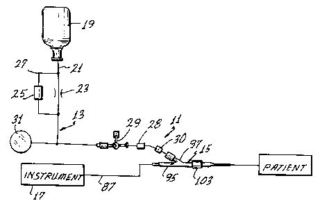
Fig. 1 is a schematic view of an assembly for the in vivo
measurement of blood parameters of interest.
Fig. 2 is a perspective view of one form of valve usable
in the assembly of Fig. 1.
Fig. 3 is an axial sectional view through the valve with
the compliant element being in communication with the conduit
leading to the lumen of the catheter.
Fig. 4 is an elevational view partially in section and
similar to Fig. 3 with the compliant element being out of
communication with the conduit.
Fig. 5 is an enlarged fragmentary sectional view of the
distal region of one form of probe and catheter usable in the
assembly of Fig. 1.
Fig. 6 is an enlarged sectional view taken generally along
line 6-6 of Fig. 5.
Fig. 7 is a longitl~inAl sectional view through the
probe-catheter assembly.

''; ! .' - i-

,
lo 1338176


Fig. 8 is a sectional view similar to Fig. 5
showing an alternate construction of the distal region of the
probe.
Fig. 9 is a schematic view similar to Fig. 1
showing another assembly for the in vivo measurement of blood
parameters of interest.


B DESCRIPTION OF THE PREFERRED EMBODIMENTS


Fig. 1 shows an assembly 11 for the in vivo
measurement of various blood parameters, and particularly the
pH value and the concentrations of oxygen and carbon dioxide.
Although the assembly 11 can be of different constructions,
in this embodiment it includes a solution introducing system
13 and a probe-catheter assembly 15. The assembly 11 may
also include an instrument 17 for providing a readout of the
blood parameters of interest.
Generally, the solution introducing system 13
introduces an appropriate anti-clotting solution, such as a
heparinized saline solution, through the probe-catheter
assembly 15 to the patient to keep the line leading to the
patient patent. Although this can be accomplished in
different ways, in the embodiment shown schematically in Fig.
1, the system 13 includes a pressurized source 19 of
heparinized saline solution, a conduit 21 leading from the
source to the probe-catheter assembly 15, a flow restrictor
23 to reduce the rate of flow through the conduit 21 to the
desired drop rate, a flush valve 25 in a bypass 27 around the
restrictor 23, a stop cock 28, a four-way valve 29, a blood

ll
~ 133~176


withdrawal site 30 and a pressure transducer 31. All of the
components of the system 13 may be conventional, and the
system 13 may include other components, if desired. In the
illustrated embodiment, solution from the pressurized source
19 flows through the restrictor 23 at a relatively slow rate,
such as 5 ml/hour. The solution flows through the valve 29
and the probe-catheter assembly 15 to the patient. If a more
rapid flow rate from the source 19 is desired, as for example
during priming, the flush valve 25 can be manually opened to
provide a relatively high-rate flow path around the
restrictor 23 in a conventional manner.
The four-way valve 29 may also be of conventional
construction. As shown in Fig. 3, the valve 29 includes a
valve body 33 having a passage 35 extending therethrough and
forming a portion of the conduit 21, a rotatable valve
element 37 in the passage 35 and a handle 39 (Fig. 2) for
manually rotating the valve element 37. The valve body 33
has a leg 41, and a closure cap 43 is attached to the leg 41
to define, along with the leg, a chamber 45 in which a
compliant element in the form of air is located. The valve
element 37 has ports 47 and 49 for communicating with the
conduit 21, and a port 51 which can communicate with the
chamber 45 as shown in Fig. 3 or which can be sealed and out
of communication with the conduit 21 and the chamber 45 as
shown in Fig. 4. In this manner, the compliant element can
be switched into, or out of, the system 13.
The pressure transducer 31 communicates with the
conduit 21 and can measure the pressure therein.
Accordingly, with the probe-catheter assembly 15 inserted
into the vascular system of a patient, the pressure
transducer 31 can provide blood pressure readings. By

12 13381 76


rotating the valve element 37 to the position of Fig. 4, the
compliance of the air within the chamber 45 cannot affect the
blood pressure readings provided by the transducer 31. The
blood withdrawal site 30 is used for taking blood samples
from the patient through the probe-catheter assembly 15.
Preferably for this kind of compliant element, the stop cock
28 is located between the valve 29 and the site 30 so that,
by closing the stop cock 28, the air in the chamber 45 cannot
be withdrawn during a blood withdrawal procedure.
The probe-catheter assembly 15 includes a catheter
53 and a probe 55 (Fig. 7). The catheter 53 may be a
conventional arterial catheter. As such, the catheter 53 may
include a proximal end 57, a distal end 59, a lumen 61
extending axially, completely through the catheter and
opening at a distal opening 63 at the distal end. The
catheter 53 has a standard lead-in taper, i.e., a -tapered
zone 65, which extends from a reference plane 66 along the
outer periphery of the catheter 53 to the distal end 59. The
diameter of the lumen 61 also decreases distally throughout
the tapered zone 65 as shown in Fig. 5. The tapered zone 65
may extend about .090 inch proximally of the distal end 59.
The catheter 53 has an externally threaded coupling 67 at its
proximal end.
The probe 55 may be of various different
constructions, and in the embodiment illustrated, includes an
oxygen sensor 69, a carbon dioxide sensor 71 and a pH sensor
73, with each of the sensors affixed to the distal ends of
single optical fibers 75, 77, and 79, respectively, (Fig. 6).
In this embodiment, the sensors 69, 71 and 73 are fluorescent
optical sensors, and they respond to the concentration of
oxygen, the concentration of carbon dioxide and the pEI value,

1338176
- 13 -

respectively, to provide continuous optical signals
indicative of the condition sensed. The optical fibers 75, 77
and 79 serve as transmission means for transmitting the
signals from the associated sensors proximally. The probe 55
is of very small cross-sectional area so that it fits within
the lumen 61 with an ample radial clearance 81 as shown in
Fig. 5-
The particular design of the probe 55 forms no part ofthis invention because the inventive subject matter is
applicable to probes of various different constructions.
Briefly, however, the sensors 69, 71 and 73 are attached to
the distal ends of the associated optical fibers 75, 77 and 79
in any suitable manner, and each of the sensors and the
associated fiber is separately encased in an inner overcoat 83
which, among other things, may assist in retA;ning the sensor
on the end of the associated fiber. The overcoat 83 is, of
course, permeable to the relevant blood parameters so that
such parameter, or one related to it, can be sensed by the
sensors. An outer overcoat B5 covers the inner overcoats 83
and a length of the fibers just proximally of the overcoats
83. Proximally of the overcoat 85, the optical fibers 75, 77
and 79 and a temperature-sensitive element, such as a
thermocouple 86 (Fig. 6), are suitably encased within an
appropriate sheath 87.
The probe 55 includes a ~Y" fitting 93 at its proximal
end as shown in Fig. 7. The optical fibers 75, 77 and 79
extend within the sheath 87 completely through one leg 95 of
the ~'Y" fitting 93 to the instrument 17 as shown in Fig. 1.
Another leg 97 of the fitting 93 has a passage 99 which
communicates with the lumen 61, and more particularly, with
the clearance 81 around the probe 55. The leg 97 is


~r
A

14 1338176


coupled to the conduit 21 of the system 13 as shown in Fig.
1. A third leg 101 of the "Y" fitting 93 carries a rotatable
internally threaded coupling 103 for attaching the "Y"
fitting of the probe 55 to the proximal end of the catheter
53 outside the cardiovascular system of the patient.
Although the details of the fitting 93 form no part
of this invention, the sheath 87 may be guided in the leg 95
by a sleeve 105 and retained in position by potting 107. The
sheath 87 extends within a flexible tube 109 suitably
attached to the leg 95, and shrink tubing 111 is provided
over the adjacent end portions of the fitting and the tube
for strain relief.
With the proximal end of the catheter 53 coupled to
the probe 55 by the coupling 103, the probe 55 is within the
lumen 6I, and the sensors 69, 71 and 73 are within the lumen
adjacent the distal opening 63 as shown in Fig. 5.
Accordingly, with the catheter within the cardiovascular
system of the patient, such as in a radial artery, the
catheter 53 keeps the sensors from contacting the wall of the
artery to thereby reduce or eliminate the wall effect and the
clot effect on the signals provided by the sensors.
In use of the assembly 11, the catheter 53 is first
inserted into the radial artery using conventional
techniques. Next, the probe 55 is inserted into the lumen 61
and attached to the proximal end of the catheter 53 with the
coupling 103. This properly positions the sensors 69, 71 and
73 within the lumen 61 to within .125 inch of the distal end
59. In priming the solution introducing system 13 prior to
insertion of the catheter into the artery, a small quantity
of air is trapped in the chamber 45. This can be
accomplished, for example, with the valve element 37 in the
position of Fig. 4, by filling the conduit 21 with solution

~_ 15
1338176


from the source 19 with the closure cap 43 removed from the
valve 29, and without allowing the solution to flow into the
leg 41. The closure cap 43 is then affixed to the leg 41 to
trap the air in the chamber 45, and then the rotatable valve
element 37 is turned to the position shown in Fig. 3. The
conduit 21 can then be connected to the probe 55.
When in use, the solution from the source 19
completely fills the lumen 61 around the probe 55. The
solution is provided under a pressure such that there is a
slow flow of solution from the lumen 61 into the patient's
artery. This introduction of the solution through the lumen
and into the artery results in an interface 113 adjacent the
distal opening 63 which has some axial length and which
includes both blood and the solution from the source 19. The
interface 113 is a partition between essentially all blood
distally of the interface and essentially all anti-clotting
solution proximally of the interface. The interface washes
axially back and forth in a tidal action as a result of the
rising and falling of the patient's blood pressure with each
heartbeat. If the solution introducing system 13 were
perfectly rigid, it would not be possible for the blood to
force the solution proximally within the lumen 61 because the
solution is essentially incompressible. However, the conduit
21 is typically in the form of flexible plastic tubing, which
has some elasticity or compliance to allow some of this tidal
action to occur. In addition, the illustrated embodiment of
the invention provides q compliant element in the form of air
within the chamber 45 which adds additional elasticity or
compliance to the system 13. Consequently, the interface can
flow back and forth to bathe the sensors 69, 71 and 73 in
blood.

16 13~8176


With this embodiment of the invention, the back and
forth travel of the interface 113 is a function of the
magnitude of the patient's blood pressure, the compliance of
the solution-introducing system 13 and the delivery pressure
of the anti-clotting solution. However, assuming that there
is some net flow of the anti-clotting solution out of the
distal opening 63, it would be necessary for at least the
distal region of the interface 113 to travel distally as far
as the distal opening, unless it is possible for some of the
solution to migrate through the blood and through the distal
opening. Because the flow rate of anti-clotting solution
into the bloodstream is extremely low, the precise manner in
which the solution enters the patient's bloodstream and the
exact extent of travel of the interface 113 is not known.
However, utilizing the tidal action of the interface, it is
possible to bathe the sensors 69, 71 and 73 in blood
sufficiently 50 that accurate readings are obtained, and it
is believed that the sensors are in essentially all blood for
a majority of the time.
Fig. 8 shows another embodiment of this invention
which is identical to the embodiment of Figs. 1-7 in all
respects not shown or described herein. Portions of the
embodiment of Fig. 8 corresponding to portions of the
embodiment of Figs. 1-7 are designated by corresponding
reference numerals followed by the letter "a."
The primary differences between the embodiment of
Fig. 8 and Figs. 1-7 is that the sensors 69a, 71a, and 73a
are at different longitudinal positions within the lumen 61a,
the sensors 71a and 73a project farther from the overcoat
85a, and there are a plurality of radial apertures 121 in the
catheter 53a leading from the lumen 61a adjacent the distal

~ ~ ~ A ~ - r, ~


17
1338176


-
opening 63a of the catheter. In this embodiment, each of the
three sensors terminates at a different axial position within
the lumen 61a, and with this construction, the total
cross-sectional area of the probe 55a reduces in step-wise
fashion from the distal end of the sensor 71a proximally.
Consequently, not all of the sensors are in the tapered zone
65a, and a larger cross-sectional area of the tapered zone
remains open for pressure sensing via the pressure transducer
31 shown in Fig. 1.
In the construction of Fig. 8, preferably the
carbon dioxide sensor 7la is the most distal sensor, and the
oxygen sensor 69a is the most proximal. The reason for this
is that carbon dioxide is the most sensitive to being even
partially out of the blood, and the oxygen sensor can provide
acceptable oxygen readings even in a fifty-fifty mixture of
the blood and the anti-clotting solution. The sensitivity of
the pH sensor 73a is intermediate the sensitivity of the
carbon dioxide sensor 71a and the oxygen sensor 73a and so is
preferably located intermediate these sensors.
The radial apertures 121 are preferably located
proximally of the sensor 73a for the purpose of allowing
blood and solution from the lumen 61a to flow out of the -
apertures. One or more of these apertures may be provided,
and in the embodiment of Fig. 8, two of the apertures are
shown. Of course, the apertures 121 may be distributed in
axially spaced relationship, as well as circumferentially
spaced relationship, along the catheter 53a. The apertures
121 may also be used in the embodiment of Figs. 1-7, if
desired. ~
Fig. 9 shows another embodiment of this invention ~-
which is identical to the embodiment of Figs. 1-7 in all

~ _ _ _, _ _ _ r l ~~ ` 1~
`:~

18
1338176


respects not shown or described herein. Portions of the ;~
embodiment of Fig. 9 corresponding to portions of the
embodiment of Figs.- 1-7 are designated by corresponding
reference numerals followed by the letter "b."
The only difference between the embodiment of Fig.
9 and Figs. 1-7 is that the valve 29 has been replaced with a
volume oscillator 131. Although the volume oscillator 131
can take different forms, including that of a conventional
syringe, in this embodiment, it is illustrated schematically
as including a cylinder 133 in communication with the conduit
21, a piston 135 slidable in the cylinder and a motor 137 for
reciprocating the piston 135 through an appropriate
reciprocating drive (not shown), such as a cam shaft. When
the piston 135 is moved upwardly as viewed in Fig. 9, a
chamber 139 below the piston is enlarged to expand the volume
of the introducing system 13b. Conversely, when the piston
135 moves downwardly, the volume of the chamber 139 is
decreased to thereby contract the volume of the introducing
system. Of course, expansion of the introducing system 13b
pulls the interface 113 (Fig. 5) proximally. Contraction of
the introducing system moves the interface distally, with the
amount of such movement, being a function of the degree to
which the volume oscillator 131 expands and contracts the
volume of the introducing system.
The motor 137 can be operated continuously,
intermittently or upon demand to create the tidal action.
There is no net or average flow of fluid in either direction
as a result of reciprocation of the piston 135. Of course,
the volume oscillator 131 can also be used with the
embodiment of Fig. 8.

~ ~ 19
1338176


Although exemplary embodiments of the invention
have been shown or described, many changes, modifications and
substitutions may be made by one having ordinary skill in the
art without necessarily departing from the spirit and scope
of this invention.

.

Representative Drawing
A single figure which represents the drawing illustrating the invention.
Administrative Status

For a clearer understanding of the status of the application/patent presented on this page, the site Disclaimer , as well as the definitions for Patent , Administrative Status , Maintenance Fee  and Payment History  should be consulted.

Administrative Status

Title Date
Forecasted Issue Date 1996-03-26
(22) Filed 1988-01-29
(45) Issued 1996-03-26
Deemed Expired 2003-03-26

Abandonment History

There is no abandonment history.

Payment History

Fee Type Anniversary Year Due Date Amount Paid Paid Date
Application Fee $0.00 1988-01-29
Registration of a document - section 124 $0.00 1988-07-14
Registration of a document - section 124 $0.00 1989-08-14
Maintenance Fee - Patent - Old Act 2 1998-03-26 $100.00 1998-03-04
Maintenance Fee - Patent - Old Act 3 1999-03-26 $100.00 1999-03-10
Maintenance Fee - Patent - Old Act 4 2000-03-27 $100.00 2000-02-17
Maintenance Fee - Patent - Old Act 5 2001-03-26 $150.00 2001-02-19
Owners on Record

Note: Records showing the ownership history in alphabetical order.

Current Owners on Record
MINNESOTA MINING AND MANUFACTURING COMPANY
Past Owners on Record
CARDIOVASCULAR DEVICES, INC.
MAXWELL, THOMAS P.
Past Owners that do not appear in the "Owners on Record" listing will appear in other documentation within the application.
Documents

To view selected files, please enter reCAPTCHA code :


To view images, click a link in the Document Description column. To download the documents, select one or more checkboxes in the first column and then click the "Download Selected in PDF format (Zip Archive)" or the "Download Selected as Single PDF" button.

List of published and non-published patent-specific documents on the CPD .

If you have any difficulty accessing content, you can call the Client Service Centre at 1-866-997-1936 or send them an e-mail at CIPO Client Service Centre.


Document
Description 
Date
(yyyy-mm-dd) 
Number of pages   Size of Image (KB) 
Representative Drawing 2001-09-13 1 7
Cover Page 1996-03-26 1 17
Abstract 1996-03-26 1 21
Description 1996-03-26 19 817
Claims 1996-03-26 19 706
Drawings 1996-03-26 3 85
Correspondence 2000-01-21 1 2
Prosecution Correspondence 1996-01-11 1 33
Office Letter 1996-01-16 1 66
Prosecution Correspondence 1993-02-11 4 135
Prosecution Correspondence 1994-09-19 7 362
Examiner Requisition 1994-06-02 2 66
Prosecution Correspondence 1991-05-30 2 74
Examiner Requisition 1991-01-30 1 51