Note: Descriptions are shown in the official language in which they were submitted.
1339067
ANTIGENIC COMPOSITIONS AND
METHODS FOR I HI',IK PRODUCTION AND USE
This invention relates to methods for detecting the presence or absence of
antibodies specific to Campylobacter pylori, and more particularly to the clinical use of
novel antigenic compositions for detecting the presence of antibodies to C. pylori
and/or for diagnosing certain gastrointestinal disorders.
Peptic ulcer disease, gastritis and other infl~mm~tory gastroduodenal conditionsare common maladies throughout the world. Numerous studies have indicated that
there is a correlation between the presence of C. pylori infection and affliction with
peptic ulcer disease or Type B gastritis (the most common form of gastritis). Hence,
determining whether or not C. pylori infection is present in patients complaining of
gastrointestinal symptoms can be useful in determining the likelihood that the symptoms
derive from gastritis or peptic ulcer.
Most current diagnostic methods for C. pylori infection are costly, difficult toperform in a clinical setting, overly time-con~llming and/or unduly invasive anduncomfortable for the patient. For instance, one test involves passage of a tube through
the mouth and into the stomach or duodenum for obtaining a biopsy of tissue. Another
test involves measuring the increase in carbon dioxide released in the breath of a patient
who has consumed a solution containing urea. (C. pylori contain the enzyme urease,
which releases carbon dioxide from ingested urea.) Costly instrumentation is required
to detect this difference. An analogous test involving carbon 14-labeled urea leads to
the production of carbon 14-labeled carbon dioxide that is easier to detect. However,
this test is undesirable because it involves exposing the patient to a radioactive isotope.
It is known that persons infected with C. pylori tend to develop antibodies
specific to the organism. Prior art methods for detecting these antibodies did not,
however, identify specific antigenic compositions capable of providing sufficient
practical utility and accuracy for widespread clinical use. Antigens which are not
sufficiently unique to substantially assure that only C. pylori-specific antibodies are
attracted render the formation of antigen/antibody complex inconclusive as to the
-
V
-2- 1339067
presence of antibody to C. pylori. Conversely, antigens which are not common to most
C. pylor~ strains or which do not produce strong immunogenic responses may not bind
the C. pylo~-specific antibodies of patients infected with certain strains. In such a
case, the failure of antigen/antibody complex to form does not necessarily indicate lack
of C. pylori infection. In the prior art, adequate sensitivity often coincided with
inadequate specificity, and vice-versa. Moreover, where sensitivity is low, practical
limits are placed on the degree by which the sample may be diluted. Hence, falsepositive signals are not as easy to elimin~te as would be the case at higher dilution.
It is an object of the present invention to obviate or mitigate the above
disadvantages.
The foregoing and other objects are achieved by providing antigenic
compositions which include at least fragments of C. pylorc and have an enriched
concentration of at least one fragment which exhibits exceptional antibody response, is
common to most strains of C. pylori, and exhibit sufficient uniqueness that it is
substantially unrecognized by antibodies present in non-infected individuals. The
phrase "at least fragments" connotes that intact bacteria, i.e. the entire organism, may
be used as well as fractional parts thereof. In certain embodiments of the invention, the
composition is enriched in at least one fragment selected from the group consisting of
63,000, 57,000, 45,000 and 31,000 dalton fragments. In another embodiment, the
antigenic composition has an enriched concentration of at least fragments of C. pylori
flagella. In another embodiment, the antigenic composition comprises at least
fragments of at least one strain of C. pylori selected from the group consisting of five
isolates, denoted by inventors' I.D. Nos 84-180, 84-182, 84-183, 86-63 and 86-86,
which have been deposited in the American Type Culture Collection (ATCC), 12301
Parklawn Drive, Rockville, MD 20852, under ATCC Nos. 53722, 53725, 53726,
53727 and 53721, respectively, on or before February 16, 1988, prior to the filing of
this patent application (hereinafter referred to collectively as the "deposited strains~).
This deposit assures performance of the deposit and ready accessibility thereto in
accordance with U.S. patent law, the Budapest Treaty and other applicable laws and
regulations.
V
I3~9067
- 3 -
The antigenic compositions described above, including specified antigens both
individually and in combination with each other, act as antigenic compositions capable
of binding C. pylo77-specific antibodies. The antigenic compositions tend to complex
with antibodies present in the systems of almost every C. pylori-infected individual
regardless of the specific strain with which he is infected. Moreover, these antigens
are seldomly recognized by antibodies present in the body fluids of non-infectedindividuals. Specific preferred antigens for the composition representing strongantibody detection capability include, but are not limited to, C. pylori fragments having
an apparent molecular weight after electrophoresis on sodium dodecyl sulfate
polyacrylamide gel (hereinafter SDS-PAGE*) of approximately 63,000, 57,000, 45,000
and 31,000 daltons. All apparent molecular weights reported herein are calculated
from calibration curves based on relative electrophoretic migration of the following
molecular weight standards: Iysozyme 14,400 daltons; soybean trypsin inhibitor 21,500
daltons; carbonic anhydrase 31,000 daltons; ovalbumin 45,000 daltons; bovine serum
albumin 66,200 daltons; phosphorylase B 92,500 daltons; beta-galactosidase 116,250
daltons; and myosin 200,000 daltons. Use of such a calibration curve typically enables
definition of molecular weight plus or minus about 1,000 daltons in this molecular
weight region. 1-2 microgram samples are applied to each lane after boiling for 5
minutes in a buffer containing sodium dodecyl sulfate (hereinafter "SDS") dithiothreitol
and glycerol. The separating gel is 10 percent acrylamide and electrophoresis isperformed at 35 mAmps for 2 hours at a constant temperature of 8~C. Bands are
resolved using a silver stain. In certain preferred embodiments, isolated flagella of C.
pylori and especially isolated fragments of said flagella having an apparent molecular
weight of approximately 63,000, 57,000 and 31,000 daltons respectively, are used,
individually or in combination with each other, as the antigen(s) of preference. The
63,000, 57,000, 45,000 and 31,000 dalton fragment from the deposited strains arefound in most C. pylori -- not just the deposited strains -- and are useful in the
antigenic composition of the invention regardless of the source from which they are
derived. Appropriate synthetic antigens homologous to the antigenic fragments
specified herein may also be used.
1339067
- 4 -
In accordance with the present invention, samples to be tested for C. pylori-
specific antibody are contacted with the antigenic compositions defined herein. In
preferred embodiments, the contacting is followed by determining whether the degree
of antigen/antibody complex formation exceeds a threshold which indicates that the
sample is positive for C. pylor~-specific antibody. The formation of antigen/antibody
complex is detected by conventional techniques. The extent of detection of the
antigen/antibody complex which should be considered a positive signal (i.e., an
indication that the test sample includes C. pylorz-specific antibody) depends upon the
detection means chosen, but may be defined generically as a value greater than the
mean plus 1 (and preferably 3) intervals of standard deviation from the results observed
from a negative control group. The negative control group should consist of
asymptomatic individuals who are members of a population which is unlikely to include
individuals infected with C. pylorc. A preferred control group, for instance, is a group
of asymptomatic children below 10 years of age. Such children form a population
unlikely to be infected.
Preferred techniques for detecting formation of antigen/antibody complexes
include, but are not limited, to enzyme-linked immunosorbent assay (ELISA), indirect
fluorescence assay, and liposome-based assay. Alternatively, a Western blot technique
may be used, in which case the bands are detected by visual inspection, and substantial
appearance of dark bands may be taken as a positive indication.
In certain preferred embodiments of the invention, clinical kits are provided
which include both antigenic compositions in accordance with the invention and ameans for detecting antigen/antibody complex.
Because of the correlation between C. pylorc infection and certain gastric
disorders such as peptic ulcer and gastritis, these disorders may be diagnosed by testing
for antibodies to C. pylor7 in accordance with the present invention. Moreover, follow-
up testing for antibodies following treatment for C. pylori infection may be used to
monitor the progress of such treatment. Rec~lse C. pylor~ infection may occur atwidely varied physical locations in the upper gastrointestinal tract, the assays of the
' V
A
1339067
invention may improve on even the accuracy obtainable by endoscopy and biopsy
which sample only specific areas.
The invention also provides, in certain aspects, fragments of C. pylorc producedwithout increasing concentration of any particular antigen wherein the antigenic mixture
is immobilized and then contacted with a test sample, and wherein the degree of
formation of antigen/antibody complex is measured either by enzyme-linked
immunosorbent assay or liposome-based assay. For this procedure, it is desirable, but
not necessary, to enrich the antigenic mixture with one or more of the preferredantigens described herein.
As used herein, an antigenic composition is considered to be "enriched" in the
concentration of one or more particular antigens whenever the concentration of the
particular antigens exceeds the natural concentration (relative to other antigens) which
results when C. pylori are extracted and fragmented under circumstances which do not
selectively increase the concentration of the particular antigens.
As used herein, the term "fragment" means a portion of a bacterium resulting
from disruption of the bacterial cell by common techniques for producing bacterial
antigens, or synthetic homologs of the said portions such as synthetic or recombinantly
produced polypeptides.
Accordingly, the present invention provides a highly specific and highly
sensitive diagnostic test for the presence of C. pylori infection. To this end, antigenic
compositions are provided which specifically and with high sensitivity attract and bind
to antibodies directed against C. pylorc. In addition, the invention provides a procedure
to aid in the diagnosis of gastrointestinal symptoms which is relatively non-invasive and
causes little patient discomfort. There are also provided within the scope of this
invention cost-effective clinical diagnostic tests for the presence of C. pylor~ which are
simple to ~(lmini.cter in a clinical or home setting, which may be quickly evaluated and
which are of a sufficient sensitivity to utilize highly dilute test samples. Kits for
performing such diagnostic tests are also contemplated within this invention. Lastly,
~r
1339067
- 6 -
there is provided a means for monitoring the effectiveness of treatments designed to
reduce or elimin~e C. pylori infection.
A preferred embodiment of this invention will be described by way of example
only, with reference to the following drawings in which:
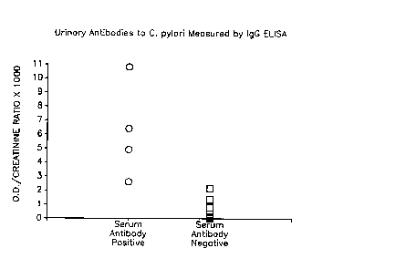
Fig. 1 illustrates the differentiation of urine specimens from persons known to
be sero-positive or sero-negative for C. pylor~ infection, by measuring
urinary IgG antibodies to C. pylori.
Figs. 2-4 relate to Example 5 and are explained therein.
Fig. 5 relates to Example 4 and is explained therein.
Fig. 6 is a whole cell profile of the deposited strains with strips 1-5 representing
deposited strains 86-63 (ATCC 53727), 84-180 (ATCC 53722), 84-182
(ATCC 53725), 86-86 (ATCC 53721) and 84-183 (ATCC 53726),
respectively,
Fig. 7 is the lipopolysaccharide profile of the five deposited strains of Fig. 6 (each lane representing the same strains as in Fig. 6)
Fig. 8 is a Western blot of the deposited strains wherein each strain has been
blotted with rabbit serum raised against that particular strain. For
instance, strip A relates to whole cell fragments of organism A reacted
with rabbit antiserum raised against organism A. Lanes A through E
represent deposited strains 84-180 (ATCC 53722), 84-182 (ATCC
53725), 84-183 (ATCC 53726), 86-63 (ATCC 53727) and 86-86 (ATCC
53721), respectively,
Fig. 9 is a Western blot of the five deposited strains (each lane representing the
same strains as in Fig. 8) wherein each lane includes proteinase
X
1339067
K-treated whole cell Iysates blotted with antiserum raised against the
homologous organism. Fig. 9 depicts primarily lipopolysaccharides
because proteins are digested away by proteinase K.
Any sample suspected of containing C. pylori antibodies may be tested in
accordance with the methods set forth herein. Preferably, the samples to be tested are
bodily fluids such as blood, serum, urine, tears, saliva and the like. Both medical and
veterinary applications are contemplated. In addition to human samples, samples may
be taken from other m~mm~l~ such as non-human primates, horses, swine, etc. Due to
the sensitivity of the test described, it is both possible and preferable to strongly dilute
the sample prior to testing. Dilution may proceed by addition of any fluid compatible
with each of the sample, the antibodies to be tested, and the immobilized antigenic
composition. Serum, when used as the sample, is preferably diluted with one or more
fluids selected from the group consisting of phosphate-buffered saline, pH 7.0-7.4
(hereinafter "PBS~), PBS-cont~ining Tween~ 20 (hereinafter "PBS T"), PBS T with
thimerosal (hereinafter, "PBS 1~"), PBS 1~ (gelatin) (hereinafter ~PBS l-rG"), and
PBS TTG with bovine gamma globulin (hereinafter ~PBS TTGG"), and is preferably
diluted when testing for IgG antibody in a ratio from about 1:500 to about 1:1000, such
as, for instance, about 1:800. Preferred dilution ratios when testing for IgA antibody
are about 1:50 to about 1:200, such as 1:100. IgG tests are preferred.
Preferred diluents and dilution ratios may vary according to the sample being
tested. Urine, for instance, is already relatively dilute and typically is not diluted
further. However, it is unnecessary to concentrate urine as is often necessary with
other assays. Prior to testing, the pH or urine is preferably adjusted to between about
7.0 and 7.4, the preferred pH for antibody function.
While dilution of sample is not required, it is believed that large dilution ratios
reduce the possibility that significant antigen/antibody complexes will be formed in the
absence of C. pylor~-specific antibodies. The extent of dilution should be taken into
~ trade-mark
-8- 1339067
account in adjusting the threshold level of antigen/antibody complex which should be
considered a positive signal.
Antigenic compositions useful in accordance with the present invention include
but are not limited to specific antigens isolated *om fragments of the five deposited
strains, one or more complete org~ni~m~ from among the deposited strains, and
mixtures of the foregoing. Isolated flagella, for instance, have proven to be effective
antigens for the antigenic composition. Antigenic fragments of the C. pylori strains,
which may be proteins, lipopolysaccharides, etc., are identified by their apparent
molecular weight derived from their electrophoretic migration on sodium dodecyl
sulfate/polyacrylamide gels as previously described.
Preferred antigenic mixtures include isolated flagella from any C. pylorz strains
or fragments of said flagella, and isolated fragments of any C. pylori strains wherein
the fragments have an apparent molecular weight on SDS-PAGE of 63,000, 57,000,
45,000 or 31,000 daltons. A mixture of antigens obtained from a pool of all fivedeposited strains is believed to include at least one antigen likely to be present in almost
all C. pylori strains. Hence, a broad specificity results, enabling the antigenic mixture
to be useful in serologic assays. It is preferred that the antigenic composition be
enriched in flagella or in at least one of the said 63,000, 57,000, 45,000 or 31,000
dalton fragments. More preferably, at least 50 percent of the composition or at least 50
percent of the C. pylori fragments are flagella or the specified molecular weight
fragments. In certain preferred embodiments the concentration reaches 85 percent or
more. For some applications, it may be desirable that the antigenic composition be
substantially free of antigens other than flagella or the specified molecular weight
fragments.
An antigenic composition is considered to be substantially free of antigens other
than the antigens of interest whenever the antigenic composition, when subjected to
electrophoresis on SDS-PAGE and appropriate staining, exhibits single well-defined
bands corresponding to the antigens of interest, and no other bands are visuallyapparent.
9 1339067
While the present disclosure provides an easy method for obtaining the
preferred antigens from the deposited C. pylori strains, it is emphasized that these
antigens are common to a large number of C. pylori strains as shown by their efficacy
in testing for the existence of C. pylori strains as shown by their efficacy in testing for
the existence of C. pylori. While the deposited strains and the description of the
present specification provide an easy manner of isolating these antigens, it is
emphasized that the present invention broadly encompasses use of these antigens
regardless of the source from which they are derived.
Antigenic compounds in accordance with the instant invention are preferably
immobilized on a substrate using conventional techniques. For instance, polystyrene
plates may be incubated with antigenic suspensions made in accordance with the
invention. Alternatively, for instance, antigens isolated as protein bands on
electrophoretic gel may be transferred to a nitrocellulose sheet by known methods. See
Towbin et al., Proc. Nat'l. Acad. Sci., 76: 4350-54 (1979); Burnette, et al., Biochem.,
112: 195-203 (1981). Numerous other techniques are known in the art for binding
antigens to substantially inert substances.
Bound antigens in accordance with the invention are preferably contacted with a
highly dilute fluid which includes the sample to be tested for presence of antibody to C.
pylori. The antigen and sample are preferably incubated for at least about one hour.
Considerably less time is needed when incubation proceeds at or near human body
temperature, a'oout 37~C. Incubation at other temperatures, for instance 4~C, is also
proper, but generally requires additional incubation time. Preferred incubation time at
37~C is from about 10 minutes to about 90 minutes. The bound antigens should then
be rinsed to remove any unbound antibodies, i.e., those which are not specific for the
antigens. Preferably, rinsing proceeds with a buffer solution such as PBS T, PBS TT
or Tris/Tween/Sodium chloride/azide. Multiple rinsings are preferred.
During incubation, C. pylori-specific antibodies bind to the immobilized
antigens to create antigen/antibody complexes. All unbound antibodies are
substantially removed during the rinsing procedure. Due to the high specificity of the
y
1339067
- 10-
antigens of the invention, antibodies which are not specific for C. pylori have been
substantially removed at this point. Naturally, if the tested sample did not contain C.
pylor~-specific antibodies, the immobilized ~ntigen~ would be substantially free of
human antibody at this point and subsequent testing for antigen/antibody complexes
should not indicate a substantial presence of such complexes. On the other hand, if the
tested sample were rich in C. pylor~-specific antibodies, these antibodies should have
bound to the immobilized antigens to form a large quantity of antigen/antibody complex
for subsequent detection.
Detection of antigen/antibody complex may be achieved by a wide variety of
known methods. Preferred methods include but are not limited to enzyme-linked
immunosorbent assay, Western blot technique, indirect fluorescence assay or liposome
based assay.
Typically, the C. pylon-specific antibodies complexed with immobilized antigen
are detected by contact with labeled or otherwise detectable second antibodies specific
for human immunoglobulin. The labeled second antibodies may be specific for any
human antibody, preferably of the IgG or IgA type, most preferably, IgG. When acute
sero-conversion is suspected, an IgM test may be appropriate. The second antibodies
are preferably incubated with the immobilized antigens for about 15 minutes to about
2 hours, preferably 30 minutes to 60 minutes at a temperature of about 20~C to about
37~C. Then, the antigens are washed with a buffer solution (preferably multiple times)
in order to remove all unbound labeled antibody. At this point, labeled antibody has
been substantially removed except where it has bound to human immunoglobulin
present on the antigens. Of course, substantially the only human immunoglobulin
present at this point should be C. pylori-specific antibody. Hence, the presence of C.
pylori-specific antibody may be indirectly measured by determining the presence or
absence of the labeled second antibody. There are many known techniques for
detecting the label. For instance, fluorescein-labeled antibody may be detected by
sc~nning for emitted light at the characteristic wavelength for fluorescein.
Alternatively, an enzyme label is detected by incubation with appropriate substrate and
detection of a color change. This can be determined by visual inspection or can be read
X
1339067
1 1
automatically by a spectrophotometer set at the appropriate wavelength. In Western
blotting, for examplet the positive signal may be detected when an enzyme is
conjugated to the second antibody. Incubation with appropriate substrate enzymatically
produces a color product in the immediate vicinity of the antigenic band resolved by
this process. The presence of a reactive band may be detected by visual inspection. In
an indirect immunofluorescence assay, fluorescein-labeled second antibodies may be
detected by fluorescence-activated detectors, or by visual inspection. A liposome-based
assay may involve the presence of fluorescein, an enzyme or a substrate inside aliposome onto which surface C. pylor~ antigens are expressed. These liposomes are
incubated with the body fluid sample to be tested, in the appropriate dilution, and are
thoroughly washed. Those liposomes with human immunoglobulins on their surface
forming an antigen/antibody complex may be recognized by incorporating a second
antibody to a specific human Ig onto the inside walls of a polystyrene tube. Those
liposomes with antibody bound to the C. pylor~ antigens will be immobilized, and non-
immobilized liposomes will be washed away. The liposomes can be Iysed with, for
instance, detergent, or complement, and the enzyme or substrate that was in the interior
is now free to react with the complementary substrate (or enzyme) in the solution in the
tube. The resulting color reaction could be detected by visual inspection or
spectrophotometric color determination. Alternatively, fluorescein present could be
detected by a fluorescence-activated detector.
Testing of certain antigenic pools of the invention with rabbit antiserum raisedto strains not in the antigenic mixture (heterologous) indicated that the pool could detect
antibodies raised to these strains, as well as detecting antibodies raised to the
homologous strains. This indicated that the pool of antigens which included both the
conserved and the diverse (strain-specific) antigens had the type of broad specificity
which should be useful in serologic assays.
The sensitivity and specificity of the antibody detection in accordance with thepresent invention have been determined using serum obtained from persons from
defined populations. The initial analysis was of 40 healthy children and antibody was
not found in this group in the IgA assay, and only once in the IgG assay (Tables 1 and
.~
- 12- 1339067
2). This is significant because both gastritis and peptic ulcer diseases are very
uncommon in this population and it serves as a negative control group. The
distribution of optical density values in the ELISA determination from this population
were used to then establish a threshold for positivity. This is significant because the
assay was then prospectively tested using high-risk and low-risk populations. Examples
1 and 2 are illustrative of the results of this ~ses~ment.
The invention is further elucidated by reference to the following examples
which are set forth only as non-limiting illustrations of the invention.
EXAMPLE 1
IgG assay using pooled suspensions of sonicates
of all five deposited strains in the antigenic composition
An antigenic composition was prepared from 5 C. pylo7i strains (ATCC deposit
numbers 53722, 53721, 53725, 53726, and 53727) which represent a diverse range of
antigens. Bacterial cells were plated onto chocolate agar, then incubated for 48 hours
at 35~C in an atmosphere containing 5 percent oxygen, 10 percent carbon dioxide, 5
percent hydrogen, and the remainder nitrogen. Cells from plates were harvested in
sterile distilled water (3 ml/plate), centrifuged twice at 5,000 x g for 10 minutes at
25~C, and then suspended in sterile distilled water. The concentration of cells from
each strain was standardized at an optical density (at 450 nanometers) of 1.5, then the
suspensions were added together in equal volumes (3 ml of each). The pooled
suspensions were sonicated on ice four times with a Branson sonifier (model S-75,~
Branson Instruments, Danbury, CT) for 30 seconds with 30 second rests. The
preparation was then centrifuged twice at 5,000 x g for 20 minutes to remove whole
cells and the supernatant was centrifuged for 1 hour at 100,000 x g at 4~C (L-78*
ultracentrifuge, Beckman Instruments, Inc., Fullerton, CA). The pellet was suspended
in sterile distilled water and brought to a standard concentration of 1-2 mg/ml.
~ trade-mark
X
1339067
- 13 -
Sonicates were aliquoted and frozen at -70~C until used. For use in the ELISA, the
sonicates were diluted in carbonate buffer (pH 9.6) to a concentration of 10 mg/ml.
Flat-bottomed wells of 96-well polystyrene plates such as IMMULON II*
available from Dynatech Laboratories of Alexandria, Virginia, were incubated
overnight at 4~C with the sonicate of pooled suspensions from these five selected C.
pylori strains at 1.0 ,ug per well in 100 ~l of 50 mM sodium carbonate buffer, pH 9.6.
The wells were aspirated dry and then washed twice with 0.01 M phosphate buffered
saline (PBS, pH 7.2) with 0.05 percent Tween-20 and 0.1 mg/ml thimerosal (PBS-TT)
and then washed twice with 200 ~l of PBS-TT with 0.1 percent gelatin (PBS-TTG) to
limit nonspecific reactivity.
Samples of blood serum were prepared from numerous patients whose known
C. pylori characteristics are reported in Table 1 below. 100 ~l of different test serum
diluted 1:800 in PBS-TTG which also includes 5 mg/ml of bovine gamma globulin
(hereinafter "PBS-TTGG") was added to each of three different wells, and plates were
incubated for one hour at 37~C. The wells were aspirated and washed three times with
PBS-TT in order to remove unbound antibodies, and then incubated for one hour at37~C with 100 microtiters horseradish peroxidase-labeled goat anti-human IgG at a
dilution of 1:5000 in PBS-TT cont~ining 1 percent bovine serum albumin and
0.1 percent bovine gamma globulin. Goat anti-human IgG is a second antibody thatbinds with the antigen/antibody complex which should have formed only in wells
exposed to positive serum, i.e., serum containing C. pylori-specific antibody. The
wells were successively washed five times with PBS-TT to remove the unbound goatantibody.
A 0.1 ml sample of developing solution containing 1.0 mg of 2,2'-azino-di-
(3-ethyl benzthiasoline sulfonic acid) (ABTS) per ml in McIlvain's buffer (pH 4.6) with
0.005 percent hydrogen peroxide was added to each well and incubated at 25~C forthirty minutes.
X
- 14- 1339067
This substrate mixture detects the peroxidase label and forms a color product
which may be detected by an ELISA reader capable of detecting light at a wavelength
of about 410 nm. The ELISA reader quantified the color reading. Assays were
performed in triplicate. Control wells on each plate were processed in an identical
fashion, except that diluent rather than test serum was added. Absorbance readings
greater than the mean + 3 intervals of standard deviation for the results observed when
a group of 40 healthy children under 10 years old were tested were taken as positive.
The positive threshold was determined to be 0.910 units of optical density at 410 nm,
where 100 microliters of developing solution is placed in the standardized flat-bottom
microtiter wells identifled above. The results are shown in Table 1.
1339067
- 15 -
TABLE 1--IgG ASSAY USING SONICATES
OF ALL FIVE DEPOSITED STRAINS
Subject No. Positive/ Percent
No. Tested Positive
Cases
Patients with gastrointestin~l ~ylllp~llls and28/29 96.6
confirmed C. pylon infection and gastritis
(confirmed by culture or by identification of
characteristic org~ni~m~ on stained
histological section
Asymptomatic persons with confirmed C. 28129 96.6
pylori infection and gastritis
Patients with confirmed duodenal ulceration 44/45 97.8
Controls
Asymptomatic persons without confirmed 2/61 3.3
C. pylon infection
Asymptomatic children 1/40 2.5
EXAMPLE 2
IgA assay using pooled suspensions of sonicates
of all five deposited strains in antigenic composition
The methods employed were identical to those indicated in Example 1 except
that the test human serum was used at 1:50 dilution for IgA determinations (reflecting
the lower IgA concentration in serum), and peroxidase-labeled goat anti-human IgA
was used as the labeled second antibody, diluted 1:1000. The positive threshold was
determined to be 0.470 units of optical density determined as in Example 1. The
results are shown in Table 2.
V
1~39067
- 16-
TABLE 2--IgA ASSAY USING SONICATES
OF ALL FIVE DEPOSITED STRAINS
Subject No. Positive/ P~.~nl
No. Tested Positive
Cases
Patients with gastrointestin~l symptoms and28/29 96.6
confirmed C. pylori infection and gastritis
(confirmed by culture or by identification of
characteristic org~nisms on stained
histological section
Asymptomatic persons with confirmed C. 28l29 96.6
pylori infection and gastritis
Patients with confirmed duodenal ulceration45/45 100.0
Controls
Asymptomatic persons without confirmed 3/61 4.9
C. pylor~ infection
Asymptomatic children 0/40 0.0
EXAMPLE 3
IgG antibodies to C. pylori in urine
of persons with serum antibodies to C. pylori
Urine from persons known to have serum antibodies to C. pylori (as determined
by the serum test of Example 1) was assessed to determine whether specific IgG
antibodies were detectable in urine. The methods employed are identical to those of
Examples 1 and 2 except that the pH of the urine specimen was neutralized to 7.4 using
lN sodium hydroxide and 100 ~l of this specimen was added to each microtiter well.
To account for differences in hydration status of the persons tested and dilution of
urine, creatinine concentration of the urine specimen was measured, and the results
were expressed as a ratio of optical density in the ELISA divided by the creatinine
V
1339067
- 17 -
concentration. This standardizes the assay regardless of variation in concentration of
urine. The results obtained from the IgG ELISA are presented in Fig. 1. There was
no overlap between the values obtained from the 4 persons who were known to be sero-
positive and the 10 known to be sero-negative. Using the mean + 3 intervals of
standard deviation for the urine specimens obtained from sero-negative persons as the
cutoff for positivity, all of the sero-positive persons were positive. The threshold
positive indicator (in units determined by multiplying optical density by 1,000, then
dividing the product by cre~tinine concentration in mg per deciliter) was 2.6 units.
EXAMPLE 4
Western blot assay
Western blot analysis of the test sera was conducted as follows: a pool of
sonicates as in Examples 1-3 was fractionated by electrophoresis on a 10 percentpolyacrylamide slab gel in the presence of sodium dodecyl sulfate (SDS). The bands
on the gel were electrophoretically transferred to a nitrocellulose sheet, according to the
procedure of Towbin et al. (Proc. Natl. Sci. USA 76:4350-54 (1979)) as modified by
Burnette (Anal. Biochem. 112:195-203 (1981)). Strip solid phase enzyme-
immunoassays were then performed.
In brief, after SDS-PAGE, the gels were covered with nitrocellulose paper
(NCP) that had been soaked in electrode buffer (192 mM glycine, 25 mM Tris base, 20
percent methanol). Electroblotting sponges were rinsed in deionized water and then
saturated with the electrode buffer. After the gel was placed on the sponge, the NCP
was laid over the gel, and then the second sponge was overlaid. This sandwich was
placed in an electroblotting apparatus, and the proteins were electrophoresed at 100 mA
for 18 hours. The NCP was rinsed in borate buffer (pH 8.0) with 0.05 percent
Tween-80 and then were incubated at room temperature for one hour with 3 percentdried non-fat milk in borate buffer. After a rinsing in borate buffer, the NCP was cut
into vertical strips containing multiple bands and each strip incubated at 25~C for 4
hours in a 1:400 dilution of the test serum samples in 3 percent dried non-fat milk in
X
1339067
- 18 -
borate buffer. After three one-hour washes in 1 percent dried non-fat milk in borate
buffer, the NCP strips were incubated for 2 hours at 25~C with peroxidase conjugated
rabbit anti-human IgG diluted 1:5000 in 1 percent milk-borate buffer. After three
twenty-minute washes in borate buffer, the NCP strips were placed in DAB solution
(50 mM Tris with 0.025 percent diaminobenzidine with two drops of hydrogen
peroxide), for 5 to 10 minutes until reaction products were optimally developed. The
reaction was stopped by washing the strips in tap water. The strips were read by visual
inspection.
Fig. 5 indicates graphically the results of these experiments. Strip A was
incubated in the absence of human antibody with only 3 percent dried non-fat milk in
borate buffer; Strip B was incubated with serum from a patient with gastrointestinal
symptoms who had neither C. pylor~ infection nor gastritis; Strip C was incubated with
serum from another patient with gastrointestinal symptoms who had neither C. pylori
infection nor gastritis; Strip D was incubated with serum from a patient with peptic
ulcer disease; Strip E was incubated with serum from a second patient with peptic ulcer
disease; Strip F was incubated with serum from a patient with gastrointestin~l
symptoms who was found to have both C. pylori infection and gastritis by endoscopy
(gastric intubation) wherein a biopsy and culture showed gastritis and presence of the
organism; Strip G was incubated with serum from another patient with gastrointestinal
symptoms who was found to have both C. pylori infection and gastritis; Strip H was
incubated with seNm from an asymptomatic person who was found to have both C.
pylori infection and gastritis; Strip I was incubated with serum from another
asymptomatic person who was found to have both C. pylor~ infection and gastritis.
EXAMPLE S
To investigate the specificity of the reactions, C. pylori cells were analyzed in
comparison with cells of other enteropathogenic org~ni.cms. The assays employed
determined whether pre-incubation of known positive sera in the C. pylorc ELISA with
C. pylori or control cells would significantly reduce optical density readings. The
serum used was a pool from C. pylorz-infected persons that had high values in the IgA,
1339067
- 19-
IgG, and IgM ELISA. The pooled serum was absorbed with whole cells of C. pylori,Escherichia coli, Campylobacterfetus, or Campylobacter jejuni. Bacterial growth from
an overnight culture on one plate was harvested, cells were suspended in distilled
water, washed twice with sterile distilled water, then mixed with 1.0 ml of the pooled
human serum and incubated at 37~C for 45 minutes. Antibodies to the bacterial
suspension were removed by centrifugation at 12,000 x g for five minutes. After
saving 100 ~4l aliquot for ELISA determination after each absorption, the supernatant
was reabsorbed five times. An unabsorbed serum control was exposed to the same
incubation and centrifugation conditions. Preincubation of the positive serum pool with
C. pylori cells significantly reduced optical density in all three immunoglobulin classes.
Absorption of the pool with C. jejuni, C. fetus, or E. coli produced minim~l decreases
in optical density in the IgA (Fig. 3) and IgG ELISA (Fig. 4). However, in the IgM
ELISA (Fig. 5), absorption with the homologous and heterologous org~ni~m~ produced
less diverse levels of inhibition. These results show that the antigens detected in the
IgA and IgG are more specific for C. pylori than are those detected in the IgM ELISA.
To further define the specificity of the C. pylori ELISAs for sero-diagnosis of
C. pylori infection, antibody levels in other control groups were compared. There
were no seroconversions between acute and convalescent-phase specimens from
30 patients with acute bacterial enteritis with fecal leukocytes present. Included among
these were 12 patients with acute C. jejuni infection, each of whom seroconverted to C.
jejuni antigens.
-20- 1339067
EXAMPLE 6
Use of single C. pylori strain
as anti~en in assay instead of Five-strain pool
Strain 84-183 (ATCC 53726) was processed exactly as indicated in Example 1.
However, for establishing the assay, instead of pooling sonicates from five strains to
reach a protein concentration of 1.0 ~g per well, the sonicate from strain 84-183
(ATCC 53726) was used alone at a concentration of 1.0 ~g per well. The results of the
comparison between the assays when the standard five-strain antigen was used andwhen the single antigen was used are shown in Table 3.
TABLE 3
Comparison of diagnostic efficacy of C. pylorc serum ELISA using
five-strain sonicates (5-Ag) versus sonicates of strain 84-183 only (1-Ag)
Number of Sera Found to be Positive for
IgG IgA
s Aga ¦ l-Agb 5 AgC ¦ l-Agd
Patients Studied
Group n
Known 14 14 14 14 14
positivee
Known 14 0 0 2 2
negativef
a. Standard assay employing five C. pylo7i strains as the antigen; optical density
above 0.910 as positive threshold.
b. Comparison assay employing one C. pylori strain (84-183) as the antigen;
optical density above 0.700 as positive threshold.
l~39o67
- 21 -
c. Standard assay employing five C. pylori strains as the antigen; optical density
above 0.470 as positive threshold.
d. Comparison assay employing one C. pylori strain (84-183) as the antigen;
optical density above 0.470 as positive threshold.
e. These patients had C. pylori present in tissue on histologic e~min~tions, had C.
pylori isolated from culture, and had gastritis on biopsy.
f. These patients had no C. pylori present in tissue or culture, and had no gastritis.
These results indicate that choice of a single C. pylori strain which possesses
certain conseNed antigens will permit an assay to be developed that has similar or
identical diagnostic efficacy as when the pooled antigen is used.
EXAMPLE 7
Use of purified C. pylori flagellae
as antigen in assay instead of five-strain pool
Flagellae of gram-negative bacteria usually possess important surface antigens
to which infected hosts generally produce antibodies. To determine whether any of
these antigens were in fact among those to which C. pylori-infected persons wereresponding, we purif1ed flagellae for further study.
Strain 84-182 (ATCC 53725) was grown exactly as indicated in Example 1.
After the cells were haNested from plates in sterile distilled water, the suspension was
centrifuged at 3,000 x g for 20 minutes. The supernatant was aspirated and the pellet
was resuspended in 0.1M Tris-HCI (pH 7.4) to achieve an optical density at the 450
nanometer setting of 1.5. This suspension was treated by passage at 0~C in a Virtis
blender at medium-high intensity for 45 seconds. This procedure shears the flagella
from Campylobacter species (Blaser et al, Infection and Immunity 1986; 53:47-52).
The suspension was then centrifuged at 12,000 x g for 10 minutes at room temperature
to pellet cells and cell debris. The supernatant was then centrifuged at 55,000 x g for
60 minutes at 5~C to sediment the sheared flagella. This pellet was resuspended in Tris
.~
-22- 1339067
buffer at 4~C and protein concentration determined. For establishing the ELISA, this
preparation was used at protein concentrations of 100 ng per microtiter plate well.
Further steps in the assay were exactly as indicated in Example 1. The results of the
comparison between the IgG assays with the standard five-strain antigen was used and
when the flagella preparations were used are shown in Table 4.
A strain other than the deposited strains was selected in an unbiased manner,
and flagella compositions were prepared as above. These compositions were tested at
100 and 500 ng per well and the results tabulated in Table 4.
TABLE 4
Comparison of diagnostic efficacy of C. pylor~ serum IgG ELISA using
five-strain sonicate versus purified flagella preparations as the antigen
Number of Sera Found to be Positive for IgG
Separated Flagellar Preparation
Flagella
Prep'n
5 Aga 100 ngb 5oo ngc 100 ngd
Patients St~ d
Group n
Known 14 14 14 14 13
positivee
Known 14 0 0 0 0
negative'
1339067
- 23 -
a. Standard assay employing five C. pylori strains as the antigen; optical density
above 0.910 as positive threshold.
b. Comparison assay employing purified C. pylori flagella from deposited strain
84-182 (ATCC 53725) as the antigen at a concentration of 100 ng/well; optical
density above 0.080 as positive threshold.
c. Comparison assay employing purified C. pylori flagellae from the non-deposited
selected strain as the antigen at a concentration of 500 ng/well; optical density
above 0.090 as positive threshold.
d. Comparison assay employing purified C. pylori flagellae from the non-deposited
selected strain as the antigen at a concentration of 100 ng/well; optical density
above 0.090 as positive threshold.
e. These patients had C. pylori present in tissue on histologic e~min~tions, had C.
pylori isolated from culture, and had gastritis on biopsy.
f. These patients had no C. pylorc present in tissue or culture, and had no gastritis.
EXAMPLE 8
Use of pooled gel filtration fractions of C. pylori
flagella as antigen in assay instead of five-strain pool
To determine which of the C. pylorc flagellae-associated antigens were those to
which C. pylori-infected persons were responding, a flagellar preparation from a C.
pylori strain selected in an unbiased manner (the same such strain as in Example 7) was
fractionated by passage through a gel f1ltration chromatographic column. The column
employed was a Superose 12~ column (Pharmacia Laboratories, Piscataway, NJ) in aPharmacia Fast Protein Liquid Chromatographic (FPLC) apparatus. The flagellar
preparation was suspended in 20 mM tris buffer (pH 8.0). The flow rate was
0.3 ml/min and fractions were collected over a period of 100 minutes. The first peak
passed through the column between 20 and 30 minutes, and other peaks were seen after
60 minutes. Analysis of the content of the first peak was by SDS-PAGE followed by
~trade-mark
1339067
- 24 -
silver staining. Using this sensitive stain, only 3 bands were resolved for the fractions
representing the peak. These migrated at 63,000, 57,000 and 31,000 daltons. For
establishing the ELISA, these fractions were pooled, and this preparation was used at a
protein concentration of 100 ng per microtiter plate well. Further steps in this assay
were exactly as indicated in Example 1. The results of the comparison between the IgG
assays when the standard five-strain antigen was used and when the gel filtration
fractions of the flagella preparation was used are shown in Table 5.
TABLE 5
Comparison of diagnostic efficacy of C. pylori serum IgG ELISA using
five-strain sonicate versus gel filtration of the flagella preparation as the antigen
Number of Sera Found to be Positive for IgG
Gel Filtration
5 Aga Pooled Fractionsb
Patients Studied
Group n
Known positiveC 14 14 14
Known negativedd 14 0 0
a. Standard assay employing five C. pylori strains as the antigen; optical density
above 0.910 as positive threshold.
b. Comparison assay employing selected fractions of C. pylon flagellae passed
through gel filtration chromatographic column at a concentration of
100 ng/well; optical density above 0.100 as positive threshold.
c. These patients had C. pylori present in tissue on histologic e~min~tions, had C.
pylori isolated from culture, and had gastritis on biopsy.
d. These patients had no C. pylori present in tissue or culture, and had no gastritis.
X
- 25 - 13390 6 7
E XAI~IPLE 9
Persistence of serum antibodies to C. ~ylori
Five persons with high IgA or IgG serum antibody levels to C. pylori were
selected for further study. From each person, a second sample obtained at least one
year after the original evaluation (mean 1.25 years) was restudied. The mean antibody
levels in the first and second sera are shown in Table 6.
TA~BLE 6
Persistence of serum antibodies to C. pylori in 5 sero-positive persons
Optical Density in C. pylori ELISAa
Antibody Class Ser~m 1 Ser~m2b
IgG 1.12 + 0.27 1.18 + 0.35
IgA 1.04 + 0.27 1.04 + 0.33
a. Mean :~: standard error of mean.
b. Serum 2 obtained at least one year after serum 1.
The levels in all five persons remained stable. None of the sera converted from
sero-positive to sero-negative during the interval.
E XA~nPLE 10
Seroconversion in a volunteer challen~ed with C. pylori
A human volunteer ingested C. pylori and developed symptoms of acute gastritis
with achlorhydria (see Morris and Nicholson; American Journal of Gastroenterology,
1987; 82:192-199). Subsequently, his Syl-lptOlllS cleared but he developed chronic
gastritis and C. pylori infection persisted. Serial serum specimens were obtained and
-26- 1339067
studied for antibodies to C. pylor~. The assays for C. pylori-specific IgG and IgA were
exactly as described in Examples 1 and 2. The assay for IgM was performed exactly as
in Example 1 except that the serum was diluted 1:400 to ~limini.~h non-specific
reactivity and the serum IgM was detected by a peroxidase-conjugated goat antibody
specific for IgM, and the reaction developed for 60 minutes. The results are shown
below in Table 7. Seroconversion in the IgA and IgG classes occurred between days
60 and 431 following experimental challenge. Although IgM seroconversion criteria
have not been specifically defined, the nearly four-fold increase in optical density
between day 8 and 22 after challenge, and gradual decline is significant. It is
noteworthy that the increase in C. pylon-specific IgM is appreciably earlier in the
course of the infection than either the IgA or IgG responses.
TABLE 7
Seroconversion to C. pylori antigens in a volunteer challenged with C. pylori
Day After Optical Density~
Day After Onset of
IgGb IgAC IgMd
Challenge Symptoms
8 5 0.154 0.114 0.163
11 8 0.111 0.138 0.227
18 15 0.185 0.179 0.591
22 19 0.049 0.165 0.647
33 30 0.157 0.177 0.524
57 0.138 0.145 0.445
431 428 >2.000 0.728 0.171
581 578 >2.000 0.476 0.223
a. Each value shown is the mean of triplicate determination.
b. Serum dilution is 1:800; threshold for positive determination is 0.910.
c. Serum dilution is 1:50; threshold for positive determination is 0.470.
-27- 1339067
d. Serum dilution is 1:400; no threshold for positivity has been established butserum obtained 22 days after challenge shows a nearly four-fold increase over
earlier samples.
EXAMPLE 11
C. pylori-specific test kits were constructed for detecting antibodies using
several different techniques for detection. One test kit for antibody detection was
comprised of a compartmented enclosure cont~ining a plurality of wells, plates which
were coated prior to use with C. pylori antigens, and ELISA materials for enzymedetection consisting of peroxidase-labeled goat anti-human IgG and a color change
indicator consisting of ABTS in McIlvain's buffer with 0.005 percent hydrogen
peroxide. Naturally, other enzymes and developers could have been used. For
instance, alkaline phosphates-labeled goat anti-human IgG could be used in conjunction
with p-nitrophenyl phosphate in diethanolamine and magnesium chloride buffer.
A second test kit for detecting antibodies using the Western blot technique was
comprised of a container, cover, nitrocellulose sheet, and a polyacrylamide slab gel in
the presence of sodium dodecyl sulfate, surfactants, pH modifiers, dried non-fat milk
and materials for enzyme detection including a color change indicator consisting of
DAB in Tris with hydrogen peroxide. This Western blot analysis kit also containsperoxidase-labeled goat or rabbit anti-human immunoglobulin and a source of C. pylori
antigens.
Another C. pylori-specific test kit for detecting antibodies using the indirect
immunofluorescence assay may include a compartmental container with C. pylori
antigens, human test serum, phosphate buffered saline and fluorescein-conjugated goat
anti-human IgG.
Finally, a different C. pylori-specific test kit for detecting antibodies uses
liposomes and comprises a container, human test serum, fluorescent marker- (or
enzyme- or substrate-) filled liposome with C. pylori antigens on their surface, and a
1339067
- 28 -
surface-active agent. In this assay the container might be a pre-coated tube or well
with goat anti-human IgG.
The terms and descriptions used herein are preferred embodiments set forth by
way of illustration only, and are not intended as limitations on the many variations
which those of skill in the art will recognize to be possible in practicing the present
invention as defined by the following claims.
~y