Note: Descriptions are shown in the official language in which they were submitted.
200049~3
`_ 1
TITLE OF THE INVENTION
Blood Perfusion System and Tube Means Used Therein
BACKGROUND OF THE INVENTION
Field of the Invention
This invention relates to a blood perfusion system for
percutaneous transluminal angioplasty to, for example,
femoral and coronary arteries for the purpose of dilating
the stenosis to improve blood flow therethrough. It also
relates to tube means used in such a blood perfusion system.
Prior Art
In treating a stenosis in a vessel such as coronary
artery, a dilation catheter having a balloon at a distal
region thereof is inserted into the~ vessel until the balloon
reaches the lesion. The balloon is then inflated to expand
the stenosis. The inflated balloon inevitably blocks the
relevant portion of the vessel to stop further blood flow.
Continued blood flow interruption for some time is dangerous
to the patient. Thus, if the operation takes a long time,
it is critical to ensure normal blood flow by transporting a
necessary amount of blood to the periphery of the lesion
through the lumen of the dilation catheter.
The following two methods are known for such blood
perfusion.
A first method is to previously collect blood in a
blood bag from the patient herself or himself or another
person. During the operation, the blood in storage is
introduced into the lumen of the dilation catheter and
injected to the periphery of the lesion.
A second method is to aspirate blood from another
vessel of the patient under operation. The blood taken in
is directly fed to the lumen of the dilation catheter and
injected to the periphery of the lesion over the entire
operation period.
~'
2al~1493
-2-
However, the first method has a problem that the blood
feed which has been in storage is less fresh and can cause
infection. A problem of compatibility with the patient
arises particularly when blood from another person is used.
The second method is advantageous in that fresh blood
can be fed. Nevertheless, in addition to the site where the
dilation catheter is inserted or the sheath is indwelled in
the patient's vessel, a cutdown or puncture must be done at
another site of the vessel (for example, a blood intake
needle be punctured) for the purpose of aspirating blood.
This adds to the burden to the patient. The additional
burden is serious to old patients, the majority of patients
who need a treatment to dilate a stenosis.
SUMMARY OF THE INVENTION
Therefore, an object of the present invention is to
eliminate the above-mentioned problems and to provide a
novel and improved blood perfusion system which can perfuse
fresh blood of the patient herself or himself through the
vessel across a lesion during operation without making an
additional cutdown or puncture for blood intake, thereby
imposing no additional burden to the patient.
Another object of the invention is to provide tube
means for use in such a blood perfusion system.
According to the present invention, there is provided a
blood perfusion system comprising a dilation catheter, a
sheath, a tube, and a pump. The dilation catheter has
leading and trailing ends and defines a longitudinal lumen
extending from the leading end to the trailing end and open
at the leading and trailing ends. The dilation catheter
further includes a dilating member at the leading end. The
sheath has leading and trailing ends and defines a
longitudinal bore through which the dilation catheter is
insertable. When the dilation catheter is inserted therein,
the sheath defines a blood intake gap between the outer
surface of the dilation catheter and the sheath bore. The
Z~ 493
sheath further includes transverse bore means in fluid
communication with the sheath bore. The tube means for
defining a continuous flowpath is connected at one end to
the transverse bore means and at another end to the trailing
end of the lumen of the dilation catheter. The pump means
is mounted in the tube means for pumping blood. When the
sheath having the dilation catheter inserted therein is
set in a blood vessel, blood is taken into the blood
intake gap in the sheath, passed through the sheath bore,
the tube means, and the dilation catheter lumen, and fed
back to the vessel through the open leading end of the
dilation catheter.
Further, a guiding catheter having a lumen through
which the dilation catheter is insertable may be provided.
In this case, the guiding catheter with the dilation
catheter received therein is inserted in the sheath.
In a preferred embodiment, the system may further
include means inserted in the flowpath of said tube means
for storing blood.
The sheath may further include a valve body mounted at
the trailing end thereof for blocking the sheath bore when
the sheath bore is empty and sealing any gap when the
dilating or guiding catheter is inserted therein.
In another aspect, the present invention provides
tube means for defining a continuous flowpath having one end
connectable to the transverse bore means and another end
connectable to the trailing end of the lumen of the dilation
catheter, the tube means including at least one section of
tubing to which blood pumping means is mountable.
According to the present invention, a gap for blood
intake is defined between the bore of the sheath to be
endermically indwelled in the vessel and the dilation
catheter. During an operation, blood is taken in through
the intake gap, passed through the sheath bore, the tube
means or blood feed circuit connected to the sheath bore,
and the lumen of the dilation catheter connected to the
2~)0Q49~3
circuit, and fed back to the vessel through the leading
opening of the dilation catheter. Since blood is taken into
the intake gap through the leading end of the sheath
endermically indwelled in the patient's vessel, it is
unnecessary to make a cutdown or puncture at another site of
the vessel, mitigating a burden to the patient.
The operation may be carried out as described above in
the case of a relatively thick vessel such as femoral
artery. In the case of a relatively thin vessel such as
coronary artery, a guiding catheter may preferably be used.
BRIEF DESCRIPTION OF THE DRAWINGS
The above and other objects, features, and advantages
of the present invention will be better understood from the
following description taken in conjunction with the
accompanying drawings, in which:
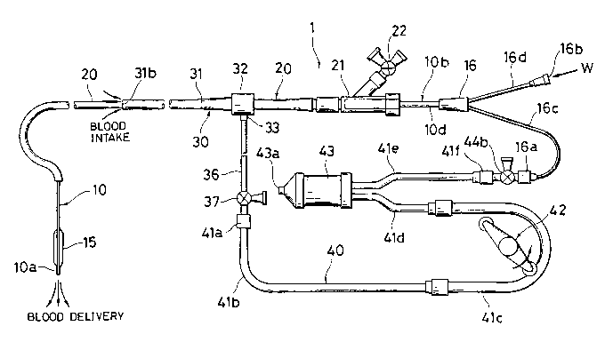
FIG. 1 is a schematic illustration of a blood perfusion
system according to one embodiment of the invention, the
system including a dilation catheter, a guiding catheter, a
sheath, and tube means;
FIG. 2 is an enlarged longitudinal cross section of a
leading portion of the dilation catheter used in the system
of FIG. l;
FIGS. 3a and 3b illustrate different examples of a
trailing portion of the dilation catheter used in the system
of FIG. l;
FIG. 4 is an enlarged, partially short cut,
longitudinal cross section of the sheath used in the system
of FIG. l;
FIGS. 5 and 6 are perspective views of the valve body
mounted in the sheath of FIG. 4, FIG. 5 showing the outer
configuration and FIG. 6 being a see-through view showing
the internal structure of the valve body;
FIGS. 7 to 10 illustrate successive steps of operation
3 5 using the blood perfusion system of the invention, FIG. 7
being a fragmental cross section showing the sheath inserted
2QO~)493
_ 5
in the vessel, FIG. 8 being a fragmental cross section
showing the dilation catheter inserted into the stenosis,
FIG. 9 being a fragmental cross section similar to FIG. 8,
but showing that the dilating member of the dilation
catheter is inflated, and FIG. 10 being a fragmental cross
section showing the dilation catheter through which blood is
fed to the periphery of the stenosis.
Like parts are designated by the same reference
numerals throughout the figures.
DESCRIPTION OF THE PREFERRED EMBODIMENTS
The blood perfusion system of the present invention and
the tube means used therein are now described in further
detail by referring to their embodiments shown in the
figures.
Although the blood perfusion system of the present
invention is illustrated herein as being applied to the
coronary artery, its application is not limited thereto.
Further in the disclosure, the terms "leading end" and
"trailing end" are generally used in connection with
catheters and associated members on a basis of the direction
of inserting the catheter into a vessel during an operation.
The terms "lumen" and "bore" have an interchangeable meaning
of the cavity of a tubular member. Reference numeral 2
designates a blood vessel under operation and 3 designates a
stenosis or lesion.
FIG. 1 is a schematic illustration of one embodiment of
a blood perfusion system according to the present invention.
Briefly stated, the blood perfusion system generally
designated at 1 includes a dilation catheter 10, a guiding
catheter 20, a sheath 30, and tube means or blood pumping
circuit 40. It will be understood from the following
description, the guiding catheter is optional.
The dilation catheter 10 is first described. FIGS. 2
and 3 are enlarged axial cross-sections of leading and
trailing portions of the dilation catheter used in the blood
200Q49~
perfusion system of FIG. 1. The dilation catheter 10 is
mainly composed of an elongated flexible tubular section of
the double wall structure which includes an inner tube 12
defining a first longitudinal bore or lumen 11 therethrough
and an outer tube 14 defining a second longitudinal bore or
lumen 13 therethrough coaxially surrounding the inner tube
12 to leave an annular space therebetween. The dilation
catheter 10 further includes a dilating member 15 attached
to a leading portion lOa of catheter 10 and a branch hub 16
of generally Y shape attached to a trailing portion lOa of
catheter 10. The catheter 10 is of a fluid tight structure
as a whole.
The first lumen 11 of inner tube 12 extends from the
leading end lOa to the trailing end lOb of dilation catheter
10. The first lumen 11 is open at its leading end in a
forward direction and communicates at its trailing end with
a first opening 16a of branch hub 16.
The second lumen 13 of the outer tube 14 also extends
from the leading end lOa to the trailing end lOb of dilation
catheter 10, and communicates at its leading end with an
interior space 15a of dilating member 15 and at its trailing
end with a second opening 16b of branch hub 16.
It is to be noted that the dilation catheter 10 is not
limited to the double tube structure of the illustrated
embodiment and may take a double lumen structure wherein two
lumens extend in juxtaposition or a double tube structure
combined with a double lumen structure.
The inner tube 12 is preferably formed from a synthetic
resin material having a rigidity imparting member 17, for
example, tubular metal mesh integrally embedded therein so
that the inner tube undergoes no twisting or bending when
rotating forces are applied thereto by a guiding catheter 20
as will be described later.
Also preferably, a length of metallic wire may be
inserted and fixedly indwelled in the second lumen 13
between the inner and outer tubes 12 and 14 so that the wire
2~1~Q493
extends from the leading end to the trailing end of the
lumen because the indwelling wire assists in inserting of
the catheter.
The outer surface portion of the inner tube 12 where it
is surrounded by the dilating member 15 is preferably
provided at two axially spaced positions, for example, with
marks 18 of radiopaque material having any desired shape in
order that the location of the dilating member 15 can be
visually identified under fluoroscopic observation.
The dilating member 15 may be formed of resinous
material such as polyethylene terephthalate (PET), polyvinyl
chloride (PVC), and ethylene-vinyl acetate copolymer (EVA).
The dilating member 15 has a forward end attached to the
outer surface of the inner tube 12 leading portion and a
rear end attached to the outer surface of the outer tube 14
leading portion as by adhesive bonding or fusion welding.
The branch hub 16 is configured in Y shape having the
first opening 16a at one branch and the second opening 16b
at the other branch as shown in FIG. 3a. A flexible guide
wire G (see FIGS. 8 and 9) is inserted into the first
opening 16a of branch hub 16 when it is desired to insert
the dilation catheter 10 to the stenosis 3 in the vessel 2.
The second opening 16b of branch hub 16 is connectable to an
admission line for dilating fluid W when it is desired to
introduce the dilating fluid into the dilating member 15.
Alternatively, as shown in FIG. 3b, the branch hub 16
may also be composed of a sleeve and flexible connecting
tubes 16c and 16d extending from the sleeve and terminating
at free ends to which the first and second connectors 16a
and 16b are attached.
The dilation catheter 10 of the above-illustrated
construction is inserted through a guiding catheter 20.
Upon insertion, the leading end portion lOa leads the
catheter 10.
As shown in FIG. 4, the guiding catheter 20 is a
tubular member defining a lumen 25 adapted to receive either
~ ()493
a guide wire (not shown) which serves to lead the guiding
catheter 20 when it is directed to a predetermined site in
the vessel or the dilation catheter 10.
In carrying out an operation for dilating the stenosis
3, the guiding catheter 20 is received for longitudinal
motion and rotation in a sheath 30 to be described later~
The guide wire (not shown) is inserted into the lumen 25 of
guiding catheter 20, if desired, through a rotary connector
21. After the leading end of the guiding catheter 20 has
reached a predetermined position, the guide wire is
withdrawn. Instead, a contrast medium is injected for
angiography.
As described above, the dilation catheter 10 or guide
wire G is introduced into the lumen 25 of guiding catheter
20 through the rotary connector 21. The rotary connector 21
defines a main bore through which the dilation catheter 10
is inserted. The connector 21 is preferably provided with a
branch having a three-way cock 22 or a multi-port manifold
which is used when it is desired to carry out another action
(for example, contrast medium injection or blood pressure
measurement) during the operation.
FIG. 4 illustrates a cross-sectional structure of the
sheath 30. As seen from the figure, the sheath 30 is an
elongated, generally cylindrical member defining a longi-
tudinal lumen or bore 35 and having leading and trailingends. The sheath 30 includes a generally cylindrical sheath
body 31 formed of a fluoro-resin material such as ethylene
tetrafluoroethylene (ETFE), perfluoroalkoxyl (PFA) resin,
and fluorinated ethylene-propylene (FEP) and a hub 32 of
metal or rigid synthetic resin fluid-tightly engaged with a
trailing portion of sheath body 31, and a transverse
protrusion 33 extending from at least one lateral site on
the hub 32 for connection to a blood feed circuit 40 which
will be described later. The transverse protrusion 33 is
connected to a three-way cock 37 through a sheath or side
tubè 36 as shown in FIG. 1. A valve body 34 to be described
2~0~493
-- g
later is mounted in the bore of hub 32 at its inlet portion
32b.
The guiding catheter 20 is loosely received in the bore
35 of sheath body 31 to define a gap 31a for blood intake
between the inner surface of bore 35 and the outer surface
of guiding catheter 20. The sheath body 31 is dimensioned
such that the inner diameter of sheath body bore 35 is
larger than the outer diameter of guiding catheter 20. The
dimensions of these members dictate the cross-sectional area
of the gap or flowpath 31a for blood intake and are selected
so as to ensure that blood enters the gap 3la at the maximum
necessary flow rate through a blood intake opening 3lb at
the leading end of sheath body 31 when the sheath 30 is set
in the brachial or femoral vessel 2. The necessary flow
rate of blood may vary with a disease case (the extent of
treatment, the type and thickness of a particular vessel to
be endermically reached, for example) and is desirably in
the range of about 40 to 50 ml/min. in the case of coronary
artery operation.
The hub 32 defines a bore 32a of an approximately equal
diameter to that of sheath body bore 35 in fluid communica-
tion with the blood intake gap 31a. The hub bore 32a at a
trailing end is diverged to form the inlet portion 32b
through which the guiding catheter 20 is inserted.
The transverse protrusion 33 on the side of hub 32 for
connection to the blood feed circuit 40 defines a trans-
versely extending bore 33a which is in fluid communication
with the blood intake gap 31a through the hub bore 32a.
Since the sheath tube 36 is fitted over the protrusion 33,
the protrusion 33 is preferably formed on the outer surface
with engaging ribs 33b for preventing accidental disengage-
ment of the tube 36 therefrom.
The valve 34 is generally mounted in the hub 32 for the
purpose of preventing the blood which has entered the bore
35 through the blood intake opening 3lb from flowing to the
exterior through the inlet portion 32a when the sheath 30 is
O- 20Q0493
dwelled in the vessel, but the guiding catheter 20 is not
inserted in the sheath bore 35. The valve 34 also plays
the role of preventing air from entering the hub bore 32a
from the exterior when the guiding catheter 20 having the
dilation catheter 10 inserted therein is inserted into the
sheath bore 35.
The valve body 34 may be a solid cylindrical disk
having a pair of opposed circular surfaces as shown in
FIGS. 5 and 6. The cylindrical valve body has a first slit
34a open only on one circular surface and a second slit 34b
open only on the other circular surface, both slits 34a and
34b axially extending a portion of the entire axial length
of the valve body. Within the valve body the slits 34a and
34b cross each other along an intersection 34c having an
axial length L. The valve body 34 is formed from a
resilient material such as various rubbers and elastomeric
resins. When the guiding catheter 20 is inserted into the
valve body 34, the valve body 34 makes a continuous surface
contact with the outer surface of the guiding catheter 20
from all directions in tight fit relation depending on the
outer diameter of the guiding catheter 20, maintaining a
fluid-tight seal between the sheath 30 and the guiding
catheter 20.
Instead of the sheath 30 of the illustrated
configuration, there may be used a sheath of the
configuration disclosed in FIG. 2 of U.S. Patent No.
4,610,674, which also discloses the detailed construction
of the valve body 34 available in the present invention.
The tube means 40 which constitutes one of the
features of the present invention provides a blood feed
circuit having a function of pumping blood from the blood
intake gap 31a to the first lumen 11 through the first
opening 16a of branch hub 16. As shown in FIG. 1, the tube
means 40 constitutes an essential portion of the blood
perfusion circuit of the blood perfusion system 1 and
basically includes a first connector 41a, a plurality of
serially
VLS:jj
~<
2~1QQ493
connected sections of tubing 41b, 41c, 41d, and 41e, and a
second connector 4lf.
The tube means 40 has one end connected to the three-
way cock 37 at the free or downstream end of the sheath tube
36 through the first connector 41a and another end connected
to the first opening 16a at the leading end of the connect-
ing tube 16c extending from the branch hub 16 through the
second connector 41f. Preferably, a three-way cock 44b is
interposed between the second connector 41f and the first
opening 16a because the cock enables admission of medica-
ment, blood sampling, or pressure measurement upon blood
pumping.
The sections of tubing 41b to 41e are usually formed
from flexible or elastic material such as polyvinyl
chloride, polyurethane, nylon, polyethylene (PE), ethylene-
vinyl acetate copolymer (EVA), and silicone. The sections
of tubing preferably have an anti-thrombotic agent such as
me~hyltrimethoxysilane coated on the inner surface thereof.
The tube means 40 further includes blood pumping means
in the form of a pump 42 and a blood reservoir 43 at
suitable locations along the line. Optionally, the tube
means 40 may further include at least one three-way cock 44b
as illustrated above. The first section of tubing 41b is
connected to the sheath tube 36 through the first connector
4la and the cock 37 and to the pumping section of tubing 41c
which is associated with the pump 42. The inlet section of
tubing 41d connects the pumping section 41c to an inlet of
the reservoir 43. The outlet section of tubing 41e extends
from an outlet of the reservoir 43.
The blood pump 42 may be a roller pump commonly used in
the medical field as shown in FIG. 1 because of its stable
flow rate. The roller pump 42 is generally of a structure
including an arm and a pair of cylindrical rollers pivoted
for free rotation at opposite ends of the arm. As the arm
is rotated at a certain revolution counter-clockwise as
shown by an arrow in FIG. 1, alternate one of the two
2~0Q493
-12-
rollers makes a continuous contact with a semi-circular
portion of the section of tubing 41c to squeeze the tubing
from the beginning to the end, thereby feeding the blood in
the tubing forward in a pulsative manner. The flow rate of
blood may vary with a disease case (the extent of treatment,
the type and thickness of a particular vessel under opera-
tion, for example) and is desirably in the range of about 40
to 50 ml/min. in the case of coronary artery operation.
Such a flow rate can be controlled by the discharge capacity
of the pump 42 which is, in turn, determined by the revolu-
tion of the arm and the inner diameter of the semi-circular
section of tubing 41c.
The blood reservoir 43 is provided mainly for the
purpose of removing bubbles from the blood. Since the
operation of pump 42 produces a negative pressure in the
bore 32a of sheath 30, some air can be sucked into the blood
inflow through the valve body 34 if the fluid blocking
function of the valve body 34 is incomplete. Since the
presence of even a trace of air bubbles in the blood feed
can cause a danger to the patient in the case of arterial
operation, bubbles should be completely removed from the
blood feed. The blood reservoir 43 is illustrated in FIG. 1
as if it lay horizontally. At least during service, the
blood reservoir 43 has to stand straight such that its vent
43a is at the top. The vent 43a of blood reservoir 43 is
preferably equipped with a two or three-way cock though not
shown.
The three-way cocks 37 and 44b are provided for various
purposes including admission of various medicaments
including a contrast medium into the vessel 2, blood
sampling, and blood pressure measurement as previously
described.
Z~QQ493
_ -13-
Now, the operation of the blood perfusion system 1 of
the above-illustrated construction is described.
(a)
Before an operation for dilating the stenosis 3 formed
in the vessel 2 is practiced, the dilation catheter 10 is
removed of as much air as possible. To this end, suction or
infusion means, most often a syringe filled with a fluid
such as a contrast medium is connected to the second opening
16b of branch hub 16, for example. By actuating the
syringe, the air in the second lumen 13 and the dilating
member 15 is purged with the fluid from the syringe.
(b)
The leading end portion of the sheath 30 is inserted
into the vessel 2 of the patient lying on a bed of fluoro-
scopic equipment (not shown) through a puncture area 4 and
indwelled thereat as shown in FIG. 7. At this point, the
blood intake opening 31b of sheath body 31 is located within
the vessel 2.
(c)
After the sheath 30 is properly secured to the puncture
area 4, the guiding catheter 20 having a guide wire
previously inserted in its lumen 25 is inserted into the
bore 35 of the sheath 30. The guiding catheter 20 is then
introduced into the vessel 2 until it reaches a predetermin-
ed site while the guide wire leads the guiding catheter 20
during the process. The guide wire is then withdrawn. A
contrast medium is injected into the vessel 2 through the
guiding catheter 20 to identify the location of the stenosis
3 by fluoroscopy.
(d) -
After the location of the stenosis 3 is identified byfluoroscopy, the dilation catheter 10 having a guide wire G
previously inserted therein is slowly inserted into the
lumen 25 through the rotary connector 21.
2~004g3
-14-
(e)
The dilation catheter 10 is inserted until it reaches
the leading end of the guiding catheter 20. Then the guide
wire G in the first lumen 11 of dilation catheter 10 is
manipulated so as to pass over the stenosis 3, and the
dilation catheter 10 is moved forward along the guide wire
G. As a result, the leading portion lOa of dilation
catheter 10 is positioned in the stenosis 3 as shown in FIG.
8.
At this point, the radiopaque marks 18 applied to the
inner tube 2 of dilation catheter 10 may be utilized to
locate the dilating member 15 in registry with the stenosis
3 as the operator desires.
(f)
After the leading portion lOa of dilation catheter 10
is settled in place, a dilating fluid W such as a contrast
medium may be injected into the dilating member 15 through
the second opening 16b of branch hub 16 to expand the
interior space 15a of dilating membér 15 as shown in FIG. 9.
Then the stenosis 3 is dilated for improved blood flow.
After the dilating procedure is completed, the dilating
fluid W is sucked out of the dilating member 15, allowing
the dilating member to contract. The guide wire G is
withdrawn from the first lumen 11. The situation is ready
for blood delivery to the periphery of the stenosis 3.
(g)
The interior of the tube means 40 is previously purged
with saline or the like. The upstream end of the tube means
40 is connected to the sheath 30 through the three-way cock
37. The pump 42 is now actuated to take in blood. The pump
42 is stopped at the time when the interior of the tube
means 40 is almost purged with blood.
Then the downstream end of the tube means or blood feed
circuit 40 is connected to the first opening 16a of dilation
catheter 10 from which the guide wire G has been withdrawn.
200Q493
,
-15-
The dilating member 15 of dilation catheter 10 is expanded
before the pump 42 is actuated again.
(h)
After the start of the pump 42, its pumping action
creates a negative pressure in a circuit portion extending
from the blood intake gap 31 of sheath 30 to the pumping
tube section 41c of the series of tubing sections 41. The
negative pressure causes the blood in the vessel 2 where the
sheath 30 is indwelled to enter the intake gap 31a through
the intake opening 31b and then reach the reservoir 43
through a route of sheath bore 32a -~ transverse protrusion
bore 33a ~ sheath tube 36 ~ three-way cock 37 -~ tube
section 41b ~ pumping tube section 41c ~ inlet tube
section 41d.
Since the negative pressure is created in the bore 32a
of sheath 30, there is a possibility that a trace of air is
introduced into the bore 32a to form bubbles in the blood
through any interstice between the outer surface of guiding
catheter 20 and the slits 34a, 34b of valve body 34.
However, such bubbles entrained in blood are separated from
the blood in the reservoir 43 and discharged through the
vent 43 if desired. Therefore, the arrangement ensures that
no bubbles harmful to the patient are present in the blood
outflow from the reservoir 43 to the outlet section of
tubing 4le.
If blood having bubbles entrained therein were pumped
into the vessel 2, such bubbles would form thrombi in
capillaries in various organs including the brain, causing
cerebropathy after the operation. It is therefore essential
to pay full attention to debubbling or air removal when
arterial or similar operation is to be performed.
(i)
During the operation of the pump 42, the fresh blood
which is taken in from the patient herself or himself
through the intake opening 3lb and debubbled in the
reservoir 43 is continuously pumped into the first lumen 11
2~ ?493
-16-
of dilation catheter 10 through a route of reservoir 43 -
~outlet tube section 41e ~ three-way cock 44b -~ first
opening 16a -~ tube 16c ~ branch hub 16.
The blood is injected to the periphery of the stenosis
3 (that is, to a distal side of the vessel) through the
leading opening of the first lumen 11 of inner tube 12 as
shown in FIG. 10. This establishes a normal flow of the
patient's own fresh blood.
Clinical ExamPle
A clinical example is given below in which a patient is
treated using the blood perfusion system 1 of the above-
illustrated arrangement.
Case: male (56 years old)
Site: LAD (left anterior descending artery), seg. 6
Procedure
(1) First, a dilation catheter Profile Plus (trade name of
USCI) having a dilating member of 3 mm in diameter was used
to dilate the stenosis under a pressure of 120 psi for one
minutes. The catheter was then contracted. It was found
that an ST-segment on an electrocardiogram rose 5 mm over a
period of 30 seconds.
(2) To smooth the inner side of the dilated portion, the
blood perfusion circuit 1 of the illustrated embodiment
which has bee primed with saline was connected to the
dilation catheter. The dilation catheter was dilated under
a pressure of 60 psi. The pump in the circuit is operated
so as to provide a presumed flow rate of about 30 ml/min.
whereby the circuit was purged with the blood taken in from
the patient herself or himself.
(3) The lesion was dilated for 3 minutes under the
conditions. The blood perfusion circuit of the invention
performed such that the ST-segment experienced no change for
the first one minute and rose 3 or 4 mm during the
subsequent two minutes. At the end of dilating operation
the ST-segment resumed the original value.
2C)0Q~93
-17-
(4) During the operation, no entry of air into the sheath
30 was recognized upon perfusion of blood taken in from the
patient. The valve body 34 was found to exert a
satisfactory air blocking function. No significant thrombus
deposition was found throughout the circuit.
In this clinical example, methyltrimethoxysilane had
been coated throughout the circuit in order to prevent
thrombus deposition.
The clinical results show that with the use of the
blood perfusion system of the invention, the blood taken in
from the patient can be perfused to the periphery of a
stenosis therein without undesirable entrainment of air
while controlling the rise of ST-segment to about 3 to 4 mm
for a period of 3 minutes.
As understood from the foregoing description, the blood
perfusion system of the invention has several benefits.
(1) It reduces a burden to the patient by eliminating the
need for another cutdown or puncture to the body.
(2) It prevents occurrence of any complication or sequela
due to blood infusion during an operation because the
patient's own fresh blood can be perfused in a real time
manner during the operation.
(3) The gas blocking function of the valve body combined
with the debubbling function of the blood reservoir prevents
entry of air into blood feed, thus preventing occurrence of
any complication due to an operation.
(4) The operator can perform an operation according to the
conventional practice without any embarrassment because the
respective steps and sequence thereof for manipulating the
blood perfusion system of the invention are little changed
from the steps and sequence commonly taken in a operation
for a similar purpose.
Although the preferred embodiments of the present
invention have been described, the invention is not limited
thereto. Many modifications and variations may be made to
-18- 2~004~3
the embodiments without departing from the scope of the
invention.
More particularly, the dilation catheter for use in
the blood perfusion system of the invention is not limited
to the illustrated embodiment. For example, use may be
made of dilation catheters of the Gruntzig type disclosed
in U.S. Patent No. 4,195,637 and the Simpson-Robert type
disclosed in U.S. Patent No. 4,323,071. In addition, a
dilation catheter of the illustrated structure, but having
a non-rigid inner tube may also be used.
There have been described the tube means and blood
perfusion system of the invention wherein the patient's own
fresh blood can be perfused during an operation by taking
in blood from a vessel through the leading end of the
sheath which is set in the vessel for allowing the dilation
catheter to be inserted, passing the blood to the lumen of
the dilation catheter through the tube means or blood feed
circuit, and feeding back the blood to the periphery of the
lesion through the leading opening of the dilation
catheter, thereby avoiding an additional cutdown or
puncture to another vessel for blood intake and thus adding
no burden to the patient than necessary.
Although some preferred embodiments have been
described, many modifications and variations may be made
thereto in the light of the above teachings. It is
therefore to be understood that within the scope of the
appended claims, the invention may be practiced otherwise
than as specifically described.
VLS : j j