Note: Descriptions are shown in the official language in which they were submitted.
CA 02001200 1999-OS-26
APPARATUS FOR REMOVING AN ELONGATED
STRUCTURE IMPLANTED IN BIOLOGICAL TISSUE
Technical Field
This invention relates to elongated structures, such as
a catheter implanted in tissue or an electrical pacemaker
lead implanted in the heart and, particularly, to apparatus
for removing such elongated structures implanted in
biological tissue.
Background of the Invention
A heart pacemaker is generally implanted subcutaneously
in the chest wall along with a coiled structure such as
an electrical wire coil lead for conducting electrical
signals such as stimulating and sensing signals between the
pacemaker and the heart. The lead is surgically implanted
through a vein leading to a cavity of the heart . A typical
lead includes one or more helical wire coils having a hollow
inner passageway that extends the entire length of the
wire coil. The coiled structures are positioned in the lead
either coaxially or laterally. The wire coils are
surrounded by an insulating material such as a flexible
1
~I~ 9~.~~.~
tube, sheath, or coating comprising, for example, silicon or
polyurethane for insulating the wire coils from body fluids
as well as each other. I-Iowever, one problem is that, over
time, fibrotic tissue commonly encapsulates the pacemaker
lead especially in areas where there is low velocity blood
flaw. When small diameter veins through which the lead
passes become occluded with fibrotic tissue, separating the
lead from the vein is difficult and causes severe damage or
destruction of the vein. Furthermore, the separation is
usually not possible without restricting or containing the
movement of the pacemaker lead.
In most cases, the useful life of a pacemaker lead lasts
for many years. However, should the pacemaker lead become
inoperative or should another heart lead be desired, the
existing pacemaker lead is typically left in place, and a
new pacemaker lead is implanted. One problem with leaving
an implanted lead in place, particularly in the heart, is
that the lead actually restricts the operation of the
various heart valves through which the lead passes. If
several leads passing through a heart valve are left in
place, the operation of the heart valve and the efficacy of
the heart is significantly impaired.
Another problem associated with leaving a pacemaker lead
in place, particularly in blood vessels, is that an
infection may develop in or. around the lead, thereby
requiring surgical removal. Surgical removal of the lead
from the heart often involves open heart surgery with
accompanying complications, risks, and significant cost.
One method for transvenous removal of a pacemaker lead
involves a prior art heart lead removal tool that utilizes
a hollow, rigid tube and a beveled rod tip .for engaging and
deforming the coiled strucaure of the heart lead. ~Iowever,
when the lead cannot be removed because of same
complication, a serious problem is that 'the tip of the tool
is locked in place and cannot be removed from the lead. As
a result, the tool and lead must be surgically removed.
2
Furthermore, the rigid tube of the tool can easily puncture
a blood vessel or, even worse, a heart cavity wall.
Another method is to transvenously extract the lead
manually without the aid of a tool. Such method is possible
only when the lead has not been encapsulated in or
restricted by a blood vessel. Even then, this method has a
number of problems. First, when the polyurethane or silicon
insulation surrounding the wire coil is damaged, the
insulation can sever and cause the coi_Led structure of the
lead to unwind and possibly to damage the heart and
surrounding blood vessels. Secondly, when both the coiled
structure and insulation are severed in the heart or a blood
vessel, surgical removal is required. Thirdly, most
pacemaker leads typically include tines or a corkscrew at
the tip or a sonically shaped tip for securing the distal
end of the pacemaker lead to a heart cavity wall. For
fibrotie tissue that has encapsulated the tip, unaided
manual removal of the heart lead from the heart cavity wall
may cause an inward extension or inversion of the wall, or
even worse, permanent damage to the heart such as tearing a
hole in the heart cavity wall.
Summary of the Invention
The foregoing problems are solved and a technical
advance is achieved with illustrative apparatus for removing
an elongated structure such as a catheter or an electrical
pacemaker lead implanted in biological tissue such as a
blood vessel or a heart cavity wall. The illustrative
apparatus includes a control unit having a longitudinal
passageway such as a flexible tube 'that is insertable in the
longiturlinal passageway of the catheter or the wire coil of
the pacemaker lead for controlling movement o:f the elongated
structure. Positioned about the distal end of the cowtrol
unit is an expandable unit that is operable to a position
for securing the control unit to the elongated structure.
The control unit passageway is used for operating the
3
expandable unit.
Tn a first embodiment, the control unit is a flexible
tube with one or more side ports or apertures for passing a
fluid therethrough for operating the expandable unit. In
this embodiment, the expandable unit is a balloon attached
about the distal end of 'the tube with the side ports leading
from the passageway for inflating or expanding the balloon
to an expanded position for securing the control unit to the
elongated structure.
In a second embodiment, the control unit again includes
a flexible tube. The expandable unit includes a number of
twisted radial projections each having a free end that is
formed from radial strips cut in the distal end of the tube.
The strips are twisted at the free end and pushed into the
passageway of the tube. The apparatus further comprises an
actuator such as a rod that is inserted into the passageway
of the tube to engage and expand the free end of the
projections into the wire coil of the pacemaker lead,
thereby securing the control tube to the 'wire coil.
In a third embodiment, a plurality of expandable strips
are longitudinally formed in the distal end of the control
tube. The actuating rod of the apparatus is inserted in the
tube passageway and attached at the distal end of the tube.
When the apparatus is inserted in the passageway of the
elongated structure, the actuator rod is pulled in a
direction out of the tube while operating the deformable
strips into an expanded position engaging the wall of the
structure passageway for securing the control tube to the
elongated structure.
In a fourth embodiment, a number of barbs or a :helical
ridge is formed at the end of the control tube. The
expandable distal end of the control tube is partially
collapsed or formed such that 'the barbs or ridge when
expanded by an actuator rod extend beyond the nominal
diameter of the tube. The actuator rod is extended through
the tube passageway to expand the distal end of the tube and
4
cause the barbs or ridge to engage the structure and secure
the control tube thereto.
In a fifth embodiment, the apparatus also includes a
hollow control tube having a longitudinal passageway
therein. An expandable slatted sleeve_ is positioned at the
distal end of the control tube. Are actuator rod is inserted
through the slotted sleeve and cantrol tube. The distal end
of the rod is enlarged to engage and expand the slotted
sleeve against the distal end of the control 'tube. When
inserted in the passageway of the elongated structure, the
actuator rod is pulled in a direction out of the control
tube passageway to force the enlarged distal end of the rod
into the passageway of the slotted sleeve and expand the
slotted sleeve into the wall of the elongated structure. As
a result, the control tubs is secured to the elongated
structure for controlling the movement thereof.
In sixth and seventh illustrative embodiments similar in
function to the fifth embodiment, an expandable sleeve
comprising a pliable material is positioned between the
distal ends of the control tube and actuating rod. In the
sixth embodiment, the pliable material sleeve is compressed
between the distal ends of the control tube and actuator rod
to expand and engage the passageway walls of the elongated
structure. In the seventh embodiment, the pliable material
sleeve is already in an expanded position to engage the
passageway walls of the elongated structure. To insert 'this
expanded pliable material sleeve into the passageway of the
elongated structure, the actuator rod is pushed into the
passageway of the control tube to longitudinally stretch the
pliable material sleeve. As a result, the outside diameter
of the sleeve is compressed to allow the apparatus to be
~.nserted unto the passageway of the elongated structure.
When inserted, the actuator rod is released allowing the
sleeve to radially expand and engage the wire coil or
passageway walls of the elongated structure.
The ~.nvention is further directed to removal apparatus
5
having a guide that i~ ir~sertable into the passageway of the
elongated structure for guiding 'the control unit in the
passageway. In those instances where the passageway of 'the
elongated structure has become blocked or occluded, the
apparatus advantageously includes this guide for breaking
through the occlusion. Furthermore, various diameter guides
are inserted into the structure passageway for determining
ttie minimum passageway diameter of the structure when the
structure has in some way been defcyrrned or damaged.
Illustratively, the guide includes a stylet wire that is
first inserted into the passageway of the elongated
structure. When the stylet guide has been inserted, the
control tube is inserted over the proximal end of the stylet
wire and inserted into the passageway of 'the structure. In
one embodiment, the expandable unit of the apparatus
includes a wire coil positioned around and attached at its
distal end to the control tube. When inserted, the control
tube is rotated to expand the wire coil and secure the
control tube to the elongated structure.
In another embodiment, the expandable unit includes a
balloon attached about the distal end of the control tube.
The control tube includes a second passageway that leads to
the balloon for inflating the balloon to secure the control
tube to the passageway wall of the elongated structure.
The invention is also directed to a removal apparatus
having a rotatable unit for securing the control unit to the
elongated structure. In one illustrative embodiment, the
removal apparatus includes a control tube insertable into
the passageway of the elongated structure for controlling
the movement thereof. Positioned about the distal end of
the control tube is a rotatabl.e unit such as a cylindrical
rod that is rotatable to a position off-centered from the
tube for securing the control tube to the elongated
structure. The apparatus also includes an actuator rod
extending through 'the control tube and attached off-centered
to the cylindrical rod for rotating the rod into the off-
6
centered position securing the control tube to the
structure.
The invention is still further directed to removal
apparatus having a control. tube 'that is insertable into the
passageway of the elongated structure and has an extended
projection at 'the distal end thereof for securing the tube
to the structure, Also included is a stylet that i.s
insertable into the passageway of the tube for operating the
extended proj ection to a retracted position for insertion or
removal of the control tube from the passageway of the
elongated structure.
The invention also includes apparatus for separating 'the
elongated structure from tissue that is restricting the
movement and, consequently, the removal of the elongated
structure. In one illustrative embodiment, 'the separating
apparatus includes a tube having a first passageway for
receiving the elongated structure. Positioned about the
distal end of the tube is a balloon that is inflatable for
separating restricting tissue from a length of the elongated
structure. A second passageway extending along the tube and
to the balloon is included for inflating the balloon.
In another embodiment, the separating apparatus includes
a first tube having a passageway for receiving the elongated
structure and a distal end for separating the structure from
the restricting tissue as the elongated structure is
received into the passageway. Also included is a second
tube having a passageway for receiving the elongated
structure and the first tube for separating the restricting
tissue from either the first tube or the elongated
structure. Advantageously, at least one of the two tubes
comprises a polypropylene material, which is much less
susceptible to kinking than teflon. Tn operation, two tubes
are alternately moved along the elongated structure to
provide tissue separation. The second tube advantageously
adding strength to the removal apparatus for separating the
restricting tissue. A control mechanism having a passageway
7
for passing the proximal end of the elongated structure
therethrough is also attached to the proximal end of the
first tube for controlling movement of the first tube in
either a rotational or longitudinal direction about the
elongated structure. To facilitate visualization of the
separating apparatus in biological tissue such as a blood
vessel, at least one of the two tubas inclixdes a radio-
opaque material such as bismuth.
The invention also includes apparatus far separating the
distal end of an elongated structure such as a pacemaker
lead from heart tissue affixed thereto. In one illustrative
embodiment, the separating apparatus includes first and
second concentric tubes each having a passageway for
receiving the structure to the distal end thereof. An
la elongated member such as stainless steel wire ar suture
material is extendable between the distal ends for cutting
the distal end of the structure from the tissue. When the
tubes are positioned at the distal end of the coiled
structure, the tubes are rotated in opposite directions to
2o wipe the wire or suture material across 'the distal ends of
the tubes arid structure, thereby cutting the distal end of
the structure from the affixed tissue. At least one of the
tubes also has a second passageway or channel for
controlling the amount and the tension of the elongated
25 means at the distal ends thereof.
In a second illustrative embodiment, the separating
apparatus includes a tube having a passageway for receiving
'the lead. The distal end of the tube is extendable to the
distal end of the pacemaker lead. Included at the distal
30 end of 'the tube is a plurality of slots for receiving the
tines of the pacemaker lead. When the tines have been
positioned in one or more of the slots, the tube is rotated
for separating the tines and distal end o:P the lead from the
encapsulating 'tissue.
35 The invention ~.s further directed to apparatus for
expandincJ the proximal end of a severed coiled structure of
8
~~; ~ ~ ~2'~'~
a pacemaker lead. hdvantageously, this expands the wire
coil structure of a pacemaker lead to insert a sizing stylet
or gauge to accurately determine the diameter of 'the wire
coil of the pacemaker lead. When the connector end is
severed from the proximal end of the pacemaker lead, the
severing operation deforms the wire coil and provides a
false indication of the true diameter of the passageway
extending to the distal end of the lead. The expanding
apparatus includes a tapered rod having distal end with a
first diameter that is easily insertable into a passageway
of the coiled structure of the pacemaker Lead. The rod has
a tapered longitudinal portion extending from the distal end
to a proximal end having a second diameter greater than the
first diameter. The tapered portion engages and expands the
proximal end of the severed coiled structure when inserted
'therein. The apparatus also includes a control mechanism
attached to the rod for controlling movement of the rod in
the passageway of the coiled structure.
Lastly, the invention includes apparatus for removing an
elongated coiled structure implanted in biological tissue
such as the wire coil of a pacemaker lead implanted in the
heart through a blood vessel leading thereto. The apparatus
includes a stylet wire that is insertable into a
longitudinal passageway of the coiled structure for
controlling movement of the structure. A wire coil is
attached at its distal end to the distal end of the stylet
wire and is expandable for securing the style't wire to the
coiled structure. The proximal end of the wire coil is
extended from the wire coil and stylet wire for engaging the
coiled structure and for controlling expansion of the wire
coil.
In another illustrative embodiment of this removal
apparatus, the proximal end of the wire coil forms a catch
wire having a folded-back segment for engaging the coiled
structure of the pacemaker lead. The folded-back segment is
illustratively attached to the catch wire using silver
9
solder. This folded-back segment of the catch wire
advantageously engages the coiled structure of the lead with
only a few turns of the stylet wire and operation of the
wire coil. rather than 10-20 as previously required. Leith
rapid engagement, the stylet wire and wire coil maintain a
relatively fixed longitudinal position in the passageway.
Previously, the relatively large number of 'turns required to
engage the wire coil with the coiled structure allowed the
removal apparatus to be withdrawn or pulled out of the
passageway one or more inches. Engagement of the wire coil
and coiled structure with only several turns of the stylet
wire prevents the stylet wire from partially pulling out of
the passageway and possibly allowing 'the distal end of the
pacemaker lead to break off and remain in the heart cavity
wall. The length of the catch wire is preferably one
quarter of an inch with the folded-back segment being a
sixteenth of an inch in length to advantageously engage the
coiled structure with the least number of rotations of 'the
stylet wire.
Brief Description of the Drawin
FIG. 1 depicts a partial cross-sectional view of a heart
having an electrical pacemaker lead implanted therein
FIG. 2 depicts a partial cross-sectional view of a prior
art tool inserted in the passageway of a heart lead for
removing the lead;
FIG. 3 illustrates sections of the apparatus of the
present invention for separating a length of a heart lead
restricted in a blood vessel and for separating the tip of
the heart lead from a heart cavity wall
FIG. 4 illustrates the leading edge of the separator
tube of the apparatus of FIG. 3 for separating the heart
lead from a blood vessel as partially shown in FIG. l;
FIG. 5 depicts a lockable mechanism for grasping the
proximal end of the pacemaker lead of FIG. 1;
FIG. 6 depicts an enlarged view of the lockable
mechanism of FIG. 5 along the lines 6-6;
FIG. 7 depicts another embodiment of the lead removal
apparatus of this invention;
FIG. 8 depicts the lead removal apparatus of FIG. 7 with
the stylet wire secured to 'the pacemaker lead;
FIG. 9 depicts a device for expanding the proximal end
of the coiled structure of FIG. 3;
FIGS. 10-21 depict alternative embodiments of the
removal apparatus of FIG. 3;
FIGS. 22 and 27 depict alternative embodiments of the
apparatus for separating encapsulating tissue from a
pacemaker lead of FIG. 3;
FIG. 23 depicts an alternative embodiment of the
apparatus for removing an elongated coiled structure
implanted in biological tissue of FIG. 3;
FIGs. 24-26 depict illustrative apparatus for separating
the distal end of an elongated structure from tissue affixed
thereto; and
FIGS. 28 and 29 depict another alternative embodiment of
the removal apparatus of FIG. 3.
Detailed Description
Depicted in FIG. 1 is a partial cross-sectional view of
2.5 heart 215 connected to a plurality of arteries and veins
such as the right subclavian vein 216 through which an
electrical heart pacemaker lead 204 has been implanted. The
lead passes internally through the right subclavian vein
216, the superior vena cave 208 and into 'the right ventricle
217 of the heart. The distal end of the lead includes an
electrode 220 for electrically stimulating the heart and is
secured to 'the apex of the right ventricle with a plurality
of tines 207, which in time become securely attached to the
ventricle wall by endothelial tissue forming around the
heart lead 'tip. Some ventricles are rela'tiv'ely smooth on
the inside, but most have trabeculae amongst which the tines
are secured into position. External to 'the right subclavian
11
vein, the proximal end 2,21 of the lead is grasped by a
lockable mechanism 222, which will be described hereinafter.
Depicted in FIG. 2 is a partial cross-sectional view of
a prior art tool 100 for removing a heart lead 111 which has
been secured to a heart cavity wall 113 via trabeculae
and/or fibrotic tissue 104. 'the lead includes an electrical
coiled structure 101 and insulating material 102 that is
formed essentially into a tube for covering the outer
surface of the coiled structure and for preventing fluids
from entering the coiled structure. At the distal end of
the heart lead are tines 103, that are formed from the
insulating material, for securing the heart lead tip
including electrode 109 to the heart cavity wall. Tool 100
includes a hollow rigid tube 105 and beveled rod 106 for
inserting in the longitudinal passageway 110 of the heart
lead coiled structure. In the passageway of hollow tube 105
is an actuating wire 107 connected to beveled rod 106. The
trailing edge of the beveled rod and the lea.ding~ edge of the
hollow tube are inclined at an angle for moving the beveled
rod across the distal end of the hollow tube when the
actuating wire is pulled. When moved, the beveled rod
engages and deforms the heart lead coiled structure as
shown. The deformed coiled structure locks the hollow tube
and beveled rod in place for limiting movement of the heart
lead. However, once secured, beveled rod 106 may not be
extracted from passageway 110 of the coiled structure since
the deformed coiled structure prevents the beveled rod and
actuating wire from traversing the passageway. The prior
art tool also includes a hollow dilator 108 for sliding over.
'the heart lead coil and separating the heart lead from the
blood vessel. A hallow explanator 112 passes over the
dilator and is rotated back and forth to explant the tip of
the heart lead from the secur.i.ng tissue and heart wall.
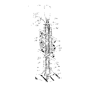
Depicted in FIG. 3 is a flexible stylet wire 200 of the
present lead removal apparatus invention that is insertable
in the longitudinal passageway 210 of a heart lead coiled
12
structure 211 for controlling and, in particular, limiting
the movement of heart lead 204 including coiled structure
211. Heart lead 204 also includes insulating material 201,
such as silicone or polyurethane, formed into a hollow tube
that surrounds the coiled structure and prevents fluids from
making contact with the coiled structure. Attached to 'the
distal end of the flexible style~t wire is an expandable wire
coil 205 consisting of approximately 25 turns of wire with
spacing between the turns. Five to seven wraps of the wire
c~i1 are attached to the distal end of the stylet wire
using, for example, solder 206. The remaining wraps of the
wire coil remain free for engaging the coiled structure when
the proximal end of the s~tylet wire is rotated in a
direction to unwind and expand the turns of the wire coil
and engage the coiled structure of the heart lead. A bead
214 of high temperature silver solder is applied to the
distal end of the stylet wire to prevent the distal end
thereof from pulling through the wire coil during separation
and removal of the heart lead. Positioned about the
proximal end of the stylet wire is control mechanism 202 for
rotating the stylet wire in either a clockwise or
counterclockwise direction or for moving the wire in a
longitudinal direction into or out of the passageway. In
this embodiment, control mechanism 202 is a loop of wire
formed from the stylet wire of which the physician may grasp
or insert his finger. The loop may also be fashioned for
attachment to another control mechanism for moving the
stylet wire. Other control mechanisms such as a slidable
chuck may be positioned at the proximal end of the stylet
wire to facilitate movement of the stylet wire. The formed
loop 202 is covered with teflon tubing 203 or other suitable
material for facilitating the easy movement of the stylet
wire. The looped end is also campressible for inserting
through a separator tube 212.
The choice of the stylet wire and wire coil varies with
the internal diameter of the coiled structure which varies
13
from .016" to about .028" for most heart leads. The
diameter of 'the stylet wire would then range from .009'° to
.015", with the coil wire ranging in diameter from .003°° to
.006". The use of stainless steel wire is preferable. The
stylet wire should be hardened wire, but ductable wire may
be used for the coil wire.
Before the stylet .wire is inserted into passageway 210
of the lead, the inside diameter of the coiled structure and
the outside diameter of the insulating material are
determined. First, lockable mechanism 222 is first applied
to the proximal end 221 of the lead between opposing
semicircular jaws 223 and 224. The details of mechanism 222
are depicted in FIGS. 5 arid 6. Semicylindrical pliable
material 225 and 226, such as latex, are affixed with
medical grade adhesive to the opposing faces of the jaws.
Semicylindrical pliable material 225 includes
semicylindrical channels 227 and 229 having different radii,
and pliable material 226 includes semicylindrical channels
228 and 230 with radii corresponding to channels 227 and
229, respectively. When jaws 223 and 224 are in a closed
position, the opposing surfaces 231 and 232 of respective
pliable material 225 and 226 are in contact with opposing
channels 227 and 228 forming one hollow cylindrical
passageway with a first diameter and opposing channels 229
and 230 forming a second hollow cylindrical passageway with
a second larger diameter. The two different~size diameter
passageways in the pliable material accommodate a number of
different size diameter pacemaker leads and are designed to
grasp and apply pressure to insulating material 201 in a
uniform manner.
When proximal end 221 of lead 204 is inserted and
grasped in the hollow passageway formed by channels 229 and
230, insulating material 201 is compressed onto coiled
structure 211, thus limiting the movement of the structure
within the insulating material.. When the physician cuts the
lead for access to the passageway of the lead, the
14
compressed insulating material prE:vents the coiled structure
from retracting into 'the passageway of the lead.
Pivotly interconnected elongated members 233 and 234 are
connected to respective opposing jaws 223 and 224 to operate
the jaws between open and closed positions. The proximal
ends 235 and 236 of the members are curved as shown in FIG.
5 to oppose each other and have a respective plurality of
teeth 237 and 238 that interlock to form a locking
mechanism. The locking mechanism is actuated by squeezing
the proximal ends of the members and opposingly positioning
the teeth thereon. When so positioned, the teeth of
mechanism 222 interlock and maintain opposing jaws 223 and
224 in a closed position.
After the lockable mechanism is applied to the proximal
end of the pacemaker lead, a pair of well-known wire cutters
or snips sever the electrical connector (not sown) .from the
proximal end 222 of pacemaker lead 204. As a result of such
severance, coiled structure 211 of the pacemaker lead is
commonly deformed, thereby presenting a false indication of
the actual diameter of longitudinal passageway 210. As a
consequence, the physician inserts expansion device 901 into
the proximal end of hollow passageway 210 to expand coiled
structure 211.
Depicted in FIG. 9 is expansion device 901 for expanding
the deformed proximal end of coiled structure 211. The
expansion device includes a tapered rod 902 having a distal
end 903 with a diameter that is easily insertable into the
passageway of the deformed coiled structure. Tapered rod
902 includes a 'tapered longitudinal portion 904 that
gradually increases in diameter to proximal end 905 that has
a diameter significantly greater than 'the diameter of the
distal end. Control handle 906 is connected to the proximal
end of the tapered rod. 'rhe physician grasps the control
handle to insert the tapered rod into the longitudinal
passageway and to expand the deformed proximal end of the
coiled structure.
~y'~: ~ ~ ~",~
With lockable mechanism 222 in a closed position and the
proximal end of the coiled structure expanded,~the physician
selects a wire guide 239, as shown in FIG. 3, having a
diameter less the diameter of the lead passageway. The
physician determines the passageway by inserting the wire
guide therein and sensing for any blockages. The guide
includes a control mechanism such as a knurled cylindrical
chuck 240 positionable about the proximal end thereof. The
physician grasps the knob to extend the guide into the lead
passageway and to rotate the guide back arid forth to clear
or break through any blockages caused by tissue or occluding
material. The guide is also used to determine or size the
inside diameter of a second coiled structure that may be
coaxially positioned inside coiled structure 211. When
utilized as a control mechanism for stylet wire 200, the
chuck may also include appendages 260 for rotating and
counting the number of times the stylet wire is rotated.
Having determined the lead passageway with the wire guide,
several other guides similar to guide 239 are individually
inserted in the passageway to determine the actual inside
diameter at the proximal end. Guide 239 is also utilized to
determine if coiled structure 2,11 has been deformed or
damaged and to determine the smallest diameter of the coiled
structure and passageway.
As shown in FTG. 3, stylet wire 200 is inserted into
longitudinal passageway 210 of coiled structure 211. The
diameter of the coil wire and stylet wire have been selected
to form a combined overall diameter which approximates the
diameter of the longitudinal passageceay of the heart lead
coiled structure within a predetermined tolerance such as
one or two thousandths of an inch. Stylet wire 200 is then
fed through the entire length o:f the passageway to the
distal end of the coiled structure which is secured to the
wall of heart cavity tissue 213 via tines 207. When fully
inserted into the heart lead, 'the distal ends of the stylet
wire and coiled structure should be in claw proximity. Tt
16
i~d
is not necessary, but probably more advantageous, that the
stylet wire be attached to the distal end of 'the heart lead.
For separating the heart lead from adjacent tissue, the
stylet wire may be secured anywhere along the passageway of
the coiled structure past the restricting tissue. To secure
the stylet wire to coiled structure 211, looped end 202 of
the stylet wire is operated in a circular direction to
unwind and expand wire coil 205. As a result, the turns of
the wire coil and coiled structure engage and intermesh,
7.0 thereby firmly securing the stylet wire to the heart lead.
This prevents any extension or stretching of the heart lead
and also controls and limits 'the movement of the lead when
separator tube 212 is moved along the length of coiled
structure 211 and insulating material 201 of the haax~t lead.
Depicted in FIG. 23 is illustrative removal apparatus
2301, which is an alternative embodiment of stylet wire 200.
Remo-ral apparatus 2301 is insertable into the longitudinal
passageway of an elongated structure such as a pacemaker
lead. The removal apparatus includes a stylet wire 2302
with a conically-shaped silver solder tip 2303 that is
positioned at the distal end thereof. Closely wrapped wire
coil 2304, similar. to wire coil 205, is attached at 'the
distal end of the stylet wire using silver solder 2305 as
previously described. The proximal end of the wire coil is
pulled to unwrap several turns of wire coil 2304. A catch
wire 2306 is formed from 'the proximal end of the wire coil
to extend in a radial direction from the wire coil and
stylet wire. Catch wire 2306 catches on or engages the
coiled structure of the pacemaker lead to engage wire coil
2304 with the coiled structure of the pacemaker lead. In
addition, the wire coil may be rotated in the opposite
direction to release the stylet wire from 'the. coiled
structure if desired.
Depicted in FTG. 28 is illustrative removal apparatus
2801, which is another alternative embodiment of the removal
apparatus depicted in FIG. 3. Removal apparatus 2801 is
1'7
~~t~~~~~v
insertable into tl-.e longitudinal passageway of an elongated
structure such as passageway 210 in pacemaker lead 204 of
FIG. 3. The removal apparatus includes a stylet wire 2802
and a wire coil having closely-spaced turns 2804 and open-
s spaced turns 2809. At the distal end 2803 of the removal
apparatus, closely-spaced turns 2804 are attached to the
distal end of stylet wire 2802 using sliver solder 2805.
The distal end 2803 is tapered or conically shaped for easy
insertion into the pacemaker lead passageway. The distal
end is tapered or shaped using any of a number of well-known
techniques such as sanding, grinding, buffing, or a
combination thereof. Several turns at the proximal end of
the wire coil are unwrapped to form a catch wire 2806 that
extends radially from the wire coil and stylet wire. Catch
wire 2806 catches on or engages the coiled structure of the
pacemaker lead when inserted in the passageway thereof for
engaging open-spaced turns 2809 with the coilad structure of
the pacemaker lead. With control mechanism 202, the stylet
wire is typically rotated in a counter-clockwise direction
for operating the wire coil and engaging the open-spaced
turns of the wire coil with the coiled structure of the
pacemaker lead. To more readily engage the coiled
structure, the proximal end 2807 of the catch wire is folded
back on itself and attached thereto with silver solder 2808.
A side view of the proximal end of catch wire 2806 taken
along the line 29-29 is depicted in FIG. 29. The proximal
end 280? is folded back and attached to catch wire 2806 so
as not to extend beyond 'the thickness of the wire as shown
in FIG. 28. This is to prevent the catch wire from engaging
the coiled structure of the pacemaker lead or binding
between the stylet wire and coiled structure when the
removal apparatus is being inserted in the passageway of the
coiled structure. Experiments in the laboratory and in
actual pacemaker lead removals indicate that 'the length of
catch wire 2806 should be preferably one-quarter of an inch
in length. The folded-back segment end' 2807 should
1. 8
.w
preferably be one-sixteenth of an inch in length. As a
result, only a few counterclockwise turns of the s~tylet wire
in laboratory 'tests were found necessary to engage the
coiled structure of the pacemaker lead. Previously, ten to
twenty turns were required to engage the coiled structure
when the catch wire did not include the folded-back proximal
end.
Depicted ir1 FIGS. 10-21 are al~teranative embodiments of
illustrative apparatus for removing the elongai~ed structure
implanted in biological tissue. All of these alternative
embodiments are for controlling the movement of an elongated
structure. The removal apparatus in each of these
alternative embodiments includes a control unit 'that is
insertable into the longitudinal passageway of the elongated
structure, such as a pacemaker lead, and securable to the
structure for controlling the movement thereof. The
apparatus also includes an expandable unit positioned about
the distal end of the control unit and operable to an
expanded position for securing the control unit to the
elongated structure. However, the control unit in each of
these alternative embodiments commonly, but not in all
cases, includes a longitudinal passageway for operating the
expandable unit to the expanded position for securing 'the
control unit to the elongated structure.
Depicted in FIG. 10 is a first alternative embodiment of
illustrative removal apparatus 1001 for removing implanted
pacemaker lead 204. The control unit of this removal
apparatus includes a flexible tube 1002 having a passageway
1003 formed longitudinally therein. Expandable balloon 1004
is positioned and attached about the distal end of the
control tube. The distal end of the control tube is also
recessed to attach to the balloon in a well-known manner at
the ends of radial recess 1005. 'fhe recess also provides a
volume in which the collapsed balloon is stored. The recess
also includes one or more side ports 1006 leading from
passageway 1003 to the balloon. A source of fluid such as
19
~~~t:~.~~~
compressed air or liquid is passed through the passageway
and into the balloon to inflate the balloon to an expanded
position as indicated by expanded balloon 1007 positioned at
the distal end of the lead.
Depicted in FIG. 11 is a second alternative embodiment
of illustrative removal apparatus 1100. In this second
alternative embodiment, the control unit also includes a
control tube 1101 fox' insertion into passageway 210 of
coiled structure 211. The expandable unit comprises a
plurality of radial projections 1102 and 1103 that have a
free end are radially formed in the distal end of the
control tube. The free end of the radial projection is
twisted and bent in an inward direction unto passageway 1104
of the control tube. As formed, these projections allow a
control tube to be easily inserted into passageway 210 of
the coiled structure. When control tube 1101 is positioned
at the distal end of the coiled structure, actuator rod 1105
is inserted in passageway 1104 of the control tube. When
inserted, the actuator rod engages the radial projections
and forces them into an expanded position extending radially
from the surface of the control tube into the coiled
structure of the pacemaker lead. When in the expanded
position, these radial projections secure the control tube
to the coiled structure, thereby controlling movement of the
coiled structure during removal from the tissue.
Depicted in FIG. 12 is a third alternative embodiment of
illustrative removal apparatus 1201 utilizing an actuator
rod 1202. The removal apparatus includes a control tube
1203 that is extendable into the longitudinal passageway of
a pacemaker lead. The expandable unit of the apparatus
comprises a plurality of longitudinal strips 1204 formed at
the distal end of the control tube. Actuator rod 1202 is
inserted in 'the passageway of the control tube and attached
to the dista:L end 1205 thereof. When the control tube .is
inserted in the longitudinal passageway of the pacemaker
lead, the actuator rod 1202 is pulled in a longitudinal
~~~Q.~~~~
direction out of passageuaay 1206 of the control tube as
shown by arrow 3.207. Typically, 'the physician will maintain
the relative pasil:ion of the proximal end of control 'tube
1203 while the actuator rod is pulled in the outward
direction. As a result, diCtal end 1205 is farted toward
'the proximal end of the control tube, as shown by arrow
1208, thereby deforming longitudinal strips 1204 in an
outward direction as imdi~.ated by arrows 1209. The
expanding strips engage the coiled structure and secure h~he
control tube to the coiled structure of the pacemaker lead.
Depicted in FIG. 13 is a fourth embodiment of
illustrative removal apparatus 1301 inserted in the
longitudinal passageway 210 of coiled structure 211.
Femoval apparatus 1301 includes a control tube 1302 having
a distal end with a spiral or helical ridge 1303 formed
therein. Alternatively, a number of barbs are fo~__°med in the
contoured distal end of control tube 1302. 'fhe distal end
includes a plurality of slits 1307 or an opening thereat for
expanding the ridge or barbs .into the coiled structure.
Actuator rod 1304 is inserted iota passageway 1305 to engage
the distal end. When engaged, actuator rod expands the
ridge or barbs in a radial direction, as shown by arrows
1308, to engage the coiled structure of the pacemaker lead.
As a result, the expanded ridge or barbs secure the control
tube to the coiled structure for controlling the movement
thereof.
Depicted in FIG. 14 is a fifth embodiment of
illustrative removal apparatus 1401 inserted in longitudinal
passageway 210 of tailed strllCture 211. The removal
apparatus includes control tubs 1402 and actuator rod 1403
extending through hollow passageway 1.404 of the control
tube. The apparatus also includes a diagonal.ly-slotaed
sleeve 1405 that .is positioned between the distal ends of
the CantrGJ. tube and actuator rod. The actuator rod also
extends through hollow pas:~ageway 1406 of the sleeve.
Attached to the distal end of thf~ actuator rod is beveled
21
tip 1407 having an outside diameter approximating the
diameter of the control tube and the nominal diameter of 'the
slotted sleeve. Similarly, the distal end of the control
tube is beveled to engage and expand the slotted sleeve. To
expand the slotted sleeve, the actuator rod is pulled, as
indicated by arrow 1.408, to engage the sleeve against the
beveled edges of the control tube and the rod. As a result,
the sleeve is expanded to a position for engaging coiled
structure 211 and securing the control tube thereto. The
slotted sleeve expands in a radial direction as indicated by
arrows 1409.
Sixth and seventh alternative embodiments of
illustrative removal apparatus 1501 and 1601 are depicted in
FIGS. 15 and 16, respectively. In FIG. 15, removal
apparatus 1501 includes a control tuba 1502 and an actuator
rod 1503 extending through longitudinal passageway 1504 of
the control tube. The distal end of the actuator rod
includes enlarged tip 1505 having a diameter approximating
the diameter of the control tube. The device also includes
expandable sleeve 1506 comprising a pliable material such as
synthetic rubber and the like which expands in a radial
direction when compressed between the distal end of the
control tube and the enlarged tip of the actuator rod. In
the relaxed state, the outside diameter of the pliable
material approximates that of the control tube and enlarged
tip of the actuator rod for insertion into longitudinal
passageway 210 of the coiled structure. When inserted into
passageway 210, the enlarged tip and distal and of the
control tube compress and radially expand the pliable
material in an outward direction toward the coiled structure
as indicated by arrows 1507. The actuator rod is pulled
through the passageway of the control tube as indicated by
arrow 1508. As a result, pliable material 1506 .is
longitudinally compressed as shown by arrows 1509 and 1510.
However, pliable material 1506 also expands in a radial
direction and engages the coiled structure, thereby securing
22
~~ -,;
~(3 4.b ~~~. ~
the control tube thereto.
Similarly, illustrative removal apparatus 1601 depicted
in FIG. 16 includes control tube 1602 having longitudinal
passageway 1610, actuator rod 1603 having an enlarged distal
tip 1604, and pliable material 1605 attached to the distal
end of control tube 1602 and enlarged actuator rod tip 1604.
However, unlike pliable material 1506, pliable material 1605
in a relaxed condition has an outside diameter greater than
the diameter of longitudinal passageway 210. Therefore, to
insert the removal apparatus in the passageway, actuator rod
is forced into passageway 1610 as indicated by arrow 1606,
thereby stretching pliable material 1605 as indicated by
arrows 1607 and 1608. As a result, the outside diameter of
the pliable material decreases as indicated by arrows 1609
for insertion into 'the passageway of the elongated
structure. When inserted, the actuator rod is released, and
the pliable material attempts to return to its relaxed
state. As a result, the pliable material engages the coiled
structure and secures the device to the pacemaker lead.
Depicted in FIGS. 17 and 18 are alternative embodiments
of illustrative removal devices 1701 and 1801 that include
a wire guide for inserting into the longitudinal passageway
of the elongated structure. In FIG. 17, removal apparatus
1701 includes wire guide 1702 that is inserted into
passageway 210 of coiled structure 211 to clear any blockage
formed therein and establish a guide for control tube 1703.
When the guide wire is fully inserted, the control tube is
inserted over the guide wire and then into passageway 210 of
the structure. The control tube also has a longitudinal
passageway 1706 for .receiving the wire guide therein. Also
included is wire coil 1704 'that is positioned and attached
at the distal ends thereof using, for example, silver solder
1705. As previously described with respect to stylet wire
200, control tube 1?03 is rotated in a direction opposite
that of coiled structure 211 for engaging and expanding wire
coil 1704, thereby securing the control tube to the coiled
23
structure.
As depicted in FIG. 18, removal apparatus 1801 includes
wire guide 1802 that is inserted into the passageway of the
elongated structure. Control tube 1803 includes two
longitudinal passageways 1804 and 1805. Passageway 1804
receives the wire guide as the control tube is inserted into
the passageway of the elongated structure. Positioned at
the distal end of the control tube is inflatable balloon
1806 with passageway 1805 leading 'thereto through sideport
ar aperture 1807. To secure the control tube to the
elongated structure, a fluid is passed through passageway
1805 to inflate the balloon to an expanded position.
Several other alternative embodiments of illustrative
removal apparatus are depicted in FIGs. 19-21. Depicted in
FIG. 19 is removal apparatus 1901 that includes control tube
1902 and cylinder 1903. The tube includes longitudinal
passageway 1904. Cylinder 1903 is positioned about the
distal end of the control tube and rotated to a position
off-center of the tube for securing the control tube to the
elongated structure. The removal apparatus includes an
actuator rod 1904 extending through the control tube and
attached to the rotatable cylinder. The rod rotates the
cylinder to an off-centered position for securing the
control tube to the elongated structure such as the coiled
structure of a pacemaker lead. Aotuator rod 1904 extends
between the rotatable cylinder and control mechanism 1905
that is positioned at the proximal end of the control. tube.
Control mechanism 1905 is rotatable between two positions
for rotating the actuator rod arid the cylinder between
expanded and retracted positions. The actuator rod is
attached to 'the cylinder at an off-centered position to
permit rotation of. the cylinder and engagement of the
elongated structure. Plug 1907 is inserted at the distal
end of the tube to maintain the off-centered position of 'the
rod in the passageway.
Depicted in FIG. 20 is illustrative removal apparatus
24
.:7(:~.~~;~~
2001 including a control tube 2002 that has a longitudinal
projection 2003 extending at the distal end thereof for
securing the control tube to the coiled structure of a
pacemaker lead. This arrangement is sometimes referred to
as a flea-clip arrangement. Depicted in FIG. 21 is a
sectioned view, taken along the lines 21-21 in FIG. 20, of
the apparatus in passageway 210 of coiled structure 211. As
shown, a stylet wire or rod 2004 is inset°ted into passageway
2005 of control tube 2002 to engage and retract the extended
projections into the wall of the control tube. When the
apparatus is inserted to the distal end of the coiled
structure, the stylet wire or rod is removed from the
passageway of the control tube. As a result, 'the spring-
like projections extend into the coiled structure of the
lead, thereby securing the control tube to the coiled
structure for controlling the movement thereof. To remove
the control tube, the rod is inserted into the control tube
passageway as shown by arrow 2101 to again engage the
projections. When the rod engages the projections extending
into the passageway, the inward extending projections move
into the wall in a direction as shown by arrows 2103,
whereas the outward extending projections move into the wall
in a direction as shown by arrows 2102.
The reader°s attention is again referred to the
preferred embodiment depicted in FIG. 3. After the stylet
wire is secured to the lead and prior to inserting separator
tube 212 over the stylet wire and lead, a tie 241 of, for
example, nylon cord or suture material is wrapped around
proximal end 221 of the lead to secure insulating material
201 to coiled structure 211. The tie controls or limits the
movement of the coiled structure within the insulating
material. With 'the insulating material secured to the
coiled structure at the proximal end, removal force is
applied not only to the coiled structure, but also 'to the
insulating material of the lead as well. This maintains the
integrity of the heart lead during subsequent tissue
CA 02001200 1999-OS-26
separation from the insulating material. In those instances
where the stylet wire has not been fully inserted to the
distal end of the lead, the tie also prevents the coiled
structure from unravelling, breaking or separating from
electrode 220 or the rest of the lead.
As previously suggested, the looped proximal end of the
stylet wire can be compressed to permit separator tube 212 to
be inserted thereover and over the insulating material of the
heart lead. Separator tube 212 comprises a semi-rigid
material, such as Teflon~, for sliding easily through the
blood vessel and over the insulating material of the heart
lead. In order to place the separator tube over the stylet,
the stylet should extend at least 12 inches beyond the
person's body so that the looped end can be grasped to apply
tension to the stylet. With the Teflon~ separator tube 10 to
12 inches long, the stylet is typically three feet long.
Depicted in FIG. 4 is fibrotic tissue 209 encapsulating
heart lead 204 in blood vessel 216. When this occurs in small
diameter veins where blood flow has been restricted or
prevented, separation and removal of the lead from the tissue
is difficult and often causes severe damage or destruction to
the vein. Without tension on stylet wire 200, separation is
usually not possible in these situations.
As shown, the distal end of the Teflon~ separator tube
212 is bevelled and includes a cutting edge or edge having a
number of teeth for separating heart lead insulating material
201 from encapsulating fibrotic tissue 209. As depicted in
FIG. 7, hollow separator tube 212 has a metal bevelled tip
242 attached to the distal end thereof with, for example, a
medical grade adhesive. The metal tip provides a more durable
edge for separating or cutting encapsulating fibrotic tissue
from the lead.
Returning the reader's attention again to FIG. 3,
separator tube 212 is moved and rotated along the outer
surface of insulating material 201 of the heart lead to
separate the lead from the blood vessel wall. After the
26
CA 02001200 1999-OS-26
separator tube has been moved along the entire length of the
heart lead, it will abut next to the heart cavity wall as
shown by phantom lines 219. The distal end of the heart lead
is typically secured to the heart cavity wall by trabeculae
or fibrotic tissue 218 that has encapsulated tines 207
positioned at the distal end of the lead. The separator tube
212 is positioned next to the heart cavity wall or pushed
slightly while the stylet wire is tensioned in the opposite
direction. The separator tube is then rotated back and forth
to dislodge and separate tines 207 and the distal end of the
heart lead from fibrotic tissue 218 and heart cavity wall
213. As a result, the heart lead has now been completely
separated from the blood vessel and the heart cavity wall for
subsequent removal. The separator tube, the stylet wire, and
the heart lead are then removed from the heart cavity and
surrounding blood vessel.
However, should the removal of the heart lead be
prevented for whatever reason, the stylet wire is rotated in
a clockwise direction to unsecure the stylet and wire coil
from the heart lead coiled structure. The time for this
operation is lessened by attaching a rotating mechanism such
as an electrical screwdriver to the proximal end of the
stylet wire.
Depicted in FIG. 27 is an alternative embodiment of
illustrative separator apparatus 2700. This separator
apparatus includes a set of separator and dilator tubes 2701
and 2702 for insertion over pacemaker lead 204. Similar to
separator tube 212, separator tube 2701 has a hollow
passageway therein for receiving the pacemaker lead. The
separator tube is advanced along the lead to engage and
separate encapsulating tissue from the lead. Dilator tube
2702 similarly has a hollow passageway therein for receiving
separator tube 2701 and the pacemaker lead therein. A
preferred material for separator and dilator tubes 2701 and
2702 is polypropylene which is more kink-resistant than
Teflon . A polypropylene tube fits easily into the blood
27
vessel for extension to the distal end of the pacemaker
lead. Furthermore, the inclusion of approximately 25=k of
bismuth provides radio-opacity for viewing w~.th, for
example, a fluoroscope during insertion of the separator
tube. When the dilator tube is inserted over the separator
tube and lead, a control mechanism 2703 having a hollow
passageway therein is inserted over the lead and connected
to the proximal end of separator tube 2101. Control
mechanism is well-known as a pinvise and is used for
controlling the movement of the separator tube in both a
longitudinal and rotational direction. The dilator tube and
separator tube are alternatively moved along the lead to
first separate the tissue from the lead and further dilate
the tissue with the dilator tube. The control mechanism
2103 provides added strength and control during the movement
of the separator tube. Dilator tube 2102 not only provides
extra dilation of the tissue but also provides additional
strength to the entire structure for separating tissue from
the pacemaker lead.
Depicted in FIG. 22 is another alternative embodiment of
illustrative separator apparatus 2201 for separating
encapsulating tissue 2205 from pacemaker lead 204. The
separator apparatus 2201 includes a tube 2202 having a
longitudinal passageway 2203 therein for receiving and
passing over the pacemaker lead including outer insulating
material 201. Distal end 2204 of 'the tube is beveled to
provide a wedge for separating encapsulating tissue 2205
from the pacemaker lead. Also positioned and attached in a
well.-known manner about the distal end of tt~e separator 'tube
is balloon 2206. The tube also includes a plurality of
hollow passageways 2207 for supplying a compressed gas or
fluid for inflating the balloon. Separator apparatus is
inserted over the insulating material sheath of the
pacemaker lead to engage encapsulating tissue 2205. The
beveled distal end provides a wedge for causing an initial
separation of the tissue from the lead. Upon initial
2s
contact and separation, the balloon is inflated to provide
further dilation and separation of the encapsulating tissue
from the pacemaker lead. The balloon is then deflated to
permit the beveled distal end to be further moved along the
pacemaker lead and engage additional encapsulating tissue.
This process is continued until all of the encapsulating
tissue is separated from the pacemaker lead.
Depicted in FIGS. 24 and 25 is separator apparatus 2401
for separating the distal end of an elongated structure such
as electrode tip 220 of pacemaker lead 204 from tissue 218
affixed thereto. This apparatus is particularly
advantageous in those instances where the electrode of the
pacemaker lead is porous allowing fibrotic tissue to grow
therein and secure the electrode tip thereto. Separator
apparatus includes a first tube 2402 having a hollow
passageway 2403 for receiving pacemaker lead 204 and
extending to the distal end thereof. Attached to the distal
end of the first tube 2402 is an elongated member such as
stainless steel wire 2404. The first tube wall also has a
hollow channel or passageway 2408 extending longitudinally
therethrough for passing 'the wire the entire length of the
tube. Alternatively, the stainless steel wire can be
affixed to the distal end using any suitable well-known
fastening means. A second tube 2405 also has a longitudinal
passageway 2406 for receiving the first tube. In addition,
the second tube similarly includes a hollow channel or
passageway 2407 for extending stainless steel wire 2404
through the entire length of the tubs and beyond the
proximal end thereof. This permits the loose end of the
wire to be controlled by the clinician to remove the distal
end of the pacemaker lead from the encapsulating or affixed
tissue. As shown in FIG. 25, the fir:~t tube is extended to
the distal end o:E the pacemaker lead and placed next to
electrode 220. The second i:ube with the stainless steel
wire is'then also positioned next to the distal. end of 'the
pacemaker lead next to the electrode. The clinician puts
29
tension on the stainless steel cutting wire and then rotates
the second tube relative to the first causing the stainless
steel wire to wipe across the face of the electrode as
shown. Rotation of the two tubes are shown by arrows 2501
and 2502. This wiping motion across the pacemaker electrode
literally cuts the electrode tip free from the encapsulating
or affixed tissue 218. In~~tead of stainless steel wire,
suture material is also used to perform the cutting action.
Depicted in I'IG. 26 is a second alternative embodiment
of illustrative separator apparatus 2601 for separating the
distal end of a pacemaker lead having a plurality of tines
such as tines 207 of pacemaker lead 204 encapsulated in
fibrotic heart tissue 218. Apparatus 2601 includes tube
2602 having a longitudinal passageway 2605 for receiving
1.5 pacemaker lead 204. The tube is inserted over the lead and
extended to the distal end thereof. The tube includes a
plurality of slots 2603 formed at the distal end for
receiving pacemaker lead tlIIE:S 207. When the tines are
received in the slots, tube 2602 is rotated back and forth
in a circular motion for dislodging and separating the tines
from the encapsulating tissue 218 extending from heart wall
tissue 213.
Depicted in FIG. 7 is another illustrative embodiment of
the lead removal apparatus of this invention. In this
embodiment, pacemaker lead 243 is similar to the lead shown
in FIG. 3; however, the distal end of the lead is of a
different configuration. In particular, electrode 244 has
two cavities therein. One cavity is for receiving 'the
coiled structure 245 of the lead. The second cavity is for
receiving and securing anchoring coil 246 secured in the
cavity with insulat.i.ng material 247 in a well-known manner.
The distal, ez~d of anchoring coil 246 is cut to form a
beveled or sharpened edge for turning or cork. ~crawing the
coil into heart cavity wa7.1 213. Anchoring coil 246, as a
rGSUlt, securely attach~a electrode 244 to the heart tissue
to pstahli;~h good oler.~c..rical. t:ontact for stimulating the
3u
CA 02001200 1999-OS-26
heart tissue with electrical pacing pulses from the
pacemaker. Insulating material 248 surrounds coiled structure
245 and partially surrounds electrode 244. Since anchoring
coil 246 is utilized in this configuration, the insulating
material is molded over the coiled structure and electrode
without forming tines for the endothelial tissue to form
therearound.
Stylet wire 249 of this lead removal apparatus and lock
wire 250 attached to the distal end thereof have a combined
diameter much less than the inside diameter of coil structure
245 of the lead. This is particularly advantageous for those
situations when the coiled structure of the lead has ben
deformed, unravelled, or in some way damaged. In this
embodiment, lock wire 250 has a plurality of turns 251
wrapped around the distal end of the stylet wire. Turns 251
of the lock wire at the distal end of the stylet wire are
closely wrapped and attached to the distal end of the stylet
wire using, for example, a silver solder. Turns 252 of the
lock wire are more loosely wrapped and are approximately 75
in number. The unwrapped proximal end 253 of the lock wire
extends beyond the passageway of the lead and is secured and
positioned by, for example, the physician's hand 258 when the
stylet wire is rotated to expand lock wire turns 252 and
engage the turns of coiled structure 245.
Control mechanism 254 such as a loop of malleable wire
is wrapped around and secured to the proximal end of the
stylet wire using, for example, silver solder 257. Slidable
chuck 240 is also suitable for use as the control mechanism
for stylet wire 249. A Teflon~ coating 255 surrounds the
interconnection to prevent possible injury to the physician
or patient. Control loop 254 is provided for the physician to
move the stylet wire in and out of the passageway of the lead
as well as rotate the stylet wire to engage the coiled
structure of the lead. When the stylet wire is secured to the
pacemaker lead, loop 254 is used to extract stylet wire
31
;~~~(~~,~~~i~
and pacemaker lead from the patient.
To unravel the turns of the lock wire, a 'tool such as an
electrical screwdriver is attached to the cowtrol mechanism
loop to rotate the stylet wire and expand the turns of the
lock wire. While the stylet wire is being rotated, the
physician secures the position of the proximal end 253 of
the lock wire to permit lock wire turns 252 to tangle and
form a bundle 259 that engages the coiled structure as
depicted in FTG. 8. The style~t may have to rotate 50 to 100
turns to form bundle 259 and engage coiled structure 245.
After the lock wire has secured the stylet wire to the
pacemaker lead, the physician grasps control loop 254 and
continues to rotate the stylet wire and pacemaker lead to
dislodge anchoring coil 246 from the heart tissue. Should
the blood vessels encapsulate the pacemaker lead, separator
tube 212 is inserted over the stylet wire and pacemaker lead
as previously described to separate the lead from the
encapsulating blood vessel tissue. The separator tube may
also be extended to the distal end of the pacemaker lead to
turn and dislodge the distal end of the pacemaker lead from
the heart tissue.
Of course, it will be understood that 'the aforementioned
lead removal apparatus and method is merely illustrative of
the application of the principles of this invention and that
numerous other arrangements may be devised by those skilled
in the art without departing from the spirit and scope of
the invention. Tn particular, a number of other control
mechanisms may be attached to the proximal end of the stylet
wire for operating the stylet wire in either a clockwise or
counterclockwise direction as well as moving the wire
longitudinally. Furthermore, this apparatus may be utilized
for removing electrical leads from body ducts and passages
as well as body tissue that has encapsulated the lead and
restricted its movement.
32