Note: Descriptions are shown in the official language in which they were submitted.
SUFtSICAL INSTRUMENTS AND ASSEMBLIES
Background of -the Invention
This invention relates to surgical 3.nstrurnents and
assemblies.
The invention is more particularly concerned with
surgical instruments for use in tracheostorny.
Where it is necessary to provide an emergency airway
via a tracheostomy, or to enable a suction catheter to be
introduced to the trachea or bronchial passages, this can
1p be achieved by means of a relatively small diameter,
uncuffed tracheostomy tube which does not hinder airflow
to the patient°s mouth and which enables the patient to
cough and clear sputum normally.
The procedure is carried out by making a stab cut
15 with a short-bladed scapel 'through the neck into -the
trachea in the region of the cricothyroid membrane.
The scapel is 'then removed and the tracheos-tomy tube is
Inserted through tho cwt by means of an introducer with a
tapered 'tip that pro,j ects from the patient end of -the
2g 'tube. Subsequently, the introducer is pulled rearwardly
cwt of the machine and of ~tha 'tube, leaving t'he tube in
placo to provide an air passage into the trachoa.
- 1 -
In an alternative technique (The Seldinger
technique), the cricothyroid membrane is pierced using a
hollow needle after having first made an incision through
'the skin with a scalpel. A guide wire is then inserted
through the needle into the trachea, 'the needle
subsequently being withdrawn to leave the guide wire
preserving access to the trachea. A dilator may -then be
fed over the guide wire to enlarge the opening into the
trachea so that a tracheostomy tube can be introduced by
sliding it over 'the dilator. Both the guide wire and the
dilator are then withdrawn leaving the tube in position.
This technique has the advantage that 'the guide wire
maintains the patency of the tracheostomy until the
tracheostomy tube is inserted. There can otherwise be
the tendency for the different layers of tissue in the
neck to become displaced, especially if the patient should
cough, making it difficult for the surgeon to insort the
tracheostomy tube.
There is a disadvantage, however, with this technique
2~ in that it is possible for the needle to be pushed too far
into the neck leading to possible damage to the posterior
wall of the 'trachea. :Ct is also possible that the noedla
could be puskied 'through the posterior wall of the trachea
into the oesophagus; this could lead to the tracheostomy
'tube being inserted into tho oesophagus rather the
'trachea.
- 2 -
CA 02001302 1999-09-09
Brief Summary of the Invention
It is an object of the present invention to provide a
surgical instrument for use in tracheostomy by which the above-
mentioned disadvantage can be alleviated.
According to one aspect of the present invention there
is provided a surgical instrument assembly for use in forming a
tracheostomy through neck tissue of a patient, wherein the
instrument assembly comprises a hollow needle and a hub, a guide
wire, a loss of resistance device and a tracheostomy tube,
wherein the needle has a rear end mounted in the hub and a
sharply pointed forward end that projects from a forward end of
the hub, wherein the length of needle projecting from the
forward end of the hub is greater than the thickness of neck
tissue between the skin surface and the anterior wall of the
trachea but less than the distance between the skin surface and
the posterior wall of the trachea, wherein the hub has at its
forward end a laterally extending face the area of said face
being at least fifty times the cross sectional area of the
needle, and wherein the hub has at its rear end a coupling, said
coupling communicating with the bore of the needle and being
shaped for coupling with the loss of resistance device, such
that insertion of the instrument assembly is limited by
engagement of the face of the hub with the skin surface of the
neck so that the forward end of the needle lies within the
trachea without contacting the posterior wall of the trachea and
such that entry of the forward end of the needle into the
trachea can be detected by flow of fluid from the loss of
resistance device through the needle, and wherein the guide wire
3
CA 02001302 1999-09-09
is insertable directly along the needle after removal of the
loss of resistance device such that the needle can be
subsequently removed leaving the guide wire in the trachea to
provide a path along which the tracheostomy tube is inserted
into the trachea.
The hub preferably includes a gripping region by which
the instrument can be gripped between finger and thumb. The
gripping region is preferably provided by two parallel walls
with stepped edges and the forward end of the needle may be
angled relative to the gripping region. The coupling at the
rear end of the hub is preferably a tapered recess adapted to
receive a cooperating tapered nose of the loss of resistance
device. The area of the face may be at least
100 mm2 and is preferably between 100-150 mm2. The hub is
preferably of a plastic material.
The loss of resistance device preferably includes a
syringe having a barrel coupled with the hub and a plunger that
is movable along the barrel when the forward end of the needle
lies within the trachea.
4
~~~~~'~.~ ~~
A surgical instrument and its method of use, in
accordance with the present invention, will now be
described, by way of example, with reference to the
accompanying drawings.
Brief Description of the Drawings
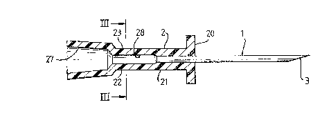
Figure 1 is a partly sectional side elevation
along the instrument;
Figure 2 is view from below of 'the
instrument;
Figure 3 is a transverse section along the
line III - III of Figure 1; and
Figures 4 illustrate successive steps in use
to 9 of the instrument.
- 5 -
Detailed Description of Preferred Embodiment
With reference first to figures 1 to 3, 'the surgical
instrument is in -the form of a needle assembly comprising
a hollow metal needle 1 joined at its rear end to a
moulded plastics hub 2.
The need7.a 1 is of 16 Gauge and is l7mm long from the
hub 2. At its forward end 3, the needle 1 is formed with
a sharply pointed angled -tip.
The hub 2 is approximately -the same length as -the
needle 1. At its forward end, the hub 2 has a laterally
extending flat face 20 which is substantially rectangular
in section but with its longer edges curved convexly. The
face 20 is about l4mm long and lOmm wide at its widest
point giving it a surface area of between 100 - 150mm
compared with the cross sectional area of the needle 1
which is about 2mm~.
The intermediate portion 21 of the hub, extending
rearwardly f~om the face 20 provides a gripping region by
which the hub can be gripped between finger and thumb.
z0 Two parallel walls 22 and 23 extend from the intermediate
portion 21 and are stepped rearwardly to reduced width,
the stepped edges 2.4 and 25 providing a nan-slap gripping
surface. The 'tip 3 of the needle 1 is angled away from
'the plane of the walls 22 so that 'the orientation of tho
noedle tip can be de-terminod from the orientation of the
walls of the hub 2.
- 6 -
Ifr~7w~~.~ ~~~~~
At its rear end, the hub 2 has a female luer--tapered
coupling bore 27 of circular section which communicates
with a smaller diameter here 23 through the hub, which in
turn communicates with the bore of the needle 1. The
coupling bore 27 is about 4mm in diameter at its rear
end and is adapted to be coupled with a loss of
resistance device 30 (Figure
With reference now also to Figures ~ and 5, 'the loss
of resistance device 30 is in the form of a syringe with a
barrel 31 along which can be slid a plunger 32. The
syringe has a luer-tapered nose 33 that can be coupled
with the bore 27 in the hub 2.
Use of the instrument will now be described with
reference to Figures 4 to 9. The loss of resistance
device 30 is first coupled with the instrument hub 2 to
form an instrument assembly, The plunger 32 is then
pulled rearwardly towards the end of 'the barrel 31 and the
-tip 3 of the needle 1 is placed against the skin of the
patient's neck in the region of the cricothyroid membrane
40, as shown in Figure 4. The hub 2 is gripped between
0 'the finger and 'thumb of one hand with the hub oriented so
that the fees on 'the t:Lp 3 of the needle is directed
generally caudally. The other hand is used -to apply a
light forward pressure on 'the plunger 32. The instrument
is pushed forwardly 'through the neck tissue and
_ 7
cricothyroid membrane 40 until -the tip 3 of the needle 1
pierces -the anterior wall 41 of -the trachea 42, as shown
in Figure 5. In this position, air in the barrel. 31 of
the loss of resistance device 30 can escape through -the
instrument and out of i-ts -tip 3 into the trachea 42. This
allows the plunger 32 to move forwardly within -the barrel
31, indicating to the surgeon that the tip of -the
instrument has entered -the trachea. The face 20 of hub 2
acts as a stop to prevent the instrument being inserted
1a too far. The area of -the face 20 is preferably at least
about fifty times the cross-sectional area of the needle 1
so that thero is no risk of the hub 2 entering the cut
made by the needle. The length of -the needle 1 projecting
from the face 20 of the hub 2 is selected, according to
15 the build of the patient (a different length of needle .
would be used for children), to be greater than the thick
ness of neck tissue between the skin surface and -the
anterior wall 41 of the trachea 42 but less -than the
distance between the skin surface and -the posterior
2o wall 43 of the 'trachea.
~~~~~~~..fl ~a
The loss of resistance device 30 is then uncoupled
from the instrument, leaving the instrument in position
in the trachea. A flexible guide wire 45, 500mm in
length, is pushed into the instrument far enough to
project out of the forward end 3 of 'the needle 1 , as
shown in Figure 6. The angled tip 3 of the needle helps
ensure that the wire is directed caudally in the trachea,
that is, towards the bronchial tract. In this respect,
the needle could have a Tuohy tip which is bent to one
side at its end.
The instrument is then pulled rearwardly out of the
trachea leaving the guide wire 45 in position, as shown
in Figure 7.
A small diame-ter_ uncuffed tracheostomy tube 46,
typically with an internal diameter of 4mm and of length
about 130mm, is slid onto a dilator 50 in the farm of a
curved rod with a pointed tip 51 which projects from the
patient end of the tube 46. The dilator 50 has a central
bore which receives the guide wire 45 so that the tube 46
2~ and dilator can be slid along 'the guide wire, as shown in
figure 8. The guide wire 45 maintains open 'the c7.zt
'through ~tha neck tissues and the painted tip 51 of the
dilator enlarges the cut as 3.t is inserted, so 'that 'the
'tuba 46 can be pushed into the trachea easily. The tube
Z5 46 has a flango .47 at its rear end which lies against the
- 9 -
,vCd ~ ! 9 V .51. ~ ~' n :~ p~~~
skin surface when it is fully inserted, with its forward
end located in the trachea.
The dilator 50 and guide wire 45 are then pulled
rearwardly out the trachea -through the tube 46 which is
left in position to provide an airway or access for
suctioning as shown in Figure 9.
The instrument of the present invention enables a
tracheostomy opening to be performed rapidly with
considerably reduced risk of damage to tho trachea
and of incorrect intubation.
- 10 -
