Note: Descriptions are shown in the official language in which they were submitted.
~~~~~ 3
_ 1 _
NOVEL POLYPEPTIDE AND PRODUCTION THEREOF
BACKGROUND OF THE INVENTION
The present invention relates to a novel polypeptide, a
DNA sequence coding for the same and a use thereof.
Many cell growth factors have been isolated and their
structures have been elucidated since the discovery of
epidermal growth factor (hereinafter referred to as EGF) and
nerve growth factor (hereinafter referred to as NGF).
1O Cell growth factors are useful for the elucidation of
the mechanism of cell differentiation and cell
multiplication mechanism, and some of them, including human
EGF, are expected to be useful as drugs. Accordingly,
studies thereon have become increasingly prevalent in recent
years.
Although the human NGF gene have been isolated, there
has been no reports concerning the production of human NGF
in large amounts by use of recombinant DNA techniques.
If a novel polypeptide promoting the growth of animal
cells is obtained, new investigations can be made thereby.
Such novel polypeptides having activities similar to known
growth factors may also be utilized as drugs.
For the purpose of discovering such a novel peptide,
the present inventors used a DNA sequence encoding NGF as a
probe and cloned a DNA sequence hybridizable therewith from
cDNA libraries of a human glioma. As a result, the
inventors succeeded in obtaining a DNA (cDNA) sequence
2~~1~~
- 2 -
coding for a novel polypeptide. The cDNA of the present
invention may be expressed in a host cell to produce the
novel polypeptide. This polypeptide may further be used as
a reagent for studies .relating to the differentiation,
growth and survival of animal cells. The polypeptide may
further be used as a drug. The present inventors have made
further investigations, based on the information described
above, and consequently completed this invention.
SUMMARY OF THE INVENTION
ZO It is therefore an object of the present invention to
provide a novel polypeptide useful as a reagent for research
investigations or as a drug. Other objects will be apparent
from the following description and appended drawings.
The present invention provides:
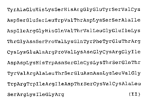
(1) a polypeptide (I) including the following amino
acid sequence tII) in a molecule thereof:
TyrAlaGluHisLysSerHisArgGlyGluTyrSerValCys
AspSerGluSerLeuTrpValThrAspLysSerSerAlalle
AspIleA.rgGlyHisGlnValThrValLeuGlyGluIleLys
ThrGlyAsnSerProValLysGlnTyrPheTyrGluThrArg
CysLysGluAlaArgProValLysAsnGlyCysArgGlylle
AspAspLysHisTrpAsnSerGlnCysLysThrSerGlnThr
TyrValArgAlaLeuThrSerGluAsnAsnLysLeuValGly
TrpArgTrpIleArgIleAspThrSerCysValCysAlaLeu
SerArgLysIleGlyArg (II)
(this amino acid sequence is hereinafter also referred to as
formula [II) for brevity),
- 3 -
(2) a DNA coding for the polypeptide described in (1),
(3) a vector including the DNA sequence described in
(2),
(4) a transformant transformed by the vector described
in (3), and
(5) a process for producing the polypeptide (I) which
comprises cultivating the transformant described in (4) in a
culture medium to produce and accumulate the polypeptide
described in (1) in a culture.
BRIEF DESCRIPTION OF THE DRAWINGS
Fig. 1 is a restriction enzyme map of a DNA including a
polypeptide (I) cDNA in plasmid pUNK5 obtained in Example 1;
Fig. 2 shows a nucleotide sequence of the DNA including
the polypeptide (I) cDNA in the plasmid pUNK5 obtained in
Example 1, and an amino acid sequence translated therefrom;
Fig. 3 shows a comparison of the amino acid sequence
(the upper row) of the polypeptide (I) of the present
invention obtained in Example 1 with an amino acid sequence
(the lower row) of human SNGF;
Fig. 4 is a restriction enzyme map of a DNA including a
polypeptide (I) cDNA in plasmid pH'NT2 obtained in Example 2;
Fig. 5 shows a nucleotide sequence of the DNA including
the polypeptide (I) cDNA in the plasmid pHNT2 obtained in
Example 2, and an amino acid sequence translated therefrom;
Fig. 6 is a schematic representation showing 'the
construction of the polypeptide (I) expression vector
pENGFT102 for Escherichia coli obtained in Example 3;
~~~~J~
- 4 - 2~5aa-46
Fig. 7 is a schematic representation showing the
construction of the polypeptide (I) expression vectors
pN'PK26 and pNTL145 for animal cells obtained in Example 5;
Fig. 8 is a schematic representation showing the
construction of the polypeptide (I) expression vector
pNTS101 .far animal cells obtained in Example 6;
Fig. 9 is a schematic representation showing the
construction of the polypeptide (I) expression vector
pANT341T for yeast obtained in Example 8;
Fig. 10 is a graph showing the influence of each sample
on the survival of chicken embryo sensory nerve cells
obtained in Example 12;
Fig. 11 shows a nucleotide sequence of a rat
polypeptide (I) gene obtained in Example 13, and an amino
acid sequence translated therefrom; and
Fig. 12 is a schematic representation showing the
construction of the polypeptide (I) expression vector
pTB1139 obtained in Example 15.
DESCRIPTION OF THE PREFERRED EMBODIMENT
The polypeptide (I) of the present invention includes a
polypeptide having the amino acid sequence of formula (II]
and a polypeptide further having a threonine residue added
to the C-terminus of the amino acid sequence of formula
(II]. Further, the polypeptide (I) of the present invention
includes a palypept,ide having several amino acid residues
added to the N-terminus and/or the C-terminus of the amino
acid sequence of formula (II]. In addition to the
- 5 -
polypeptides described above, the polypeptide (I) of the
present invention includes portions o.f the above
polypeptides which have the same activity as the above
polypeptides, and polypeptides in which portions of the
above amino acid sequences are replaced with one or more
different amino acids or amino acid sequences, or in which
one or more different amino acids or amino acid sequences
are added to or inserted into the above amino acid
sequences, and which have the same activity as the abave
polypeptides.
The polypeptide (I) having the following amino acid
sequence (II') in which Thr is added to the C-terminus of
the amino acid sequence (II) was produced by E. coli
transformants in Examples mentioned below.
TyrAlaGluHisLysSerHisArgGlyGluTyrSerValCys
AspSerGluSe.rLeuTrpValThrAspLysSerSerAlaIle
AspIleArgGlyHisGlnValThrValLeuGlyGluIleLys
ThrGlyAsnSerProValLysGlnTyrPheTyrGluThrArg
CysLysGluAlaArgProValLysAsnGlyCysArgGlylle
AspAspLysHisTrpAsnSerGlnCysLysThrSerGlnThr
TyrValArgAlaLeuThrSerGluAsnAsnLysLeuValGly
TrpArgTrpIl2ArgIleAspThrSerCysValCysAlaLeu
SerArgLysIleGlyArgThr (II')
The polypeptide (I) having the amino acid sequence (II)
or (II') may also be expressed using animal cell
transformants.
When the polypeptide (I) is produced by using
- 6 -
recombinant DNA techniques, a methionine residue
corresponding to initiation codon ATG upstream from a gene
coding for the polypeptide (I) may be added to the
N-terminus of the polypeptide (I).
The DNA coding for the polypeptide (I) of the present
invention can be obtained, for example, by the fallowing
process:
(1) Messenger RNA (mRNA) is isolated from polypeptide
(I)-producing cells.
(2> Single stranded complementary DNA (cDNA) is
synthesized .from the mRNA, followed by synthesis of double
stranded DNA.
(3) The complementary DNA is introduced into a plasmid
or a phage.
(4) A host cell is transformed with the recombinant
phage or plasmid thus obtained.
(5) After culturing of the transformants thus obtained,
the plasmid or the phage containing the desired DNA is
isolated from the transformants by an appropriate method
such as plaque hybridization or colony hybridization.
(6) The desired cloned DNA sequence is cut out from the
plasmid or the phage.
(7> The cloned DNA is subcloned into an appropriate
plasmid.
The mRNA coding for the polypeptide (I) can be obtained
by polypeptide (I>-producing cells, for example, cells,
tissues and organs of animals such as human and rat,
~~~~J~~
_ 7 _
specifically by human gliomas, human filial cells, human
placenta, rat gliomas, kidneys, livers, hearts, brains,
spleens, thymuses, lungs and submandibular glands.
Suitable methods for preparing the mRNA from the
polypeptide (I)-producing cells include the guanidine
thiocyanate method [J. M. Chirgwin et al., Biachemistry 18,
5294 (1979)] and the like.
Using the mRNA thus obtained as a template, cDNA is
synthesized by use of reverse transcriptase, for example, in
ZO accordance with the method of H. Okayama et al. [Molecular
and Cellular BiologY 2, 161 (1979); ibid. 3, 280 (1983)] or
the method of U. Gubler and B. J. Hoffman [Gene 25, 263
(1983)]. The cDNA thus obtained is introduced in the
plasmid to produce human cDNA libraries.
The plasmids into which the cDNA may be introduced
include, for example, pBR322 [Gene 2, 95 (1977 » , pBR325
[Gene 4, 121 (1978)], pUCl2 [Gene 19, 259 (1982)], pUCl3
[Gene 19, 259 (1982)], pUCl8 [Gene 33, 103 (1985)]; pUCl9
[Gene 33, 103 (1985)], pUC118 [Methods in Enzymology 153, 3
(1987)], pUC119 [Methods in Enzymoloqy 153r (1987)].
However, any other plasmid can be used as long as it is
replicable and sustainable in the host cell. The phage
vectors into which the cDNA may be introduced include, for
example, tZgtll [R. Young and R. Davis, Proc. Nat]. Acad.
Sci. U.S.A. 80, 1194 (1983)]. However,.any other phage
vector is suitable, if growable in the host cell.
The methods for inserting the cDNA into the plasmid
~~~~~t~ J
include, for example, the method described in T. Maniatis et
al., Molecular Cloning, A Laboratory Manual, Cold Spring
Harbor Laboratory, p.239 (1982). The methods for inserting
the cDNA into the phage vector include, for example, the
method of 'I'. V. Hyunh et al. [DNA Cloning, A Practical
Approach h, 49 (1985)]. 'fhe plasmid or the phage vector
thus obtained is introduced in the appropriate cells such as
E. coli.
Examples of E. coli described above include E. coli
K12DH1 [Pros. Nat]. Acad. Sci. U.S.A. 60, 160 (1968)), JM103
[Nucl. Acids Res. 9, 309 (1981)], JA221 [Journal of
Molecular Biology 120, 517 (1978)), HB101 [Journal of
Molecular Biology 41, 459 (1969)1 and C600 [Genetics 39, 440
(1954)].
Suitable methods of transformation of the host cell
with the plasmids include, for example, the calcium
chloride method or the calcium chloride/rubidium chloride
method described in T. Maniatis et al., Molecular Cloning,
Cold Spring Harbor Laboratory, p.249 (1982). Further, for
example, phage vectors can be transduced into multiplied E.
coli. using the in vitro packaging method.
The cDNA libraries containing the polypeptide (I) cDNA
can be obtained by the above-mentioned methods. However,
they are also available as commercial products. For
example, a human glioma-derived cDNA library and a human
placenta- derived cDNA library are available from Clontech
Laboratories, Tnc., U.S.A. Examples of suitable methods for
- 9 -
cloning polypeptide (I) cDNA from the cDNA library include
the plaque hybridization method using a labeled probe or the
colony hybridization method [T. Maniatis et al., Molecular
Cloning, A T~aboratory_Manual, Cold Spring Harbor Laboratory
(1982)]. Any DNA can be employed as a DNA used as the probe
in the above hybridization as long as it is hybridizable
with the DNAs coding fox the polypeptide (I). Such DNA
include, for example, cDNA coding for all or part of NGF,
genomic DNA, chemically synthesized DNA, and
oligonucleotides chemically synthesized on the basis of the
amino acid sequence of NGF. Examples of the above-mentioned
NGFs include mouse NGF [Proc. Natl. Acad. Sci. U.S.A. 68,
2417 (1971), Nature 302, 538 (1983)], human NGF [Nature 303,
821 (1983)] and NGFs of other animals.
The polypeptide (I) cDNA thus cloned may be subcloned
into, for example, pBR322, pUCl2, pUCl3, pUCl8, pUCl9,
pUC118 and pUC119 to express the polypeptide (I) cDNA, if
necessary.
The nucleotide sequence of the DNA thus obtained is
determined by, for example, the Maxam-Gilbert method [A. M.
Maxam and W Gilbert, Proc. Natl. Acad. Sci. U.S.A. 74, 560
(1977)] or the dideoxy method (J. Messing et al., Nucleic
Acids Research 9, 309 (1981>] to confirm the existence of
the polypeptide (I) cDNA. As a result, if the whole region
coding for the polypeptide (I) is not covered, the cDNA may
be cloned again by plaque hybridization using that DNA
fragment as the probe or colony hybridization to obtain any
- la -
region not covered.
As described above, the DNA coding for the
polypeptide (I) can be obtained.
In addition to the above methods, the DNA including the
DNA segment coding for the polypeptide (I) of the present
invention can also be obtained by cloning .from genomic DNA
libraries of human, rat, mouse and 'the like. Further, the
DNA coding fox the polypeptide (I) may be obtained by
chemical synthesis based on the amino acid sequence of the
polypeptide (I) elucidated from the nucleotide sequence of
the DNA from the nucleotide sequence of the DNA thus cloned.
Any DNA may be used as the DNA coding for the
polypeptide (I) of the present invention as long as it codes
for the polypeptide (I). Illustrative examples include a
DNA represented by the nucleotide sequence of the following
formula [III] and a DNA in which ACA is further added to the
3'-terminus of the nucleotide sequence of the following
formula [III]:
TACGCGGAGC ATAAGAGTCA CCGAGGGGAG TACTCGGTAT
GTGACAGTGA GAGTCTGTGG GTGACCGACA AGTCATCGGC
CATCGACATT CGGGGACACC AGGTCACGGT GCTGGGGGAG
ATCAAAACGG GCAACTCTCC CGTCAAACAA TATTTTTATG
AAACGCGATG TAAGGAAGCC AGGCCGGTCA AAAACGGTTG
CAGGGGTATT GATGATAAAC AC'rGGAACTC TCAGTGCAAA
ACATCCCAAA CCTACGTCCG AGCACTGACT TCAGAGAACA
ATAAACTCGT GGGCTGGCGG TGGATACGGA TAGACACGTC
CTGTGTGTGT GCCTTGTCGA GAAAAATCGG AAGA (III)
J t
- 11 -
(this nucleotide sequence is hereinafter also referred to as
formula (III] for brevity).
In some cases, portions of the nucleotide sequence
constituting this DNA may be removed or replaced. Further,
one or more additional bases may be added to or inserted
into this DNA. It is preferable that the removal,
replacement or addition of bases is carried out by a codon
unit corresponding to the expression of the corresponding
amino acid or acids.
The DNA coding for the polypeptide'(I) thus obtained
can be used as it is, or cut out with a restriction enzyme
if desired, depending upon the intended use.
Suitable methods for obtaining the polypeptide (I) of
the present invention include (1> isolating the polypeptide
(I) from the organisms of animals including human, (2)
preparing the polypeptide (I) by peptide synthesis and (3)
producing the polypeptide (I) by using gene recombination.
The third method is industrially preferable.
Examples of expression systems (host-vector systems)
for producing the polypeptide (I) using recombinant DNA
techniques include expression systems of bacteria,
actinomycetes, yeast, molds, insect cells and animal cells.
Suitable expression methods include (a) producing and
accumulating gene products in cells, (b) secreting gene
2~ products out of cells and accumulating them in culture
media, and (c) secreting gene products into periplasms.
!J~r~
- 12 -
In order to secrete the polypeptide (I) in the above
methods of (b) and (c), a DNA coding for a signal peptide or
a DNA coding .fox a signal peptide and a propeptide (prepro)
may be ligated to the 5'-terminus of the DNA coding for the
polypeptide (I). Any peptide can be used as the
above-mentioned signal peptide as long as it can induce
secretion of the polypeptide (I). Examples of such signal
peptides include the signal peptides of E. coli enterotoxin
and mutants thereof, signal peptides of Bacillus
amyloliquefaciens neutral protease and a-amylase, signal
peptides of Bacillus brevis middle wall proteins, signal
peptides of Saccharomyces cerevisiae invertase, phosphatase,
a-factor and killer factor, a signal peptide of Aspergillus
awamori glucoamylase, a signal peptide of the polypeptide
(I), a signal peptide of egg-white lysozyme and its mutants
thereof, a signal peptide of human interleukin-2, and signal
peptides of human, mouse, rat, chicken and bovine NGFs.
Examples of suitable propeptides include propeptides of S.
cerevisiae a-factor and killer factor, a propeptide of A.
awamori glucoamylase, a propeptide of the polypeptide (I?,
and prapeptides of human endothelin, human, mouse, rat,
chicken and bovine NGFs.
In addition to the above methods, the polypeptide (I)
can also be obtained by producing a fused protein of. the
polypeptide (I) and another protein and then cleaving it
with an appropriate protease.
An initiation codon may be added to the 5'-terminus of
~~~~Ju
- 13 -
the above DNA containing the DNA segment coding for the
polypeptide (I) such as the DNA coding for the polypeptide
(I), the DNA coding .for the signal peptide and the
polypeptide (T>, or the DNA coding for the signal peptide,
the propeptide and the palypeptide (I), and a termination
codon may be added downstream therefrom. The resulting DNA
may be inserted downstream from a promoter in a vector,
thereby constructing a polypeptide (I) expression vector.
As the vector used for expression of the polypeptide
(I), any vector can be used as long as it functions in the
host cells chosen. Examples of E. coli expression vectors
include pBR322, pBR325, pUCl2 and pUCl3, pUCl8, pUCl9,
pUC118, pUC119 and derivatives thereof. Examples of
Bacillus subtilis expression vectors include pUB110, pC194,
pE194, pTB5 and derivatives thereof, and examples of B.
brevis expression vectors include pUB110, pHY481, pC194,
pHY500, pNU200 and derivatives thereof. Examples of S.
cerevisiae expression vectors include pSHl9, pSHlS and
derivatives thereof, and examples of Schizosaccharomyces
0~ expression vectors include pDB248, pPA-4 and
derivatives thereof. Examples of animal cell expression
vectors include retrovirus vectors, vaccinia virus vectors,
bovine papilloma virus vectors and SV40-series vectors (such
as pKSV-10, pSV2-dhfr and pTB389).
Any promoter is suitable as long as it functions in the
host cells chosen.
For example, when E. coli vectors are used, suitable
r t~
- 14 -
promoters include the trp promoter, the lac promoter, the
tac prornoter, the ~,PL promoter, the recA promoter and the
T7 promoter. When B. subtilis vectors are used, examples of
suitable promoters include the SPO1 promoter, the P1
promoter and the neutral protease gene promoter. When B.
brevis vectors are used, examples of suitable promoters
include the extracellular major protein gene promoter and
the SPO1 promoter. When S. cerevisiae vectors are used,
examples of suitable promoters include the GLD promoter, the
pH05 promoter, the GAL10 promoter, the GALI promoter, the
PGK promoter and the a--factor promoter. When S. op mbe
vectors are used, examples of suitable promoters include the
GLD promoter and an SV40 promoter. When animal cell vectors
are used, examples of suitable promoters include an SV40
promoter, the LTR promoter and the metallothionein promoter.
In order to increase the expression efficiency, it is
preferable in yeast to use a terminates (such as a PGK
terminates) downstream from the DNA coding for the
polypeptide (I), and it is preferable in an animal cell to
use an enhances, an RNA splicing signal, a poly A addition
signal or a selected marker.
Methods for constructing the expression vector of 'the
present invention are known per se and described, far
example, in Molecular Cloninct, A Laboratory Manual, Cold
Spring Harbor Laboratory (1982).
Using the polypeptide (I) expression. vector thus
prepared, the host cell may be transformed.
~~~~~J
- 15 -
Suitable host cells include bacteria such as E, coli,
B. subtilis and B. brevis, actinomycetes such as
Streptornyces lividans, yeast such as S, cerevisiae,
Schizosaccharomyces pombe and Pichia pastoris, molds such as
Aspergillus orizae, A_sper ig llus nidulans and Aspergillus
niger, and animal cells such as monkey cell COS-7 cell, Vero
cell, Chinese hamster ovary cell (CHO) and mouse L cell.
Mare particularly, suitable E. coli strains include
DH1, JM103, JA221, HB101, C600, MV1184 and mutants thereof.
Suitable B. subtilis strains include MI114, 1A274 and
mutants thereof. Suitable B. brevis strains include 47,
47-5, HPD31 and mutants thereof. Suitable S. cerevisiae
strains include AH22R , NA47-3AP-, TB39P and mutants
thereof. Suitable S. pombe strains include ATCC38399, TH168
and mutants thereof.
Methods for the transformation of host cells using the
DNA sequence of the present invention such as the
polypeptide (I) expression plasmid, are known in the art.
E. coli may be transformed, for example; by the method of
Cohen et al. (Pros. Natl. Acad. Sci. U.S.A. 69, 2110
(1972)]. B. subtilis may be transformed, for example, by
the protoplast method (Molecular & General Genetics 168, 111
(1979)1 or the competent method (J. Mol. Biol. 56, 209
(1971)]. B. brevis may be transformed, for example, by the
method of Takahashi et al. (J. Bacteriol. 156, 1130 (1983)].
S. cerevisiae and S. pombe may be transformed, for example,
by the method of Hinnen (Proc. Natl. Acad. Sci. U.S.A. 75,
~~~~Jc~
- is -
1929 (1978)] or the lithium method (J. Bacteriol. 153, 163
(1983)]. Animal cells may be transformed, for example, by
the method of Graham [Viralogy 52, 456 (1973)].
As described above, the transformants transformed with
the DNA containing the DNA segment coding for the
polypeptide (l) may be obtained in accordance with the
present invention.
When transformants wherein the host cells are bacteria,
actinomycetes, yeast or mold are cultivated, a liquid medium
is suitable as a medium used for culture. Carbon sources,
nitrogen sources, inorganic compounds and other nutrients
necessary for growth of the transformant are contained
therein. Suitable carbon sources include, for example,
glucose, dextrin, soluble starch and sucrose. Suitable
nitrogen sources include inorganic or organic materials such
as ammonium salts, nitrates, amino acids, corn steep liquor,
peptone, casein, meat extracts, soybean meal and potato
extract solution. Suitable inorganic compounds include, for
example, calcium chloride, sodium dihydrogenphosphate and
magnesium chloride.
The pH of the medium is preferably about 5 to 8.
When the host is E. coli, it is preferable that the
medium used for cultivation is, for example, M9 medium
containing glucose and Casamino Acids (Miller, Journal of
~eriments in Molecular Genetics, 431- 433, Cold Spring
Harbor Laboratory, New York, (1972)]. The cultivation is
usually carried out at J.4 to 43aC for about 3 to 24 hours,
- 17 -
with aeration or shaking if necessary.
When the host is Bacillus, the cultivation is usually
carried out at about 30 to 40°C for about 16 to 96 hours,
with aeration or agitation if necessary.
When yeast transformants are cultivated, examples of
suitable media include Burkholder minimum medium [K. L.
Bostian et al., Proc. Natl. Acad. Sci. U.S.A., 77, 4505
(1980)]. The pH of the medium is preferably adjusted to
about 5 to 8. The cultivation is usually carried out at
about 20 to 35°C fox about 24 to 144 hours, with aeration or
shaking if necessary.
When the animal cell transformants are cultivated,
examples of suitable media include MEM medium containing
about 5 to 20~ fetal calf serum [Science 122, 501 (1952)],
DMEM
medium [Virology 8, 396 (1959)]r RPMI1640 medium [,1. Am.
Med. Assac. 199, 519 (1967)] and 199 medium [Proc. Soc. Exp. .
Biol. Med. 73, 1 (1950)]. The pH is preferably about 6 to
8. The cultivation is usually carried out at about 30 to
40°C for about 15 to 60 hours, with aeration or shaking if
necessary.
The polypeptide (I) of the present invention may be
produced and accumulated inside or outside the cells.
When intracellular polypeptide (I) is extracted from
the cultivated cells, the cells are collected after
cultivation by methods known in the art. Then, the
collected cells are suspended in an appropriate buffer
~~~ ~~ ~~_i
- 18 - 27580-46
solution containing a protein denaturant such as urea or
guanidine hydrochloride, or a surface-active agent such as
Triton X-100, followed by centrifugation to obtain a
supernatant containing the polypeptide (T). Alternatively
the collected cells may be disrupted by ultrasonic
treatment, treatment with an enzyme such as lysozyme or
freeze-thawing, followed by centrifugation to obtain a
supernatant containing the polypeptide (I).
The purification of the polypeptide (I) contained in
the culture supernatant or produced and accumulated in the
cells can be performed by an appropriate combination of
known purification methods. These known purification
methods include methods utilizing a difference in solubility
such as salt precipitation and solvent precipitation;
methods mainly utilizing a difference in molecular weight
such as dialysis, ultrafiltration, gel permeation
chromatography and SDS-polyacrylamide gel electrophoresis;
methods utilizing a difference in electric charge such as
ion-exchange column chromatography; methods utilizing
specific affinity such as affinity chromatography; methods
utilizing a difference in hydrophobicity such as reverse-
phase high performance liquid chromatography; and methods
utilizing a difference in isoelectric point such as
isoelectro focusing electrophoresis.
If the polypeptide (T) thus obtained has activity, it
may be used as it is. If it does not exhibit activity, it
may be used after activation by an enzymatic or nonenzymatic
Trade-mark
r~; r; :3
- 19 -
method.
The activity of the polypeptide (T) of the present
invention can be determined by enzyrne immunoassays, radio
immunoassays or the like.
The polypeptide (I) has the functions of promoting the
differentiation and growth of animal cells, promoting the
survival of animal. cells, enhancing gene expression, and
inducing the production of proteins and enzymes. Hence, the
activity of the polypeptide (I) can be assayed, taking these
functions as indices. Because of its homology to NGF,
polypeptide (I) may have activities and functions similar to
those of NGF. Illustrative examples o.f such activities and
functions include the promoting neurite outgrowth in PC12
cells (L. A. Greene, Brain Research 133, 350 (1977); R.
Heumann et al., Experimental Cell Research 145, 179 (1983)1
and the promoting function of the survival of chicken embryo
sensory ganglia (dorsal root ganglio) (A. M. Davies & R. M.
Lindsay, Developmental Biology 111, 62 (1985)).
The polypeptide (T) of the present inventian is useful
as a reagent for studies relating to the differentiation,
growth and survival of animal cells. LVhen the polypeptide
(I) is used for these studies, for example, it is preferable
to add the polypeptide (I) to a culture medium for animal
cells to give a final concentration of about 0.1 to 1,000
ng/ml, more preferably about 1 to 100 ng/ml. The animal
cells may be cultivated in the culture medium containing the
polypeptide (I), and thereby the degree of the
- 20 -
differentiation, growth and survival of the animal cells can
be determined.
Polypeptide (I) may also function in the repair of
damaged tissues and organs, and therefore the polypeptide
(I) may be useful as a drug.
Furthermore, the DNA coding for the polypeptide (I) can
be utilized as a probe for detection and determination of
polypeptide (I) mRNA and for cloning of NGF genes.
When the DNA encoding the polypeptide (I) is used as a
probe, for example, 0.5 ~g of the DNA (about 300 bp) coding
for the polypeptide (I) is labeled with [a-32P1dCTP
()400 Ci/mmol) (Amersham, UK) by using a nick translation
kit supplied by Amersham tabout 107 cprn). In cloning by
plaque hybridization, the hybridization is performed using
0.005 ug (105 cpm) of the above labeled probe per filter.
When bases, amino acids and so on are indicated by the
abbreviations in this specification and drawings, the
abbreviations adopted by IUPAC-IUB Commission on Biochemical
Nomenclature or commonly used in the art are employed. For
example, the following abbreviations are used. When the
optical isomers are capable of existing with respect to the
amino acid, the L-form is represented unless otherwise
specified.
DNA . Deoxyribonucleic acid
A . Adenine
C . Cytosine
G > Guanine
- z1 -
T . Thymine
Ala . Alanine
Arg . Arginine
Asn . Asparagine
Asp . Aspartic acid
Cys . Cysteine
Gln . Glutamine
Glu . Glutamic acid
Gly . Glycine
His . Histidine
Ile . Isoleucine
Leu . Leucine
Lys . Lysine
Met . Methionine
Phe . Phenylalanine
Pro . Proline
Ser . Serine
Thr . Threonine
Trp . Tryptophan
Tyr . Tyrosine
Val . Valine
Boc . t-Butyloxycarbonyl
MeBzl: p-Methylbenzyl
Bzl . Benzyl
-p . Polystyrene resin for solid synthesis of
peptide
PAM . p-Oxymethylphenylacetamidomethyl resin
AcOH : Acetic acid
.~
- 22 -
OBzl . Benzyl ester
'fos . Tosyl
Br-z . 2-Bromobenzyloxycarbonyl
C1-z . 2-Ch~.orobenzyloxycarbonyl
The rnicroorganisms obtained in Reference Example 1
described below and the t.ransformants obtained in Examples
described below were deposited at the Institute for
Fermentation, Osaka, Japan (IFO), and further at
Fermentation Research Institute, Agency of Industrial
Science and Technology, Ministry of International Trade and
Industry, Japan (FRI) under the Budapest treaty. Their
accession numbers and deposit dates are shown in Table 1.
Table 1
Microorganism IFO FRI
Escherichia cola. IF'0 14832 FERM BP-2304
MV1184/pUNKS (February 10, 1989)(February 22, 1989)
(Example 1)
Escherichia coli IFO 14874 FERM BP-2420
BL21(DE3)/ (May 11, 1989) (May 17, 1989)
pENGFT102
(Example 4)
Escherichia coli IFO 14873 PERM BP°2421
DH1/pNTL145 (May 11, 1989) (May 17, 1989)
(Example 5)
Saccharomyces _ IFO 10467 PERM BP-2399
cerevisiae TB39~ (April 24, 1989) (April 25, 1989)
(Reference
Example 1)
Saccharomyces IFO 10475 FERM BP°2530
cerevisiae (July 18, 1989) (July 26, 1989)
TB39~-/pANT341T ,
(Example 9)
~~~~t.lc:j~
- 23 - 27580-46
10
Escherichia coliIFO 14903 FERM BP-2529
BL21(DE3)/pLysS,(July 14, (July 26, 1989)
1989)
pENGFT102
(Example 10)
Excher.ichia IFO 14934 FERM BP-2618
coli
DH1/pRNTl8 (September 7, 1989) (September 30,
1989)
(Example 13)
L-H14-1 IFO 50223 FERM BP-2754
(Example 15> (January 30, 1990)(February 7,1990)
The present invention will hereinafter be described in
detail with the following Reference Examples and Examples.
It is understood that these Reference Examples and Examples
are not intended to limit the scope of the present
invention.
Reference Example 1 (Preparation of S. cerevisiae TB39 P )
S, cerevisiae NA74-3A (a, pho9, his4, leu2) (IFO 10430,
FERM BP-1947) (refer to Japanese Patent Publication
(Laid-open) No. 63-283716/1988 corresponding to EP-317,209A)
was crossed with S. cerevisiae DK-13D (a, leu2, trpl, his3)
[Molecular and Cellular Biology 4, 771 (1984)]. One of the
resulting strains was treated with ethidium bromide to
obtain its respiratory-deficient strain _S> cerevisiae TB39~-
(a, MAta, leu2, his3, pho9, ~ -) (IFO 10467, FERM BP-2399).
Reference Example 2 (Preparation of Anti-N-Terminal Peptide
Antibody)
(1) Synthesis of H-TVr-Ala-Glu-His-LVS-Ser-His-ArQ-Gly-Glu-
Tyr-Ser-Val-Cy_s-OH
This peptide was synthesized by a solid synthesizing
method using an automatic peptide synthesizer Model 430A
(Applied Biosystems). As a program, "Standard 1" was used.
- 24 27580-46
The synthesis was basically conducted in accordance with the
method described in R. B. Merrifield, Adv. Enz my o1. 32,
221-296 (1969). Boc-Cys(MeBzl)'PAM-P (0.5 mmol/g) was used
as a resin, and the synthesis was carried out sequentially
from 'the carboxyl terminus. As Boc-amino acids, there were
used Boc-Val, Box-Ser(Bz1>, Boc-'fyr(Br-Z), Boc-Glu(OBzl),
Boc-Gly, Boc-Arg(Tos), Boc-I~Iis('.Cos), Boc-Lys(C1-Z) and
Boc-Ala. A peptide resin was synthesized up to the amino
terminus Tyr, and then taken out of the synthesizer,
followed by drying.
To 1 g of the peptide resin were added 1.5 m1 of p-
cresol and 0.5 ml of 1, 2-ethandithiol, and about 8 ml of
liquid hydrogen fluoride was further added thereto, followed
by reaction at 0°C for 2 hours. After the reaction was
completed, hydrogen fluoride was removed under reduced
pressure in a desiccator, and washed with a 0.1~ solution of
2-mercaptoethanol in diethyl ether, followed by washing with ,
diethyl ether to remove most of the included reagents. The
peptide was extracted with 10 ml of 3~ acetic acid, and the
resin included in the extracted solution was removed by
filtration. The filtrate was purified by gel permeation
chromatography using a Sephadex G-25 column. The conditions
of the gel permeation chromatography were as follows:
Column size: 2.8 X 60 cm; Detecting wavelength: 280 nm;
Solvent: 3~ acetic acid; Flow rate: 40 ml/hr
Fractions containing the peptide were collected and
lyophilized to obtain a powdery sample. The resulting
*
Trade-mark
.~ ~ J e~ J
- 25 - 2?580-46
powdery sample was further purified by reverse-phase high
performance liquid chromatography under the following
conditions:
Column: YMC*pack, A-324 ADS 10 X 250 mm;
Column temperature: 25°C;
Eluent A: 0.1~ trifluoroacetic acid-99.9 distilled
water;
Eluer~t B: 0.1~ trifluaroacetic acid--99.9 acetonitrile;
Elution program: 0 minute (90~ A + 10$ B), 30 minutes
t60~ A + 40~ B>;
Elution rate: 2 ml/minute;
Detecting wavelength: 230 nm
Main peak fractions eluted at a retention time of 23.0
minutes under these conditions were collected, and passed
through a Bio RAD*AGl X 8 column (AcOH type, 1.8 X 5 cm).
Washings were also collected. Then, acetonitrile was
removed by distillation, followed by lyophilization. Thus,
56 mg of white powder was obtained. The resulting product
showed a sharp single peak at 23.0 minutes under the same
conditions as with the above-mentioned high performance
liquid chromatography
Determination of free SH groups by the method described
in G. L. Elman, Arch. Biochem. B_iophys. 82, 70-77 (1959):
114
Values of analysis of ammo acids: Ser 1. 65 ( 2 ) ; Glu
2.13(2); Gly 1.00(1); Ala 1.04(1); 1/2Cys 0.82(1); Val
1.03(1); Tyr 1.97(2); Lys 0.95(1); His 1.72(2); Arg 1.00(1)
Recovery: 74~
Trade-mark
'J .J r~
- 26 -
2%580-46
1/2Cys was determined by the performic oxidation
method. The values in parentheses show theoretical values.
(2) Preparation of Conjugate of N-terminal Peptide and
Hemocyanin
In 4 m1 of U.2 M phosphate buffer (pH 7.3> were
dissolved 5 mg of the N-terminal peptide obtained in (1)
described above and 10 mg of hemocyanin, and 400 u1 of 2.5~
glutaraldehyde cooled in ice water was added thereto drop by
drop while stirring. After stirring under ice cooling for 3
hours, the dialysis against distilled water was carried out
to obtain a conjugate of the N-terminal peptide and
hemocyanin.
(3) Preparation of Conjugate of N-terminal Peptide and
Bovine Serum Albumin
To 3 ml of 0.1 M phosphate buffer (pH 7.5) was added
132 mg of bovine serum albumin (BSA) (solution A). To 200
u1 of dimethylformamide was added 11.2 mg of N-(Y-
maleimidebutyloxy)succinimide (GMBS) (solution B). The
solution B was added dropwise to the solution Awhile
stirring with a stirrer, and the mixture solution was
reacted at 30oC for 30 min~;tes> Then, the reaction product
was purified by a Sephadex G-25 column (1.5 X 20 cm) using
0.1 M phosphate buffer (pH 6.5)-0.1 M NaCl as an eluent to
obtain bovine serum albumin in which maleimide groups were
introduced.
In 0.1 M phosphate buffer-5 mM ~DTA was dissolved 5 mg
of the peptide obtained in (1) described above, and 20 mg of
*Trade-mark
~'' ~'7~"~')
2 ~ ~~. .:. ~m ..~
- 27
the maleimide group-introduced bovine serum albumin was
added thereto (the total volume is not more than 5 ml),
followed by reaction at 30°C :Eor 60 minutes. Then, PBS
tphosphate-buffered saline) was added thereto until the
total volume is 12 ml, and thereby a conjugate of the
N-terminal peptide and bovine serum albumin was obtained.
(4) Preparation of Anti-Polypeptide (I) N-Terminal Peptide
The conjugate of the N-terminal peptide and hemocyanin
obtained in (2) described above was thoroughly mixed with
Freund°s complete adjuvant, and the resulting mixture was
subcutaneously injected into the rabbits. Thereafter, at 2-
week intervals, the conjugate of the N-terminal peptide and
bovine serum albumin obtained in (3> described above was
mixed with Freund°s incomplete adjuvant, and the resultant
mixture was injected into the same rabbits.
Blood collected from the rabbits immunized as described
above was centrifuged to obtain an anti-polypeptide (I) N-
terminal peptide antibody.
Example 1 (Cloning of Polypeptide (I) cDNA)
Escherichia coli Y1090 was infected with the human
glioma-derived ~gtll cDNA libraries (Clontech Laboratories,
Inc.), and then about 6 X 10⁵ phage were spread on an'agar
plate containing NZCYM medium described in Molecular
Cloning, A Laboratory Manual, Cold Spring Harbor Laboratory
(1982), followed by cultivation at 37°C for 5 hours. Then,
a nylon membrane was placed on the plate, arid removed after
it was allowed to stand for 1 minute. This nylon membrane
_ 2g _
was soaked in 0.5 M NaOH- 1.5 M NaCl, then in 1.5 M NaCl-0.5
M Tris-HG1 (pH 8.U>, and further in 2 X SSC [Refer to
Molecular Cloning, A laboratory Mannual, Cold Spring Harbor
Laboratory (1982)]. After air drying, the membrane was
allowed to stand at 80oC for 2 hours.
A DNA (about 0.38 kb) coding for human SNGF [Nature
303, 821 (1983)] was chemically synthesized and labeled with
[a-32P]dCTF by nick translation, thereby preparing a probe.
Using the nylon membrane and the probe obtained in the
above, hybridization was carried out according to the method
described in Molecular Cloning, A Laboratory Manual, Cold
Spring Harbor Laboratory (1982). Namely, the nylon membrane
was soaked in a hybridization solution containing the probe,
and maintained at 65°C for 16 hours. The nylon membrane was
washed with 2 X SSC-0.1~ SDS at room temperature, and then
with 1 X SSC-0.1~ SDS at 60°C. Thereafter, positive clones
were detected by autoradiography.
A cDNA portion was cut out with EcoRI from the clone
SGN1321 thus obtained arid inserted into the EcoRI site of
plasmid pUC118 (Takara Shuzo) to obtain plasmid pUNK5.
Using the plasmid pUNK5 thus obtained, E. coli MV1184
(Takara Shuzo) was transformed by the method of Cohen et al.
(previously described) to obtain transformant E. coli
MV1184/pUNK5 (IFO 14832, FERM BP-2304).
Fig. 1 shows the restriction enzyme map of the cDNA
portion including the polypeptide (I) cDNA contained in the
plasmid pUNK5 and having a whole length of about 0.78 kb.
~ ~ nry~)
~. .L .J e: ~~
- 29 -
In Fig. l, ~ shows an untranslated region, ~ shows
a propeptide code region, and ~ shows a region coding
for a polypeptide further having a threonine residue at the
C- terminus of the amino acid sequence of formula [II).
The nucleotide sequence of the cDNA portion obtained in
the above was determined by the dideoxy method [Messing et
al., Nucl. Acid. Res. 9, 309 (1981)1. Fig. 2 shows the
determined nucleotide sequence and the amino acid sequence
translated thereby. In Fig. 2, the region extending from
position -1 to the N-terminus of the amino acid sequence is
a portion of the propeptide, and the region of positions 1
to 118 or positions 1 to 119 shows the polypeptide having
the amino acid sequence of formula [II] and the polypeptide
further having a threonine residue at the C-terminus of the
amino acid sequence of formula [II).
Fig. 3 shows the amino acid sequence of the polypeptide
(I) determined by the above method, in comparison with the
amino acid sequence of the human ~NGF described in Ullrich
et al., Nature 303, 821 (1983). In Fig. 3, the upper row
indicates the sequence of 119 amino acids of the polypeptide
(T), and the lower row indicates the amino acid sequence of
the human SNGF. The same amino acid residue portions are
boxed. In the figure, "-" only shows a chemical bond.
As apparent from this comparison, the sequence of 119
amino acids of the polypeptide (I) of the present invention
has a homology of about 60~ with the amino acid sequence of
the above human SNGF.
,, s~
.~ t ' ~. i ::; -:~
- 30 -
Further, when the sequence of 119 amino acids of the
polypeptide (I) determined as described above is compared
with the amino acid sequence of the mouse SNGF shown in
Angeletti et al., Proceedings of National_Academy of
Sciences, U.S.A. 68, 2417 (1971) and Scott et al., Nature
302, 538 (1983), it has a homology of about 60~.
From the above comparison, the polypeptide (I) of the
present invention is considered to be a novel polypeptide.
Exams 2 (Recloning of Polypeptide (I) cDNA>
Using the EcoRI-AhaIII fragment containing the 5'-
terminal side of the polypeptide (I) cDNA portion contained
in the pUNK5 obtained in Example 1 as a probe, one of the
human glioma--derived cDNA libraries (Clontech Laboratories,
Inc.) was cloned in a manner similar to that of Example 1.
A cDNA portion was cut out with EcoRI from one of many
positive clones, ~ HNT31, thus obtained, and inserted into
the EcoRI site of plasmid pUC119 (Takara Shuzo) to obtain
plasmid pHNT2. Fig. 4 shows the restriction enzyme map of a
polypeptide (I) cDNA (about 1.1 kb) inserted into the
plasmid pHNT2. In Fig. 4, ~ shows a signal peptide
code region, ~ shows a propeptide code region, and
shows a region coding for a polypeptide further having a
threonine residue at the C-terminus of the amino acid
sequence of formula [II7.
The nucleotide sequence of the cDNA portion obtained in
the above was determined by the dideoxy method (previously
described). Fig. 5 shows the determined nucleotide sequence
- 31 -
and the amino acid sequence translated thereby. In Fig. 5,
"Signal" indicates the signal peptide, "Pro" indicates the
propeptide and "Mature" indicates the polypeptide (I)
(mature protein>.
Example 3 (Construct.ion of Polypeptide (I) Expression Vector
for Escherichia coli)
The polypeptide (I) cDNA inserted unto the plasmid
pUNK5 obtained in Example 1 has an ScaI site near the region
coding for the 11th tyrosine residue from the N-terminus of
polypeptide (I), and an Nsil site downstream from a
termination codon of the polypeptide (I) by 50 bases (refer
to Figs. 2, 4 and 5). A 0.3-kb Scat-NsiI segment was
isolated from the plasmid PUNKS, and adapters NGFTE-1
(35mer), NGFTE-2 (33mer>, NGFTE-3 (7mer) and NGFTE-4 (l5mer)
were ligated thereto with T4 DNA ligase, followed by
treatment with restriction enzymes NdeI and BamHI. Thus, a
0.3-kb NdeI-BamH2 fragment was obtained (refer to Fig. 6).
These adapters are as follows:
NGFTE-l: 5' TATGTACGCGGAGCATAAGAGTCACCGAGGGGAGT 3' 35mer
NGFTE-2: 5' ACTCCCCTCGGTGACTCTTATGCTCCGCGTACA 3' 33mer
NGFTE-3: 5' TGCCAGG 3' 7mer
NGFTE-4: 5' GATCCCTGGCATGCA 3' l5mer
The expression vector PET-3C having a T7 promoter
[Rosenberg et al., Gene 56, 125 (1987)] was similarly
cleaved with Nde2 and BamHI to isolate a 4.4-kb NdeT-BamH2
fragment.
The 4.4-kb Nde2-BamHI fragment obtained above was
A :'
a
32 -
ligated to the 0.3-kb NdeI-BamHI fragment with T4 DNA
ligase, and then the ligated fragment was inserted into E.
coli DH1 to prepare a transformant. A plasmid isolated from
the resulting ampicillin-resistant transfo.rmant E. coli
DHl/pENGFT102 was named pENGFT102 (Fig. 6).
Example 4 (Isolation of Transformant and Expression)
Using the polypeptide (I) expression vector
pENGFT102 obtained in Example 3, E. coli BL21(DE3) LGene 56,
125 (1987)] was transformed to obtain transformant E. coli
BL21(DE3)/pENGFT102 (IFO 14874, FERM BP-2420).
The transformant E. coli BL21(DE3)/pENGFT102
was cultivated on 5 ml of LB culture medium containing 50
ug/ml ampicillin and 0.2~ glucose in a test tube at 37°C for
16 hours. 1 ml of the resulting culture solution was
transferred into a 200-ml flask containing 20 ml of the same
medium, and cultivated at 37oC. When the Klett value
reached 170 to 200, IPTG was added thereto to give a final
concentration of 0.4 mhi, and the cultivation was further
continued for 3 hours. Cells collected from 30 u1 of the
resulting culture solution were suspended in a sample buffer
(50 mM Tris-HCl (pH 6.8), 2 mM EDTA, 1'~ SDS, l~
mercaptoethanol, 8~ glycerol, 0.025 Bromophenol Blue], and
heated for 5 minutes, followed by electrophoresis on 16~
polyacrylamide gels containing 0.1~ SDS. After
electrophoresis, the gels were dyed with Coomassie Brilliant
Blue. As a result, a 15-kilodalton (kDa) protein which was
riot detected in E. coli BL21(DE3)/pET-3C obtained by
- 33 -
transforming E. coli BL21(DE3) by use of the above vector
pET-3C was detected in E. coli BL21(DE3)/pENGFT102. The
amount of the l5kDa protein produced was about 10$ of the
total protein. This protein was also detected by the
Western blotting method using a rabbit anti-mouse NGF
antibody (Collaborative Research, Inc. U.S.A.).
Example 5 (Construction of Polypeptide (I) Expression Vector
for Animal Cells)
A 1.1-kb EcoRI fragment containing the polypeptide (I)
cDNA was isolated from the plasmid pHNT2 obtained in Example
2. The expression vector pTB389 (described in Japanese
Patent Unexamined Publication (Laid-open) No. 64-2572/1989
corresponding to EP-251,244A) was similarly cleaved with
EcoRI. The resulting fragment was ligated to the above
1.1-kb EcoRI fragment containing the polypeptide (T) cDNA
with T4 DNA ligase, and then the ligation mixture was used
for the transformation of E. coli DH1 (Molecular Cloning A
Laboratory Manual, Cold Spring Harbor Laboratory, p.505,
1982). A plasmid was isolated from the resulting
ampicillin.-resistant transformant (E. coli DH1/pNTK26>, and
this plasmid was named pNTK26.
A 1.1-kb ClaI-HindIII fragment containing an Abelson
mouse leukemia virus (A-MuLV) LTR region was isolated from
plasmid pTB505 (described in Japanese Patent Unexamined
Publication tLaid-open) No. 62-175182/1987 corresponding to
EP-225,701A). The plasmid pNTK2E was similarly cleaved with
restriction enzymes ClaI and HindIII, and the smaller
- 34 -
fragment was removed. Then, the resulting fragment was
ligated to the above 1.1-kb CIaI-HindIII .fragment containing
the A-MuLV LTR region with T4 DNA ligase, and the licJation
mixture was used for the transformation of E. coli DH1 to
give an ampicillin-resistant transformant E. coli
DH1/pNTL145 (IFO 14873, FERM BP-242.1). Plasmid pNTL145 was
isolated from the transformant thus obtained (Fig. 7).
Example 6 (Construction of Polypeptide (I) Expression Vector
fox Animal Cells)
A 0.83-kb EcoRI-AhaIII fragment containing the regions
coding for the signal peptide, the propeptide and the
polypeptide (I) in polypeptide (I) cDNA was isolated from
the plasmid pHNT2 obtained in Example 2 (as to the location
of the AhaIII site, refer to Figs. 4 and 5). The
5'-terminus (EcoRI) of the resulting fragment was made flush
with Klenow fragment, and then an XhoI linker pCCTCGAGG was
ligated to each terminus thereof with T4 ligase, followed by
treatment with Xhol. Thus, a 0.86-kb XhoI fragment was
obtained.
The expression vector pKSV-10 (Pharmacia) for animal
cells was cleaved with restriction enzyme BgIII, and then
both ends (XhoI) of the resulting fragment were made flush
with Klenow fragment. The XhoI linker (previously
described) was added thereto, and this fragment was ligated
to the above 0.86-kb XhoI fragment with T4 DNA ligase. The
ligated fragment was used to transform E. coli DHl. Plasmid
pNTS101 was isolated from the resulting ampicillin-resistant
transformant E. coli DH1/pNTS101 (Fig. 8).
- 35 -
Example 7 (Expression of Polypeptide (I) cDNA in Animal
Cells)
Monkey COS-7 cells were cultivated in monolayer in
Dulbecco's modified Eagle's medium (DMEM medium) (Flow
Laboratories) containing 10~ fetal calf serum, followed by
exchanging the medium for the same medium. After 4 hours
from the exchange, calcium phosphate gels containing the
expression vector pTB389, 10 ug of the polypeptide (I>
expression vector pNTK26 and the.polypeptide tI) expression
vector pNTL145, respectively, were prepared according to the
known method [Graham et al., Virology 52, 456 (1973)7, and
added to cells to obtain transformants COS-7/pTB389, COS-
7/pNTK26 and COS-7/pNTLl45, respectively. These cells were
cultivated in a carbon dioxide incubator for 4 hours, and
then treated with glycerol [Gorman et al., Science 221, 551
(1983)], followed by cultivation for 3 days. Cultures after
cultivation were centrifuged to obtain culture supernatants.
PC12 cells were cultivated in the presence of the
respective supernatants according to the method described in
Brain Research 133, 350 (1977) and Experimental Cell
Research 145, 179 (1983), and the proportions of cells whose
neurites were more than 2 times the diameters of the cells
were calculated. The results are shown in Table 2.
Table 2
Culture Proportion of Cells with
Supernatant Neurites
Vector (u7) ($)
pTB389 40 11
17
. pNTK26 40
pNTLl45 40 20
,r
~~1~
- 36 -
Using a culture supernatant obtained by a method
similar to that described above, an effect on acetylcholine
(ACh) content of co-cultured septal and basal forebrain
neurons [M. Kakihana and M. Suno, Nerve Chemistry 27, 166
(1988)] was investigated.
Septum and basal forebrain were dissected from 17-day
fetal brains, and nerve cells were isolated therefrom in
accordance with the method of Hatanaka et al. [Develop.
Brain Res. 30, 47 (1986)]. The cells were seeded on a 48-
well plate pretreated with 100 ug/ml of poly--L-ornithine at
a density of about 1 X 10⁶ cells/cm2/well, and cultivated in
500 u1 of serum-free DME/F12/N2 medium for 24 hours. After
removing by suction, 500 u1 of DME/F12/10~ FCS and the
supernatant of the specimen were added. After 2 days, the
culture solution was exchanged for 750 u1 of the same
culture solution, and the supernatant was added again,
followed by cultivation for 2 days. The supernatant was
added in two kinds of ways. Namely, 50 1~1 of the
supernatant was added for the former two days and 75 u1
thereof for the latter two days to give a final
concentration of 10~. When mouse NGF (7S-NGF) purchased
from Wako Junyaku was used, it was diluted with 0.1~
avalbumin/PBS, and 101 thereof was added.
After 4 days from the addition of the supernatant, the
supernatant was removed by suction, and 500 u1 of 0.3 N PCA
cooled and 20 to 60 pmol/20 u1 of EHC (ethylhomocholine) for
measurement of ACh were added thereta. After gentle
!". r ')
~. !J z-i ~~
- 37
stirring, 500 u1 of the solution was transferred to an
Eppendolf microtube. Subsequent operations were carried out
in accordance with previously reported methods, and the
amount of ACh was measured by use of EIPLC/ECD (high
performance liquid ch.romatography/electrochemical detector
sy stem). After ex traction of ACh, the cells were dissolved
in 500 u1 of 1N NaOH, and the amount of protein was
determined (Bio-RAD protein assay). A Dunnett's t-test was
used for statistical treatment.
The results are shown in Table 3.
Table 3
Number Acetylcholine
Experiment Sample of Wellscontent
(pmol/mg protein)
1 Mouse NGF 0 ng/ml 6 492+31
Mouse NGF 0.1 ng/ml 6 526+14
Mouse NGF 1 ng/ml 6 600+31
Mouse NGF 10 ng/ml 6 775+29
Supernatant (10$) of
COS-7/pTB 389 6 582+22
Supernatant (10~) of
COS-7/pNTL 145 6 652+13
2 Supernatant (10~) of
COS-7/pTB389 4 332+ 7
Supernatant (10~) of
COS-7/pNTL 145 6 395+ 7
Example 8 (Construction of Polypeptide (I) Expression Vector
for Yeast)
Human lysozyme expression vector pGEL125 (produced by
the method described in European Patent Publication No.
~~~~a
-- 38 -
255,233) was cleaved with HindIII, and the resulting
fragment was made .flush with Klenow fragment, followed by
ligation with T4 DNA ligase to obtain plasmid pGEL125H
having no HindIII site. Then, the plasmid pGEL125Fi was
cleaved with XhoT, and the resulting fragment was ligated to
XhoI-HindIII adapter 5'TCGAGGCCA with T4DNA ligase,
CCGGT'fCGA5'
whereby an 8.3-kb HindTII-BamHI fragment was obtained. A
1.6-kb EcoRI fragment-containing a-factor gene was isolated
from the plasmid p69A [Cell 30, 933 (1982)], and made flush
with Klenow fragment. 'thereafter, the resulting fragment
was ligated to BamHI linker 5'CCGGATCCGG3' with T4 DNA
ligase, followed by treatment with BamHI and HindIII. The
0.9-kb BamHI-HindIII fragment thus obtained (containing a
promoter of the a-factor gene and a DNA coding for a prepro
region) was ligated to the above 8.3-kb HindIII-BamHI
fragment with T4 DNA ligase, and E. coli DH1 was transformed
using this reaction mixture. A plasmid isolated from the
resulting ampicillin-resistant transformant was named
pALFA103 (9.2 kb).
The 0.9-kb BamHI-HindIII fragment containing the
promoter of the a-factor gene and the DNA sequence coding
for the prepro region was isolated and then inserted into
phage vector M13mp18. In order to make a new Hina111 site
upstream from the 3'-terminus (HindIII site) of a DNA coding
for a pro region of the a-factor by 24 bases, the colon TCT
of serine at the 81st position in the pro region was
converted to AGC. Namely, using primer
~~~~J
39 _
5'TTTATCCAAGCTTACCCCTTC3' and the above phage vector Ml3mp1$
containing the 0.9-kb BamHI-HindIII fragment, site-directed
mutagenesis was conducted by use of an Amersham kit to
obtain a desired clone. A 0.9-kb BamHT-HindIII fragment 24
by shorter than that before the rnutagenesis was isolated
from the resulting clone, and ligated to the 8.3 kb BamHI-
HindIII .fragment (previously described) derived from
pGEL125H to obtain plasmid pALFA310.
A 0.29-kb AhaTII-SaII fragment containing a PGK
terminates was isolated from the plasmid pGLDp31-RcT
(European Patent Publication No. 0235430). Xhol linker
pCCTCGAGG was ligated to this fragment with T4 DNA ligase,
followed by treatment with XhoI and SalI to obtain a 0.29-kb
XhoI-Salt fragment containing the PGK terminates. This
0.29-kb Xhol-SalI fragment was inserted into an XhoI site
positioned downstream from the DNA coding for the pro region
of the a-factor in the plasmid pALFA 310 (previously
described). Thus, plasmid pALFA 310T was obtained.
A l.l-kb EcoRI fragment containing the polypeptide (I)
cDNA was isolated from the plasmid pHNT2 obtained in Example
2. This 1.1-kb EcoRI fragment was cleaved with AhaIII,
followed by addition of the Xhol linker. The resulting
fragment was cleaved with ScaI to obtain a 0.36-kb ScaI-XhoI
fragment. To this 0.36-kb ScaI-XhoI fragment was ligated
the following synthetic DNA and the XhoI-HindIII adapter
(previously described), followed by treatment with HiridIII.
5'AGCTTGGATAAAAGATACGCGGAGCATAAGAGTCACCGAGGGGAGT3'
3'ACCTATTTTCTATGCGCCTCGTATTCTCAGTGGCTCCCCTCAS°
HindIII ScaI
- 40 -
Thus, a 0.4-kb HindIII fragment coding for the polypeptide
(z).
The 0.4-kb HindIII fragment coding for the polypeptide
(I) was inserted into the IIindIII site positioned at the 3'-
terminus of the DNA coding for the prepro region of the
a-factor of the plasmid pALFA310T, and thereby a polypeptide
(I> expression vector pANT341T was obtained (F'ig. 9).
Exam 1p a 9 (Isolation of Transformant and Expression of
Polypeptide (I) cDNA)
Using the polypeptide tI) expression vector pANT341T
obtained in Example 8, S. cerevisiae TB39~- (IFO 10467, FERM
BP-2399) obtained in Reference Example 1 was transformed by
the lithium method (J. Bacterial. 153, 163 (1983)), whereby
transformant S. cerevisiae TB39P-/pANT341T (2F0 10475, FERM
BP-2530) was obtained.
The transformant S. cerevisiae TB39P-/ pANT341T was
inoculated into 5 ml of modified Burkholder medium
(containing 89 g of sucrose, 11 g of glucose, 5.6 g of
asparagine and 0.44 g of KH2P04 per litter) [Amer. J. Bot.
30, 206 (1943)] in a test tube, and cultivated at 30°C for 3
days with shaking. 1 ml of the resulting culture was
transferred into a test tube containing 4 ml of the above
medium, and cultivated at 30oC for 1 day with shaking. 2 ml
of this culture was further transferred into a 200-ml
Erlenmeyer flask containing 18 ml of the above medium, and
cultivated at 30°C for 3 days with shaking.
The culture thus obtained was centrifuged, and
~~~~JJ~
-- 41 -
trichloroacetic acid was added to 750 u1 of its supernatant
to precipitate proteins. The precipitate was dissolved in a
sample buffer [Laemmli, Nature 227, 680 (1970)], and heated
at 100°C for 5 minutes, .followed by electrophoresis on 15~
polyacrylamide gels containing 0.5$ SDS. The proteins on
the gels were transferred to a nitrocellulose membrane
according to the method of Burnette [Analytical Biochemistry
112, 195 (1981)].
Western blotting was carried out using a rabbit
anti-mouse NGF antibody (Collaborative Research Inc. U.S.A.)
and an affinity-purified HRP-linked goat anti-rabbit IgG
(Bio RAD, U.S.A.). As a result, a band corresponding to a
molecular weight of about 15 kilodaltons (kDa) of the
polypeptide (I) was detected. On the other hand, for the
supernatant of S. cerevisiae TB39P /pALFA310T, this band was
not detected.
Example 10 (production of Polypeptide (I) by E. coli)
Escherichia coli BL21(DE3) [Gene 56, 125 (1987)] was
transformed by use of the polypeptide (I) expression vector
pENGFT102 obtained in Example 3 and T7 lysozyme expression
vector pLysS to obtain transformant E. coli
BL21/(DE3)/pLysS, pENGFT102 (IFO 14903, FERNI BP-2529).
The transformant E. coli BL21(DE3)/pLysS, pENGFT102 was
cultivated in LB medium [1~ tryptone (Difco), 0.5~ yeast
extract, 0.5~ NaCl] containing 50 ug/ml of ampicillin, 10
ug/ml of chloramphenicol and 0.2~ glucose at 37°C far 16
hours with shaking. The culture (12.5 ml) was transferred
- 42 -
into a 1-liter Erlenmeyer flask containing 250 ml of the
same medium, and cultivated at 30°C for 3 hours with
shaking. Thereupon, the Klett value of the culture solution
reached 170. Isopropyl-S-D(-)-thiogalactopyronoside was
added to this culture at a final concentration of 0.1 mM,
and the cultivation was continued at 30oC for 3 hours with
shaking. Cells collected from 30 ~1 of the culture thus
obtained were suspended in 30 u1 of sample buffer [Laemmli,
Nature 227, 680 (1970)], and heated at 100°C for 5 minutes,
followed by electrophoresis on 16~ polyacrylamide gels
containing 0.1~ SDS. The proteins on the gels were
transferred to a nitrocellulose membrane according to the
method of Burnette [Analytical Biochemistry 112, 195
(1981)], and then, Western blotting was carried out using
the rabbit anti-mouse NGF antibody (Collaborative Research
Inc. U.S.A.) and the affinity-purified HRP-linked goat
anti-rabbit IgG (Bio RAD, U.S.A.). As a result, the
polypeptide (I) having a molecular weight of 15 kilodaltons
(kDa) was detected.
When gels obtained in a manner similar to that
described above and subjected to electrophoresis were dyed
with Coomassie Brilliant Blue, a l5-kDa protein
corresponding to the polypeptide (I) was detected, and its
production amount was estimated to be about l0~ based on the
total amount of proteins.
Example 11 (Isolation of Polypeptide (I))
The culture (3.75 liter) of the transformant E. coli
- 43 -
27580-46
BL21(DE3)/pLysS, pEI:IGFT102 obtained in Example 10 was
centrifuged to give 17 g (wet) of cells. The cells were
suspended in 375 ml of 50 mM Tris-HC1 (pEI 8.0) and
freeze-thawed, followed by treatment with a sonic oscillator
(Kaijo Denki, 2A, 2 minutes) 3 'times. The broken cell
suspension was centrifuged, arid the resulting precipitate
was dissolved in 60 ml of 5 M guanidine hydrochloride-5 mM
EDTA-1 mM PMSF-0.1 mM APMSF-20 mM dithiothreitol (DTT)-50 mM
sodium phosphate buffer (pH 6.0). The solution thus
ZO obtained was applied to a Sephacryl S-200 column
equilibrated with 2 M guanidine hydrochloride-5 mM EDTA-0.1
mM APMSF-5 mM DTT-25 mM sodium phosphate buffer (pH 6.0),
and the fractions in which the polypeptide (I) was detected
by the Western blotting method (previously described) were
collected (volume= 300 ml). This solution was concentrated
by use of an ultrafilter equipped with a YM5 membrane
(Amicon), and 50 ml of the resulting concentrated solution
was applied to the Sephacryl S-200 column as described
above. Thus, 164 ml of a solution containing 328 mg of the
purified polypeptide (I) was obtained. The purity was
investigated by SDS-polyacrylamide gel electrophoresis. As
a result, it was confirmed that the resulting purified
polypeptide (I) was substantially homogeneous:
A solution containing the above purified polypeptide
(I) was loaded onto an Ultrapore ~tPSC column (0.46 X 7.5 cm,
Altex), and chromatographed by high-performance liquid
chromatography (HPLC) with a trifluoroacetic acid-
Trade-mark
_ 44 -
acetonitrile eluent solvent system to obtain the homogeneous
polypeptide (I). The N-terminal amino acid sequence of the
resulting polypeptide (I) was determined with a gas phase
protein sequences (Model 470A, Applied Biosystems).
Consequently, the N-terminal amino acid sequence of the
purified polypeptide (I) agreed with the N-terminal amino
acid sequence of the polypeptide (I) deduced from the
nucleotide sequence of cDNA except that a methionine residue
was added to the N-terminus (Table 4).
Table 4 N-terminal Amina Acid Sequence
1 2 3 4 5 6 7 8 9 10
Sequence Deter-
mined from Met Tyr Ala Glu His Lys Ser His Arg Gly
Purified Sample
Sequence Deduced
from cDNA Tyr Ala Glu His Lys Ser His.Arg Gly Glu
The amino acid composition of the purified sample
obtained above was determined by the ninhydrin method. As a
result, the observed values substantially agreed with the
theoretical values calculated from the of a tide (I) to
P Yp p
the N-terminus of which a methionine residue was added
(Table 5).
- 45 -
Table 5 Amino Acid Composition
Experimental 1) Theoretical2)
Value Value
Asp 10.3 11
Thr 8.3 9
Sex 10 .0 12
Glu 11.0 11
Pro 1.8
Gly 7.9 8
Ala 5.1 5
Cys 5.9 6
Val 8.4 9
Met 1.0 1
T1e 6.8 7
Leu 5.1 5
Tyr 5.2 5
Phe 1.1 1
Lys 9.6 10
a0
His 3.6 4
Arg 9.3 10
Trp 3.6 4
1) Calculated taking Glu as 1l.
2) Calculated with a methi,onine residue was added to the
N-terminus of the polypeptide (I).
~~~.~.%
- 46 --
A solution (protein concentration: 2 mg/ml) containing
the above purified polypeptide (I) was diluted with 2 M
guanidine hydrochloride-5 mM EDTA-0.1 mM APMSF-5 mM DTT-25
mM sodium phosphate buffer (pH 6.0) so as to give a protein
concentration of 10 ug/ml. The diluted solution was
dialyzed against a 50-fold amount of 1 mM EDTA-50 mM NaHC03-
Na2C03 (pH 10.0) at 4°C for 16 hours and further dialyzed
against the same buffer for 4 hours. The effect of the
resulting dialyzed fluid on PC12 cells was examined in
accordance with the method described in Brain Research 133.
350 (1979) and Experimental Cell Research 145, 179 (1983).
As a result, it was observed that 6~ of the PC12 cells had
neurites by addition of the inner dialyzed fluid, and 2~
thereof had neurites having a length of at least 2 times the
diameter of the cell body. On the other hand, far 1 mM
EDTA-50 mM NaHC03-Na2C03 (pH 10.0) as a control, not more
than 0.5~ of the cells had neurites. It was observed that
the purified polypeptide (I) obtained by a methad similar to
that described above had the activity (previously described)
of promoting the survival of chicken embryo sensory neurons
(dorsal root ganglia).
Example 12 (Expression of Polypeptide (I) cDNA in Animal
Cells)
Monkey COS-7 cells were cultivated in monolayer in
Dulbecco's modified Eagle's medium (DMEM medium) containing
10~ fetal calf serum in a carbon dioxide incubator, followed
by exchanging the medium for the same medium. After 4 hours
~~~~~z~jv
- 47 _
from the exchange, calcium phosphate gels containing 10 ug
of pTB389 (described in Japanese Patent Unexamined
Publication (Laid-open) No. 64-2572/1989 corresponding to
EP-251,244A> or 10 ug of pNTL145 (refer to Example 5) were
prepared according to a known method [Graham et al.,
Virology 52, 456 (1973)l, and added to cells. These cells
were cultivated for 4 hours, and then treated with glycerol
[Gorman et al., Science 221 , 551 (1983)], followed by
cultivation in DMEM containing 10~ fetal calf serum for 16
hours. After the medium was exchanged for DMEM containing
0.5~ fetal calf serum, the cells were further cultivated for
2 days, and the resulting culture was centrifuged. The thus
obtained culture supernatant (sample 1) of the COS-7 cells
transfected with pTB389 and the culture supernatant (sample
2) of the COS-7 cells transfected with pNTLl45 were used for
the following experiments.
Trichloroacetic acid was added to 0.5 ml of each sample
to precipitate proteins. The resulting precipitate was
dissolved in a sample buffer [Laemmli, Dlature 227, 680
(1970)], and heated at 100°C for 5 minutes, followed by
electrophoresis on 17~ polyacrylamide gels containing 0.5$
SDS. The proteins on the gels were transferred to a
nitrocellulose membrane according to the method of Burnette
[Analytical Biochemistry 112, 195 (1981)]. Western blotting
was carried out using the anti-polypeptide (I> N-terminal
peptide antibody obtained in Reference Example 2 and
affinity-purified HRP-linked goat anti-rabbit xgG (Bio RAD,
- 48 -
U.S.A.). As a result, for the culture supernatant (sample
2) of the COS-7 cells transfected with pNTL145, a band of
molecular weight of about 15 kilodaltons (kDa) corresponding
to polypeptide (I) was detected. However, :Eor the culture
supernatant (sample 1) of the COS-7 cells transfected with
pTB389, the band corresponding to the polypeptide (I) was
not detected. When an anti-mouse NGF antibody
(Collaborative Research, U.S.A.) was used in place of the
above anti-polypeptide~(I) N-terminal peptide antibody, the
band corresponding to the polypeptide (I) was detected.
Sensory neurons (dorsal root ganglia) were isolated
from 8-day-old chicken embryos and treated with 0.1~ trypsin
(swine pancreas crystallized trypsin, Wako Junyaku) in CMF
tcalcium-magnesium free) - PBS at 37°C for 20 minutes to
disperse cells. The pre- cultivation of the cells was
performed in DMEM containing 10$ fetal calf serum (FCS) on a
plastic culture dish fox 2 hours, and thereby non-nerve
cells were adhered. Then, cells not adhered were collected
by centrifugation, and seeded on a 24-well plate coated with
poly-L-ornithine at a density of 10~ to 105 cells/well.
Each sample dialyzed against DMEM was immediately added
thereto for cultivation, using a mixture culture medium
(DMEM containing 10~ FCS, 1 uM Ara-G and 50 ug/ml of
kanamycin : Ham's F12 = 1 . 1) as a culture medium. After
cultivation for 9 days, the numbers of surviving nerve cells
were determined with respect to l0 visual fields per well,
on the scale that the cell had a smooth surface arid a
~~~~JiJ
- 49 -
neurite with a length of at least 2 times the diameter of
the cell body.
The configurations of the nerve cells were compared to
one another, 3 days after the cultivation was initiated.
When 105 cells were placed in each well and each sample
was added thereto in an amount of 10$ by volume, based on
the culture solution, in the sample 2, a number of surviving
nerve cells were observed and neurites were densely
distributed. To contrast, in the sample l, a number of dead
cells (floating cells having uneven contours and no
neurites) were observed and the surviving cells were smaller
in number than that in the example 2.
When the cells seeded at a density of 104 cells/well
were cultivated, all samples exhibited the high development
of the neurites in sample concentrations of 5$ and 10$.
However, in a sample concentratian of 2~, the nerve cells
having highly developed neurites were observed in the sample
2, whereas the development of the neurites was poor and the
cell bodies were small in the sample 1.
When the cells seeded at a density of 104 cells/well
were cultivated, the number of the surviving nerve cells was
counted, 4 days after the cultivation was initiated (Fig.
10). Referring to Fig. 10, open circles ( 0) show the
culture supernatant (sample 1) of the COS-7 cells
transfected with pTB3$9, and closed circles ( ) show the
culture supernatant (sample 2) of the COS-7 cells
transfected with pNTLl45. The sample 2 increased the
- 50 -
survival of the sensory nerve cells and its effect was
dependent on concentration, compared to the sample 1.
Example 13 (Cloning of Rat Polypeptide (I) Gene)
A 1.1-kb EcoRT DNA fragment containing polypeptide (I)
cDNA was isolated from the p7.asmid pHNT2 obtained in Example
2, and labelled by the oligolabelling reaction (Nippon Gene)
to obtain a probe.
Total RNA was prepared from each organ of 5-week-old
rats by the Guanidium-CsCl method, and poly(A> RNA was
obtained by use of oligo-dt cellulose. Using the probe
described above, Northern blotting of the poly(A) RNA
obtained from each tissue was carried out. Consequently, a
1.4-kb messenger RNA (mRNA> of polypeptide (I) was detected
in the kindney, liver, heart, brain, spleen, thymus, lung
and submandibular gland. The above result suggested that
the polypeptide (I) gene also existed in rat and was
expressed in many tissues.
A 0.45-kb EcoRI-AhaIII fragment coding for.the human
polypeptide (I) was isolated from the plasmid pUN~t5 obtained
in Example l, and Southern hybridization of rat genomic DNA
was carried out using this fragment as a probe. This probe
hybridized to an approximately 7.4-kb EcoRI fragment, an
approximately 3.8-kb BglII fragment and an approximately
3.8-kb HindIII fragment, and this suggested that a
polypeptide (I) gene also existed in rat.
~~~~_J~f~
- 51 -
Then, a l.l-kb EcoRI fragrnent containing polypeptide
(I> cDNA was isolated from the plasmid pHNT2 obtained in
Example 2, and the rat polypeptide (I) gene was cloned using
this fragment as a probe. A rat genomic DNA library used
for cloning, which was constructed by partially digesting
DNA derived from liver of female rat (Sprague-Dawley) and
introducing the regulti.ng fragment into a Charon 4A phage,
was purchased from Clontech. E. coli LE362 was infected
with this phage library to form about 5 X 10⁵ plaques per
plate. The phage DNAs were transferred from 10 independent
plates to a nitrocellulose membrane according to the known
method [T. Maniatis et al., Molecular Cloning, A Laboratory
Manual], and hybridized with the above probe. As a result,
7 positive clones were obtained. One positive clone
(ArNGF2-8) contained an approximately 12-kb inserted DNA
fragment. It was deduced from the results of the Southern
hybridization that a region coding for the polypeptide (I)
existed in a 0.95-kb BgIII-HindIII fragment i:n the DNA
fragment. Then, the 0.95-kb BglII-HindIII fragment was
subcloned in plasmid pUC118 (Takara Shuzo) to obtain plasmid
pRNTl8. Using the plasmid pRNTlB, E. coli DH1 was
transformed to obtain transformant E. coli DHl/pRNTlB (IFO
14934, FERM BP-2618).
The above 0.95-kb BglII-HindIII fragment was cleaved
with various restriction enzymes, and the resulting
fragments were subcloned in pUC118, Ml3mpl8 and the like,
respectively. Then, their nucleotide sequences were
~~~~ 3
- 52 - 27580-96
determined by use of Seaqunase*(Toyobo) (Fig. 11).
Consequently, it was repealed that the 0.95-kb BglII-HindIII
fragment contained a region coding for a signal peptide, a
pro regian and a mature protein of the rat polypeptide (1),
and that an intron did not exist.
Comparing the amino acid sequence of the rat
polypeptide (I) deduced from the nucleotide sequence to that
of the human pol.ypeptide (I), differences were observed at
11 residues for the signal sequence and the pro .region, but
there was no difference for the mature protein (polypeptide
(I)). It was thus proved that the amino acid sequence of
the rat polypeptide (I) completely agreed with that of the
human polypeptide (I).
Example 14 (Cloning of Polypeptide (I) cDNA)
A 0.83°kb DNA fragment coding for a signal sequence, a
pro region and a polypeptide (T) was isolated from
polypeptide (I) cDNA to prepare a probe. Using the
resulting probe, 0.73-kb arid 1.1-kb polypeptide (I) cDNAs
were cloned from a human placenta library (Clontech
Laboratories, Inc.) in a manner similar to those in Examples
1 and 2. The nucleotide sequence of the polypeptide (I)
cDNA thus obtained agreed with the nucleotide sequence of
the polypeptide (I) cDNAs cloned in Examples 1 and 2.
Example 15 (Establishment of Polypeptide (I)-Producing
Animal Cell Strain by Tntroduction of
Polypeptide (I) Expression Vector>
(1) Construction of Expression Vector
Trade-mark
~~~~.~~~3'3
.d .l ~~ ~~
53 -
A 0.86-kb EcoRI-AhaIII fragment containing regions
coding for a signal peptide, a propeptide and a polypeptide
(I) of the polypeptide (I) cDNA was isolated from the
plasmid pEINT2 obtained .in Example 2. On the other hand, the
plasmid pTB399 CCell Struct. Funct. 12, 205 (1987)] for
expression of interleukin (IL)-cDNA was cleaved with BglII,
and then treated with DNA polymerase Klenow fragment,
followed by further cleavage with EcoRI to obtain a fragment
(about 3.8 kb) from which the IL-cDNA portion was removed.
To this fragment was ligated the above 0.86-kb EcoRI-AhaIII
fragment by the T4 DNA ligase reaction to obtain plasmid
pTB1091.
Then, a 1.0-kb BamHI fragment containing a hygromycin
B-resistant gene was isolated from plasmid pLG89 (Gene 25,
179 (1983)], and replaced with a region (1.0-kb BglII-SmaI)
containing the neomycin-resistant gene of pTB6 (Cell Struct.
Funct. 12, 205 (198?)l. Thus, the hygromycin-resistant gene
expression vector pTB681 having an HSV TK gene promoter was
constructed. A HindIII linker was added to a l.8-kb
fragment obtained by cleaving the plasmid pTB681 with PvuII,
and then the resulting fragment was inserted into the
HindIII site of the polypeptide (I) expression vector
pTB1091 obtained above to construct the polypeptide (I)
expression vector pTB1139 having the hygromycin-resistant
gene (Fig. 12).
(2) Establishment of Polypeptide (I)-Producing Animal Cell
Strain
Mouse L cells (TK-deficient strain) were seeded on a
- 54 - ~~~~~~'3
!~ nJ r~
Falcon Schale 6 cm in diameter (7 X 10⁵ cells/Schale), and
cultivated in Eagle's MEM containing lUg FCS. The next day,
the cells were transfected with 10 ug of the expression
vector pTB7.139 in accordance with the method of Graham et
al. [Vi.rology 52, 456 (1.973)1, followed by cultivation in
the above medium for 2 days. After treatment with trypsin,
the resulting cells were seeded on a new Schale again, and
the cultivation was continued in 10$ FCS-MEM containing 500
ug/ml of hygromycin B(Sigma). After 2 to 3 weeks,
hygromycin-resistant cells multiplied in a colony form were
obtained. The hygromycin-resistant L cells thus obtained
were cloned according to a known method such as the limited
dilution method to obtain clones L-H1-1, L- H6-l, L-H11-1,
L-H13-l, L-H14-1 (IFO 50223, FERM BP-2754), L-H18-1,
L-H19-1, L-H35-1, L-H36-1 and L-H43-1. The cells of each
clone were seeded on a 24-well plate and cultivated. When
the cells became approximately confluent, the medium was
exchanged for 0.5 ml/well of MEM medium containing 0.1~ FCS.
After the cultivation for 2 days, a supernatant was
subjected to SDS-polyacrylamide gel electrophoresis, and the
polypeptide (I) was detected by Western blotting using the
polypeptide (I) N-terminal peptide antibody prepared in
Reference Example 2. As a result, it was revealed that
about 1 mg of the polypeptide (I) was produced in the medium
of each clone described above.